The transcription factor unc 130/FOXD3/4 contributes to the biphasic calcium response required to optimize avoidance behavior
←
→
Page content transcription
If your browser does not render page correctly, please read the page content below
www.nature.com/scientificreports
OPEN The transcription factor
unc‑130/FOXD3/4 contributes
to the biphasic calcium response
required to optimize avoidance
behavior
Sayaka Hori & Shohei Mitani*
The central neural network optimizes avoidance behavior depending on the nociceptive stimulation
intensity and is essential for survival. How the property of hub neurons that enables the selection of
behaviors is genetically defined is not well understood. We show that the transcription factor unc-
130, a human FOXD3/4 ortholog, is required to optimize avoidance behavior depending on stimulus
strength in Caenorhabditis elegans. unc-130 is necessary for both ON responses (calcium decreases)
and OFF responses (calcium increases) in AIBs, central neurons of avoidance optimization. Ablation
of predicted upstream inhibitory neurons reduces the frequency of turn behavior, suggesting
that optimization needs both calcium responses. At the molecular level, unc-130 upregulates the
expression of at least three genes: nca-2, a homolog of the vertebrate cation leak channel NALCN;
glr-1, an AMPA-type glutamate receptor; and eat-4, a hypothetical L-glutamate transmembrane
transporter in the central neurons of optimization. unc-130 shows more limited regulation in
optimizing behavior than an atonal homolog lin-32, and unc-130 and lin-32 appear to act in parallel
molecular pathways. Our findings suggest that unc-130 is required for the establishment of some AIB
identities to optimize avoidance behavior.
Behavioral optimization has a vital function shared by many species: it enables the central nervous system to
take reasonable action in response to environmental information1. Optimization of avoidance responses to
harmful stimuli plays a critical role in defense, and failure directly leads to life-threatening s ituations1. Most
animals choose various avoidance behaviors, such as reflexes, retreats, and U-turns, depending on the s ituation2.
The neural circuit that evaluates risk to decide the appropriate behavior is not fully understood because of the
complexity of these responses compared to simple all-or-none r esponses3. Thus, elucidating the neural and
molecular basis is a great challenge.
The zebrafish (Danio rerio) and the nematode Caenorhabditis elegans (C. elegans) have been used as model
animals to elucidate the mechanism at the synaptic level. In zebrafish, two Mauthner cells in the hindbrain
directly control the muscles and determine the escape d irection4,5. Mauthner cells can alter glycine receptor
expression in a stimulus-dependent manner in addition to following the genetic and developmental programs5.
The excitation levels of the Mautner cells vary in response to stimulus intensity, but their association with behav-
ioral changes remains unclear.
Previously, we reported that C. elegans primarily uses three types of avoidance behaviors depending on the
stimulus intensity: short reversals, long reversals, and omega turns6. C. elegans has an advantage as a model
animal because all synaptic connections are anatomically d escribed7. The following question remains: how
is information processed, and how does it drive behavioral output at the synaptic level? We aimed to reveal a
simple prototypical neural circuit at the synaptic level that optimizes avoidance behavior in C. elegans by using
a complete neural wiring d iagram7,8 and genes with human orthologs in O rthoList9,10.
As part of a neural circuit, ASH sensory neurons mainly perceive nociceptive stimuli, which is the first step,
and excite at least three downstream circuits8. In the first circuit, ASHs directly form glutamatergic synapses with
AIB interneurons, which increases the probability of omega t urns11. In the second circuit, ASHs indirectly drive
Department of Physiology, Tokyo Women’s Medical University School of Medicine, Tokyo 162‑8666, Japan. *email:
mitani.shohei@twmu.ac.jp
Scientific Reports | (2022) 12:1907 | https://doi.org/10.1038/s41598-022-05942-0 1
Vol.:(0123456789)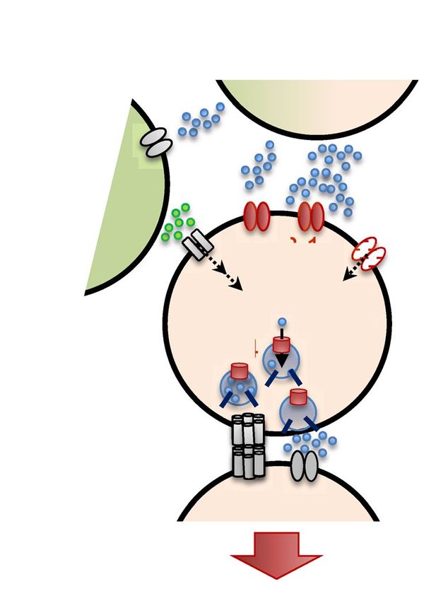
www.nature.com/scientificreports/

          Scientific Reports |   (2022) 12:1907 |   https://doi.org/10.1038/s41598-022-05942-0   2

Vol:.(1234567890)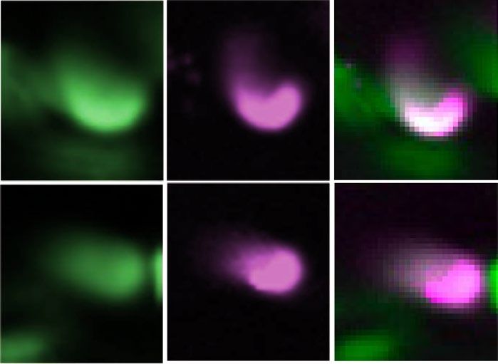
www.nature.com/scientificreports/
◂Figure 1. unc-130 is necessary for turning behavior during optimization of avoidance behavior. (a) A schematic
of optimization of avoidance behavior in C. elegans. During exposure to a mild osmotic stimulus, the animal
exhibits a reversal behavior, such as body retraction or backward movement. Strong stimulation induces body
bending, causing an omega turn so that the animal can return to its original location26. (b) In wild-type C.
elegans, the total frequency of avoidance increased with increasing sorbitol concentration (P < 0.001 in 0 M vs.
2–6 M, 1 M vs. 2–4 M, n = 11, 12, 15, 17, 20 (0–6 M, respectively)). In particular, the omega turn frequency was
significantly higher under 2–6 M compared to 0 M, under 4 M compared to 1 M, and under 6 M compared to
1 M (P < 0.001, P < 0.01, P < 0.001). (c) Nematodes with a null mutation in the unc-130 had a lower omega turn
frequency than wild-type animals (Fig. 1b) (P < 0.05 in 1 M and 6 M; n = 12, 12, 14, 20). On the other hand, the
total avoidance rate (sum of reversals and omega turns) was similar to that of wild-type animals because of the
increase in reversal frequency in all conditions (P > 0.05). (d) A schematic of the harsh tap assay. A platinum
wire placed in the forward direction is lightly applied to the tip of the nematode’s nose. e unc-130 mutants
showed the omega turn rate comparable to the wild-type animals. P = 0.392 (n = 10, 10, t-test). (f) Schematic
of the analysis method using channelrhodopsin 2 (ChR2(H134)). ChR2 (H134) was selectively expressed in
primary nociceptive sensory neurons (ASH sensory neurons)27. Exposure to blue light can selectively activate
ASH sensory neurons and their downstream neurons. (g) Adding all-trans retinoic acid (ATR) and blue light
triggered turn behavior. unc-130 mutants had a significantly lower turn frequency than the wild-type animals.
*** indicates P < 0.001, * indicates P < 0.05 (n = 13, 14, 14, 14). (h) Self-promoter-driven UNC-130 expression
rescued the decreased turn frequency of unc-130 mutants. See Fig. S1i–l. *** indicates P < 0.001, * indicates
P < 0.05, ns indicates P > 0.05 (n = 6, 6, 4). All statistical analyses were performed by Kruskal–Wallis test with
Dunn’s multiple comparisons test as a post hoc test. The error bars in the figures represent the ± SEM.
AIB activity through cholinergic AIA inhibitory interneurons11,12, but the neurotransmitter receptor between
AIAs and AIBs is unidentified. In the last circuit, ASHs form glutamatergic synapses with AVA interneurons,
whose ON responses and OFF responses (calcium increases) induce reversals and omega turns6. AIBs can also
affect the OFF-calcium response in the AVA neurons via chemical synapses. AIBs and AVAs coordinate muscle
contractions required for turns via motor/interneurons7,11,12. We have reported that a lack of AIB cells or gap
junction innexin-1 (inx-1) in AIBs reduces turn frequency but increases reversal frequency. These results suggest
that AIBs play a central role in optimization and that electrical synapses are components involved in turning6.
Calcium imaging using a microfluidic system13 has revealed that AIBs show strong calcium induction after a
hyperosmotic stimulus following a decrease during s timulation6.
AIB neurons develop in two steps. First, transcription factors encoded by proneural genes determine progeni-
tors’ neuronal fates, and then the progenitors differentiate into mature neurons with unique properties through
the combined activity of several transcription f actors14–17. Previously, we screened 210 neural transcription factors
and found that abnormal cell LINeage 32, lin-32, a nematode homolog of the proneural gene atonal/ATOH19,18, is
essential for the optimization of avoidance behaviors via AIB neuron development. lin-32 mutants lack a number
of neural function genes in AIBs6, and lin-32 may contribute to the initiation of neurogenesis rather than the
determination of AIB neuron identity. Therefore, transcription factors that establish the unique features of AIBs
have been left unexplored.
Here, we report that unc-130, an ortholog of the forkhead transcription factor 3 or 4, FOXD3/49,10, plays an
essential role in avoidance optimization, mainly by adjusting turn frequency. unc-130 null mutants lack both
ON responses (calcium decreases) and OFF responses (calcium increases) in AIBs. Inhibitory AIAs affect turn
frequency, implying their involvement in AIB ON responses (calcium reductions). OFF responses (calcium
increases) in the AVAs directly downstream of the AIBs may depend on the AIBs. We report that unc-130
upregulates three genes: the putative Nematode CAlcium channel 2 (nca-2)19, a homolog of the vertebrate cation
leak channel NALCN; AMPA-type glutamate receptor 1 (glr-1)20,21; and eat-4, a hypothetical L-glutamate trans-
membrane transporter22. However, it does not upregulate synaptobrevin (snb-1)23–25 or electrical synapse innexin
1 (inx-1)9,10. Our results suggest that unc-130 partially determines AIB neuron identity to aid in the optimization
of avoidance behaviors.
Results
A FOXD3/4 ortholog, unc‑130, affects the frequency of turn behavior during avoidance opti-
mization. C. elegans tends to show reversal behavior in response to mild osmolarity changes but is biased
toward omega turns in a manner correlating with stimulus strength (Fig. 1a)6. In our previous paper, we men-
tioned that unc-130 null mutants exhibited reduced avoidance frequency in response to noxious stimuli6. How-
ever, it remained unclear whether unc-130 affected the choice of appropriate behavior or simple locomotion. To
clarify this issue, we analyzed the avoidance behavior patterns in response to four gradient osmotic concentra-
tions of noxious stimuli. The results clarified that the unc-130 mutants were less likely to take omega turns upon
hyperosmotic stimulation than wild-type animals (Fig. 1b and c; Fig. S1a–h). Instead, the number of reversals
increased in the mutants (Fig. 1c). The critical point is that the unc-130 mutants had no significant changes in
the sum of omega turn and reversal frequencies, suggesting that they can avoid stimuli but not optimize how to
do so (Fig. 1b and c; Fig. S1a–h).
Meanwhile, such behavioral phenotype left the possibility that the unc-130 mutant was unable to make the
omega turn because of its uncoordinated movements. We hypothesized that the omega turn might occur by a
more intense stimulus. If this were the case, we could conclude that the uncoordinated movements were not
the reason for the inability to turn. In the drop test, it was difficult to use a sorbitol solution thicker than 6 M
due to the viscosity. Therefore, to investigate whether the unc-130 mutant can mediate the omega turn as well as
the wild-type animals, we established and performed an original harsh tap assay with a platinum wire. In this
Scientific Reports | (2022) 12:1907 | https://doi.org/10.1038/s41598-022-05942-0 3
Vol.:(0123456789)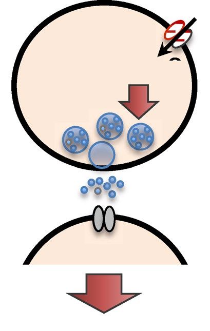
www.nature.com/scientificreports/

          Scientific Reports |   (2022) 12:1907 |   https://doi.org/10.1038/s41598-022-05942-0   4

Vol:.(1234567890)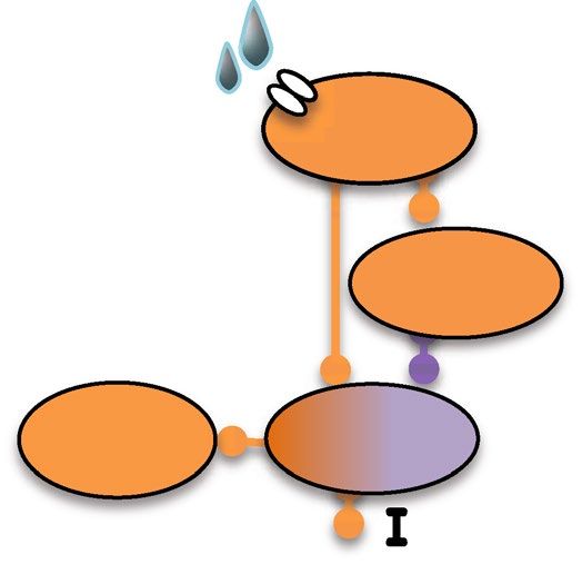
www.nature.com/scientificreports/
◂Figure 2. unc-130 affects both biphasic neural responses of the central neurons for optimization of avoidance
behavior. (a) Biphasic neural responses in AIBs. IP2.0 is an indicator to monitor the calcium decrease. In
wild-type animals, an ON response (calcium decrease) occurred during stimulation (5–35 s; light blue area in
the figure) and was followed by an OFF response (calcium increase) immediately after the stimulus (n = 16). In
unc-130 mutants, both of these ON and OFF calcium responses were absent (n = 15). (b) Detailed schematic
of the downstream neural circuit of ASH sensory neurons. ASH sensory neurons sense osmotic stimuli at
receptors and directly excite two types of interneurons (AIBs and AIAs) via glutamate release. Cholinergic AIAs
inhibit calcium induction of downstream AIBs. AIBs release glutamate or signal via electrical synapses to excite
downstream motor/interneurons, resulting in turn behavior. (c) Cell ablation of inhibitory AIA interneurons
resulted in reduced turn frequency. **indicates P < 0.01 (n = 12, 8, t-test). (d) Analysis of calcium responses using
G-CaMP6s in ASH sensory neurons (n = 20, 19). (e) Calcium imaging of AIA neurites using G-CaMP6s. There
was no significant difference in the peak of ON calcium increase in the wild-type animals compared with unc-
130 mutants (P = 0.892, n = 21, 23, t-test). (f) Calcium imaging of AVA neurons, one of the types of downstream
AIB neurons. Wild-type animals showed a small ON calcium increase during stimulation and a large increase
after stimulation (n = 20). In the unc-130 mutants, OFF calcium responses were abolished (n = 19). The area in
orange color is enlarged in the right view. The error bars in these figures represent the ± SEM values.
experiment, we tapped nematodes on the tip of their nose with the platinum wire on NGM plates, and observed
their avoidance behavior (Fig. 1d). As a result, the unc-130 mutant also mediated omega turns in an average of
84.0%, and there was no statistically significant difference compared to the wild-type animals, which mediated
omega turns in 89.0% (P = 0.392, n = 10, 10, t-test) (Fig. 1e). This data clearly shows that unc-130 mutants have the
physical ability to perform omega turn. Based on the above results, we concluded that unc-130 affects the behavio-
ral choice. The behavioral phenotype in the null mutants corresponds to “hypoesthesia” in psychological t erms28.
In the drop test, a sorbitol solution accompanying touch or mechanical stimuli might trigger sensory neurons
other than ASHs. To confirm whether the optimization defect in unc-130 mutants is dependent on noxious
stimuli, we observed the behavior induced by selective excitation of ASH sensory neurons using channelrhodop-
sin-2 (Fig. 1f)29. Optogenetic excitation separate from touch or mechanical stimuli reproduced the optimization
defect (P < 0.001, n = 13, 14, 14, 14, Kruskal–Wallis test with Dunn’s multiple comparisons test as a post hoc test)
(Fig. 1g), suggesting that unc-130 optimizes avoidance patterns in response to the strength of the noxious stimuli.
UNC-130 driven by its promoter rescued the behavior of the null mutants, indicating that the optimization
defect was due to the loss of unc-130 gene function (P < 0.05, n = 6, 6, 4, Kruskal–Wallis test with Dunn’s multiple
comparisons test as a post hoc test) (Fig. 1h; Figs. S1i–l and S2a–e).
unc‑130 is necessary for biphasic calcium responses of the central neurons for behavioral opti-
mization. Previously, we showed that AIB neurons are the central neurons for turning and the biphasic
response involving a calcium reduction during osmotic stimulation and an increase after stimulation6. Consid-
ering that unc-130 expression is relatively restricted in cell lineages including AIB cells (ABplaapa, ABpraapa),
during embryogenesis30, we hypothesized that the optimization defect of unc-130 mutants likely resulted from
AIB neuronal dysfunction. To test this hypothesis, we attempted to analyze AIB neural responses, including
calcium reductions during stimulation, using the calcium indicator inverse-pericam 2.0 (IP2.0), whose fluores-
cence intensity increases as the calcium concentration d ecreases31. As we expected, the fluorescence intensity of
IP2.0 increased during osmotic stimulation and decreased after stimulation in wild-type animals, showing that
AIBs received inhibitory input during stimulation and excitatory input after the stimulus was removed (Fig. 2a
and Fig. S3a). In contrast, the unc-130 mutants showed no responses (Fig. 2a). Baseline fluorescence values
before correction was close to minimum IP2.0 fluorescence values (Fig. S3bc and c) (P = 0.077, n = 14, 18, t-test),
implying relatively high resting calcium level in both wild-type and unc-130 animals (P > 0.05, n = 14, 18, t-test).
These data suggest that reduced responses in AIB neurons cause the optimization defect in unc-130 mutants.
Increases in calcium in AIBs induce t urning6,18, but it remains unclear whether the AIB ON response (cal-
cium decrease) contributes to the turning behavior. In C. elegans, ASH sensory neurons indirectly inhibit AIBs
via predicted AIA inhibitory interneurons (Fig. 2b)32. Therefore, we analyzed whether ablation of AIAs reduces
turning. Animals in which AIAs were ablated by caspase CED-3-mediated cell death33 exhibited significantly
decreased turn frequencies (P < 0.01, n = 12, 8, t-test), and AIA-ablated unc-130 mutants exhibited indifferent
turn frequencies (Fig. 2c; Fig. S3d). These results imply that AIA neurons are required for turning, potentially
via inhibition of AIBs during the stimulus.
We compared the responses in ASHs using the calcium indicator GCaMP6s34,35 to confirm that the other
neuron responses in the avoidance circuit remained unaltered, and we found no differences between unc-130
mutants and wild-type animals (Fig. 2d). This result supports the occurrence of “secondary hypoesthesia” based
on a central neuron defect rather than "primary hypoalgesia,” a simple decrease in perception. We also analyzed
the calcium responses of the AIAs using GCaMP6s. Since calcium responses in AIAs to noxious osmolarity
stimuli have not yet been reported, we started by observing the detailed patterns in wild-type animals. We found
increased calcium concentrations in AIA neurites during osmotic stimulation (Fig. 2e; Fig. S3e). In previous
studies, calcium imaging has been performed in AIA neurites36. One reason is that AIA neurites, but not the
cell bodies, show large calcium fluctuations. Fortunately, we were able to observe calcium responses in AIA cell
bodies using our original transgenic strain (Fig. S3f). We found that AIA cell bodies show a triphasic pattern: a
sustained increase in calcium concentration during stimulation, a decrease for 30 s after the end of stimulation,
and a subsequent increase (Fig. S3f). This response pattern appears to correlate more strongly with calcium
decrease responses in AIB neurites than with responses in AIA neurites (Fig. S3a, e and f). Considering that the
AIA-AIB synapse is a predicted inhibitory circuit, the responses in the AIA cell body might affect the biphasic
Scientific Reports | (2022) 12:1907 | https://doi.org/10.1038/s41598-022-05942-0 5
Vol.:(0123456789)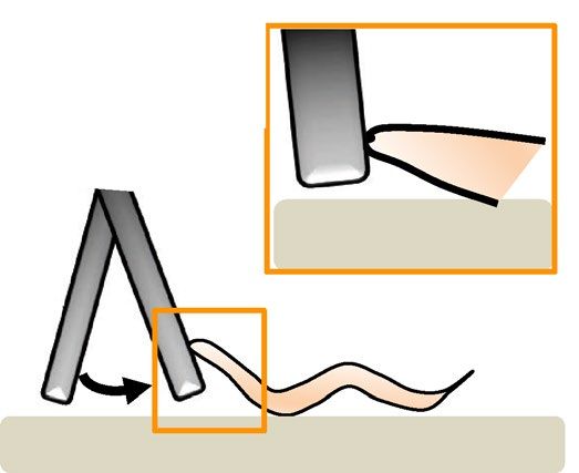
www.nature.com/scientificreports/

          Scientific Reports |   (2022) 12:1907 |   https://doi.org/10.1038/s41598-022-05942-0   6

Vol:.(1234567890)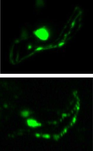
www.nature.com/scientificreports/
◂Figure 3. unc-130 upregulates the expression of putative cation channels in AIB neurons. (a) All-trans
retinoic acid and blue light induce turning. The turning frequency was 77.9% in the wild type but only 15.3%
in unc-130 mutants. ***indicates P < 0.001, *indicates P < 0.05 (n = 14, 14, 17, 15, Kruskal–Wallis test with Dunn’s
multiple comparisons test as a post hoc test). (b) nca-2 mutants showed significantly reduced turn frequencies
in response to 4 M sorbitol than wild-type animals. ***indicates P < 0.001 (n = 13, 11, t-test). (c) Expression of
nca-2 promoter-driven GFP in AIB cell bodies (arrowheads). inx-1 promoter-driven mCherry was coexpressed
as a cell identification marker for AIBs. nca-2 showed weaker AIB expression in a typical unc-130 animal than
in a typical wild-type animal. Scale bar = 5 µm. (d) The ratiometric analysis of the intensity of nca-2 promoter-
driven GFP expression was significantly lower in AIBs of unc-130 mutants than in those of wild-type animals.
***
indicates P < 0.001 (n = 10, 11, t-test). (e) AIB-selective expression of NCA-2a rescued the decreased turn
frequency of nca-2 mutants. ** indicates P < 0.01 (n = 12, 12, 12, one-way ANOVA followed by Tukey’s post
hoc test). See Fig. S7a–d. The error bars in these figures represent the ± SEM values. (f) Model diagram of the
hypothetical function of nca-2 in AIB. NCA-2 may involve the release of neurotransmitters from synaptic
vesicles via cation influx.
response in the AIB. Then, we observed the AIA reaction in the unc-130 mutants and clarified that there was no
statistically significant variation in peak ΔF/F amplitude (P = 0.892, n = 21, 23, t-test), although the response was
slightly dampened in mutants compared within wild-type animals (Fig. 2e).
Finally, to understand the output function of AIBs on the avoidance circuit, we observed the calcium response
in AVAs. AVAs receive input from the presynaptic AIB neurons (Fig. 2b). As expected, unc-130 mutants showed
remarkably impaired OFF responses (calcium increases) in AVAs (Fig. 2f). In our previous paper, we discussed
how low ON responses (calcium increases) in AVAs during osmotic stimulation might induce reversal behav-
ior independent of A IBs6. Since the unc-130 mutants can perform reversal behavior (Fig. 1c; Fig. S1f and g),
we explored whether the AVA ON response (calcium increase) remains in the unc-130 mutants. The unc-130
mutants showed an ON response (calcium increase), although it was reduced in size (see the enlarged view of
the area in Fig. 2f, inset), consistent with our hypothesis. These results suggest that AIBs are critical neurons for
determining appropriate behaviors in response to various stimuli.
The inhibitory synaptic receptors on AIBs have not yet been identified. Since AIAs are cholinergic neurons37,
we first considered the possibility of the downregulation of inhibitory acetylcholine-gated chloride channels
expressed on AIBs in the unc-130 mutants. We observed the expression patterns of six hypothetical acetylcholine-
gated or ligand-gated chloride channels to focus on the inhibitory channels expressed on AIB neurons. In this
experiment, we found that all unc-130 mutant animals expressed innexin 1 (inx-1) on AIBs (n = 25); we used the
inx-1 promoter to mark AIBs and identify AIB-specific gene expression. AIBs expressed acc-1 (Acetylcholine-
gated Chloride Channel 1)38, lgc-46 (Ligand-Gated ion Channel 46, predicted to have chloride channel activity),
and lgc-49 (Ligand-Gated ion Channel 49, predicted to have chloride channel activity)39 promoter-driven GFP
(Fig. S4a). However, AIBs did not express acc-2, acc-3, or acc-4 (Acetylcholine-gated Chloride Channel 2, 3, or 4,
respectively)38 (Fig. S4b), nor did they express lgc-47 or lgc-48 (Ligand-Gated ion Channel 47 or 48, respectively,
predicted to have chloride channel activity)10. We analyzed the turn behavior of deletion mutants of acc-1, lgc-46,
and lgc-49, but there were no differences between the single or double mutants and wild-type animals (Fig. S4c
and d).
We next assumed a contribution of G protein-coupled acetylcholine receptors. We observed turn behavior
in deletion mutants of goa-1 (G protein O, alpha subunit 1; an ortholog of human GNAO110), which exhibits G
protein-coupled acetylcholine receptor activity40. In C. elegans, only goa-1 encodes a member of the mammalian
Gi/o class of Gα subunits, and the predicted amino acid sequence of C. elegans GOA-1 is over 80% identical to
that of mammalian Gαo40. Hypothetical goa-1 null mutants showed a lower turn frequency (13.3 ± 4.41%) than
wild-type animals (56.3 ± 5.96) (P < 0.001, n = 9, 8, t-test) (Fig. S5a), but the expression levels of goa-1 promoter-
driven GFP in AIBs were comparable in both strains (Fig. S5b and c) (P = 0.450, n = 10, 9, t-test). Thus, we
concluded that the reduction in turning was independent of the goa-1 function in AIB neurons. In C. elegans,
there are three types of muscarinic-type acetylcholine receptors: gar-1 (G-protein-linked Acetylcholine Receptor
1)41, gar-242, and gar-343. Although we analyzed the behavior of triple gene mutants44, turn frequency was not
significantly lower in these animals than in wild-type animals (Fig. S5d) (P = 0.226, n = 9, 8, t-test). Consequently,
we could not identify the AIA-AIB inhibitory receptors, but the possibility remains that an unknown G-protein
coupled acetylcholine receptor participates.
unc‑130 upregulates the expression of a predicted cation channel. unc-130 mutants showed no
stimulus-dependent OFF responses (calcium increases) (Fig. 2a). Therefore, we tested whether the turn behavior
could be recovered by cation flux through ChR2(H134R) selectively expressed in AIBs. Contrary to our expecta-
tions, turn behavior induction was minor in unc-130 mutants (Fig. 3a). In optogenetics, cation flux is reported
to be milder than that under natural stimulation45. Therefore, we proposed three hypotheses: 1. that there is
dysfunction in the calcium signaling pathway in AIBs, 2. that there are reductions in presynaptic output from
AIBs, and 3. AIBs are developmentally altered in unc-130 mutants.
First, we tested the former. There are eight of nine predicted voltage-gated calcium channel subunits in C.
elegans46. We excluded three genes from our analysis. One of them, ccb-1 (Calcium Channel, Beta subunit 1,
IBs46. We performed a drop
predicted to have high voltage-gated calcium channel activity), is not expressed in A
test for mutants of the other seven channels and found that only nca-2 (putative Nematode CAlcium channel 2, a
homolog of the vertebrate cation leak channel NALCN) mutants showed a lower turn frequency in response to a
4 M sorbitol drop than wild-type animals (Fig. 3b; Fig. S6a and b) (P < 0.001, n = 13, 11, t-test). The mutants for
Scientific Reports | (2022) 12:1907 | https://doi.org/10.1038/s41598-022-05942-0 7
Vol.:(0123456789)
www.nature.com/scientificreports/

          Scientific Reports |   (2022) 12:1907 |   https://doi.org/10.1038/s41598-022-05942-0   8

Vol:.(1234567890)
www.nature.com/scientificreports/
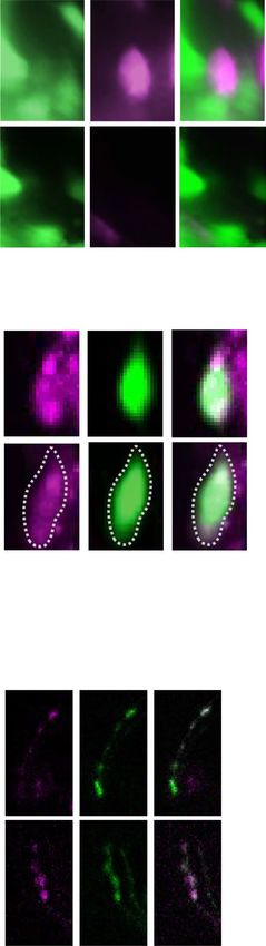
◂Figure 4. unc-130 affects the expression of two molecules in glutamate synapses in AIB neurons. (a) Expression
analysis of glr-1 promoter-driven GFP. DsRedx driven by the promoter of the olfactory receptor odr-2 was
coexpressed as a cell identification marker for AIBs. glr-1 was weakly expressed in AIBs in a wild-type animal
(arrowhead), while both glr-1 and odr-2 were absent in an unc-130 mutant. Scale bar = 5 µm. (b) Frequency
of animals expressing GFP in AIB neurons. Because we used the mosaic Ex strain, GFP was expressed in two
AIB cells at 65.5% and in one cell at 35.0% in wild-type animals. The frequencies of animals with GFP-positive
AIBs were 4.76% for two cells and 47.6% for one cell; 47.6% had no expression among the unc-130 mutants.
***
indicates P < 0.001 (n = 20, 21, Fisher’s exact test). (c) The expression of mCherry driven by the promoter of
the vesicular glutamate transporter eat-4 was weak in an unc-130 mutant. Scale bar = 5 µm. (d) The intensity
of mCherry expression in AIBs of unc-130 mutants was significantly reduced. **indicates P < 0.05 (n = 20, 20,
Mann–Whitney test). (e) Comparison of synaptic localization patterns. The arrowheads indicate accumulated
SNB-1::mCherry in AIB neurites. Scale bar = 5 µm. (f) The distribution of the maximum accumulation size
of SNB-1::mCherry was not different between wild-type and unc-130 mutants. “ns” indicates no significant
difference (P = 0.262, n = 28, 24, t-test). (g) The ratiometric analysis of the total intensity of mCherry expression
in AIB presynaptic regions was not different between wild-type and unc-130 mutants. “ns” indicates no
significant difference (P = 0.326, n = 20, 20, t-test). The error bars in these figures represent the ± SEM values.
the other five channels, tag-180 (an ortholog of human voltage-gated calcium channel auxiliary subunit alpha
2 delta 2)47 (Fig. S6a), unc-2 (UNCoordinated 2, a predicted voltage-gated calcium channel)48, nca-1 (putative
Nematode CAlcium channel 1)19, cca-1 (Calcium Channel Alpha subunit 1)49, ccb-2 (Calcium Channel, Beta subunit
2)19, and unc-36 (UNCoordinated 36)47, showed no reductions in turn frequency (Fig. S6b). However, it should
be noted that nca-1 (gk9) and ccb-2 (ok862) provide no evidence for null alleles only because they are predicted
to have hypothetical reduced expression and partial loss of a domain, respectively (Fig. S13). So we could not
completely exclude the possible roles of these channels.
To uncover whether unc-130 regulates nca-2 expression in AIBs, we quantified GFP intensity driven by the
nca-2 promoter. The results showed that AIBs expressed nca-2, but the unc-130 mutants showed significantly
reduced fluorescence intensity compared with wild-type animals (Fig. 3c and d). The neural subsets expressed
nca-2. To test whether reduced nca-2 expression in the AIBs correlates with reduced turn frequency, we per-
formed a rescue experiment with the AIB-selective expression of the NCA-2a protein. The results revealed that
the defect in nca-2 mutants was substantially alleviated such that the mutants recovered an optimization pattern
relatively similar to that of wild-type animals (Fig. 3e; Fig. S7). Thus, we conclude that nca-2 expression in AIBs
is necessary for avoidance behavior optimization, suggesting a voltage-gated cation channel or a cation leak
channel expression in the AIBs may upregulate synaptic transmission from AIBs to downstream inter-/motor
neurons (Fig. 3f).
unc‑130 upregulates the expression of functional molecules for glutamatergic synapses. Tran-
scription factors frequently determine neural identity by regulating the expression of multiple genes46. To deter-
mine whether unc-130 contributes to the excitatory input from upstream neurons to the AIB, we observed glr-1
(GLutamate Receptor family (AMPA) 1) promoter-driven GFP expression. glr-1 is one of the primary excitatory
receptors in AIB neurons50. GLR-1 is the primary glutamate receptor between ASHs and AIBs. Among the wild-
type animals, 65.0% of individuals expressed GFP in two AIB cells, while 35.0% expressed it in one (Fig. 4a and
b). Notably, transgenic C. elegans carrying extrachromosomal transgenes frequently display mosaic expression47.
Among the unc-130 mutants, only 5.0% of individuals expressed GFP in two AIB cells, while 52.4% expressed
it in one (Fig. 4a and b); mutants showed a significant difference compared with the wild-type animals. This
result indicates a reduction in excitatory synaptic input from the ASHs. We also tried to use the Pinx-1-driven
markers, but the transgenic strains could not be maintained, so we used Podr-2 as the marker promoter in this
experiment. AIBs are glutamatergic n eurons51. To determine whether typical glutamatergic synaptic vesicles are
formed in AIBs, we analyzed the expression level of eat-4 (a predicted L-glutamate transmembrane transporter),
which fills vesicles with glutamate37. We detected a significant reduction in the fluorescence intensity of eat-4
promoter-driven mCherry in the AIBs of unc-130 mutants (P < 0.05, n = 20, 20, Mann–Whitney test) (Figs.4c
and d), suggesting that unc-130 is required for the release of proper amounts of glutamate. A decrease in the
expression of eat-4 might cause the decrease in turns upon AIB::ChR2 stimulation shown in Fig. 3a.
We suspected that the unc-130 mutant forms abnormal presynapses. Then, we observed the localization
of mCherry-tagged synaptobrevin (SNB-1), a typical presynaptic marker, at synaptic sites. However, both the
maximum size and the total fluorescence intensity of SNB-1::mCherry fusion puncta indicated no significant
differences between the unc-130 mutants and wild-type animals (Fig. 4e–g). RAB-3, a component of synaptic
vesicles52, also showed no differences in accumulation size and fluorescence intensity between the two strains.
These results indicate that unc-130 is required for both the input and output of glutamatergic synaptic transmis-
sion at AIB neurons but not for the fundamental function of synapse formation.
unc‑130 does not alter the expression of electrical synapses in AIBs. Previously, we reported that
lin-32 upregulates a broad spectrum of genes in AIB neurons, including the dominant electric synapses inx-1,
and that its mutation causes secondary hypoesthesia similar to that caused by unc-130 mutation6. Therefore, we
analyzed the property of inx-1 positive cells, expression rate, cell morphology, and expression intensity, as one
AIB indicator of functional overlap between unc-130 and lin-32. First, in terms of expression rate, GFP-positive
cells were observed in all unc-130 mutants (n = 128) (Fig. S8a and b). 116 (90.6%) animals had normal AIB loca-
tion and morphology, while 12 animals (9.38%) had an ectopic dendrite extending to the tip of the nose (Fig. S8a
and b). However, the occurrence rates were not significantly different (P = 0.0714, n = 40, 128, Fisher’s exact test),
Scientific Reports | (2022) 12:1907 | https://doi.org/10.1038/s41598-022-05942-0 9
Vol.:(0123456789)www.nature.com/scientificreports/
Figure 5. unc-130 does not regulate electrical synapses in the AIBs. (a) Comparison of the expression of inx-1
promoter-driven GFP. unc-130 mutants showed similar expression levels. Scale bar = 5 µm. (b) There were
no differences in the intensity of inx-1 promoter-driven GFP expression measured in cell bodies between the
wild-type animals and the unc-130 mutants. “ns” indicates no significant difference (P = 0.960, n = 20, 20, Mann–
Whitney test). (c) The double mutants of unc-130 and lin-32 almost completely lacked turn behavior (n = 26),
whereas unc-130 and lin-32 single mutants exhibited moderately lower turn frequencies (n = 19, 25) than
wild-type animals (n = 14). Each mutant had an increased reversal frequency instead of exhibiting turning. (d)
unc-130 and lin-32 single mutants (n = 19, 25) showed significantly different turn frequencies than the wild-type
animals (n = 14) and the double mutants (n = 26). ***indicates P < 0.001, *indicates P < 0.05 (Kruskal–Wallis test
with Dunn’s multiple comparisons test as a post hoc test). The error bars in these figures represent the ± SEM
values.
Scientific Reports | (2022) 12:1907 | https://doi.org/10.1038/s41598-022-05942-0 10
Vol:.(1234567890)www.nature.com/scientificreports/
Figure 6. Schematic model of the AIB calcium response regulated by unc-130. (a) Relationship between the
ON/OFF stimulus and calcium dynamics in AIB. During stimulation, excitatory/inhibitory (E/I) balance from
ASHs and AIAs could bias inhibition, leading to ON responses (calcium decreases) in AIBs. After stimulation,
the ASH OFF response (calcium increase) is a possible trigger for the OFF response (calcium increase) in the
AIBs. unc-130 impaired both ON responses and OFF responses (red line). (b) Schematic of the molecules used
for optimization and the contribution of unc-130 (shown in red). unc-130 is necessary for the glutamatergic
signaling pathway because it regulates at least glr-1, nca-2, and eat-4 expression in the AIBs to cause defects in
the calcium response and turn optimization dynamics.
and there was no association between the presence of ectopic neurites and turn/reversal frequency (Fig. S8c)
(P = 0.892, n = 40, 128, Fisher’s exact test). They suggest that this morphological is not related to the optimiza-
tion of avoidance behavior. Next, to determine whether unc-130 might affect the amount of inx-1 expression,
we quantified the intensity of GFP driven by the inx-1 promoter in the mutants. The unc-130 mutants expressed
the same amount of inx-1 as wild-type animals (Figs. 5a and b) (P = 0.326, n = 20, 20, Mann–Whitney test),
indicating that unc-130 regulates genes, inx-1 different from those regulated by lin-32. Finally, we performed
a drop test on the double mutants to confirm that unc-130 and lin-32 act in different molecular pathways. The
double mutants exhibited more remarkable optimization defects than the single mutants (Fig. 5c), and the turns
almost completely disappeared (Fig. 5d). Meanwhile, the reversal ability remained (Fig. 5b; Fig. S12), suggest-
ing that unc-130 and lin-32 regulate parallel molecular pathways for turning. We conclude that unc-130 impacts
behavioral optimization by regulating the characteristic gene expression of calcium dynamics and glutamatergic
synapse function, unlike lin-32.
unc-130 starts to be expressed in early embryogenesis but not detected in neurons from the larvae stage
onward53. In order to clarify whether unc-130 contributes to AIB development and determination decisions in
embryogenesis or gene expression in the adult stage, we examined behavioral rescue in the adult driven unc-130
by the inx-1 or npr-9 promoters, which cause AIB-selective expression from larva onward. As a result, all three
or four independent transgenic lines driven UNC-130 from the larva onward could not rescue (Figs. S9 and
S10), suggesting that function during embryogenesis is essential. Behavioral optimization defects in unc-130
adults were not rescued by overexpression of NCA-2a or EAT-4 driven by the inx-1 promoter (Figs. S10 and
S11). Co-transduction with multiple genes in AIB regulated by unc-130—at least nca-2, eat-4 and glr-1 – might
be able to rescue behavioral defects.
Discussion
unc‑130 regulates the restricted genes for specific neuronal phenotypes to optimize behav-
ior. We have reported that the proneural gene lin-32 promotes the expression of a wide range of genes in
AIBs, including gap j unctions6. However, unc-130 seems to play a more limited role in AIB identity. The unc-130
mutants had reduced expression of glr-1, nca-2, and eat-4 in the AIBs but not the electrical synapse component
inx-1 and standard chemical synaptic component SNB-1 in AIB neurons, suggesting that the unc-130 mutation
causes behavioral optimization defects by disrupting selective glutamatergic synapse functions (Fig. 6a).
During intense osmotic stimulation, the AIBs receive excitatory inputs through a direct circuit between
the ASHs and AIBs via glutamate and GLR-1 receptors as well as inhibitory inputs mediated by AIAs (Fig. 6a).
Scientific Reports | (2022) 12:1907 | https://doi.org/10.1038/s41598-022-05942-0 11
Vol.:(0123456789)www.nature.com/scientificreports/
Considering the AIB suppression that occurs during osmotic stimulation (Fig. 2a), inhibitory input may exceed
GLR-1-mediated excitatory input during hyperosmolar stimulation. We could not identify the inhibitory recep-
tors on AIBs in this study (Figs. S4 and S5). In addition to the unanalyzed inhibitory acetylcholine receptors,
neuropeptide receptors might be involved in AIA-AIB suppression. The unc-130 mutation may regulate either
inhibitory receptors or genes in the downstream calcium signaling cascade.
The molecular basis of optimization after intense osmotic stimulation is also intriguing. After stimulation,
OFF responses (calcium increases) in ASHs can induce glutamate release. GLR-1, a receptor for glutamate on
AIBs, induces a change in conductance to open NCA-2, a predicted voltage-gated cation channel or a cation
leak channel, in order to promote the intracellular influx of cations. This contributes to the increase in the OFF-
calcium concentration response in AIBs. We showed that overexpression of NCA-2a in AIBs of unc-130 mutants
rescued the optimization defect (Fig. 3e), and unc-130 partially downregulated nca-2 and glr-1 (Figs. 3d and 4b).
These results imply that excitatory input is reduced and suggest the amplification mechanism.
The vesicular glutamate transporter EAT-4 transports glutamate into synaptic vesicles. Calcium induction
promotes the secretion of glutamate-releasing synaptic vesicles that transmit information to downstream inter-/
motor neurons, including neck motor neurons. We have shown that the contraction strength of the neck posi-
tively correlates with turn behavior6. Thus, the AIBs appear to integrate the two excitatory and inhibitory (E/I)
pieces of information from the AIAs and ASHs to evaluate the intensity and exposure time of the stimulus and
to output avoidance behavior with appropriate intensity and timing.
We demonstrated that unc-130 mutants show reduced expression of glr-1 and nca-2 (Figs. 3c, d, 4a, b), sug-
gesting that insufficient excitatory input from ASHs and voltage-dependent calcium influx occur. Additionally,
the reduced expression of EAT-4 may lead to insufficient glutamate loading in the synaptic vesicles, resulting in
reduced transmission of information to the downstream neural circuit.
On the other hand, since AIB-specific driven unc-130 does not rescue behavioral abnormalities (Figs. S3 and
S4), showing the importance of an act of unc-130 in a developmental stage. Often, with the loss of transcription
factors during developmental stages, unexpected circuits may form or differentiate into different cells that are
similar in lineage. It has also been reported that unc-130 is required to make a difference between AWA and ASG,
chemosensory neurons generated from the ABp(l/r)aapap lineage from which AIB is d erived30. unc-130 specifies
two glial types that arise from the neighboring lineage (ABp(l/r)aapa)54. As for the AIBs that we are focusing
on in this study, they express at least inx-1, which is selective for AIBs, and show a consistent number, location,
and interneuron-like morphology. We consider that they are incomplete differentiated AIBs rather than sister
cells of sensory neurons or glial cells.
Now we conclude that unc-130 is involved in both the function and development of AIBs, and comprehensive
discussion summarizing the importance of both sides is more appropriate for the role of unc-130. unc-130 is
required for the establishment of some AIB identities (e.g., biphasic response, expression of nca-2, glr-1, and eat-
4, and role as a behavioral optimization center), and unc-130 contributes to behavioral optimization through its
role in regulating the expression of a group of genes that are necessary for AIB identities. unc-130 has the defect
in the lineage determination, so the possibility remains that it reflects an unidentified reorganization of neurons
in addition to AIB defects. It might explain the synthetic defect in the behavior of unc-130;lin-32 double mutants.
Biphasic neural response of AIB neurons. We provide the first evidence for a biphasic neural response
of AIBs: calcium decreases during osmotic stimulation followed by a calcium increase after the stimulus (Fig. 2a
and Fig. S3a). First, we discuss the neural mechanisms of the calcium decrease in wild-type C. elegans during
stimulation. Calcium imaging results showed that ASHs and AIAs were excited and that AIBs were inhibited
during the stimulus (Fig. 2a, d, and e; Fig. S1a, e and f). Since ASHs and AIAs form synapses and since AIA-
AIB connections are likely to be inhibitory (Fig. 2b)7,32, we speculate that ASHs may directly excite AIAs during
stimulation and that excited AIAs inhibit AIBs (Figs. 2b and 6a), resulting in decreased intracellular calcium
concentrations in AIBs (Fig. 6b). It has been implied that excitation of AWC chemosensory neurons might cause
calcium decreases in AIBs during stimulus exposure55,56, but this has not been stringently verified. This study
clearly showed such responses with the indicator IP2.0, which monitors the decrease in intracellular calcium
concentration associated with osmotic stimulation received by ASHs.
Next, we considered the neural mechanisms of calcium increases in wild-type C. elegans after stimulation.
Calcium imaging results showed that ASHs and AIBs, but not AIAs, were excited after the stimulus (Figs. 2a, d,
and e; Fig. S3a, e and f). Although removing the C O2 stimulus evokes an AIA OFF response (calcium increase)57,
it is likely due to neural circuits distinct from those involved in response to noxious osmolality. Anatomical
analysis showed a direct synaptic connection between the ASHs and AIBs (Fig. 2b)58, but the details of ASH-
AIB synapses are not well understood. Since ASHs show the biphasic (ON/OFF-increase) response (Fig. 2d)46,
we speculate that the ASH OFF response (calcium increase) engages the disinhibition of AIB after stimulation6.
Physiologically, such a biphasic interneuron response has also been observed in mammals. In rodents, over
60% of suprachiasmatic nucleus neurons may use such “rebound responses” or “postinhibitory rebounds,” and
the probability of the response is positively correlated with the duration of hyperpolarization59. One of the next
questions will be to determine whether the duration of ON-hyperpolarization of AIBs is correlated with the
probability of OFF-calcium responses in C. elegans, so that similarity with a property of suprachiasmatic nucleus
neurons can be clarified. Rebound responses are also observed in striatal neurons and imply the existence of
an essential mechanism for fear processing and decision-making60. C. elegans AIBs share similarities in that
they are involved in escape behavior optimization and behavioral choice in response to harmful stimuli. The
biphasic neural response with excitatory-inhibitory association seems likely to have been evolutionarily acquired
for behavioral diversity. We consider that the biphasic neural response in C. elegans may be one of the neural
mechanisms of primitive excitation-inhibition association. In addition, rebound firing is also associated with
Scientific Reports | (2022) 12:1907 | https://doi.org/10.1038/s41598-022-05942-0 12
Vol:.(1234567890)www.nature.com/scientificreports/
diseases; for example, drug addiction pathogenesis in rodents and thalamic neurons generate unusual rebound
umans60,61. In the future, C. elegans AIB neurons may be
firing at the end of inhibition in Parkinson’s disease in h
used as a neuron model for drug addiction pathogenesis and neurodegenerative diseases.
Furthermore, AIB biphasic responses may biologically contribute to temporal control of the initiation of turn
behavior. Direct AIB excitation by optogenetics facilitates turning without r eversal6, suggesting that AIA-AIB
inhibition suppresses time-consuming turn behavior under conditions of toxic osmolality to expedite escape.
Involvement of FOXD3/4 in avoidance behavior optimization. In this study, we demonstrated, for
the first time, that a FOXD3/4 ortholog, unc-130, specifies avoidance behavior patterns using C. elegans. FOXD3
is widely conserved from invertebrates to vertebrates, including humans10. FOXD3 defines the early pluripotency
uscles62–64. Even
of neural crest stem cells in vertebrates to differentiate into diverse cells, such as neurons and m
in C. elegans, unc-130 has a similarity to FOXD3/4 with regard to its expression in both neural progenitors and
adult muscle cells30,65,66. Future unc-130 studies may lead to universal molecular insights into primary neuro-
genesis. In addition, whole-human genome analysis has implied that FOXD4 is a risk factor for suicide and
obsessive–compulsive disorder67. Such outcomes may be attributable to vulnerability to stress. unc-130 might
be useful for illustrating a prototypical circuit for improved coping behavior under exposure to harmful stimuli.
During development, combinations of transcription factors determine neuron identities. Hobert et al. have
comprehensively mapped the combination of transcription factors expressed in all neurons of C. elegans37,68. They
speculate that combinations of homeobox (Hox) transcription factors can code for almost all neural identities69;
these findings will accelerate the understanding of individual neural characters. In addition, basic helix-loop-
helix (bHLH) transcription factors, including proneural genes, act in the initial phase and have a crucial role in
neurogenesis61. However, forkhead box (Fox) transcription factors were grouped relatively recently in 200061,
and their functions in the nervous system are still less known than those of Hox and bHLH molecules.
Conclusion
Our findings suggest that a FOXD3/4 ortholog, unc-130 contributes to behavioral optimization mediated by pre-
and postsynaptic function to mediate biphasic neural responses. In summary, reductions in the ON and OFF
calcium responses required for integrating this information and producing behavioral outputs result in incorrect
behavioral choices in response to stimuli of different intensities in unc-130 mutants.
Methods
Nematodes and maintenance. We cultured C. elegans strains using modified standard techniques70.
NGM agar plates containing 67 mg/ml antibiotic streptomycin and 10 µg/ml nystatin were used. Escherichia coli
OP50-1 was seeded as food. unc-130(tm320), acc-1(tm3268), nca-2(tm1305), unc-2(e55), and lin-32(tm2044)
mutants were backcrossed twice with N2. lgc-46(ok2949), lgc-49(tm6556), and goa-1(sa734) mutants were back-
crossed three, four, and five times with N2, respectively. cca-1(gk30), nca-1(gk9), unc-36(e251), unc-36(ok862),
and ccb-2(ok862) were not backcrossed because of the pilot screening (Fig. S6b). The deletion and point muta-
tion sites are described in Fig. S13. The strain information is summarized in Table S1.
Plasmid construction. For the own-promoter rescue experiment (Fig. 1h; Figs. S1i–l), pPD_Punc-
130::UNC-130 was constructed by subcloning the sequence from 5854 bp upstream of the ATG to the end of the
3’ UTR of the unc-130 genomic sequence into the pPD95.75 vector instead of gfp. For the AIB-specific promoter
rescue experiment (Figs. S10 and S11), promoters of inx-1 or Pnpr-96 were transferred into the pPD_Punc-
130::UNC-130 plasmid instead of Punc-130. For calcium imaging, we constructed pPD_Pinx-1::IP2.0 (Fig. 2a;
Fig. S3a) or pPD_Psra-6::GCaMP6s (Fig. 2d) by subcloning IP2.0 from pDEST-IP2.0 (codon for C. elegans) or
both the 2409 bp sra-6 promoter and GCaMP6s sequences from N2 genomic DNA and pGP-CMV-GCaMP6s
into the pPD95.75_Pinx-16 or pPD95.75 plasmids instead of gfp, respectively. For expression analysis of inhibi-
tory acetylcholine receptors (Fig. S4a and b), the 5022 bp goa-1 promoter, 5354 bp acc-1 promoter, 5007 bp lgc-46
promoter, 3485 bp lgc-49 promoter, 7838 bp acc-2 promoter, 5777 bp acc-3 promoter, and 2385 bp acc-4 pro-
moter sequences upstream of the ATG from N2 genomic DNA were cloned into pPD95.75_Pinx-16, respectively.
For expression analysis of nca-2 (Figs. 3c and d), pPD_Pnca-2::gfp was constructed by subcloning a total of
9990 bp containing the nca-2 promoter and the first exon and intron of the nca-2a region using the N2 genome
as a template into the pPD95.75 v ector6. For rescue analysis of nca-2 (Fig. 3e; Fig. S7), the 5611 bp nca-2 coding
region using the cDNA template into the pPD_Pinx-1::gfp instead of gfp. For rescue analysis of eat-4 (Fig. S13),
the 2218 bp eat-4 coding region using the cDNA template into the pPD_Pinx-1::gfp instead of gfp. For observa-
tion of synaptic localization (Figs. 4e–g), we constructed pPD_Pinx-1::snb-1::mCherry by subcloning the 327 bp
snb-1 coding region using the cDNA template except for the stop codon into the pPD_Pinx-1::mCherry vector6.
We constructed all plasmids using In-Fusion HD Cloning Plus (Takara Bio USA, 638909). pPD95.75 was a gift
from Dr. Andrew Fire. lin-44p::gfp was a gift from Dr. Yuichi Iino. pPD_gcy-28dp::ced-3(p15), pPD_gcy-28dp::ced-
3(p17), pPD_Pins-1(short)::mCherry (Fig. 2c; Fig. S3b) and pFX_Pgcy-28.d::GCaMP6s (Fig. 2e; Fig. S3c and d)
were gifts from Dr. Yuji Suehiro. pcDNA3.1/hChR2(H134R)-mCherry (Plasmid #20938), G-CaMP3 (Plasmid
#22692), and pGP-CMV-GCaMP6s (Plasmid #40753) were obtained from Addgene (www.addgene.org).
Transgenic lines and strains. For all rescue experiments, we created three or more independent trans-
genic lines. For unc-130 rescue experiments, to generate tm320;tmEx5292 (Fig. 1g; Fig. S1i–l), tm320;jskEx0002,
tm320;jskEx0003, tm320;jskEx0024, tm320;jskEx0028, tm320;jskEx0029, tm320;jskEx0011, tm320;jskEx0013
and tm320;jskEx0014 transgenic animals, pPD_Punc-130::UNC-130::3’ UTR(2 ng/μl), pPD_Pinx-
1::UNC-130::3’ UTR(2 ng/μl or 0.2 ng/μl) or pPD_Pnpr-9::UNC-130::3’ UTR(2 ng/μl), pPD95.75_Pinx-1
Scientific Reports | (2022) 12:1907 | https://doi.org/10.1038/s41598-022-05942-0 13
Vol.:(0123456789)www.nature.com/scientificreports/
(20 ng/μl), and pBluescript KS( +)T1 (140 ng/μl) were coinjected with lin-44p::gfp (20 ng/μl) as an injection
marker into tm320 mutants, respectively. For calcium imaging, to generate tmEx5274 transgenic animals,
pPD_Pinx-1::IP2.0 (20 ng/μl) and pBluescript KS( +)T1 (160 ng/μl) were coinjected with lin-44p::gfp (20 ng/
μl) into N2 animals. For AIA ablation experiments, to generate tmEx3494 transgenic animals, pPD_odr-
2p::ced-3(p15) (80 ng/μl), pPD_ser-2p::ced-3(p17) (80 ng/μl), and pFX_DsRedxT-gpa-2(aa1 + int.) (20 ng/μl)
were coinjected with lin-44p::gfp (20 ng/μl) into N2 animals. To generate tmEx5137 transgenic animals for
ASH calcium imaging, pPD_Psra-6::GCaMP6s (180 ng/μl) was coinjected with lin-44p::gfp (20 ng/μl) into
N2 animals. To generate the tmEx5293 transgenic animals, pFX_Pgcy-28.d::GCaMP6s (50 ng/μl), pPD_Pins-
1(short)::mCherry (30 ng/μl), and pBluescript KS( +)T1 (100 ng/μl) were coinjected with lin-44p::gfp (20 ng/
μl) into N2 animals. For expression analysis of inhibitory acetylcholine receptors, acc-1, acc-2, acc-3, and acc-4,
to generate tmEx5408, tmEx5409, tmEx5411, and tmEx5412 transgenic animals, pPD_Pacc-1::gfp (100 ng/μl)
or pPD_Pacc-2::gfp (100 ng/μl), pPD_Pacc-3::gfp (100 ng/μl) or pPD_Pacc-4::gfp (100 ng/μl), and pPD_Pinx-
1::mCherry (80 ng/μl) were coinjected with lin-44p::gfp (20 ng/μl) into N2 animals. For lgc-46 and lgc-49, to
generate tmEx5454 and tmEx5449 transgenic animals, pPD_Plgc-46::gfp (100 ng/μl) or pPD_Plgc-49::gfp
(100 ng/μl), pPD_Pinx-1::mCherry (40 ng/μl), and pBluescript KS( +)T1 (40 ng/μl) were coinjected with lin-
44p::gfp (20 ng/μl) into N2 animals. To generate tmEx5614 transgenic animals, pPD_Pgoa-1::gfp (100 ng/μl),
pPD_Pinx-1::mCherry (20 ng/μl) and pBluescript KS( +)T1 (60 ng/μl) were coinjected with lin-44p::gfp (20 ng/
μl) into N2 animals. For expression analysis of the hypothetical voltage-dependent calcium channels and nca-
2, to generate tmEx5615 and transgenic animals, pPD_Pnca-2::gfp (100 ng/μl) and pPD_Pinx-1::mCherry
(40 ng/μl) were coinjected with lin-44p::gfp (20 ng/μl) and pBluescript KS( +)T1 (40 ng/μl) into N2 animals.
For AIB-specific rescue, to generate tm1305; tmEx5629, tm1305;jskEx0017 and tm1305;jskEx0018 transgenic
animals, pPD_Pinx-1::NCA-2a (20 ng/μl) and pPD_Pinx-1::gfp (20 ng/μl) were coinjected with lin-44p::gfp
(20 ng/μl) and pBluescript KS( +)T1 (140 ng/μl) into nca-2(tm1305) animals. To generate tm1305;jskEx0019,
tm1305;jskEx0020 and tm1305;jskEx0021 transgenic animals, pPD_Pinx-1::EAT-4 (20 ng/μl) and pPD_Pinx-
1::gfp (20 ng/μl) were coinjected with lin-44p::gfp (20 ng/μl) and pBluescript KS( +)T1 (140 ng/μl) into unc-
130(tm320) animals. For precise synaptic localization, to generate tmEx5377 transgenic animals, pPD_Pinx-
1::SNB-1::mCherry (80 ng/μl) and pPD_Pinx-1::gfp (20 ng/μl) were coinjected with unc-122p::mCherry (100 ng/
μl) into N2 animals. The strains tmIs825[Psra-6::ChR2(H134R)::mCherry + Pges-1::EGFP]), tmEx4532[Pnpr-
4::G-CaMP6s + Pnmr-1::mCherry + Pin-44::gfp], tmEx4456[Pinx-1::ChR2(H134R)::mCherry + Pinx-1::gfp + Plin-
44::gfp], rhIs4[glr-1::GFP + dpy( +)]; tmEx3532[DsRedxT-odr-2(aa1) + pBluescript KS + T1] (Fig. 4a and
b), otIs292[eat-4::mCherry + rol-6]; tmEx3958[Pinx-1::gfp + Plin-44::gfp + pBluescript] (Fig. 4c and d), and
tmIs1260[Pinx-1::gfp + Punc-122::mCherry] were generated in our previous study6. tmIs825, tmIs1260, and rhIs4
were backcrossed twice, and otIs292 was backcrossed once with N2. Supplemental Table 1 summarizes the strain
names, genotypes, injected plasmid concentrations, injected recipients, number of outcross with N2, and meth-
ods of crossing with mutants of the alleles used in the paper.
Drop test. We performed a drop test as we have previously described6. In this study, we used 1–6 M sorbitol
dissolved in S basal6. “n = x” in the figure legends indicates the number of plates analyzed. Responses were classi-
fied as omega turns, long reversals, or short reversals, as previously d escribed6. Each score was calculated as the
average percentage for 10 ± 3 animals. For a rescue experiment, we selected Pinx-1::gfp marker-positive animals
as AIB-rescued animals since extrachromosomal transgenic animals exhibited a mosaic expression pattern. The
experimenter was blinded to the nematode strains during the experiment.
Harsh tap assay with a platinum wire. We tapped the nematodes on the tip of their noses with a plati-
num wire (diameter: 0.25 mm) on NGM plates, respectively. The platinum wire has a smooth polished surface to
avoid damaging the nematode. “n = x” in the figure legends indicates the number of plates analyzed. Responses
were classified as omega turns and others, as previously described6. Each score was calculated as the average
percentage for 10 animals. The experimenter was blinded to the nematode strains during the experiment.
Channelrhodopsin 2‑induced avoidance assay. We performed a ChR2(H134R)-induced avoidance
escribed6,71. The animals were individually irradiated with 100% blue light (approximately
assay as previously d
2.47 μW/cm2) using a CFP filter (365 nm) at their heads for 2 s. Each score was calculated for 10 ± 3 animals. We
performed the experiments on at least three different days and calculated the average percentage. “n = x” in the
figure legends indicates the number of plates analyzed. The experimenter was blinded to the nematode strains
and to whether all-trans retinoic acid (ATR) was added or not.
Calcium imaging of neurons. As previously described, we performed calcium imaging using an olfac-
tory chip6,13. We used 2 M sorbitol dissolved in S basal as a stimulus. All optical recordings of neurons were
performed on an IX71 microscope with a 40X immersion objective (Olympus Optical) and an ORCA-Flash2.8
CMOS camera (Hamamatsu Photonics) and analyzed with MetaMorph software (Molecular Devices). We cap-
tured time stacks of the fluorescence images at one frame per second. The images were analyzed as previously
described18. We calculated the percent change in the fluorescence intensity relative to the average intensity dur-
ing the 5 s before stimulation. In the IP2.0 analysis, the same baseline fluorescence was seen in both wild-type
animals and unc-130 mutants, we shifted them to 0 for subtraction and normalization as corrected baselines. In
the statistical analysis of AIA, we compared the maximum ΔF/F values from the 5 s before stimulation to 100 s
after stimulation. We performed image tracking using a custom ImageJ (NIH, https://imagej.nih.gov/ij/) plugin.
We drew a rectangular region of interest (ROI) surrounding the cell body, and the ROI was shifted according to
the new position of the center for every frame.
Scientific Reports | (2022) 12:1907 | https://doi.org/10.1038/s41598-022-05942-0 14
Vol:.(1234567890)You can also read