The Potential Utility of Prebiotics to Modulate Alzheimer's Disease: A Review of the Evidence
←
→
Page content transcription
If your browser does not render page correctly, please read the page content below
microorganisms

Review
The Potential Utility of Prebiotics to Modulate Alzheimer’s
Disease: A Review of the Evidence
Jea Woo Kang              and Angela M. Zivkovic *

                                          Department of Nutrition, University of California, Davis, CA 95616, USA; jwkkang@ucdavis.edu
                                          * Correspondence: amzivkovic@ucdavis.edu; Tel.: +1-(530)-752-3973

                                          Abstract: The gut microbiome has recently emerged as a critical modulator of brain function, with
                                          the so-called gut-brain axis having multiple links with a variety of neurodegenerative and mental
                                          health conditions, including Alzheimer’s Disease (AD). Various approaches for modulating the gut
                                          microbiome toward compositional and functional states that are consistent with improved cognitive
                                          health outcomes have been documented, including probiotics and prebiotics. While probiotics
                                          are live microorganisms that directly confer beneficial health effects, prebiotics are oligosaccharide
                                          and polysaccharide structures that can beneficially modulate the gut microbiome by enhancing
                                          the growth, survival, and/or function of gut microbes that in turn have beneficial effects on the
                                          human host. In this review, we discuss evidence showing the potential link between gut microbiome
                                          composition and AD onset or development, provide an overview of prebiotic types and their
                                          roles in altering gut microbial composition, discuss the effectiveness of prebiotics in regulating gut
                                          microbiome composition and microbially derived metabolites, and discuss the current evidence
                                          linking prebiotics with health outcomes related to AD in both animal models and human trials.
                                Though there is a paucity of human clinical trials demonstrating the effectiveness of prebiotics in

                                          altering gut microbiome-mediated health outcomes in AD, current evidence highlights the potential
Citation: Kang, J.W.; Zivkovic, A.M.
                                          of various prebiotic approaches for beneficially altering the gut microbiota or gut physiology by
The Potential Utility of Prebiotics to
                                          promoting the production of butyrate, indoles, and secondary bile acid profiles that further regulate
Modulate Alzheimer’s Disease: A
                                          gut immunity and mucosal homeostasis, which are associated with beneficial effects on the central
Review of the Evidence.
                                          immune system and brain functionality.
Microorganisms 2021, 9, 2310.
https://doi.org/10.3390/
microorganisms9112310                     Keywords: gut microbiome; Alzheimer’s disease; gut-brain axis; prebiotics

Academic Editor: Francesco Di Pierro

Received: 14 October 2021                 1. Introduction
Accepted: 2 November 2021                      Microbiota dysbiosis, characterized as the disproportional increase or decrease in
Published: 6 November 2021                abundance of certain bacterial strains, has been associated with multiple complications,
                                          including obesity [1], type 2 diabetes (T2DM) [2], and neurodegenerative diseases such as
Publisher’s Note: MDPI stays neutral      Alzheimer’s Disease (AD) [3]. AD is the most common neurodegenerative disease affecting
with regard to jurisdictional claims in
                                          about 5 million people in the U.S., and about 25 million people worldwide [4]. Only
published maps and institutional affil-
                                          about 5–10% of AD patients present with early onset dementia directly linked to genetic
iations.
                                          mutations that are causal for AD development [5]. The vast majority of AD patients, on the
                                          other hand, develop neurodegenerative disease due to a combination of factors including
                                          but not limited to apolipoprotein E genotype [6,7], presence of metabolic syndrome and
                                          certain lifestyle factors [8], and, as recently revealed, microbiome composition [3]. AD is a
Copyright: © 2021 by the authors.         neurodegenerative disease characterized by memory loss and a progressive loss of cognitive
Licensee MDPI, Basel, Switzerland.        function involving the extracellular accumulation of pathogenic amyloid-β (Aβ) peptides
This article is an open access article    that oligomerize and aggregate, forming plaques [9], and the intracellular accumulation of
distributed under the terms and
                                          hyperphosphorylated tau proteins that form neurofibrillary tangles [10]. The causes for
conditions of the Creative Commons
                                          the formation of Aβ plaques and neurofibrillary tangles are not clear. However, chronic
Attribution (CC BY) license (https://
                                          neuroinflammation and dysfunctional microglia have emerged as key drivers of these
creativecommons.org/licenses/by/
                                          processes [11,12]. Notably, neuroinflammation has recently been found to be modulated by
4.0/).

Microorganisms 2021, 9, 2310. https://doi.org/10.3390/microorganisms9112310                        https://www.mdpi.com/journal/microorganisms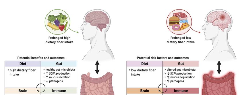
Microorganisms 2021, 9, 2310                                                                                            2 of 19

                               the gut microbiome via the gut-brain axis [13]. The links between microbiome composition
                               and AD are intriguing and provide potential ways to ameliorate or even prevent AD
                               progression through modifying the microbiome. This could be achieved via various ways,
                               including fecal transplant and consumption of probiotics or prebiotics. Prebiotics are
                               oligosaccharide molecules that are non-digestible to the human host, and which serve
                               as substrates for microorganisms in the gut, and thus modulate the composition and/or
                               function of gut microbes in a manner that is beneficial to the host [14,15]. In this review, we
                               discuss the evidence linking gut microbiome composition and function with AD and its
                               associated co-morbidities, provide an overview of prebiotic types and their effects, discuss
                               evidence for the effectiveness of prebiotics in modulating gut microbiome composition
                               and microbial metabolite production, and discuss the potential for prebiotics to induce a
                               beneficial shift in the gut microbiome and modify health outcomes relevant for individuals
                               with AD.

                               2. Links between Gut Microbiome Composition and AD and Associated
                               Co-Morbidities
                                     The importance of diet in modifying the gut microbiome has been emphasized through
                               many intervention studies in humans and animal models. Studies have demonstrated
                               that diet affects gut microbiota composition and diversity [16–25]. Diet composition and
                               duration of intervention are the two most relevant diet-related factors in shaping the
                               gut microbiome. The most well-studied dietary interventions thus far have involved the
                               comparison of high-fat or Western diets enriched in animal-derived foods vs. lower-fat
                               or plant-based diets (Figure 1). From animal studies to human studies the diversity and
                               proportion of microbes have been found to be consistently altered by diets depleted vs.
                               enriched in plant substrate. Specifically, diets depleted in non-digestible fiber and enriched
                               in protein and fat have been consistently linked with an increase in protein- and fat-
                               degrading bacteria belonging to the phyla Firmicutes, Proteobacteria, and Deferribacteres,
                               and a decrease in Bacteroidetes and butyrate-producing species, which are generally
                               known to be beneficial for human health [26–30]. Conversely, fiber-enriched diets are
                               typically associated with increases in the abundance of species in the phylum Bacteroidetes,
                               the genus Prevotella, and Bifidobacterium spp. [31–35]. These changes in gut microbiota
                               composition are closely associated with host health and disease. The health effect is not only
                               attributed to the enrichment of beneficial gut microbes but to the production of secondary
                               metabolites such as short chain fatty acids (SCFAs) from the degradation of non-digestible
                               carbohydrates by specific fiber-fermenting taxa [36–38]. The presence of these taxa is
                               associated with protection from AD, and a number of associated co-morbidities including
                               T2DM and cardiovascular disease (CVD). In the next several paragraphs we review the
                               evidence linking gut microbiome alterations to AD, as well as associated co-morbidities.
                                     Studies have shown a connection between the composition and diversity of gut
                               microbes and AD (Figure 1) [3,39,40]. In a recent study a reduction in overall gut micro-
                               biome richness as well as decreases in Bifidobacterium and Adlercreutzia under Actinobac-
                               teria, SMB53 (family Clostridiaceae), Dialister, Clostridium, Turicibacter, and cc115 (family
                               Erysipelotrichaceae) under Firmicutes were observed in AD participants [3]. On the other
                               hand, Blautia, Phascolarctobacterium, and Gemella under Firmicutes, Bacteroides and Alistipes
                               under Bacteroidetes, and Bilophila under Proteobacteria were increased in AD patients [3].
                               In addition, 13 genera were associated with cerebrospinal fluid (CSF) biomarkers of AD [3],
                               showing that gut microbiome composition or diversity may contribute to AD development.
                               Firmicutes and Bacteroidetes are two dominant phyla in the human gut [41] and it has been
                               observed that the Firmicutes/Bacteroidetes ratio is associated with obesity, gut dysbiosis,
                               and a number of diseases including diabetes and CVD. However, the use of this ratio as an
                               assessment of the health state of the gut microbiota is controversial, as contradictory results
                               have been reported [3,39,42–45]. Gut microbiota composition assessment metrics that are
                               based on measurements at the phylum level are unlikely to be useful since individual
                               genera and species, even strains, under a particular phylum can play opposite roles in
                               overall gut health, taking on different metabolic roles, producing different metabolites, and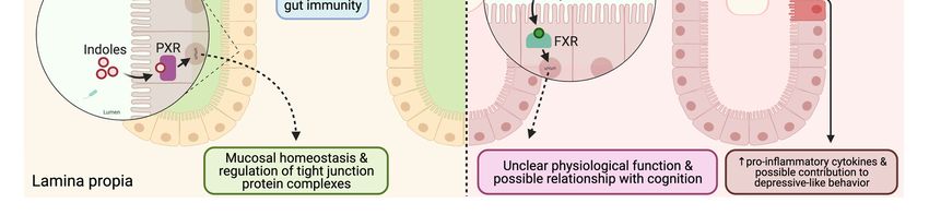
Microorganisms 2021, 9, 2310 3 of 19
Microorganisms 2021, 9, x FOR PEER REVIEW 3 of 20
interacting with other microbes in the gut in different ways such that the overall effect of
all individual species of that phylum is complex (Figure 1).
Figure 1. The potential association of prebiotic-gut-Alzheimer’s Disease (AD) in individuals with prolonged high vs.
Figure 1. The potential association of prebiotic-gut-Alzheimer’s Disease (AD) in individuals with prolonged high vs. low
low fiber diet. The intake of dietary fiber may further influence gut health, immune system, and brain function. High
fiber diet. The intake of dietary fiber may further influence gut health, immune system, and brain function. High dietary
dietary fiber intake may help maintain healthy gut microbiota, which is associated with increase in SCFA production,
fiber intake may help maintain healthy gut microbiota, which is associated with increase in SCFA production, mucus
mucus secretion
secretion and decrease
and decrease in pathogens.
in pathogens. Healthy Healthy gut physiology
gut physiology leads
leads to to regulated
regulated gut immune
gut immune systemsystem and immune
and immune home-
homeostasis
ostasis in which positively affects the brain. Anti-inflammatory metabolites signal the brain and its central immuneimmune
in which positively affects the brain. Anti-inflammatory metabolites signal the brain and its central system,
system,
which may which may potentially
potentially contribute contribute to functional
to functional brain andbrain and prevention
prevention of AD onsetof AD onset or development.
or development. Bacterial
Bacterial genera that
are shown
genera thatto arebeshown
less abundant
to be lessin AD patients
abundant in were
AD patients were Bifidobacterium
Bifidobacterium and Adlercreutzia Adlercreutzia
andunder Actinobacteria, SMB53 (family
under Actinobacteria,
Clostridiaceae),
SMB53 (familyDialister, Clostridium,
Clostridiaceae), Turicibacter,
Dialister, and Turicibacter,
Clostridium, cc115 (familyand
Erysipelotrichaceae) under Firmicutes.under
cc115 (family Erysipelotrichaceae) Low dietary fiber
Firmicutes.
intake
Low may alter
dietary fibergut microbiota
intake may alterleading to dysbiosisleading
gut microbiota in the gut, decrease in
to dysbiosis in SCFA production,
the gut, decrease and increase
in SCFA in pathogens.
production, and
Dysbiosis
increase inin the gut may
pathogens. cause compromised
Dysbiosis in the gut may gut cause
immune system and gut
compromised inflammation in the gut.
immune system and Pro-inflammatory
inflammation in the metab-
gut.
olites signal the brain and its central immune system and potentially bring chronic damage to the brain, which may result
Pro-inflammatory metabolites signal the brain and its central immune system and potentially bring chronic damage to
in dysfunctional brain, AD onset or development. Bacterial genera that are shown to be more abundant in AD patients
the brain, which may result in dysfunctional brain, AD onset or development. Bacterial genera that are shown to be more
were Blautia, Phascolarctobacterium, and Gemella under Firmicutes, Bacteroides and Alistipes under Bacteroidetes, Bilophila
abundant in AD patients were Blautia, Phascolarctobacterium, and Gemella under Firmicutes, Bacteroides and Alistipes under
under Proteobacteria.
Bacteroidetes, Bilophila under Proteobacteria.
Studies have shown a connection between the composition and diversity of gut mi-
The onset and progression of AD has been linked directly to neurodegenerative pro-
crobes and AD (Figure 1) [3,39,40]. In a recent study a reduction in overall gut microbiome
cesses secondary to the deposition of Aβ plaques and aggregation of hyperphosphorylated
richness as well as decreases in Bifidobacterium and Adlercreutzia under Actinobacteria,
tau tangles [46]. Recently, the pathogenesis of AD has been hypothesized further to be trig-
SMB53 (family Clostridiaceae), Dialister, Clostridium, Turicibacter, and cc115 (family Erysip-
gered by amyloid fibers of bacterial origin, which induce a proinflammatory response [47].
elotrichaceae) under Firmicutes were observed in AD participants [3]. On the other hand,
A recent study found that amyloid-positive cognitively impaired patients had higher Es-
Blautia, Phascolarctobacterium, and Gemella under Firmicutes, Bacteroides and Alistipes un-
cherichia/Shigella and lower Eubacterium rectale and Bacteroides fragilis abundances compared
der Bacteroidetes, and Bilophila under Proteobacteria were increased in AD patients [3]. In
to amyloid-negative cognitively normal controls, and these compositional changes were
addition, 13
correlated genera
with were associated
an increased with
production ofcerebrospinal fluidcytokines
pro-inflammatory (CSF) biomarkers of AD [3],
and a reduction of
showing that gut microbiome composition or diversity may contribute to
anti-inflammatory cytokines [48]. In a cross-sectional study in Australian women consump-AD develop-
ment.
tion of Firmicutes and(high
a “junk food” Bacteroidetes arefat)
sugar, high two dominant
diet phyla
was highly in the human
associated with Aβgutdeposition,
[41] and it
has been observed that the Firmicutes/Bacteroidetes ratio is associated
whereas consumption of the Mediterranean diet was associated with higher cognitive with obesity, gut
dysbiosis, and a number of diseases including diabetes and CVD. However, the
scores than other diet groups [49]. Interestingly, in a small study participants with mild use of this
ratio as animpairment
cognitive assessmentconsuming
of the health state of the
a modified gut microbiota is controversial,
Mediterranean-ketogenic as contra-
diet consisting of
dictory results have been reported [3,39,42–45]. Gut microbiota composition
less than 20 g/d of carbohydrate were found to have higher abundances of Enterobacteri- assessment
metrics
aceae, that are based
Akkermansia, on measurements
Slackia, at the
Christensenellaceae phylum
and level are unlikely
Erysipelotrichaceae to beabundances
and lower useful since
individual genera and species, even strains, under a particular phylum
of saccharolytic Bifidobacterium and Lachnobacterium compared to cognitively normalcan play opposite
partic-
roles in overall gut health, taking on different metabolic roles, producing different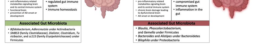
Microorganisms 2021, 9, 2310                                                                                            4 of 19

                               ipants [50]. In a follow-up study the low-carbohydrate modified Mediterranean-ketogenic
                               was found to have a potential beneficial effect in AD patients in preventing memory de-
                               cline [51]. However, these studies were conducted in small cohorts (e.g., 17 individuals,
                               11 MCI patients and 6 controls), thus the effects of low-carbohydrate, low-fiber diets, even
                               in the context of high monounsaturated and polyunsaturated vs. saturated fat ratios such
                               as those seen in the Mediterranean diet, need to be further investigated in larger trials.
                                     T2DM and AD have been known to share several pathophysiological features includ-
                               ing hyperglycemia leading to increased Aβ production, and impaired glucose transport
                               and subsequent glucose metabolism [52]. A new potential AD biomarker, S100B, has been
                               investigated to learn the common pathophysiology of these diseases [53]. A cross-sectional
                               study conducted with 100 South Indian AD patients showed that elevated levels of S100B
                               protein in serum were significantly associated with clinical dementia rating scores com-
                               pared to healthy controls [54]. Serum S100B protein levels in T2DM patients were also
                               shown to be positively correlated with cognitive function [55]. In patients with clinically
                               diagnosed T2DM a high-fiber diet composed of whole grains and prebiotics promoted
                               strain specific growth of acetate and butyrate producing bacteria Faecalibacterium prausnitzii,
                               Lachnospiraceae bacterium, and Bifidobacterium pseudocatenulatum [56]. The treatment
                               group had improved levels of hemoglobin A1c, as well as increased glucagon-like peptide-
                               1 production compared to the control group [56]. These results suggest the high-fiber diet
                               induced gut microbial alteration is correlated with improvement of blood glucose regula-
                               tion in T2DM patients. These findings have important implications for the management of
                               AD due to the high rates of T2DM comorbidity in AD patients.
                                     In addition to a link with T2DM, CVD has also been linked with AD [57,58]. The
                               occlusion of blood vessels that support the deep brain result in silent brain infarcts [59].
                               This type of infarct is shown to be associated with lower cognitive function related to
                               attention, memory, and language [60]. CVD may directly affect poor blood flow to the
                               brain causing cerebrovascular disease [61]. Meta-analyses of prospective cohort studies
                               exploring the association of coronary heart disease with dementia or cognitive impair-
                               ment found that coronary heart disease is associated with an increased risk of dementia
                               or cognitive impairment [62,63]. It is well-established that as much as 80% of the risk
                               for CVD is attributable to diet and lifestyle factors [64–66]. Many human studies have
                               demonstrated an inverse association between the consumption of dietary fiber and the
                               incidence of CVD [67–71]. Patients with primary hypertension showed a high frequency
                               of opportunistic pathogens such as Klebsiella spp., Streptococcus spp., and Parabacteroides
                               merdae, whereas Roseburia spp. and F. prausnitzii which are SCFA-producers were abundant
                               in healthy individuals [72]. Another study found that total and LDL-cholesterol levels were
                               lowered after the consumption of flaxseed fiber [73]. However, although the consump-
                               tion of maize-derived whole grain cereal led to increases in bifidobacteria, no significant
                               changes were observed in serum lipids [74]. Further studies examining the role of dietary
                               fiber and specific increases or decreases of gut microbes as well as their metabolites on
                               CVD endpoints are needed.
                                     Taken together, the overall findings from the published literature suggest that mod-
                               ifying gut microbial composition and diversity toward a profile associated with healthy
                               individuals consuming healthy diets may help attenuate AD progression. Diets and prebi-
                               otic approaches that aim to increase beneficial bacterial species that have been found to be
                               depleted in AD patients such as Bifidobacterium spp., and approaches that aim to decrease
                               the abundance of deleterious bacterial species such as Bilophila may be beneficial for the
                               prevention of AD (Figure 1).

                               3. Overview of Prebiotic Types and Their Roles in Modifying Gut Microbiota
                                    Dietary fibers, which are somewhat difficult to define, can be classified according
                               to their solubility. Insoluble fiber, which does not dissolve in water, passes through the
                               digestive tract providing bulking by absorbing water. Soluble fiber, on the other hand,
                               dissolves in water and is mostly fermented by commensal bacteria residing in the colon
Microorganisms 2021, 9, 2310 5 of 19
and contributing to satiety [75,76]. Although this general categorization of fibers according
to their solubility may be useful, insoluble fibers are fermented to a certain degree and
some soluble fibers may be non-viscous. Recently, the classification of fiber according
to functionality is gaining attention. The functionality depends on the structure and
fermentability of the specific dietary fiber. Thus, types of dietary fiber and subsequent
gut microbial composition, diversity, and richness changes are highly intriguing areas for
further research. It is especially relevant to patients with AD given that particular dietary
fibers may modify the gut microbiome in a beneficial direction, increasing the levels of
metabolites that improve cognitive function and attenuate neurotoxicity [77]. Here, we
have listed a number of dietary fibers with known impacts on enrichment of certain gut
microbes, suggesting their potential as prebiotic supplements for AD patients (Table 1).
Cellulose and hemicellulose are major water-insoluble, non-starch polysaccharides
found in plant cell walls. Cellulose degradation is known to be conducted by Ruminococcus
spp. and Bacteroides spp. producing SCFAs as a byproduct [78–80]. Some species of gut
microbes, including Butyrivibrio spp. Clostridium spp. and Bacteroides spp. are observed to
break down hemicellulose [81]. Lignin is also a water-insoluble, non-starch polysaccharide
that constitutes plant cell walls together with cellulose and hemicellulose, however its
interaction with gut microbes is not well-documented. One study has shown that lignin
supports the prolonged survival of bifidobacteria in an in vitro condition [82]. Resistant
starch, another type of dietary fiber that is water-insoluble is a starch polysaccharide which
is not degradable by the α-amylase enzyme of the host. Resistant starch was shown to
increase the ratio of Firmicutes to Bacteroidetes [92]. At the genus level, Bifidobacterium and
Ruminococcus have been identified to relatively thrive when exposed to resistant starch [83].
Fructan is a polymer of five carbon membered ring fructose molecules, which consists
of several different types depending on the chemical bond. Fructo-oligosaccharide (FOS)
and inulin are major forms of fructan considered as dietary fibers that are capable of being
fermented by multiple members of the gut microbiota community [93]. FOS is a short chain
oligosaccharide of fructose linked by β (2→1) glycosidic bonds. Inulin is a heterogeneous
polysaccharide with β (2→1) linkage and terminal glucose. These fructan molecules have
a bifidogenic effect that enhances the relative abundance of Bifidobacterium spp. in the
host gut [84–86,94]. Similarly, galacto-oligosaccharide (GOS) is a short chain polymer of
mainly galactose linked with a β (1→4) bond and terminal glucose [95]. FOS and GOS are
commercially used to produce infant formula to mimic the properties of human milk [96].
These oligosaccharides are important nutrients to develop the gut microbiome of infants
leading to colonization of beneficial bifidobacteria [97,98]. The promotion of these gut
microbiota in infants decreases the niche for pathogenic bacteria and helps to enhance
gut barrier function [87,99–101]. FOS supplementation in chronically stressed mice was
demonstrated to prevent intestinal barrier impairment and neuroinflammation along with
improved depression-like behavior and significant changes in the abundance of Lactobacillus
reuteri [102]. FOS from Morinda officinalis were also tested in rats with AD-like symptoms
and mice with inflammatory bowel disease showing the potential of FOS as a prebiotic
that improved gut barrier integrity, alleviated neuronal degradation, downregulated AD
markers, and maintained the diversity and stability of the gut microbiome of the host [103].Microorganisms 2021, 9, 2310 6 of 19
Table 1. Types of dietary fibers associated with the growth of certain gut microbiota.
Main Natural/Food Associated
Fiber Types References
Features Sources Gut Microbiota
Ruminococcus spp.
Cellulose Bulking Plant cell wall [78–80]
Bacteroides spp.
Butyrivibrio spp.
Hemicellulose Bulking Plant cell wall Clostridium spp. [81]
Bacteroides spp.
Lignin Bulking Plant cell wall Bifidobacterium spp. [82]
Seeds and
Bifidobacterium spp.
Resistant Starch Fermentable unprocessed whole [83]
Ruminococcus spp.
grains
Fructan
Fructo- Jerusalem artichoke,
oligosaccharide Fermentable chicory, Bifidobacterium spp. [84]
(FOS) and the Blue Agave
Wheat, bananas,
Inulin Fermentable asparagus, Jerusalem Bifidobacterium spp. [85,86]
artichoke, and chicory
Galacto- Enzymatic conversion
Bifidobacterium spp.
oligosaccharide Fermentable of lactose, added in [87]
Lactobacillus spp.
(GOS) infant formula
Bacteroides spp.
Viscous, Bran of cereals such as
β-glucan Prevotella spp. [88,89]
Fermentable oats and barley
Bifidobacterium spp.
Bifidobacterium spp.
Viscous, Pears, apples, berries,
Pectin Lactobacillus spp. [90]
Fermentable and oranges
Enterococcus spp.
Substances that are
Bifidobacterium spp.
Gums (gum Viscous, secreted from plant
Lactobacillus spp. [91]
arabic) Fermentable cells in response to
Bacteroides spp.
injury (gum arabic)
Beta-glucan is a polysaccharide that contains β-D-glucose linked by glycosidic bonds.
A linear, non-branched β-glucan mostly found in the bran of cereals such as oats and
barley is water-soluble and consists of β-D-glucose with (1→3), (1→4)-linkage [104]. This
physicochemical property of β-glucan results in increased viscosity and a thickening effect
on feces, and it provides beneficial, saccharolytic gut microbes with fermentable substrate
to consume [105–107]. Consumption of high molecular weight β-glucan increased the
proportion of Bacteroides and Prevotella [88]. Supplementation of either whole grain oats or
oat bran elevated the production of SCFAs and produced a bifidogenic effect [89].
Pectin is a water-soluble dietary fiber mainly found in the skin of apples. Pectin is a
component of the primary cell wall and middle lamella which contribute to adherence of
adjacent plant cells. The structure of pectin is very complex and the pectic polysaccharides
are abundant in galacturonic acids. Homogalacturonan is a polymer of galacturonic acid
bonded with α-1,4-linkage and the types of pectin may vary according to its side chain
sugars [108]. These complex pectins are known to be degraded by gut microbiota whose
diversity is found to be preserved by pectin in ulcerative colitis patients [109]. Pectins
derived from apples were found to be utilized by beneficial colonic bacteria including
Bifidobacterium, Lactobacillus, Enterococcus, suggesting a prebiotic capacity of pectin [90].
Gums are commonly found in food thickeners because of their capability of gel
formation and emulsion stabilization. Particularly, gum arabic is well determined for
its solubility in water, becoming viscous depending on its concentration. Gum arabic is
a complex heteropolysaccharide mainly containing 1,3-linked β-D-galactose units withderived from apples were found to be utilized by beneficial colonic bacteria including
Bifidobacterium, Lactobacillus, Enterococcus, suggesting a prebiotic capacity of pectin [90].
Gums are commonly found in food thickeners because of their capability of gel for-
Microorganisms 2021, 9, 2310 mation and emulsion stabilization. Particularly, gum arabic is well determined for its sol-
7 of 19
ubility in water, becoming viscous depending on its concentration. Gum arabic is a com-
plex heteropolysaccharide mainly containing 1,3-linked β-D-galactose units with 1,6-
linked β-D-galactose side chains attached to rhamnose, glucuronic acid and arabinose res-
1,6-linked β-D-galactose
idues [110,111]. side chains
It is accessible to the attached to rhamnose,
gut microbes having aglucuronic
potential toacid and arabinose
increase probiotic
residues
bacteria in the human gut. At a dose of 10 g for 4 weeks gum arabic resultedtoinincrease
[110,111]. It is accessible to the gut microbes having a potential signifi-
probiotic bacteria
cantly higher in theof
numbers human gut. At a dose
Bifidobacterium, of 10 g for
Lactobacillus, and4 Bacteroides
weeks gumspp.arabic
in aresulted
human
in trial [91].higher numbers of Bifidobacterium, Lactobacillus, and Bacteroides spp. in a
significantly
clinical
human clinical trial [91].
The structural complexity of dietary fibers and the associated diversity of gut mi-
crobesThe structural
that consume complexity of dietary
them require fibers
further and the
research. It associated
is importantdiversity of gut microbes
to determine the utili-
that consume them require further research. It is important to determine
zation of specific fibers by distinct microbiota and to demonstrate which structural the utilization
traits
of specific fibers by distinct microbiota and to demonstrate which structural traits and/or
and/or components of these fibers affect cognitive function via altering the gut microbi-
components of these fibers affect cognitive function via altering the gut microbiome in
ome in future studies.
future studies.
4. Effectiveness
4. Effectiveness of
of Prebiotics
Prebiotics in
in Modulating
Modulating Gut
Gut Microbiome
Microbiome Composition
Composition and
and Mi-
crobial Metabolite
Microbial MetaboliteProduction
Production
The overall
The overall impact
impact of
of the
the gut
gut microbiome
microbiome on
on the
the production
production of
of microbial
microbial metabolites
metabolites
function is
and gut barrier function is summarized
summarized in
in Figure
Figure 2.
2.
Figure
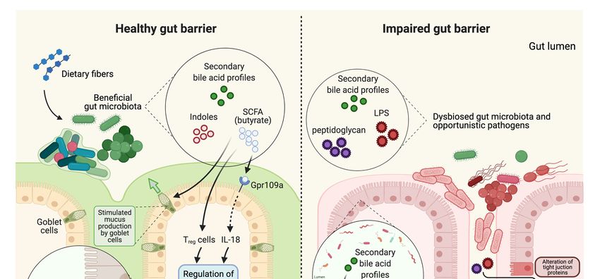
Figure 2.2. Gut
Gut barrier
barrier integrity
integrity changes
changes andand differences
differencesininsignaling
signalingmolecules
moleculesin inhealthy
healthyvs.
vs. unhealthy
unhealthy gut.gut. In
In healthy
healthy gut
gut
barrier,
barrier, dietary fiber from diet is digested by the beneficial gut microbiota which produces secondary metabolites such
dietary fiber from diet is digested by the beneficial gut microbiota which produces secondary metabolites such as
as
SCFA
SCFA(butyrate),
(butyrate),indoles,
indoles,and
andsecondary
secondarybile acid
bile profiles.
acid Butyrate
profiles. is known
Butyrate is known to use Gpr109a
to use as aasreceptor
Gpr109a expressed
a receptor in the
expressed in
enterocyte which
the enterocyte produces
which IL-18,
produces or itormay
IL-18, directly
it may affect
directly T regulatory
affect (Treg
T regulatory (T)regcells. IL-18
) cells. IL-18and
andTreg
Tregcells
cellscan
canboth
bothregulate
regulate
gut immunity.
gut immunity. SCFA
SCFAalsoalsostimulates
stimulatesmucus
mucusproduction
productionby bygoblet
gobletcells
cellsfor
for healthy
healthy mucosal
mucosal barrier.
barrier. Indoles
Indoles are
are ligands
ligands for
for
pregnaneXXreceptor
pregnane receptor(PXR)
(PXR)acting
actingasas transcription
transcriptionfactor
factorinin sustaining
sustaining mucosal
mucosal homeostasis
homeostasisand and regulation
regulationof of tight
tight junction
junction
complexes. Secondary bile acid profiles are ligands for farnesoid X receptor (FXR) and can be found in both healthy and
unhealthy gut. The physiological roles of secondary bile acid profiles are unclear and may have possible relationship with
cognition. In impaired gut barrier, gut microbiota are dysbiosed and byproducts such as peptidoglycan and LPS are released
from opportunistic pathogens. The mucosal barriers are attenuated which provides more close contact of pathogens near
enterocytes altering tight junction proteins. The peptidoglycan and LPS may pass through compromised tight junction
increasing pro-inflammatory cytokines and possibly contributing to depressive-like behavior.Microorganisms 2021, 9, 2310 8 of 19
The fermentation of dietary fiber or prebiotics by gut microbiota and the major metabo-
lites from that process have been elucidated in many studies [112–115]. Particularly, bu-
tyrate is the preferred energy source of apical colonocytes [116]. Furthermore, SCFA lower
the pH of the gut, suppressing the growth of pathogens [117], mediate gut immune reg-
ulation [118], and influence gut motility [119]. Thus, SCFAs act as signaling molecules
that induce downstream pathways modulating the physiology, immunity, and metabolism
of enterocytes. Gpr109a is a type of G protein-coupled receptor specifically activated by
butyrate and is expressed in enterocytes, immune cells, and even in microglia [120–122].
Butyrate binding to the gpr109a receptor triggers several cellular signaling pathways
(Figure 2) including those involving the colonic epithelium, macrophages, and dendritic
cells. For example, Gpr109a signaling is known to promote anti-inflammatory proper-
ties by inducing IL-18 and IL-10 production, which induces differentiation of naïve T
cells to T regulatory cells, thus supporting overall gut immunity by preventing colonic
inflammation [123].
Neurotransmitters are another class of signaling molecule that plays an important
role in the gut-brain axis. Serotonin, for example, is known to be mostly released from
epithelial enterochromaffin cells [124,125]. The gut microbiota play a key role in promoting
serotonin synthesis by host enterochromaffin cells. SCFA or secondary bile acids produced
by gut microbes mediate serotonin production by enterochromaffin cells, which can further
affect gut motility via the enteric nerve and brain serotonergic systems [126,127]. These
findings suggest that certain prebiotic supplements, which stimulate the production of
SCFA and secondary bile acids by specific microbes, can improve neurological function
and behavior via upregulation of serotonin [128]. Another interesting neurotransmitter
that connects gut and brain function is Gamma-aminobutyric acid (GABA). GABA is a
crucial inhibitory neurotransmitter in the central nervous system and its alteration in
GABAergic mechanisms is related to central nervous system disorders [129]. A recent
study demonstrated the link between the gut microbiome (Bacteroides spp.) and GABA
production, a response negatively correlated with depression [130]. Fecal microbiota from
healthy control and schizophrenia patients were compared and each were transplanted to
germ-free mice. Gut microbial dysbiosis shown in schizophrenia was related to changes in
the GABA cycle which, in turn, may affect neurobehavioral status such as schizophrenia-
relevant behaviors [131]. The production of neurotransmitters, particularly serotonin and
GABA was distinctly linked with Bifidobacterium and Lactobacillus genera [132]. These
findings highlight the potential role of prebiotics that promote the composition of these
specific microbes, because their presence has been linked with decreased dysbiosis in the
gut and the production of functional neurotransmitters, which may contribute to enhancing
enteric health and attenuating AD-related neurobehavioral disorders.
In addition to neurotransmitters, prebiotics may also play an important role in reg-
ulating cytokine expression. Soluble fiber (pectin) treatment in mice resulted in faster
recovery from endotoxin-induced sickness behaviors along with changes in the concen-
trations of cytokines, including IL-1RA, IL-4, IL-1β and TNF-α in the brain [133]. The
pectin-supplemented mice also had increased concentrations of cecal acetate, propionate,
and butyrate as a byproduct of pectin fermentation, which was associated with increased
gastrointestinal IL-4 [133]. These findings suggest that soluble fiber not only affects the gas-
trointestinal tract and peripheral immune system but also neuroimmune system function.
In another study in adult and aged mice a high fiber diet with inulin led to increased levels
of cecal SCFA production including butyrate and acetate [134]. A reduction in inflammatory
infiltrate was observed in the aged mice on the high fiber diet, and researchers specifically
showed that sodium butyrate had anti-inflammatory effects on microglial profile, lowering
inflammatory gene expressions [134]. These data suggest that butyrate produced from
prebiotic fermentation may be a potent modulator of gut immune function and directly
linked to microglial function in the brain.
Gut microbiota derived metabolites such as SCFA and indole are critical for sustaining
intestinal barrier function (Figure 2). Acetate and butyrate, for example, improve gobletMicroorganisms 2021, 9, 2310 9 of 19
cell differentiation and stimulate mucus production by goblet cells to maintain healthy
mucosal barrier [135]. Mice fed a low-fiber Western style diet were found to have a
defect in mucin production, which was prevented by supplementation with a synbiotic of
Bifidobacterium longum and inulin [136], suggesting that when SCFA-producing microbes
are present in the gut along with a preferred substrate, the net effect is enhanced mucosal
barrier function. In addition to a decrease in fiber-fermenting microbes and thus SCFA
production, a diet deficient in fiber can also promote the enrichment of mucus-degrading
gut microbes such as Akkermansia muciniphila [137]. Bifidobacterium bifidum, which has
the ability degrade mucin [138] may protect thinning of the mucus layer by inhibiting
Akkermansia muciniphila, as was shown in mice with omeprazole-induced small intestine
injury [139]. Paradoxically, the presence of Akkermansia muciniphila has been linked with
beneficial health effects [140–143], as well as negative health effects in individuals with
certain health conditions [144,145]. The roles of specific microbes and their metabolites in
the maintenance vs. degradation of the mucosal barrier are context-specific and require
further study. Prebiotics may be a useful strategy to prevent mucus degradation by
supporting the growth of SCFA-producing microbes and thus increasing mucin production,
as well as sustaining the homeostasis of mucolytic vs. non-mucolytic bacteria in the gut.
Butyrate is known to regulate the expression of tight junction protein complexes [146].
Sodium butyrate was shown to increase Claudin-1 expression and induced redistribution
of ZO-1 and Occludin in vitro [147]. Butyrate treatment accelerated the assembly of tight
junctions by reorganizing the tight junction proteins in a Caco-2 cell monolayer model [148].
No studies have demonstrated a direct link between butyrate derived from the gut on tight
junctions supporting endothelial cells that form the blood–brain barrier. However, these
findings of a beneficial effect of butyrate on barrier function in the gut epithelium raises
the question of whether a similar benefit may also be found in endothelial cells. A link
between butyrate and brain function has been suggested. Bourassa et al. hypothesized that
butyrate could be used as an important alternative energy substrate in the Alzheimer’s
brain where glucose utilization has been found to be reduced [149–151].
Indoles are a class of molecules produced by gut microbes that have the potential to
affect gut and brain function. In a germ-free mouse model, oral administration of indole
led to up-regulation of tight and adherens junction-associated molecules in the epithelial
cells of the colon [152]. Indole 3-propionic acid acts as a ligand for pregnane X receptor
and increased expression of junctional protein-coding mRNAs while decreasing TNF-α
in a mouse model [153]. The effect of indole 3-propionic acid was also tested in the Caco-
2/HT29 coculture model and showed an increase in tight junction proteins, mucins, and
goblet cell secretion products [154]. However, the role of indole and its derivatives is
controversial in terms of the gut-brain axis [155,156]. Studies have demonstrated potent
neuroprotective properties of indoles, which cross the blood–brain barrier and protect
the brain from oxidative stress [157] as well as prevent electron leakage from neuronal
mitochondria [158,159]. However, other studies report excessive production of indole by
gut microbes may negatively affect emotional behavior in rats due to the neurodepressive
properties of oxidized derivatives of indole, oxindole and isatin [160]. Indoxyl sulphate,
an oxidized and sulphated form of indole produced from the liver, may reduce the efflux
of neurotransmitters through the organic anion transporter 3, causing accumulation of
metabolites [161,162]. Thus, the effects of indoles on gut barrier and brain function require
further study, as the variety of indole metabolites produced by the gut microbes and their
co-metabolism by the host generate a complex suite of molecules with differential effects.
Bile acids are a category of metabolite that is modulated by gut microbial metabolism,
and which may have effects on the gut-brain axis. Bile acids are produced in hepatocytes
and play a critical role in fat digestion and absorption. Most (95%) bile acids are recycled
back to the liver via enterohepatic recirculation after reaching the terminal ileum. How-
ever, bile acids that are not recycled are excreted in feces or may be metabolized by the
colonic microbiota, forming secondary bile acids via a series of microbial enzyme activities
including deconjugation and 7α-dehydroxylation [163]. Thus, secondary bile acids areMicroorganisms 2021, 9, 2310 10 of 19
gut microbe-derived metabolites that may further regulate bile acid signaling of the host,
affecting the activation of the enteroendocrine bile acid receptor, farnesoid X receptor
(Figure 2) [164]. Several papers have shown a connection between bile acid metabolism
and AD. In AD patients, significantly lower serum concentrations of a primary bile acid
(cholic acid) and increased secondary bile acid (deoxycholic acid) were observed compared
to cognitively normal older adults [165]. Increased deoxycholic acid to cholic acid ratio
is known to be strongly associated with cognitive decline [166]. The ratio of primary to
secondary bile acids was positively correlated with the abundance of Bifidobacterium in a
human clinical trial [167]. Recently, alteration in bile acid profiles was shown to have an
association with cognitive decline and AD-related genetic variants [165].
There are likely hundreds if not thousands of microbially produced molecules that
likely play important roles in host health. Among these, butyrate, indole, and bile acids,
are to date, the most well-studied, and their roles in gut health, brain function, and specific
roles in the pathophysiology of AD, are starting to emerge. As we gain knowledge on both
short-term and long-term effects of diet on the brain mediated by the gut microbiome, it
will be important to establish a dossier of evidence of benefit of specific prebiotics for the
pathophysiology of AD. In the following section, we discuss potential prebiotic approaches
to supplement AD patients.
5. Current Evidence for Effectiveness of Prebiotics in AD Animal Models and
Human Trials
The effectiveness of prebiotics for the treatment of AD will ultimately need to be eval-
uated on the basis of their ability to either improve or prevent cognitive decline. However,
other symptoms of AD related to behavioral and emotional changes are also viable targets
of prebiotic intervention studies in AD patients. The current literature showing the poten-
tial effects of prebiotics on cognitive function in both animal models and human studies
mainly focuses on the effects of fructans, both in the form of oligosaccharides and inulin,
β-glucan from yeast or the bran of cereals, plant polysaccharides, and polysaccharides
synthesized from sugars. This evidence is summarized below.
5.1. Animal Models
Animal models have been used in several studies to evaluate the effect of prebiotics
on AD, particularly mice due to their reliability on intervention and ease of sampling. In
this section, animal studies on administration of prebiotics that led to improvement in
AD associated brain disorders are summarized. Bimuno-GOS intake in pregnant mice
affected the offspring’s exploratory behavior and brain gene expression as well as re-
ducing anxiety [168]. Additionally, fecal butyrate and propionate levels were increased
after Bimuno-GOS supplementation in postnatal mice [168]. In another study, behavioral
testing was performed on mice from the least stressful (three-chamber test) to the most
stressful (forced swim test) for 5 weeks during a 10-week prebiotic administration period
including lead-in and lead-out periods [169]. The prebiotic treatment with a FOS+GOS
combination resulted in a reduction of stress-related (depression and anxiety) behaviors,
and reversed chronic stress (elevations in corticosterone and proinflammatory cytokine
levels) in the supplemented mice compared to the control mice with no prebiotic treat-
ment [169]. In a rat model exhibiting oxidative stress, mitochondrial dysfunction, and
cognitive decline in the brain induced by high fat diet-induced obesity these outcomes were
improved and cognitive function was restored by 12-week supplementation of either prebi-
otic (xylo-oligosaccharide), probiotic (Lactobacillus paracasei HII01), or combined treatment
with similar efficacy [170]. The effectiveness of mannan-oligosaccharide was tested in a
5xFamilial AD transgenic mouse model [171]. The treatment with mannan-oligosaccharide
reduced Aβ accumulation in the brain and suppressed neuroinflammatory responses [171].
Mannan-oligosaccharide not only improved cognitive and behavioral disorders, but also
gut barrier integrity by reshaping the composition of gut microbiota, specifically increases in
the relative abundances of Lactobacillus and decreases in Helicobacter [171]. Importantly, theMicroorganisms 2021, 9, 2310 11 of 19
observed changes in gut microbiota composition and butyrate production were negatively
correlated with oxidative stress in the brain and behavioral deficits [171].
5.2. Human Trials
Studies on the effects of prebiotic supplementation directly on cognitive and behav-
ioral outcomes in Alzheimer’s patients are currently lacking. However, a few human
intervention studies were conducted to test the effectiveness of certain prebiotics alone or
with probiotics on improving symptoms associated with AD such as behavioral, mood,
memory, anxiety, and cognitive disorders.
Fructan and GOS-based prebiotics show promising and consistent results in clinical
trials in decreasing anxiety and improving cognitive and behavioral outcomes. The pre-
biotic Bimuno-GOS improved antisocial behaviors in autistic children [172]. Trans-GOS
stimulated bifidobacteria in the gut of irritable bowel syndrome patients and lowered anxi-
ety [173]. Short chain FOS enhanced fecal bifidobacteria and reduced anxiety scores [174].
Inulin in healthy participants resulted in better recognition and improved recall [175].
In obese patients adhering to calorie restrictions for 3 months supplementation with 16
g/d of inulin had moderate impact on mood and cognition, with responders who ex-
perienced an increase in Coprococcus and Bifidobacterium having stronger benefits than
non-responders [176]. Importantly, in most of these intervention studies, subjects supple-
mented with fructan or GOS prebiotics showed increases in bifidobacteria in general along
with improvement in their symptoms. Many studies have already reported the connection
between the increase in bifidobacteria and beneficial health outcomes (Table 1). Indeed,
the growth of bifidobacteria is selectively stimulated by fructans [177]. The increase in Bifi-
dobacterium longum 1714 strain in healthy mice showed stress resistance and pro-cognitive
effects [178,179]. The same Bifidobacterium strain from this preclinical study displayed asso-
ciation with reduction in stress and improvement in memory in healthy volunteers [180].
The results from these studies suggest a strong connection between prebiotics, the gut
microbiome, particularly bifidobacteria, and brain function.
Other studies provide supporting evidence that prebiotics modulate brain function
in a manner that would be consistent with desired improvements in symptoms of AD
but were not necessarily linked with or did not examine gut microbiome composition.
Beta-glucans from yeasts, plants or cereals have been shown to have beneficial health
effects on the profile of mood state in healthy individuals [181,182]. Plant polysaccharides,
which mainly consist of non-starch polysaccharides found in foods were shown to have
effect on healthy adults, improving their recognition and memory performance [183,184].
Polydextrose, which is a synthesized prebiotic, was supplemented in healthy females and
showed moderate improvement in cognition as well as significant change in abundance of
Ruminiclostridium 5 compared to the placebo group [185]. Other studies have found 30–60
mL of lactulose for 3 months improved cognitive function and health-related quality of life
in patients with minimal hepatic encephalopathy [186].
6. Concluding Remarks
Although human clinical studies examining the effects of specific prebiotics on gut
microbiome-mediated cognitive health outcomes in AD patients are lacking, there is mount-
ing evidence that prebiotics have the potential to be a viable approach for ameliorating
symptoms associated with AD. Promoting the growth and activity of beneficial, SCFA-
producing microbes such as bifidobacteria is emerging as a clear therapeutic target for
improving gut barrier function, decreasing inflammation, and improving cognitive and
behavioral outcomes. A variety of prebiotic types, particularly fructans, have been found
to be effective in modulating gut microbiome composition and microbial metabolite pro-
duction, and modifying health outcomes relevant for individuals with AD. More research
is needed to determine which prebiotics, at what dosages, and in which context (e.g., on
what dietary background, in combination with specific probiotics, at what frequency, etc.)
are the most effective for not only decreasing AD-associated symptoms such as anxiety andMicroorganisms 2021, 9, 2310 12 of 19
depression, but also potentially improving cognition or preventing the loss of cognitive
function in individuals at risk for AD. Further mechanistic research to determine how
changes in the gut microbiome related to prebiotic supplementation alter neuroinflamma-
tory signaling are also needed so that targeted, effective, potentially personalized therapies
can be developed to treat and prevent the progression of neurodegenerative processes
in AD.
Author Contributions: Conceptualization, J.W.K. and A.M.Z.; writing—original draft prepara-
tion, J.W.K.; writing—review and editing, J.W.K. and A.M.Z.; visualization, J.W.K.; supervision,
A.M.Z.; funding acquisition, A.M.Z. All authors have read and agreed to the published version of
the manuscript.
Funding: This research and APC was funded by the Jastro Shields fellowship and the USDA National
Institute of Food and Agriculture, Hatch project CA-D-NUT-2242-H.
Acknowledgments: All figures were created with BioRender.com (accessed on 2 November 2021).
Conflicts of Interest: The authors declare no conflict of interest.
References
1. Turnbaugh, P.J.; Ley, R.E.; Mahowald, M.A.; Magrini, V.; Mardis, E.R.; Gordon, J.I. An Obesity-Associated Gut Microbiome with
Increased Capacity for Energy Harvest. Nature 2006, 444, 1027–1031. [CrossRef]
2. Forslund, K.; Hildebrand, F.; Nielsen, T.; Falony, G.; Le Chatelier, E.; Sunagawa, S.; Prifti, E.; Vieira-Silva, S.; Gudmundsdottir, V.;
Krogh Pedersen, H.; et al. Disentangling Type 2 Diabetes and Metformin Treatment Signatures in the Human Gut Microbiota.
Nature 2015, 528, 262–266. [CrossRef]
3. Vogt, N.M.; Kerby, R.L.; Dill-McFarland, K.A.; Harding, S.J.; Merluzzi, A.P.; Johnson, S.C.; Carlsson, C.M.; Asthana, S.; Zetterberg,
H.; Blennow, K.; et al. Gut Microbiome Alterations in Alzheimer’s Disease. Sci. Rep. 2017, 7, 13537. [CrossRef]
4. Qiu, C.; Kivipelto, M.; von Strauss, E. Epidemiology of Alzheimer’s Disease: Occurrence, Determinants, and Strategies toward
Intervention. Dialogues Clin. Neurosci. 2009, 11, 111–128.
5. Selkoe, D.J. Alzheimer’s Disease: Genes, Proteins, and Therapy. Physiol. Rev. 2001, 81, 741–766. [CrossRef]
6. Mahley, R.W.; Weisgraber, K.H.; Huang, Y. Apolipoprotein E4: A Causative Factor and Therapeutic Target in Neuropathology,
Including Alzheimer’s Disease. Proc. Natl. Acad. Sci. USA 2006, 103, 5644–5651. [CrossRef] [PubMed]
7. Strittmatter, W.J.; Saunders, A.M.; Schmechel, D.; Pericak-Vance, M.; Enghild, J.; Salvesen, G.S.; Roses, A.D. Apolipoprotein E:
High-Avidity Binding to Beta-Amyloid and Increased Frequency of Type 4 Allele in Late-Onset Familial Alzheimer Disease. Proc.
Natl. Acad. Sci. USA 1993, 90, 1977–1981. [CrossRef]
8. Baumgart, M.; Snyder, H.M.; Carrillo, M.C.; Fazio, S.; Kim, H.; Johns, H. Summary of the Evidence on Modifiable Risk Factors for
Cognitive Decline and Dementia: A Population-Based Perspective. Alzheimer’s Dement. 2015, 11, 718–726. [CrossRef] [PubMed]
9. Glenner, G.G.; Wong, C.W. Alzheimer’s Disease: Initial Report of the Purification and Characterization of a Novel Cerebrovascular
Amyloid Protein. Biochem. Biophys. Res. Commun. 1984, 120, 885–890. [CrossRef]
10. Wischik, C.M.; Novak, M.; Edwards, P.C.; Klug, A.; Tichelaar, W.; Crowther, R.A. Structural Characterization of the Core of the
Paired Helical Filament of Alzheimer Disease. Proc. Natl. Acad. Sci. USA 1988, 85, 4884–4888. [CrossRef]
11. Sochocka, M.; Diniz, B.S.; Leszek, J. Inflammatory Response in the CNS: Friend or Foe? Mol. Neurobiol. 2017, 54, 8071–8089.
[CrossRef] [PubMed]
12. Tohidpour, A.; Morgun, A.V.; Boitsova, E.B.; Malinovskaya, N.A.; Martynova, G.P.; Khilazheva, E.D.; Kopylevich, N.V.; Gertsog,
G.E.; Salmina, A.B. Neuroinflammation and Infection: Molecular Mechanisms Associated with Dysfunction of Neurovascular
Unit. Front. Cell. Infect. Microbiol. 2017, 7, 276. [CrossRef] [PubMed]
13. Carabotti, M.; Scirocco, A.; Maselli, M.A.; Severi, C. The Gut-Brain Axis: Interactions between Enteric Microbiota, Central and
Enteric Nervous Systems. Ann. Gastroenterol. 2015, 28, 203–209. [PubMed]
14. Bindels, L.B.; Delzenne, N.M.; Cani, P.D.; Walter, J. Towards a More Comprehensive Concept for Prebiotics. Nat. Rev. Gastroenterol.
Hepatol. 2015, 12, 303–310. [CrossRef]
15. Carlson, J.L.; Erickson, J.M.; Lloyd, B.B.; Slavin, J.L. Health Effects and Sources of Prebiotic Dietary Fiber. Curr. Dev. Nutr. 2018, 2,
nzy005. [CrossRef] [PubMed]
16. Filippo, C.D.; Cavalieri, D.; Paola, M.D.; Ramazzotti, M.; Poullet, J.B.; Massart, S.; Collini, S.; Pieraccini, G.; Lionetti, P. Impact of
Diet in Shaping Gut Microbiota Revealed by a Comparative Study in Children from Europe and Rural Africa. Proc. Natl. Acad.
Sci. USA 2010, 107, 14691–14696. [CrossRef] [PubMed]
17. Claesson, M.J.; Jeffery, I.B.; Conde, S.; Power, S.E.; O’Connor, E.M.; Cusack, S.; Harris, H.M.B.; Coakley, M.; Lakshminarayanan,
B.; O’Sullivan, O.; et al. Gut Microbiota Composition Correlates with Diet and Health in the Elderly. Nature 2012, 488, 178–184.
[CrossRef]Microorganisms 2021, 9, 2310 13 of 19
18. Kashyap, P.C.; Marcobal, A.; Ursell, L.K.; Larauche, M.; Duboc, H.; Earle, K.A.; Sonnenburg, E.D.; Ferreyra, J.A.; Higginbottom,
S.K.; Million, M.; et al. Complex Interactions Among Diet, Gastrointestinal Transit, and Gut Microbiota in Humanized Mice.
Gastroenterology 2013, 144, 967–977. [CrossRef] [PubMed]
19. Parks, B.W.; Nam, E.; Org, E.; Kostem, E.; Norheim, F.; Hui, S.T.; Pan, C.; Civelek, M.; Rau, C.D.; Bennett, B.J.; et al. Genetic
Control of Obesity and Gut Microbiota Composition in Response to High-Fat, High-Sucrose Diet in Mice. Cell Metab. 2013, 17,
141–152. [CrossRef]
20. Daniel, H.; Gholami, A.M.; Berry, D.; Desmarchelier, C.; Hahne, H.; Loh, G.; Mondot, S.; Lepage, P.; Rothballer, M.; Walker, A.;
et al. High-Fat Diet Alters Gut Microbiota Physiology in Mice. ISME J. 2014, 8, 295–308. [CrossRef] [PubMed]
21. Carmody, R.N.; Gerber, G.K.; Luevano, J.M.; Gatti, D.M.; Somes, L.; Svenson, K.L.; Turnbaugh, P.J. Diet Dominates Host Genotype
in Shaping the Murine Gut Microbiota. Cell Host Microbe 2015, 17, 72–84. [CrossRef] [PubMed]
22. Filippis, F.D.; Pellegrini, N.; Vannini, L.; Jeffery, I.B.; Storia, A.L.; Laghi, L.; Serrazanetti, D.I.; Cagno, R.D.; Ferrocino, I.; Lazzi, C.;
et al. High-Level Adherence to a Mediterranean Diet Beneficially Impacts the Gut Microbiota and Associated Metabolome. Gut
2016, 65, 1812–1821. [CrossRef] [PubMed]
23. Vaughn, A.C.; Cooper, E.M.; DiLorenzo, P.M.; O’Loughlin, L.J.; Konkel, M.E.; Peters, J.H.; Hajnal, A.; Sen, T.; Lee, S.H.; de La
Serre, C.B.; et al. Energy-Dense Diet Triggers Changes in Gut Microbiota, Reorganization of Gut-Brain Vagal Communication and
Increases Body Fat Accumulation. Acta Neurobiol. Exp. 2017, 77, 18–30. [CrossRef]
24. Zou, J.; Chassaing, B.; Singh, V.; Pellizzon, M.; Ricci, M.; Fythe, M.D.; Kumar, M.V.; Gewirtz, A.T. Fiber-Mediated Nourishment of
Gut Microbiota Protects against Diet-Induced Obesity by Restoring IL-22-Mediated Colonic Health. Cell Host Microbe 2018, 23,
41–53.e4. [CrossRef]
25. Beilharz, J.E.; Kaakoush, N.O.; Maniam, J.; Morris, M.J. Cafeteria Diet and Probiotic Therapy: Cross Talk among Memory,
Neuroplasticity, Serotonin Receptors and Gut Microbiota in the Rat. Mol. Psychiatry 2018, 23, 351–361. [CrossRef] [PubMed]
26. Hildebrandt, M.A.; Hoffmann, C.; Sherrill-Mix, S.A.; Keilbaugh, S.A.; Hamady, M.; Chen, Y.; Knight, R.; Ahima, R.S.; Bushman, F.;
Wu, G.D. High-Fat Diet Determines the Composition of the Murine Gut Microbiome Independently of Obesity. Gastroenterology
2009, 137, 1716–1724.e2. [CrossRef] [PubMed]
27. Walker, A.; Pfitzner, B.; Neschen, S.; Kahle, M.; Harir, M.; Lucio, M.; Moritz, F.; Tziotis, D.; Witting, M.; Rothballer, M.; et al.
Distinct Signatures of Host–Microbial Meta-Metabolome and Gut Microbiome in Two C57BL/6 Strains under High-Fat Diet.
ISME J. 2014, 8, 2380–2396. [CrossRef] [PubMed]
28. Kim, K.-A.; Gu, W.; Lee, I.-A.; Joh, E.-H.; Kim, D.-H. High Fat Diet-Induced Gut Microbiota Exacerbates Inflammation and
Obesity in Mice via the TLR4 Signaling Pathway. PLoS ONE 2012, 7, e47713. [CrossRef] [PubMed]
29. de La Serre, C.B.; Ellis, C.L.; Lee, J.; Hartman, A.L.; Rutledge, J.C.; Raybould, H.E. Propensity to High-Fat Diet-Induced Obesity
in Rats Is Associated with Changes in the Gut Microbiota and Gut Inflammation. Am. J. Physiol.-Gastrointest. Liver Physiol. 2010,
299, G440–G448. [CrossRef]
30. Lecomte, V.; Kaakoush, N.O.; Maloney, C.A.; Raipuria, M.; Huinao, K.D.; Mitchell, H.M.; Morris, M.J. Changes in Gut Microbiota
in Rats Fed a High Fat Diet Correlate with Obesity-Associated Metabolic Parameters. PLoS ONE 2015, 10, e0126931. [CrossRef]
31. Kovatcheva-Datchary, P.; Nilsson, A.; Akrami, R.; Lee, Y.S.; De Vadder, F.; Arora, T.; Hallen, A.; Martens, E.; Björck, I.; Bäckhed, F.
Dietary Fiber-Induced Improvement in Glucose Metabolism Is Associated with Increased Abundance of Prevotella. Cell Metab.
2015, 22, 971–982. [CrossRef]
32. Wu, G.D.; Chen, J.; Hoffmann, C.; Bittinger, K.; Chen, Y.-Y.; Keilbaugh, S.A.; Bewtra, M.; Knights, D.; Walters, W.A.; Knight, R.;
et al. Linking Long-Term Dietary Patterns with Gut Microbial Enterotypes. Science 2011, 334, 105–108. [CrossRef] [PubMed]
33. So, D.; Whelan, K.; Rossi, M.; Morrison, M.; Holtmann, G.; Kelly, J.T.; Shanahan, E.R.; Staudacher, H.M.; Campbell, K.L. Dietary
Fiber Intervention on Gut Microbiota Composition in Healthy Adults: A Systematic Review and Meta-Analysis. Am. J. Clin. Nutr.
2018, 107, 965–983. [CrossRef]
34. Bibbò, S.; Ianiro, G.; Giorgio, V.; Scaldaferri, F.; Masucci, L.; Gasbarrini, A.; Cammarota, G. The Role of Diet on Gut Microbiota
Composition. Eur. Rev. Med. Pharmacol. Sci. 2016, 20, 4742–4749. [PubMed]
35. Jefferson, A.; Adolphus, K. The Effects of Intact Cereal Grain Fibers, Including Wheat Bran on the Gut Microbiota Composition of
Healthy Adults: A Systematic Review. Front. Nutr. 2019, 6, 33. [CrossRef]
36. Holscher, H.D. Dietary Fiber and Prebiotics and the Gastrointestinal Microbiota. Gut Microbes 2017, 8, 172–184. [CrossRef]
[PubMed]
37. Ramirez-Farias, C.; Slezak, K.; Fuller, Z.; Duncan, A.; Holtrop, G.; Louis, P. Effect of Inulin on the Human Gut Microbiota:
Stimulation of Bifidobacterium Adolescentis and Faecalibacterium Prausnitzii. Br. J. Nutr. 2008, 101, 541–550. [CrossRef]
[PubMed]
38. De Vuyst, L.; Leroy, F. Cross-Feeding between Bifidobacteria and Butyrate-Producing Colon Bacteria Explains Bifdobacterial
Competitiveness, Butyrate Production, and Gas Production. Int. J. Food Microbiol. 2011, 149, 73–80. [CrossRef] [PubMed]
39. Brandscheid, C.; Schuck, F.; Reinhardt, S.; Schäfer, K.-H.; Pietrzik, C.U.; Grimm, M.; Hartmann, T.; Schwiertz, A.; Endres, K.
Altered Gut Microbiome Composition and Tryptic Activity of the 5xFAD Alzheimer’s Mouse Model. J. Alzheimer’s Dis. 2017, 56,
775–788. [CrossRef] [PubMed]
40. Wu, S.-C.; Cao, Z.-S.; Chang, K.-M.; Juang, J.-L. Intestinal Microbial Dysbiosis Aggravates the Progression of Alzheimer’s Disease
in Drosophila. Nat. Commun. 2017, 8, 24. [CrossRef]You can also read


