The orthopedic characterization of cfap298tm304 mutants validate zebrafish to faithfully model human AIS
←
→
Page content transcription
If your browser does not render page correctly, please read the page content below
www.nature.com/scientificreports
OPEN The orthopedic characterization
of cfap298tm304 mutants validate
zebrafish to faithfully model
human AIS
Laura Marie‑Hardy 1*, Yasmine Cantaut‑Belarif2, Raphaël Pietton1, Lotfi Slimani3 &
Hugues Pascal‑Moussellard1,2
Cerebrospinal fluid (CSF) circulation relies on the beating of motile cilia projecting in the lumen of the
brain and spinal cord cavities Mutations in genes involved in cilia motility disturb cerebrospinal fluid
circulation and result in scoliosis-like deformities of the spine in juvenile zebrafish. However, these
defects in spine alignment have not been validated with clinical criteria used to diagnose adolescent
idiopathic scoliosis (AIS). The aim of this study was to describe, using orthopaedic criteria the
spinal deformities of a zebrafish mutant model of AIS targeting a gene involved in cilia polarity and
motility, cfap298tm304. The zebrafish mutant line cfap298tm304, exhibiting alteration of CSF flow due
to defective cilia motility, was raised to the juvenile stage. The analysis of mutant animals was based
on micro-computed tomography (micro-CT), which was conducted in a QUANTUM FX CALIPER, with
a 59 µm-30 mm protocol. 63% of the cfap298tm304 zebrafish analyzed presented a three-dimensional
deformity of the spine, that was evolutive during the juvenile phase, more frequent in females, with a
right convexity, a rotational component and involving at least one dislocation. We confirm here that
cfap298tm304 scoliotic individuals display a typical AIS phenotype, with orthopedic criteria mirroring
patient’s diagnosis.
Zebrafish is a frequently used animal model in genetic and could be used to investigate
AIS. In human, adolescent idiopathic scoliosis (AIS) is by far the most common form of non-congenital
scoliosis seen in practice, occurring in the absence of associated p athologies1–3. It is the most frequent muscu-
loskeletal disorder occurring in 1–3% of the population, arising in childhood and worsening before the peak
of growth during adolescence. If the frequency of AIS is higher in female patients (80%), no major differences
in the curves pattern has been demonstrated yet4,5. If untreated, AIS can lead to chronic low back pain, pulmo-
nary restrictive syndrome and severe disabilities along life2,6–10. While bracing is an efficient way to contain the
progression of spinal curves, surgical correction may be needed to correct severe and resistant curves. Thus, a
clear understanding of patients’ physiopathology is essential to appreciate the clinical behavior and may help to
prevent the disease by identifying the progression factors and then individually adapt treatments.
Among genetic model organisms helping to understand AIS pathogenicity and etiology, zebrafish has recently
emerged as an advantageous animal m odel11–13. A biomechanical study in finite elements recently showed that
the zebrafish spine is a relevant model of human spine d eformation14. While scoliosis occured rarely in quadru-
pedal animals, the longitudinal shape of zebrafish coupled with their ability to experience spinal loads in water
make them naturally sensitive to three-dimensional spine deformities, which they even naturally develop with
umans15,16. Recent studies have demonstrated a link between spinal curves and
elderliness, highly similarly to h
cilia motility involved in cerebrospinal fluid (CSF) c irculation11–13,17,18, as mutants defective in cilia motility
develop AIS phenotypes. A cilium-linked physiopathology in AIS patients is highly suspected, as mutations
related to cilium has been found in AIS cohorts (POC5, PAX1) and due to the correlation between CSF-flow
1
Orthopedic Surgery and Trauma Center, Pitié-Salpêtrière Teaching Hospital, 47 Boulevard de l’Hôpital,
75013 Paris, France. 2Paris Brain Institute, ICM, Inserm U 1127, CNRS UMR 7225, Sorbonne Université, 75013 Paris,
France. 3EA 2496 Laboratory Orofacial Pathologies, Imaging and Biotherapies, Dental School University Paris
Descartes Sorbonne Paris Cité, and Life Imaging Platform (PIV), Montrouge, France. *email: laura.marie-hardy@
aphp.fr
Scientific Reports | (2021) 11:7392 | https://doi.org/10.1038/s41598-021-86856-1 1
Vol.:(0123456789)
www.nature.com/scientificreports/
atients19–22. Cfap298 is a gene involved in cilium
and AIS illustrated by the higher prevalence of scoliosis in AIS p
dysfunction, that has been investigated on a Zebrafish model, concluding to spine deformities mimicking AIS,
as well as ptk7 mutants for example11–13,23. However, in these studies, the orthopedic characterizations of spinal
deformities are sometimes lacking analysis through the eyes of clinicians used to analyze spine deformities,
scoliosis, among others.
Indeed, although zebrafish are susceptible to develop spinal curvatures naturally or upon targeted gene muta-
tions, it is not clear whether these AIS animal models faithfully recapitulates the attributes of human pathologies
of the spine. Here, using micro-computed tomography and clinical criteria, we characterized spine deformities
developed in the zebrafish mutant line cfap298tm304 that affects cilia motility and polarity to confirm the link
between these deformities and A IS12,24,25.
Methods
A genetically modified zebrafish for cilium related gene and exhibiting spine deformities was
investigating in micro‑CT. Animal husbandry. All procedures were performed on juvenile and adult
zebrafish in accordance with the European Communities Council Directive (2010/63/EU) and French law
(87/848) and approved by the Paris Brain Institute (Institut du Cerveau). An approval agreement was obtained
from the French ethic committee for experimentation on juvenile and adult zebrafish (APAFIS agreement num-
ber 2018071217081175). All experiments were performed on Danio rerio of AB, Tüpfel long fin (TL) and nacre
background. The study was carried out in compliance with the ARRIVE guidelines. Animals were obtained from
a natural mating and were raised under a 14/10 light/dark cycle.
This study did not involve human subjects. All methods were carried out in accordance with relevant guide-
lines and regulations.
Induction of cilia motility defects and spine deformities. 27 sibling animals were obtained from the natural
mating of a cfaptm304/+ male and a cfap298tm304/tm304 female24,25, resulting in the progeny in 50% of homozygous
mutant animals (n = 17 animals analyzed) and 50% of heterozygous siblings (n = 10 animals analyzed) . As the
cfap298tm304 mutation is thermosensitive, the induction of cilia motility defects was based on a temperature shift
as described in a previous work12. Animals were first raised at 25 °C until 6 days post-fertilization (dpf) to allow
normal embryonic development and then switched at 30 °C from 18 to 23 dpf as described in a previous work12.
Homozygous mutant animals subjected to this restrictive temperature (30 °C) developed defects in spine align-
ment as previously reported. Heterozygous animals were subjected to the same temperature shift and used as
control siblings.
Scannographic analysis. Juvenile zebrafish were imaged at 8 and 12 weeks old under general anesthesia using
0.02% MS-222 (Sigma). Micro-Computed Tomography (micro-CT) was performed using a QUANTUM FX
CALIPER, with a 59 µm – 30 mm protocol. The DICOM images were analyzed with RADIANT DICOM
VIEWER 5.0.0 software. All scannographic analyses were performed on three-dimensional reconstructions,
oriented in sagittal and coronal planes as described i n26 and were performed by an orthopedic surgeon to avoid
bias. The thoracic and lumbar segments of the zebrafish’s spines were defined according to the sagittal alignment:
zebrafishes present one long thoracic kyphosis followed by a lumbar lordosis. The classical criteria for scoliosis
characterization taken into account in this study were the number of spine curves, the side of the main curve,
the Cobb angles, the number of dislocations, the Lenke classification27, apical vertebral rotation (AVR) and api-
ces of the curves. The Lenke classification was applied to the zebrafish’s spines considering the main curves and
the contra-curves (I: main thoracic, II: double thoracic, III: double major, IV: triple major, V: thoraco-lumbar/
lumbar and VI: thoraco-lumbar/lumbar/main thoracic), without the sagittal modifier rules. Dislocations were
analyzed in 3D reconstruction, as known to be more precise than 2D measurements and defined by an AVR > 10°
associated to a lateral listhesis on the frontal plane between two adjacent v ertebras28. They were measured from
frontal reconstructions as described in Marie-Hardy et al.26.
Statistical analysis. All values are represented as histogram distributions or mean ± SEM (stated for each in the
figure legend). Differences were analyzed with two-tailed-t-tests. Significance was set at 0.05. Statistical details
related to sample size in each group and p-values are reported in the figures and figure legends. Asterisks denote
the statistical significance: *p < 0.05; **p < 0.01; ***p < 0.001; ns, p > 0.05.
Results
63% of the zebrafish cohort exhibits a three‑dimensional spinal deformity of the spine with
right convexity, similar to AIS. cfap298tm304 zebrafish carries a temperature sensitive mutation in the cilia
motility gene cfap298, where cilia beating can be inactivated in a temporally controlled manner to alter CSF
flow in brain and spinal cord c avities12. Shifting the environmental temperature of the animals from permissive
(25 °C) to restrictive (30 °C) results leads to the development of three-dimensional spinal deformity of the spine
during growth12. While the zebrafish appears to be an effective model for exploiting the pathogenicity hypoth-
eses underlying the development of AIS, its usefulness for orthopaedic research lacks validation with the clinical
criteria used for the diagnosis of human patients.
Here, we took advantage of the cfap298tm304 mutation to characterize spinal deformities on the basis of
orthopedic criteria and to fully validate this AIS model. We performed micro-CT imaging on a cohort of 17
homozygous mutant animals (cfaptm304/tm304) and 10 heterozygous siblings (cfap298tm304/+), which were subjected
to a temperature shift to allow the development of spinal misalignment in the homozygous mutant experimental
group (see “Methods” for details).
Scientific Reports | (2021) 11:7392 | https://doi.org/10.1038/s41598-021-86856-1 2
Vol:.(1234567890)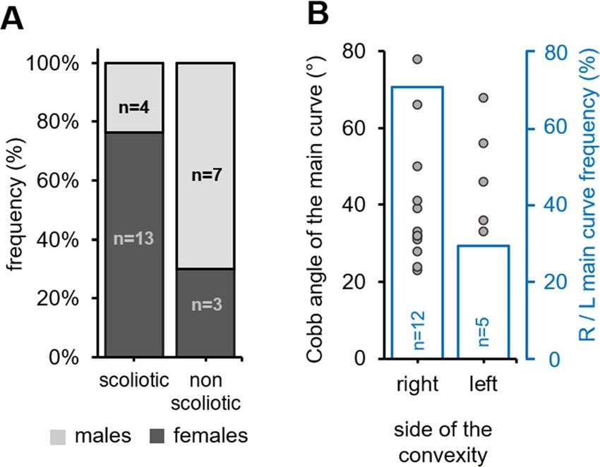
www.nature.com/scientificreports/
Figure 1. Three examples of frontal 3D-reconstructions of zebrafish’s spine deformities, classified according
to Lenke and compared to corresponding human scoliosis. Left: Reconstructions from RaDiant Dicom Viewer
5.0.0 software (https://www.radiantviewer.com/); Right: Clinical plain radiographs (personal collection of
LMH).
Radiological confirmation of a suspected scoliosis relies on the observation of a three-dimensional and rota-
tional spinal deformity characterized by a thoracic curvature in the frontal plane with a Cobb angle greater than
10 degrees on frontal r adiographs1,3. Here, we observed that 63% of the cfap298tm304 population analyzed (n = 17
out of 27 animals) had three-dimensional spinal deformity in the frontal and sagittal planes (Fig. 2A, B), with
curves exceeding 10 degrees in the frontal plane (42.1° ± 4.1°, [min–max: 23°–78°]). In the scoliotic population
analyzed, we observed that most of the animals had double or triple curves (n = 13 out of 17 scoliotic fishes)
(Fig. 2C). If classified as human adolescent idiopathic scoliosis, 29% of the curves were Lenke 3, 29% Lenke 4,
18% Lenke 5, 6% Lenke 1 and 6 or 1 and 2 animals were unclassifiable according to Lenke classification27. The
Fig. 1 displays some example of the Lenke classification applied to Zebrafish spine.
To fully describe the curve pattern of scoliotic animals, we measured the amplitude of the main curve at
8 weeks old, and observed it was larger when the apex was located in the thoraco-lumbar junction (Fig. 2D),
reflecting the anatomical location of the main curve observed in the patients. Moreover, the magnitude of the
main curve for the 17 scoliotic zebrafishes was 42.1° ± 4.1°, [min–max: 23°–78°], while we observed a mean
angle of 36.5° ± 6.7° [min–max: 13°–77°] for the first minor curve and 27.9 ± 4.8°, [min–max: 16°–43°] for the
second compensatory curve.
Spinal deformities associated with AIS in human pathology are also characterized by a progression of the
curve severity over time, most notably during the period preceding the pubertal growth s purt29. Thus, we ana-
lyzed the evolution of the curve severity on scoliotic cfap298tm304 animals between 8 and 12 weeks of age. The
mean length of the zebrafishes (all cohort) at 8 weeks old was 15.3 mm ± 1.86; [11.7;18.9 mm] and 19.8 mm ± 3.37
[12.4;25.5 mm] at 12 weeks old. We observed that the mean Cobb angle of the scoliotic cfap298tm304 zebrafish
increased by 5.5° ± 7.3° (mean ± SEM) during this period, showing the progressive development of spine tor-
sion. The mean Cobb angle for the second curve at 12 weeks was 47.6° ± 13° [29; 82°]. Moreover, the mean
apical vertebral rotation for the main curve was 31° ± 13° [min–max: 20°–69°], reflecting its three-dimensional
shape. The analysis of the curve patterns at 12 weeks also showed that spine curves remained mainly thoracic
or thoraco-lumbar with double or triple curves and that 82% of the scoliotic juvenile fishes presented at least
one dislocation (Fig. 2E). The dislocations were located at the thoraco-lumbar junction or at the lumbar spine
for 79% of the animals.
Another feature of AIS is its sexual dimorphism. While female are a least more likely to develop scoliosis by
a ratio of t wo30,31 and most severe curves are ten times most prevalent in females than males32 and the risk of
progressive deformity requiring surgical treatment is five times higher in girls than in boys33. Thus, we compared
the frequency of scoliosis occurrence in male and female siblings obtained from the cross of cfap298tm304/+ and
cfaptm304/tm304 parents (Fig. 3A). At 8 weeks old, 76% (n = 13 out of 17 animals) of scoliotic fish were females,
compared to 30% in the non-scoliotic population (n = 3 out of 10), suggesting a female bias in the penetrance of
scoliotic curves. Moreover, only 4/13 (31%) of the male fishes developed scoliosis, compared to 13/14 (93%) of
the females, that difference being statistically significative according to Fischer’s test (p = 0.001). Although curve
patterns may vary in AIS patients, right-sided thoracic deformities are by far the most common34. Figure 3B
shows the distribution of the convexity of the main thoracic curve in cfap298tm304 scoliotic animals, which was
located to the right in 71% of the scoliotic fish. Overall, these results showed that the cilia-defective cfap298tm304
mutant displayed characteristics of spinal curvature defects observed in human AIS patients.
Scientific Reports | (2021) 11:7392 | https://doi.org/10.1038/s41598-021-86856-1 3
Vol.:(0123456789)www.nature.com/scientificreports/
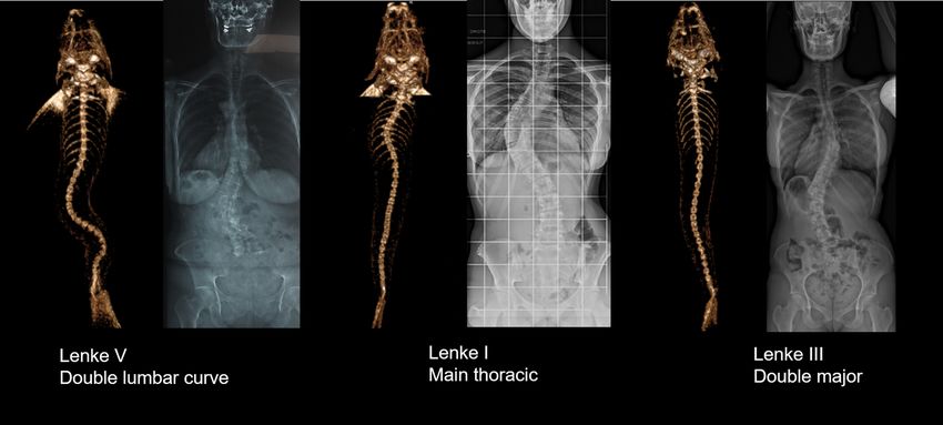
Figure 2. Juvenile cfap298tm304/tm304 zebrafish mutants develop an evolutive thoraco-lumbar curvature of the
spine. (A) Frontal views of scannographic reconstructions of 8-week-old non-scoliotic (top) and cfap298tm304
scoliotic sibling (bottom). (B) Frontal and sagittal view of the same cfap298tm304 scoliotic fish after scannographic
reconstruction showing a 3-dimensional torsion of the spine reminiscent of AIS. (C) Distribution of the
frequency of the number of spine curves observed in the sagittal plane of scoliotic (grey) and non-scoliotic
(white) animals. The population analyzed was raised from a cross of cfap298tm304/+ with cfap298tm304/tm304. Most
of cfap298 scoliotic animals display double or triple curves in the frontal plane. (D) Distribution of the Cobb
angle of the main curve in cfap298 scoliotic fish depending on the localization of the apex of the main curve.
Bars represent the average Cobb angles ± SEM. Each point represents a single fish. Most of the main curve apices
are located between T6 and L10 (11 out of 17 fish) (E) Histogram showing the distribution of the number of
dislocations observed in the scoliotic population (frequency, %).
Conclusions
The cfap298tm304 mutant seems to be after orthopaedic analysis of the curves a relevant model
of AIS. The scannographic analysis of cfap298tm304 mutants at two different ages (8 and 12 weeks) conducted
in this study is based on orthopedic criteria. It indicates the presence of dislocations for 82% of the cohort as
well as a three-dimensional deformity of the spine with a frontal Cobb angle above 10° for 63% of the scoliotic
animals analyzed. The presence of an apical rotation is a clue element to define scoliosis rather than other spine
deformities and was found at 31° ± 13° in this cohort, which is a strong argument for scoliosis. As previously
described12, spine deformities are seen precociously, at 8 weeks old in cfap298tm304 mutants35. Consistently with
previously reported AIS phenotype, we characterize here the evolutive severity of spinal deformities associated
with scoliosis between 8 and 12 weeks. An increase of 5.5° was observed in four weeks, for an average Cobb angle
of the main curve reaching 47.6° ± 13°.The predominance of a right thoracic convexity more frequent in female
is also coherent with A IS36,37. Thus, our orthopedic analysis of spinal deformities confirms the relevance of the
mutant cfap298 tm304
as a model for human AIS12,38,39.
One limitation of this study is of course the number of animals in the cohort, that render some statistics
regarding male–female dimorphism and analysis of the curves weak. However, the analysis in an orthopaedic
fashion does provide the clue elements to firmly link this phenotype to AIS.
How can we go further on the link between the cfap298tm304 mutation and the pathogenicity of AIS? The
cfap298tm304 mutation affects a cytoplasmic protein that is expressed at the base of cilia, little sensory and motile
organelles protruding from the surface of specialized cells in the organism24. Cilia dysfunction may be clini-
cally linked to AIS, since variants in ciliary genes, especially involved in cellular mechanotransduction (LBX1:
spinal cord differentiation, somatosensory signal transduction, POC5: centrin and inversin interaction in the
centrioles, GRP126: axons myelinization), were found in AIS patients20,21,40,41. Recent literature focusing on
AIS etiology also highlighted a link with elongated osteoblasts cilium, related to several transduction genetic
Scientific Reports | (2021) 11:7392 | https://doi.org/10.1038/s41598-021-86856-1 4
Vol:.(1234567890)www.nature.com/scientificreports/
Figure 3. cfap298tm304 mutants exhibit a sexual and a right convexity bias for scoliosis development. (A) Stacked
histogram showing the distribution of scoliotic and non-scoliotic phenotypes (frequency, %) in the analyzed
cohort according to the gender (males: light grey, females: dark grey). Spinal curves were more prevalent in
females (dark grey). (B) Distribution of the Cobb angle of the main curve in cfap298tm304 scoliotic fish according
to the side of the convexity in the frontal plane (right versus left, grey points; each point represents a single fish).
The frequency of the convexity of the main curve is represented in blue. Note that most of the curves are biased
to the right side (12 out of 17 fishes).
defects have been found in AIS patients, suggesting a complex molecular and cellular cilium involvement in
scoliosis17,42. Impairments in the inner ear system (lateral semi-circular canal asymmetry and vestibular canals
morphology) possibly linked to cilia defects have also been identified in AIS patients and suggest the involve-
ment of the vestibular system to keep the spine properly aligned43–45. no clear mechanism has emerged from
these tissue-specific candidates.
On contrary, cilia beating in the brain and spinal cord cavities have been thoroughly explored and investigated
recently in zebrafish. A link between CSF flow and AIS seems particularly relevant due to the frequent associa-
tion of type I Chiari malformation with scoliosis and the possible regression of the spinal curves observed in
ecompression19. The initial finding that disturbing CSF flow generates three-
these patients after a sub-occipital d
dimensional spine deformities placed cilia and CSF as essential players for keeping the spine straight during
the juvenile period of growth. This explanation had recently been clarified and deepened by the generation of
zebrafish mutants targeting the SCO-spondin protein forming the Reissner fiber. While cilia beating is necessary
to form this acellular thread bathing in C SF46, zebrafish mutants devoid of this fiber have recently been shown to
develop AIS-like spinal c urves47,48, possibly with an involvement of the signaling pathway of Urotensin-related
peptides39,49. These early forays into the genetics of AIS in zebrafish together with our orthopedic characteriza-
tion of the cfap298tm304 mutant bolsters now the use of this model to find novel mechanisms regulating spine
alignment.
Received: 1 January 2021; Accepted: 15 March 2021
References
1. Campos, M. A. & Weinstein, S. L. Pediatric scoliosis and kyphosis. Neurosurg. Clin. N. Am. https://doi.org/10.1016/j.nec.2007.04.
007 (2007).
2. Lonstein, J. E. Adolescent idiopathic scoliosis. Lancet https://doi.org/10.1016/S0140-6736(94)90572-X (1994).
3. Dickson, R. A. The etiology and pathogenesis of idiopathic scoliosis. Acta Orthop. Belg. https://doi.org/10.14531/ss2006.4.84-93
(1992).
4. Horne, J. P., Flannery, R. & Usman, S. Adolescent idiopathic scoliosis: Diagnosis and management. Am. Fam. Phys. 89, 193–198
(2014).
5. Smorgick, Y. et al. Clinical and radiographic characteristics in male and female adolescent candidates for idiopathic scoliosis
surgery. Isr. Med. Assoc. J. (2019).
6. Bezalel, T., Carmeli, E., Been, E. & Kalichman, L. Scheuermann’s disease: Current diagnosis and treatment approach. J. Back
Musculoskelet. Rehabil. https://doi.org/10.3233/BMR-140483 (2014).
7. Altaf, F., Gibson, A., Dannawi, Z. & Noordeen, H. Adolescent idiopathic scoliosis. BMJ https://doi.org/10.1136/bmj.f2508 (2013).
8. Daffner, S. D., Beimesch, C. F. & Wang, J. C. Geographic and demographic variability of cost and surgical treatment of idiopathic
scoliosis. Spine https://doi.org/10.1097/BRS.0b013e3181d88e78 (2010).
9. Du, C. et al. Relevant areas of functioning in people with adolescent idiopathic scoliosis on the international classification of
functioning, disability and health: the patients’ perspective. J. Rehabil. Med. https://doi.org/10.2340/16501977-2147 (2016).
10. Kotwal, S., Pumberger, M., Hughes, A. & Girardi, F. Degenerative scoliosis: a review. HSS J. https://doi.org/10.1007/s11420-011-
9204-5 (2011).
11. Boswell, C. W. & Ciruna, B. Understanding idiopathic scoliosis: a new Zebrafish school of thought. Trends Genet. https://doi.org/
10.1016/j.tig.2017.01.001 (2017).
Scientific Reports | (2021) 11:7392 | https://doi.org/10.1038/s41598-021-86856-1 5
Vol.:(0123456789)www.nature.com/scientificreports/
12. Grimes, D. T. et al. Zebrafish models of idiopathic scoliosis link cerebrospinal fluid flow defects to spine curvature. Science https://
doi.org/10.1126/science.aaf6419 (2016).
13. Hayes, M. et al. Ptk7 mutant zebrafish models of congenital and idiopathic scoliosis implicate dysregulated Wnt signalling in
disease. Nat. Commun. https://doi.org/10.1038/ncomms5777 (2014).
14. Newham, E. et al. Finite element and deformation analyses predict pattern of bone failure in loaded zebrafish spines. J. R. Soc.
Interface https://doi.org/10.1098/rsif.2019.0430 (2019).
15. Hayes, A. J. et al. Spinal deformity in aged zebrafish is accompanied by degenerative changes to their vertebrae that resemble
osteoarthritis. PLoS ONE https://doi.org/10.1371/journal.pone.0075787 (2013).
16. Ouellet, J. & Odent, T. Animal models for scoliosis research: State of the art, current concepts and future perspective applications.
Eur. Spine J. https://doi.org/10.1007/s00586-012-2396-7 (2013).
17. Baschal, E. E. et al. Idiopathic scoliosis families highlight actin-based and microtubule-based cellular projections and extracellular
matrix in disease etiology. G3 Genes Genomes Genet https://doi.org/10.1534/g3.118.200290 (2018).
18. Wang, Y. et al. Coding variants coupled with rapid modeling in Zebrafish implicate dynein genes, dnaaf1 and zmynd10, as ado-
lescent idiopathic scoliosis candidate genes. Front. Cell Dev. Biol. https://doi.org/10.3389/fcell.2020.582255 (2020).
19. Brockmeyer, D., Gollogly, S. & Smith, J. T. Scoliosis associated with Chiari I malformations: the effect of suboccipital decompres-
sion on scoliosis curve progression. A preliminary study. Spine https://doi.org/10.1097/01.BRS.0000092381.05229.87 (2003).
20. Oliazadeh, N., Gorman, K. F., Eveleigh, R., Bourque, G. & Moreau, A. Identification of elongated primary cilia with impaired
mechanotransduction in idiopathic scoliosis patients. Sci. Rep. https://doi.org/10.1038/srep44260 (2017).
21. Patten, S. A. et al. Functional variants of POC5 identified in patients with idiopathic scoliosis. J. Clin. Invest. https://doi.org/10.
1172/JCI77262 (2015).
22. Xu, L. et al. Genetic variant of PAX1 gene is functionally associated with adolescent idiopathic scoliosis in the Chinese population.
Spine https://doi.org/10.1097/BRS.0000000000002475 (2018).
23. Austin-Tse, C. et al. Zebrafish ciliopathy screen plus human mutational analysis identifies C21orf59 and CCDC65 defects as caus-
ing primary ciliary dyskinesia. Am. J. Human Genet. https://doi.org/10.1016/j.ajhg.2013.08.015 (2013).
24. Jaffe, K. M. et al. C21orf59/kurly controls both cilia motility and polarization. Cell Rep. https://doi.org/10.1016/j.celrep.2016.01.
069 (2016).
25. Brand, M. et al. Mutations affecting development of the midline and general body shape during zebrafish embryogenesis. Develop-
ment 123, 129–142 (1996).
26. Marie-Hardy, L., Khalifé, M., Slimani, L. & Pascal-Moussellard, H. Computed tomography method for characterising the zebrafish
spine. Orthop. Traumatol. Surg. Res. https://doi.org/10.1016/j.otsr.2018.12.008 (2019).
27. Lenke, L. G., Edwards, C. C. & Bridwell, K. H. The Lenke classification of adolescent idiopathic scoliosis: how it organizes curve
patterns as a template to perform selective fusions of the spine. Spine https://doi.org/10.1097/01.brs.0000092216.16155.33 (2003).
28. Ferrero, E. et al. Tridimensional analysis of rotatory subluxation and sagittal spinopelvic alignment in the setting of adult spinal
deformity. Spine Deform. https://doi.org/10.1016/j.jspd.2017.01.003 (2017).
29. Ponseti, I. V., Pedrini, V., Wynne Davies, R. & Duval Beaupere, G. Pathogenesis of scoliosis. Clin. Orthop. Relat. Res. https://doi.
org/10.1097/00003086-197610000-00034 (1976).
30. Carter, O. D. & Haynes, S. G. Prevalence rates for scoliosis in US adults: Results from the first national health and nutrition exami-
nation survey. Int. J. Epidemiol. https://doi.org/10.1093/ije/16.4.537 (1987).
31. Kruse, L. M., Dobbs, M. B. & Gurnett, C. A. Polygenic threshold model with sex dimorphism in clubfoot inheritance: the Carter
effect. J. Bone Jt. Surg. Ser. A https://doi.org/10.2106/JBJS.G.01346 (2008).
32. Miller, N. H. Cause and natural history of adolescent idiopathic scoliosis. Orthop. Clin. North Am. https://doi.org/10.1016/S0030-
5898(05)70091-2 (1999).
33. Hresko, M. T. Clinical practice. Idiopathic scoliosis in adolescents. N. Engl. J. Med. https://d oi.o
rg/1 0.1 056/N
EJMcp
12090 63 (2013).
34. Compton, J., Vander Voort, W. & Weinstein, S. Scoliosis curvature follows thoracic organ orientation. Spine https://doi.org/10.
1097/BRS.0000000000002731 (2020).
35. Parichy, D. M., Elizondo, M. R., Mills, M. G., Gordon, T. N. & Engeszer, R. E. Normal table of postembryonic zebrafish develop-
ment: Staging by externally visible anatomy of the living fish. Dev. Dyn. https://doi.org/10.1002/dvdy.22113 (2009).
36. Weinstein, S. L., Dolan, L. A., Cheng, J. C., Danielsson, A. & Morcuende, J. A. Adolescent idiopathic scoliosis. Lancet https://doi.
org/10.1016/S0140-6736(08)60658-3 (2008).
37. Coonrad, R. W., Murrell, G. A. C., Motley, G., Lytle, E. & Hey, L. A. A logical coronal pattern classification of 2000 consecutive
idiopathic scoliosis cases based on the scoliosis research society-defined apical vertebra. Spine https://doi.org/10.1097/00007632-
199806150-00016 (1998).
38. Van Gennip, J. L. M., Boswell, C. W. & Ciruna, B. Neuroinflammatory signals drive spinal curve formation in zebrafish models of
idiopathic scoliosis. Sci. Adv. https://doi.org/10.1126/sciadv.aav1781 (2018).
39. Zhang, X. et al. Cilia-driven cerebrospinal fluid flow directs expression of urotensin neuropeptides to straighten the vertebrate
body axis. Nat. Genet. https://doi.org/10.1038/s41588-018-0260-3 (2018).
40. Jiang, H. et al. Association between ladybird homeobox 1 gene polymorphisms and adolescent idiopathic scoliosis: a MOOSE-
compliant meta-analysis. Medicine (Baltimore) https://doi.org/10.1097/MD.0000000000016314 (2019).
41. Liu, G. et al. Genetic polymorphisms of GPR126 are functionally associated with PUMC classifications of adolescent idiopathic
scoliosis in a Northern Han population. J. Cell. Mol. Med. https://doi.org/10.1111/jcmm.13486 (2018).
42. Oliazadeh, N., Franco, A., Wang, D. & Moreau, A. Abnormalities in primary cilium of osteoblasts of adolescent idiopathic scoliosis
patients. Cilia https://doi.org/10.1186/2046-2530-4-S1-P6 (2015).
43. Catanzariti, J. F. et al. Does adolescent idiopathic scoliosis relate to vestibular disorders? A systematic review. Ann. Phys. Rehabil.
Med. https://doi.org/10.1016/j.rehab.2014.04.003 (2014).
44. Hawasli, A. H., Hullar, T. E. & Dorward, I. G. Idiopathic scoliosis and the vestibular system. Eur. Spine J. https://doi.org/10.1007/
s00586-014-3701-4 (2014).
45. Hitier, M. et al. Lateral semicircular canal asymmetry in idiopathic scoliosis: an early link between biomechanical, hormonal and
neurosensory theories?. PLoS ONE https://doi.org/10.1371/journal.pone.0131120 (2015).
46. Cantaut-Belarif, Y., Sternberg, J. R., Thouvenin, O., Wyart, C. & Bardet, P. L. The reissner fiber in the cerebrospinal fluid controls
morphogenesis of the body axis. Curr. Biol. https://doi.org/10.1016/j.cub.2018.05.079 (2018).
47. Troutwine, B. R. et al. The reissner fiber is highly dynamic in vivo and controls morphogenesis of the spine. Curr. Biol. https://doi.
org/10.1016/j.cub.2020.04.015 (2020).
48. Rose, C. D. et al. SCO-spondin defects and neuroinflammation are conserved mechanisms driving spinal deformity across genetic
models of idiopathic scoliosis. Curr. Biol. https://doi.org/10.1016/j.cub.2020.04.020 (2020).
49. Lu, H., Shagirova, A., Goggi, J. L., Yeo, H. L. & Roy, S. Reissner fibre-induced urotensin signalling from cerebrospinal fluid-
contacting neurons prevents scoliosis of the vertebrate spine. Biol. Open https://doi.org/10.1242/bio.052027 (2020).
Scientific Reports | (2021) 11:7392 | https://doi.org/10.1038/s41598-021-86856-1 6
Vol:.(1234567890)www.nature.com/scientificreports/
Acknowledgements
We thank the SOFCOT (Societé française de Chirurgie Orthopédique et Traumatologique) for a grant to LMH;
This work was supported by an ANR-11-INBS-0011 (NeurATRIS: Translational Research Infrastructure for
Biotherapies in Neurosciences).
Author contributions
L.M.H. and Y.C.-B. wrote the manuscript, L.M.H. and L.S. did the experimentations, Y.C.B., L.M.H. and R.P.
review the manuscript, H.P.M. supervised the work.
Competing interests
The authors declare no competing interests.
Additional information
Correspondence and requests for materials should be addressed to L.M.-H.
Reprints and permissions information is available at www.nature.com/reprints.
Publisher’s note Springer Nature remains neutral with regard to jurisdictional claims in published maps and
institutional affiliations.
Open Access This article is licensed under a Creative Commons Attribution 4.0 International
License, which permits use, sharing, adaptation, distribution and reproduction in any medium or
format, as long as you give appropriate credit to the original author(s) and the source, provide a link to the
Creative Commons licence, and indicate if changes were made. The images or other third party material in this
article are included in the article’s Creative Commons licence, unless indicated otherwise in a credit line to the
material. If material is not included in the article’s Creative Commons licence and your intended use is not
permitted by statutory regulation or exceeds the permitted use, you will need to obtain permission directly from
the copyright holder. To view a copy of this licence, visit http://creativecommons.org/licenses/by/4.0/.
© The Author(s) 2021, corrected publication 2021
Scientific Reports | (2021) 11:7392 | https://doi.org/10.1038/s41598-021-86856-1 7
Vol.:(0123456789)You can also read