The Na+/H+ Exchanger NHX1 Controls H+ Accumulation in the Vacuole to Influence Sepal Color in Hydrangea macrophylla
←
→
Page content transcription
If your browser does not render page correctly, please read the page content below
Article
The Na+/H+ Exchanger NHX1 Controls H+ Accumulation in the
Vacuole to Influence Sepal Color in Hydrangea macrophylla
Gaitian Zhang, Suxia Yuan, Hui Qi, Zhiyun Chu and Chun Liu *

                                             Institute of Vegetables and Flowers, Chinese Academy of Agricultural Sciences, Beijing 100081, China
                                             * Correspondence: liuchun@caas.cn; Tel.: +86-010-82109510

                                             Abstract: Hydrangea macrophylla is popular for its unique physiological characteristics and changeable
                                             colors. Previous studies have shown that the pH of the vacuoles of the sepal cells of hydrangea affects
                                             the color of the sepals. Located on the vacuolar membrane, NHX1 is an important H+ proton pump
                                             that drives the exchange of metal ions. This proton pump affects the physiological environment by
                                             controlling the accumulation of H+ in the vacuole. In hydrangea, the HmNHX1 gene has an open
                                             reading frame of 1626 bp and encodes a total of 541 amino acids. Bioinformatic analysis showed that
                                             HmNHX1, which encodes a Na+ /H+ exchanger, is located on the vacuolar membrane. Tissue-specific
                                             expression analysis showed that the expression of this gene in the treatment group was higher than
                                             that in the control group. The ion flux in the vacuoles of colored hydrangea in the treatment group
                                             and the control group were measured, and the results showed that HmNHX1 was indeed a Na+ /H+
                                             exchanger. When the results of the HmNHX1 expression analysis and ion flux measurements are
                                             combined, it can be seen that HmNHX1 regulates the accumulation of H+ in the vacuole, ultimately
                                             affecting the color of the plant.

                                             Keywords: Hydrangea macrophylla; NHX1; flower color; ion flux measurements

                                             1. Introduction
Citation: Zhang, G.; Yuan, S.; Qi, H.;
                                                  Hydrangea macrophylla is a unique ornamental plant species, and its main ornamental
Chu, Z.; Liu, C. The Na+ /H+
                                             parts are the sepals of sterile flowers [1]. This species is native to East Asia [2]. Hydrangea
Exchanger NHX1 Controls H+
                                             is famous for its rich and variable colors, and the color of the sepals of some varieties
Accumulation in the Vacuole to
                                             can change to blue after aluminum application [3]. In vitro simulation experiments have
Influence Sepal Color in Hydrangea
macrophylla. Int. J. Plant Biol. 2023, 14,
                                             shown that the aluminum ion content, the type of co-pigment, and the pH of the simulation
266–275. https://doi.org/10.3390/
                                             solution affect the color of the sepals. The average pH of the vacuole of blue hydrangea
ijpb14010022                                 plants is 4.1, while that of the vacuole of red plants is 3.3 [4,5]. In other species, the pH of the
                                             vacuole of purple-blue flowers is higher than that of red flowers [6–8]. This occurs because
Academic Editor: Adriano Sofo
                                             when the pH is higher, the absorbance of the chromogenic substance, i.e., co-pigmentation
Received: 3 February 2023                    of anthocyanins and co-pigments, is bathochromically shifted [9,10].
Revised: 18 February 2023                         Studies have shown that the vacuolar pH in relation to plant flower color is mainly
Accepted: 18 February 2023                   regulated by two different types of proton pumps that rely on a H+ concentration gradient
Published: 20 February 2023                  to transport H+ : P-type ATPase and Na+ (K+ )/H+ exchangers [6,11]. The Na+ (K+ )/H+
                                             exchanger is a CPA (monovalent cation reversal protein) that is involved in the regulation
                                             of the cell cycle and proliferation, salt tolerance, vesicle trafficking, and biogenesis [12].
                                             In most related studies, NHX1 has been found to encode a sodium–hydrogen antiporter
Copyright: © 2023 by the authors.            involved in salt tolerance; its main role is to transport Na+ (K+ ) into the vacuole and,
Licensee MDPI, Basel, Switzerland.
                                             at the same time, replace H+ . This method not only ensures the stability of the solution
This article is an open access article
                                             environment inside the vacuole but also effectively isolates the metal ions inside the vacuole
distributed under the terms and
                                             to maintain the normal biological activities of cells [13–15]. However, this also increases
conditions of the Creative Commons
                                             the pH of the vacuole.
Attribution (CC BY) license (https://
                                                  The color of Ipomoea tricolor changes from pink to purple or blue from the bud stage to
creativecommons.org/licenses/by/
                                             the opening stage. The pigments do not change; the vacuole’s pH continues to increase.
4.0/).

Int. J. Plant Biol. 2023, 14, 266–275. https://doi.org/10.3390/ijpb14010022                                          https://www.mdpi.com/journal/ijpb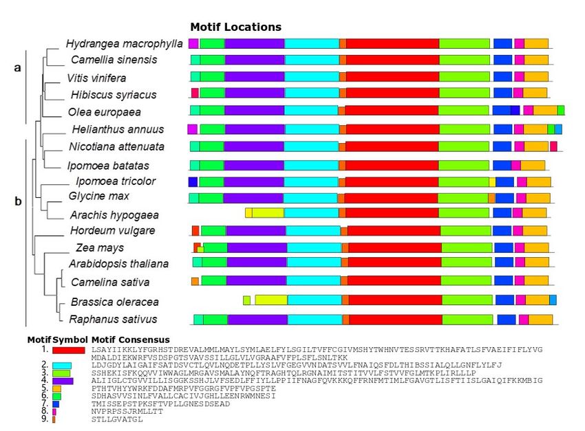
2.1. Plant Materials and Sequencing
            This experiment used two-year-old ‘Bailmer’ cuttings
Int. J. Plant Biol. 2023, 14                                                                                         267
                                                                                                                         as te
       als were used). All plants were grown in the greenhouse of the I
       Flowers, Chinese           Academy
                    Related research   has pointed out of     Agricultural
                                                        that the key gene that causes theSciences,               and all
                                                                                             blue corollas of Japanese
                    morning glory is NHX1 [16]. LnNHX1 was also the first protein identified to regulate the
       Huaduoduo increase
                     compound                  fertilizer
                              in pH of the vacuole                was
                                                      and cause the   flowerregularly
                                                                              color to become blue provided
                                                                                                       [6]. Obviously, as t
                    NHX1 is related to changes in flower color. Therefore, this study took HmNHX1 as the
       Aluminum was researchsupplied
                              object to explore to    the
                                                 the role     control,
                                                          of NHX1   in the colorstarting          when
                                                                                 change of hydrangea           the hydr
                                                                                                          sepals.

       longer elongated,
                    2. Materials terminal
                                   and Methods buds were evident, and plant height
                    2.1. Plant Materials and Sequencing
       mL of 6 g/L Al2This (SO      4)3·18H
                                experiment        2O, pH‘Bailmer’
                                            used two-year-old    = 4.5)        was
                                                                           cuttings      applied
                                                                                    as test materials (onlyonce
                                                                                                             the sepalsa we
                    were used). All plants were grown in the greenhouse of the Institute of Vegetables and
       had formed. Flowers,
                     EightChinese  pots       (with
                                          Academy          three pots
                                                     of Agricultural   Sciences, constituting
                                                                                  and all pots were 15 cm     one      biolo
                                                                                                                in size.
                    Huaduoduo compound fertilizer was regularly provided as the experimental fertilizer.
       cluded in theAluminum
                       aluminum   was supplied treatment             group
                                                to the control, starting  when theand        in internodes
                                                                                     hydrangea    the control  were no gro
                    longer elongated, terminal buds were evident, and plant height was fixed. Aluminum
       out with a mixture
                    (500 mL of 6 g/L ofAlthree
                                            (SO ) ·18H
                                                     2different
                                                         O, pH = 4.5) was
                                                           4 3      2     plants
                                                                            applied once anda weekperformed
                                                                                                    after the terminal     at
                    buds had formed. Eight pots (with three pots constituting one biological replication) were
       bud stage (S1),     the
                    included   in thecoloring          period
                                      aluminum treatment     group and(S2),       and
                                                                        in the control       the
                                                                                        group.       blooming
                                                                                                Sampling   was carried    pe
                    out with a mixture of three different plants and performed at three different stages (the bud
       material hadstage
                      three
                          (S1), thebiological           replicates.
                                     coloring period (S2), and the blooming period (S3)) (Figure 1). Each material
                               had three biological replicates.

                               Figure 1. ‘Bailmer’ materials used in this study. CK, control group; Tr, treatment group; S, stage.
       Figure 1. ‘Bailmer’ materials used in this study. CK, control group; Tr,
                               2.2. RNA Extraction and cDNA Synthesis
                                    RNA was extracted with an EasySpin Plus Plant RNA Kit (Aidlab, Beijing, China).
                               The RNA concentration was measured by an ultraviolet-visible spectrophotometer (Miulab,
       2.2. RNA Extraction and cDNA Synthesis
                               ND-100, Hangzhou, China). The RNA was reverse-transcribed with a PrimeScript RT
                               Reagent Kit with gDNA Eraser (Takara, Dalian, China). The reaction volume for reverse
            RNA was extracted with an EasySpin Plus Plant RNA Ki
                               transcription was 20 µL. Each reaction system included 500 ng of RNA.

       The RNA concentration was measured by an ultraviolet-visibl
       ulab, ND-100, Hangzhou, China). The RNA was reverse-trans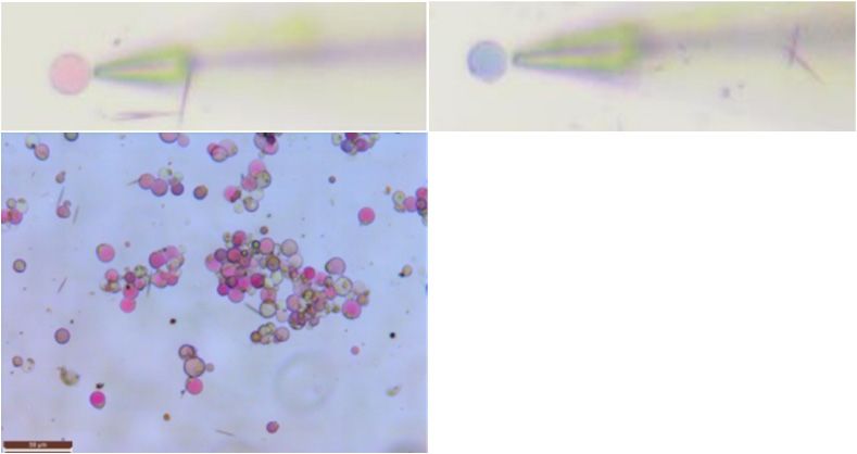
Int. J. Plant Biol. 2023, 14                                                                                                  268

                               2.3. Full-Length Amplification of the HmNHX1 Coding DNA Sequence (CDS)
                                     The sequence of ItNHX1 (Ipomoea tricolor NHX1, AB292774) [17] was queried (via
                               BLAST), and the sequence was obtained (https://www.ncbi.nlm.nih.gov) (accessed on
                               17 September 2020). We used the sequence of ItNHX1 to search for HMNHX1 in the
                               single-molecule real-time (SMRT) sequencing results.
                                     We used Bailmer plants (at S3) as the experimental materials. According to the isoform
                               data of HmNHX1 screened by single-molecule real-time sequencing, the obtained sequence was
                               used to predict the open reading frame (ORF) via NCBI ORFfinder (https://www.ncbi.nlm.
                               nih.gov/orffinder) (accessed on 14 January 2021), and the results were inputted into the NCBI
                               database to compare the length of the CDS of HmNHX1 with that of the homologs in other
                               species. Primers containing the HmNHX1 ORF were designed (HmNHX1-F, ACATGTGAT-
                               GTGATGCTTAGTTCGGAAG; HmNHX1-R, GACCAACAAGTGGGCGACAATCTGTAT) via
                               Integrated DNA Technologies (https://sg.idtdna.com/calc/analyzer) (accessed on 16 January
                               2021). A KAPA HiFi HotStart ReadyMix PCR Kit (Roche, Indianapolis, IN, USA) was then used
                               to amplify the target fragment (the reaction system consisted of 25 µL of 2× KAPA HiFi HotStart
                               ReadyMix, 3 µL of 5 µM forward primer, 3 µL of 5 µM reverse primer, 3 µL of cDNA template,
                               and 16 µL of sterilized ddH2O). The PCR program was as follows: 95 ◦ C predenaturation for
                               3 min; 35 cycles of 98 ◦ C denaturation for 20 s, 60 ◦ C annealing for 15 s, and a 72 ◦ C extension
                               for 1.5 min; and a 72 ◦ C final extension for 1.5 min. A Bio-Rad T-100 PCR thermal cycler
                               instrument was used. The PCR products were subsequently detected via 1% (w/v) agarose
                               gel electrophoresis.

                               2.4. Gel Extraction
                                    The target bands were removed and placed into a preweighed 2 mL centrifuge tube.
                               The weight of the gel was approximately 400 mg. An EasyPure Quick Gel Extraction Kit
                               (TransGen, Beijing, China) was used to recover the gels.
                                    A Zero Background pTOPO-Blunt Simple Cloning Kit (Aidlab, Beijing, China) and
                               DH5α chemically competent cells (Tsingke, Beijing, China) were used for transformation.
                               Luria–Bertani medium was used for the colony culture (100 mg/L ampicillin). We used the
                               bacterial liquid as the amplification template to perform PCR amplification and sent the
                               bacterial liquid corresponding to the correct amplification length for sequencing (Sangon
                               Biotech, Shanghai, China).

                               2.5. Bioinformatic Analysis
                                    The sequence of HmNHX1 was predicted via the NCBI ORFfinder (https://www.
                               ncbi.nlm.nih.gov/orffinder) (accessed on 2 February 2021) program. Cell-PLoc 2.0 (http:
                               //www.csbio.sjtu.edu.cn/bioinf/Cell-PLoc-2/) (accessed on 2 February 2021) was used
                               to predict the subcellular location of the protein encoded by the HmNHX1 gene. SOPMA
                               (https://npsa-prabi.ibcp.fr/cgi-bin/npsa_automat.pl?page=npsa_sopma.html) (accessed
                               on 2 February 2021)was then used to predict the secondary structure of the protein encoded
                               by HmNHX1. SMART (http://smart.embl.de/) was used to predict the conserved regions
                               and basic functions of the protein encoded by HmNHX1. ClustalX and ESPript (https:
                               //espript.ibcp.fr/ESPript/cgi-bin/ESPript.cgi) (accessed on 2 February 2021) were used
                               for sequence alignment. MEGA 7 was subsequently used to construct a phylogenetic tree.
                               MEME (https://meme-suite.org/) (accessed on 23 February 2022) was used to analyze the
                               motif of HmNHX1. Figures were drawn with Origin95 and Inkscape.

                               2.6. Tissue-Specific Expression Analysis of HmNHX1
                                    The standard curve was used to calculate the copy number of HmNHX1 in each sample.
                               The concentration of the plasmids returned by Sangon Biotech was 223.3 ng/µL, which was
                               used as a benchmark for different dilution factors (100 , 10−1 , 10−2 , 10−3 , 10−4 , 10−5 , 10−6 ,
                               10−7 , 10−8 , and 10−9 ). We chose the sample data with dilutions of 10−5 , 10−6 , 10−7 , 10−8 ,
                               and 10−9 to construct the standard curve because their Cq values were similar. Plasmid
                               copy numbers were calculated by the plasmid copy number calculation formula. The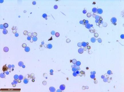
Int. J. Plant Biol. 2023, 14 269
logarithmic value of the plasmid copy number for different concentrations of plasmids
was used as the abscissa, and the Cq value of the corresponding plasmid concentration
was used as the ordinate (we included the Cq values of each sample). The instrument
used for real-time fluorescence quantification was a Light Cycler 480 II (Roche, Basel,
Switzerland), and the fluorescence was quantified with Forget-Me-Not qPCR Master Mix
(Biotium, Fremont, CA, USA). The reaction volume was 10 µL, which consisted of 1 µL
of the cDNA template (25 ng of RNA substrate template in each reaction), 0.5 µL of the
amplification primers (5 µM), 5 µL of 2× Forget-Me-Not qPCR Master Mix, and 3 µL of
sterile dH2 O. The PCR program was as follows: enzyme activation at 95 ◦ C for 2 min,
denaturation at 95 ◦ C for 5 s, annealing at 60 ◦ C for 10 s, and extension at 72 ◦ C for 20 s.
The sequences of the primers used for fluorescence quantification included TGATGCCA-
CATCAGTTGTGCTG (forward) and CACCCAGCAAAGTGCTTGTGAG (reverse). All
materials had three experimental replicates.
2.7. Ion Flux Measurements of Bailmer Vacuoles
Sepal protoplast isolation was performed according to the method of Yoshida et al. [4],
with modifications. We prepared 0.008% poly-L-lysine (w/v, FW: 150,000–300,000, Sigma,
St. Louis, MO, USA) [18], and then smeared onto disposable petri dishes, which were sub-
sequently placed in a refrigerator to dry for later use. A noninvasive microtest technology
(NMT-YG-100, Younger USA, LLC., Amherst, MA, USA, 01002) was used to measure the
H+ , Na+ , and K+ fluxes of the vacuoles of pink and blue sepals of Bailmer plants.
3. Results
3.1. HmNHX1 Bioinformatic Analysis
The results were analyzed, after which the primers were designed and amplified.
The amplified product was sequenced, and its full length was 1861 bp. ItNHX1 was the
first gene identified to be associated with flower color and pH changes. The amino acid
sequence similarity between lnNHX1 and HmNHX1 was 78.29%.
ORFfinder predicted that the HmNHX1 CDS has a total length of 1626 bp and encodes
541 amino acids. The protein encoded by HmNHX1 is located on the vacuolar membrane,
and its secondary structure consists of 44.92% α-helices, 31.79% random coils, 18.85%
extended chains, and 4.44% β-turns. This protein functions as a Na+ /H+ exchanger. The
conserved functional region is formed by amino acids located from 24 to 444.
HmNHX1-related sequences in the NCBI database were queried, after which multiple
sequence alignment and phylogenetic tree analysis were performed. We used the sequences
of 17 species to construct evolutionary trees. These species included woody plant species
and herbaceous plant species (gramineous plant species, model plant species, etc.). From
the results of the phylogenetic tree analysis, Hydrangea macrophylla, Camellia sinensis, Vitis
vinifera, Hibiscus syriacus, and Olea europaea clustered on the same three-level branches;
these results contrast with those of herbaceous plant species, for which the clustering was
obviously more diversified and had high similarity in the conserved structure interval.
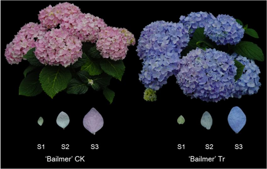
However, Motifs 1–9 showed that NHX1 of all species had high similarity in the conserved
structure interval (Figure 2).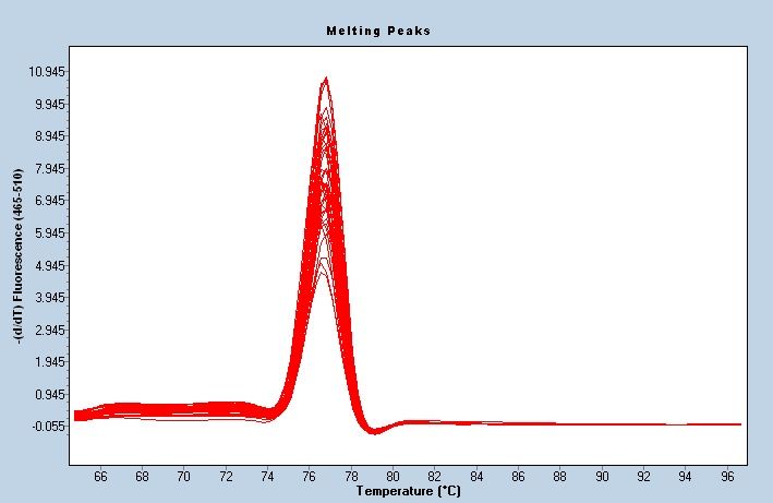
Int. J. Plant Biol. 2023, 14, FOR PEER REVIEW
Int. J. Plant Biol. 2023, 14 270
Figure
Figure 2. Phylogenetictree
2. Phylogenetic tree of
of NHX1.
NHX1. PartPart(a)
(a)includes
includeswoody
woodyplants, and Part
plants, and(b) includes
Part herba- herba
(b) includes
ceous plants. The figure on the right shows the analysis of the NHX1motif for
ceous plants. The figure on the right shows the analysis of the NHX1motif for the correspondingthe corresponding
species:
species: Hydrangeamacrophylla,
Hydrangea macrophylla, Camellia
Camelliasinensis (XM_028205587.1),
sinensis Vitis vinifera
(XM_028205587.1), Vitis(NM_001280886.1), Hi-
vinifera (NM_001280886.1)
biscus syriacus (XP_039020832.1), Olea europaea (XM_022986696.1), Helianthus annuus (XM_022146020.2),
Hibiscus syriacus (XP_039020832.1), Olea europaea (XM_022986696.1), Helianthus annuu
Nicotiana attenuata (XM_019398501.1), Ipomoea batatas (AFQ00709.1), Ipomoea tricolor (AB292774.1), Glycine
(XM_022146020.2), Nicotiana attenuata (XM_019398501.1), Ipomoea batatas (AFQ00709.1), Ipomoea tri
max (NM_001250237.2), Arachis hypogaea (XP_025680083.1), Hordeum vulgare (ANS57040.1), Zea mays
color (AB292774.1), Glycine max (NM_001250237.2), Arachis hypogaea (XP_025680083.1), Hordeum vul
(AAP20428.1), Arabidopsis thaliana (NM_122597.3), Camelina sativa (XP_010455152.1), Brassica oleracea
gare (ANS57040.1), Zea mays (AAP20428.1), Arabidopsis thaliana (NM_122597.3), Camelina sativ
(XP_013611175.1), and Raphanus sativus (XP_018440589.1).
(XP_010455152.1), Brassica oleracea (XP_013611175.1), and Raphanus sativus (XP_018440589.1).
3.2. Expression Analysis of HmNHX1
3.2. Expression Analysis
A standard curve of
wasHmNHX1
constructed to analyze the expression of HmNHX1 in Bailmer
hydrangea
A standardsepals (Figure
curve was3a). The melt curve
constructed showed the
to analyze thatexpression
the primer was well specified
of HmNHX1 in Bailme
(Figure 3b). The expression patterns in the control and treatment groups showed that the
hydrangea sepals (Figure 3a). The melt curve showed that the primer was well specified
expression of HmNHX1 at S1 and S2 was significantly higher than that in the control group.
(Figure 3b). The expression
The expression of HmNHX1patterns
at S3 was in the control
higher than thatand treatment
in the groups
control group, showed
although the that th
expression of HmNHX1
results were at S1
not significant. and S2the
In general, was significantly
expression level ofhigher
HmNHX1 than that
in the in the contro
treatment
group. The expression of HmNHX1 at S3 was higher than that in the control group,
group was higher than that in the control group in the same period. The expression of alt
HmNHX1 gradually increased with plant growth in both the treatment and control
hough the results were not significant. In general, the expression level of HmNHX1 in the groups.
These results suggest that the amount of H+ in the vacuole required to maintain the stability
treatment group was higher than that in the control group in the same period. The expres
of the blue chromogenic substance is less than that required for the pink chromogenic
sionsubstance
of HmNHX1 gradually increased with plant growth in both the treatment and contro
(Figure 3c).
groups. These results suggest that the amount of H+ in the vacuole required to maintain
the stability of the blue chromogenic substance is less than that required for the pink chro
mogenic substance (Figure 3c).Int.Int. J. Plant
J. Plant Biol. Biol. 2023, 14, FOR PEER REVIEW
2023, 14 271
Equation y = a + b*x
Intercept 42.90509 ± 0.40295
34 Slope -3.5785 ± 0.08172
Residual sum of squares 0.20036
32 -0.99922
Pearson's r
30 R squared 0.99844
Adjusted R squared 0.99792
28
Cq value
26
24
22
20
18
2 3 4 5 6 7
log(Quantity)
(a) (b)
Control group
Treatment group
650,000 a
600,000
Quantification of NHX1
550,000
500,000
450,000 a a
400,000 a
350,000
300,000
250,000 b
200,000
150,000 b
100,000
50,000
0
S1 S2 S3
(c)
Figure 3. Quantification of the expression of HmNHX1. (a) Standard curve of quantity. The range of
Figure
concentration 3. stable
with Quantification
fluorescenceof the expression
values of HmNHX1.
and good linear (a) Standard
ratios was selected. curve of
The logarithm of quantit
the copyconcentration
number was used with stable
as the fluorescence
x−axis, values
and the number and good
of reaction linear
cycles (Cq) ratios was
was used as selected.
the Th
y−axis. (b) Melting curve of HmNHX1. (c) Expression of HmNHX1
the copy number was used as the x−axis, and the number of reaction cycles (Cq) w
at different developmental stages
of sepalsy−axis.
under two
(b)different
Meltingtreatments
curve ofinHmNHX1.
Bailmer hydrangeas (p < 0.05).
(c) Expression of HmNHX1 at different develo
of sepalsbetween
3.3. Relationship underNHX1
two different treatments
and Ion Flux in Bailmer hydrangeas (p < 0.05).
Measurements
Protoplasts were isolated from pink and blue sepals of Bailmer hydrangeas at the full
blooming3.3. Relationship
stage, between of
and measurements NHX1
the H+and
, K+ ,Ion
andFlux
Na+ Measurements
currents were performed. The
results of the Protoplasts
ion flux measurements
were showed
isolated fromthat pink
the vacuolar
and H+ sepals
blue of blue sepals tendedhydrang
of Bailmer
+ +
to out the vacuole, K tended to exit, and Na tended to enter, whereas the vacuolar H+
blooming stage, and measurements + of the H+, K+, and Na +
+ currents were pe
of pink sepals tended to enter the vacuole, K tended to enter, and Na tended to exit.
results
Combining theseof the ion
results withflux
thosemeasurements
of the analysis ofshowed thatexpression
the HmNHX1 the vacuolar H of
+
patterns, weblue sep
inferredout
that the vacuole,
the amount of HK that
+ + tended to exit,inand
accumulated Na tended
+
the vacuoles of pinktosepals
enter, whereas
was higher the v
than thepink + that accumulated in the vacuoles of +blue sepals, and that NHX1 is +a
amountsepals tended to enter the vacuole, K tended to enter, and Na tended
of H
Na+ /H+bining
exchange pump,
these not a K
results
+ /H+ exchange pump (Figure 4).
with those of the analysis of the HmNHX1 expression
inferred that the amount of H+ that accumulated in the vacuoles of pink sepa
than the amount of H+ that accumulated in the vacuoles of blue sepals, and
a Na+/H+ exchange pump, not a K+/H+ exchange pump (Figure 4).Int.Int.
J. Plant Biol.
J. Plant 2023,
Biol. 2023,14,
14FOR PEER REVIEW 272 7
(a)
(b) (c) (d)
Vacuole Na+ flux(pmol·cm-2·s-1)
Vacuole H+ flux(pmol·cm-2·s-1)
Vacuole K+ flux(pmol·cm-2·s-1)
1400 200
35 Bailmer blue Bailmer blue Bailmer blue
1200 150
30 Bailmer pink Bailmer pink 100 Bailmer pink
1000
25 800 50
20 600 0 efflux
15 400 -50
10 200 efflux -100 influx
5 0 -150
efflux -200 influx
0 -200
influx -400
-5 -250
1 2 3 4 5 6 7 1 2 3 4 5 6 7 1 2 3 4 5 6 7
Figure 4. Protoplast and ion flux in Bailmer hydrangea. The ion flux of 7 vacuoles was measured in
Figure
each Protoplast
4. and
pink and ion
blue sepal. flux in Bailmer
(a) Protoplasts andhydrangea. The ionvacuole
the corresponding flux of 7ion
vacuoles was measured(bar
flux measurements
in each pink and
+ blue sepal. (a) Protoplasts+ and the corresponding vacuole
+ ion flux measurements
= 50 µm). (b) H flux measurements. (c) K flux measurements. (d) Na flux measurements. Positive
(bar = 50
values µm).that
show H+ flux
(b)ions measurements.
flowed (c) K+ flux
out of the vacuoles, measurements.
and negative values Na+ flux
(d)show thatmeasurements.
ions flowed into
Positive values
the vacuoles. show that ions flowed out of the vacuoles, and negative values show that ions flowed
into the vacuoles.
4. Discussion
4. Discussion
For
For most plants,flower
most plants, flowercolor
colorserves
serves asas a visual
a visual signal
signal to attract
to attract insects
insects to spread
to spread pollen pol-
len
and promote reproduction [19]. Anthocyanins, which are secondary metabolites, are theare
and promote reproduction [19]. Anthocyanins, which are secondary metabolites,
the
keykey substances
substances for flower
for flower color color formation
formation and areand are in
stored stored in the vacuoles
the vacuoles of plants
of plants [20,21].
[20,21]. Research related to blue flower formation in hydrangeas
Research related to blue flower formation in hydrangeas is continuing, and key genes is continuing, and key
genes regulating the DFR pathway have been identified
regulating the DFR pathway have been identified [22]. However, the color of flowers[22]. However, the color of flow-
is
ers
notisonlynot based
only based on anthocyanins.
on anthocyanins. The pHThe pHvacuole
of the of the vacuole
also is aalso
key is a key
factor in factor in main-
maintaining
taining the stability
the stability of anthocyanins
of anthocyanins in the vacuole,
in the vacuole, and the andpHthe haspH anhas an important
important influenceinfluence
on
thethe
on colorcolorof of
plants’
plants’ floral organs
floral organs [23]. In In
[23]. in in
vitro
vitrosimulation
simulation experiments,
experiments, it was
it wasfound
found
thatthe
that the absorbance
absorbance of a solution
solutionchanged
changedwhen whenthe thepH pHchanged,
changed, even
even when
when thetheamount
amount
of pigments was the same [24]. In the process of ion flux
of pigments was the same [24]. In the process of ion flux measurements, we found thatmeasurements, we found thatthe
the Hthe+ ofvacuoles
the vacuoles of blue sepals
H + of of blue sepals hadhad
an an obvious
obvious efflux
efflux trend
trend comparedwith
compared withthatthatofofthe
vacuoles of pink sepals, suggesting that the pH of the vacuoles of blue sepals was was
the vacuoles of pink sepals, suggesting that the pH of the vacuoles of blue sepals higher
higher
than thatthan thatvacuoles
of the of the vacuoles
of pinkofsepals.
pink sepals.
TheseThese
resultsresults
were were consistent
consistent withwiththosethose of
of previ-
previous studies [4].
ous studies [4].
In contrast to H++ -ATPase and H++-PPase, which uses the H+ concentration gradient to
In contrast to H -ATPase and H -PPase, which uses the H+ concentration gradient to
pump H++ outside of the vacuoles and generate energy, NHX1 consumes energy and pumps
pump H outside of the vacuoles and generate energy, NHX1 consumes energy and
H+ to the outside of the vacuoles. The Na+ /H+ antiporter is crucial for the regulation of
pumps
cellularHsalt
+ to the outside of the vacuoles. The Na+/H+ antiporter is crucial for the regulation
and pH [25]. In the process of studying Ipomoea tricolor, researchers found that
of cellularwas
ItNHX1 saltthe
and pH important
most [25]. In theregulatory
process ofgene studying Ipomoeathe
that controls tricolor, researchers
increase in pH in foundthe
that ItNHX1 was the most important regulatory gene that
vacuole of tricolor morning glory and the formation of the blue color of the corolla controls the increase in[18].
pH in
the
After vacuole of tricolor
quantitative morning
analysis glory it
of HmNHX1, and
wasthe formation
found that theof the blue trend
expression colorofofHmNHX1
the corolla
[18].
was After
consistentquantitative
with theanalysis
phenotype of HmNHX1,
of the change it was found that
in flower color;the expression
that is, at the trend
same of
HmNHX1
time point, was
theconsistent
expressionwith leveltheof phenotype
HmNHX1 was of the change
higher in flower
in the vacuoles color; that sepals
of blue is, at the
same
than in timethosepoint, the expression
of pink level ofhave
ones. Experiments HmNHX1shownwas thathigher
Ipomoeaintricolor
the vacuoles of blue sep-
NHX1 encodes a
+ + + +
K /H
als thanexchanger,
in those of pinkInNHX1
while encodes a Na have
ones. Experiments /H exchanger;
shown thatnonetheless, the homology
Ipomoea tricolor NHX1 en-
between
codes a Kthese two exchangers
+/H+ exchanger, whilewas as high
InNHX1 as 92.7%
encodes [18,26].
a Na +/H+ exchanger; nonetheless, the ho-
mology between these two exchangers was as high as 92.7% [18,26].Int. J. Plant Biol. 2023, 14 273
The ItNHX1 sequence was used for sequence alignment together with transcriptome
data from Hydrangea. The results of the monoclonal sequencing of the obtained sequence
were subjected to bioinformatic analysis, and the results showed that HmNHX1 in Hy-
drangea species encodes a unique eukaryotic Na+ /H+ exchanger. To further validate the
results of bioinformatic analysis, we performed ion flux measurements. If the protoplasm
was still present in the cell wall, we could not easily judge whether the probe was accurately
close to the vacuole or other organelles, so we isolated the protoplasm to facilitate our de-
tection of vacuolar ion fluxes. The noninvasive microtest technology can measure the flow
rate and concentration of various ions over a period of time while maintaining the activity
of the cells [27]. Ion flux was closely related to various cellular life activities, and many life
activities are altered differently by the ion flux. This assisted in verifying the functionality
of some ion pumps [27]. After measurements and identification of the ion fluxes, according
to the existing results of NHX1-related research [28] and in combination with the results of
the quantitative expression analysis, it was proven that NHX1 in hydrangea is a Na+ /H+
exchanger and is related to color changes.
The main anthocyanin component of hydrangea is delphinidin. In in vitro simulation
experiments with hydrangea, co-pigments in the simulated vacuolar solution were deter-
mined to be 5-caffeoylquinic acid (5cq) or 5-p-coumarinic acid (5pcq); moreover, when
the vacuolar solution included enough Al3+ and when the pH was approximately 4, the
simulated solution appeared blue [5]. In other words, the complexes of delphinidin and
5cq (5pcq) and Al3+ together produced a blue color in the solution at pH 4. Indeed, related
studies have shown that this process of Al3+ absorption in hydrangea, which results in
the sepals turning blue, is a way for the plants to cope with aluminum stress and alleviate
the effects of aluminum toxicity. Similar to the in vitro simulation experiments, hydrangea
transports and isolates Al3+ within the vacuoles to avoid aluminum stress. Al3+ , together
with delphinidin and the co-pigments, formed a chromogenic substance capable of pro-
ducing a purple-blue color [29,30]. According to the results of the present experiment,
when the Bailmer plants were stressed with aluminum, the main role of HmNHX1 may
have been to adjust the concentration of H+ in the vacuole, affecting the vacuolar solution
content and maintaining homeostasis of the vacuole. Then, because of changes in the
solution content inside the vacuole, the material state of the chromogenic substance of
hydrangea underwent some degree of change, which established conditions to ensure that
the hydrangea formed a blue chromophore, ultimately leading to phenotypic changes in
the hydrangea sepals. There have been many analytical chemistry-related reports on color
changes in hydrangea, and the related components and formation processes have been
thoroughly elucidated via in vitro simulation experiments. However, in terms of the genes
related to color regulation in hydrangea sepals and their functional verification, further
investigations and research are needed.
5. Conclusions
This study showed that the blue sepal formation mechanism of hydrangea is not
exactly the same as the mechanism of the change in the corolla color from purple to blue
in Japanese morning glory. The key to blue color formation in hydrangea sepals is that
after the hydrangea has absorbed a certain amount of aluminum ions, the plant responds
to a series of biological reactions that may cause poisoning, and this reaction lays the
physiological and biochemical foundation for the hydrangea’s sepals to turn blue. NHX1
is one of the genes involved in this biological regulation and is mainly responsible for
Na+ /H+ replacement in the vacuoles, which affects the hydration inside the vacuole of the
hydrangea sepal and lays the foundation for the formation of blue hydrangea flowers.Int. J. Plant Biol. 2023, 14 274
Author Contributions: Conceptualization, C.L., S.Y. and G.Z.; methodology, C.L., S.Y. and G.Z.; soft-
ware, G.Z.; investigation, G.Z., H.Q. and Z.C.; resources, C.L.; data curation, G.Z.; writing—original
draft preparation, G.Z.; writing—review and editing, C.L.; supervision, C.L.; project administra-
tion, C.L.; funding acquisition, C.L. All authors have read and agreed to the published version of
the manuscript.
Funding: This research was funded by the Central Public Interest Scientific Institution Basal Research
Fund (IVF-BRF2020021) and the Science and Technology Innovation Program of the Chinese Academy
of Agricultural Science (CAAS-ASTIP-2020-IVFCAAS).
Institutional Review Board Statement: This study does not involve humans or animals.
Informed Consent Statement: Not applicable.
Data Availability Statement: The data that support the findings of this study have been deposited
into the CNGB Sequence Archive (CNSA) of the China National GeneBank database (CNGBdb)
and the NCBI Sequence Read Archive (SRA) of the National Center for Biotechnology Information.
The mRNA sequence data have been submitted to the NCBI GenBank database. Because of data
confidentiality issues, public inquiries will be available after 10 May 2024.
Acknowledgments: We are in gratitude to the National Flower Improvement Center and Laboratory
of Horticultural Crop Biology and Germplasm Creation for providing the facilities.
Conflicts of Interest: The authors declare no conflict of interest.
References
1. Galopin, G.; Codarin, S.; Viemont, J.D.; Morel, P. Architectural Development of Inflorescence in Hydrangea macrophylla cv.
Hermann Dienemann. Hortscience 2008, 43, 361–365. [CrossRef]
2. Ito, T.; Aoki, D.; Fukushima, K.; Yoshida, K. Direct Mapping of Hydrangea Blue-complex in Sepal Tissues of Hydrangea macrophylla.
Sci. Rep. 2019, 9, 5450. [CrossRef] [PubMed]
3. Schreiber, H.D. Curious chemistry guides hydrangea colors. Am. Sci. 2014, 102, 444. [CrossRef]
4. Yoshida, K.; Toyama-Kato, Y.; Kameda, K.; Tadao, K. Sepal Color Variation of Hydrangea macrophylla and Vacuolar pH Measured
with a Proton-Selective Microelectrode. Plant Cell Physiol. 2003, 44, 262–268. [CrossRef] [PubMed]
5. Ito, T.; Oyama, K.; Yoshida, K. Direct Observation of Hydrangea Blue-Complex Composed of 3-O-Glucosyldelphinidin, Al3+ and
5-O-Acylquinic Acid by ESI-Mass Spectrometry. Molecules 2018, 23, 1424. [CrossRef] [PubMed]
6. Fukada-Tanaka, S.; Inagaki, Y.; Yamaguchi, T.; Saito, N.; Iida, S. Colour-Enhancing Protein in Blue Petals. Spectacular Morning
Glory Blooms Rely on a Behind-the-Scenes Proton Exchanger. Nature 2000, 407, 581. [CrossRef]
7. Quattrocchio, F.; Verweij, W.; Kroon, A.; Spelt, C.; Mol, J.; Koes, R. PH4 of petunia is an R2R3 MYB protein that activates vacuolar
acidification through interactions with Basic-Helix-Loop-Helix transcription factors of the anthocyanin pathway. Plant Cell 2006,
18, 1274–1291. [CrossRef]
8. Ryoji, T.; Noriko, Y.; Nobuyuki, Y. A MYB transcription factor controls flower color in soybean. J. Hered. 2013, 104, 149–153.
[CrossRef]
9. Asen, S.; Stewart, R.N.; Norris, K.H. Co-Pigmentation of Anthocyanins in Plant Tissues and Its Effect on Color. Phytochemistry
1972, 11, 1139–1144. [CrossRef]
10. Asen, S.; Stewart, R.N.; Norris, K.H. Anthocyanin, Flavonol Copigments, and pH Responsible for Larkspur Flower Color.
Phytochemistry 1975, 14, 2677–2682. [CrossRef]
11. Verweij, W.; Spelt, C.; Di Sansebastiano, G.P.; Vermeer, J.; Reale, L.; Ferranti, F.; Koes, R.; Quattrocchio, F. An H+ P-ATPase on the
Tonoplast Determines Vacuolar pH and Flower Colour. Nat. Cell Biol. 2008, 10, 1456–1462. [CrossRef]
12. Brett, C.L.; Donowitz, M.; Rao, R. Evolutionary Origins of Eukaryotic Sodium/Proton Exchangers. Am. J. Physiol. Cell Physiol.
2005, 288, C223–C239. [CrossRef]
13. Darley, C.P.; Van Wuytswinkel, O.C.M.; Karel, V.; Mager, W.H.; De Boer, A.H. Arabidopsis thaliana and Saccharomyces cerevisiae
NHX1 genes encode amiloride sensitive electroneutral Na+ /H+ exchangers. Biochem. J. 2000, 351, 241–249. [CrossRef] [PubMed]
14. Qiao, W.H.; Zhao, X.Y.; Li, W.; Luo, Y.; Zhang, X.S. Overexpression of AeNHX1, a root-specific vacuolar Na+ /H+ antiporter from
Agropyron elongatum, confers salt tolerance to Arabidopsis and Festuca plants. Plant Cell Rep. 2007, 26, 1663–1672. [CrossRef]
[PubMed]
15. Borrero, V.B.; Leidi, E.O.; Andrés, Z.; Rubio, L.; De Luca, A.; Fernández, J.A.; Cubero, B.; Pardo, J.M. Ion Exchangers NHX1 and
NHX2 Mediate Active Potassium Uptake into Vacuoles to Regulate Cell Turgor and Stomatal Function in Arabidopsis. Plant Cell
2012, 24, 1127–1142. [CrossRef]
16. Yoshida, K.; Kondo, T.; Okazaki, Y.; Katou, K. Cause of blue petal colour. Nature 1995, 373, 291. [CrossRef]
17. Yoshida, K.; Mori, M.; Kondo, T. Blue Flower Color Development by Anthocyanins: From Chemical Structure to cell. Nat. Prod.
Rep. 2009, 26, 884–915. [CrossRef] [PubMed]Int. J. Plant Biol. 2023, 14 275
18. Chen, X.; Bao, H.; Guo, J.; Jia, W.; Tai, F.; Nie, L.; Jiang, P.; Feng, J.; Lv, S.; Li, Y. Na+ /H+ Exchanger 1 Participates in Tobacco
Disease Defence Against Phytophthora Parasitica Var. Nicotianae by Affecting Vacuolar pH and Priming the Antioxidative System.
J. Exp. Bot. 2014, 65, 6107–6122. [CrossRef] [PubMed]
19. Koes, R.E.; Quattrocchio, F.; Mol, J. The flavonoid biosynthetic pathway in plants: Function and evolution. BioEssays 1994,
16, 123–132. [CrossRef]
20. Holton, T.A.; Cornish, E.C. Genetics and Biochemistry of Anthocyanin Biosynthesis. Plant Cell 1995, 7, 1071–1083. [CrossRef]
21. Alfenito, M.R.; Souer, E.; Goodman, C.D.; Buell, R.; Mol, J.; Kose, R.; Walbot, V. Functional Complementation of Anthocyanin
Sequestration in the Vacuole by Widely Divergent Glutathione S-transferases. Plant Cell 1998, 10, 1135–1149. [CrossRef]
22. Peng, J.; Dong, X.; Xue, C.; Liu, Z.; Cao, F. Exploring the Molecular Mechanism of Blue Flower Color Formation in Hydrangea
macrophylla cv. “Forever Summer”. Front. Plant Sci. 2021, 12, 585665. [CrossRef] [PubMed]
23. Goto, T.; Tamura, H.; Kawai, T.; Hoshino, T.; Kondo, T. Chemistry of metalloanthocyanins. Ann. N. Y. Acad. Sci. 1987, 471, 155–173.
[CrossRef]
24. Griesbach, R.J. Correlation of pH and Light Intensity on Flower Color in Potted Eustoma grandiflorum Grise. HortScience 1992,
27, 817–818. [CrossRef]
25. Arkin, I.T.; Xu, H.F.; Jensen, M.Ø.; Arbely, E.; Bennett, E.R.; Bowers, K.J.; Chow, E.; Dror, R.O.; Eastwood, M.P.; Flitman-Tene, R.; et al.
Mechanism of Na+ /H+ antiporting. Science 2007, 317, 799–803. [CrossRef] [PubMed]
26. Yamaguchi, T.; Fukada-Tanaka, S.; Inagaki, Y.; Saito, N.; Yonekura-Sakakibara, K.; Tanaka, Y.; Kusumi, T.; Iida, S. Genes Encoding
the Vacuolar Na+ /H+ Exchanger and Flower Coloration. Plant Cell Physiol. 2001, 42, 451–461. [CrossRef]
27. Matthew, G.; Wendy, S.; Mark, T.; Stephen, D.T. Simultaneous Flux and Current Measurement from Single Plant Protoplasts
Reveals a Strong Link between K+ Fluxes and Current, but No Link between Ca2+ Fluxes and Current. Plant J. 2006, 46, 134–144.
[CrossRef]
28. Rauf, M.; Shahzad, K.; Ali, R.; Ahmad, M.; Habib, I.; Mansoor, S.; Berkowitz, G.A.; Saeed, N.A. Cloning and Characterization of
Na+ /H+ Antiporter (LfNHX1) Gene from a Halophyte Grass Leptochloa fusca for Drought and Salt Tolerance. Mol. Biol. Rep. 2014,
41, 1669–1682. [CrossRef]
29. Ma, J.F.; Ryan, P.R.; Delhaize, E. Aluminium Tolerance in Plants and the Complexing Role of organic acids. Trends Plant Sci. 2001,
6, 273–278. [CrossRef]
30. Negishi, T.; Oshima, K.; Hattori, M.; Kanai, M.; Mano, S.; Nishimura, M.; Yoshida, K. Tonoplast- and Plasma Membrane-Localized
Aquaporin-Family Transporters in Blue Hydrangea Sepals of Aluminum Hyperaccumulating Plant. PLoS ONE 2012, 7, e43189.
[CrossRef]
Disclaimer/Publisher’s Note: The statements, opinions and data contained in all publications are solely those of the individual
author(s) and contributor(s) and not of MDPI and/or the editor(s). MDPI and/or the editor(s) disclaim responsibility for any injury to
people or property resulting from any ideas, methods, instructions or products referred to in the content.You can also read