The Molecular Evolution of Type 2 Vaccine-Derived Polioviruses in Individuals with Primary Immunodeficiency Diseases - MDPI
←
→
Page content transcription
If your browser does not render page correctly, please read the page content below
viruses
Review
The Molecular Evolution of Type 2 Vaccine-Derived
Polioviruses in Individuals with Primary
Immunodeficiency Diseases
Kouichi Kitamura                and Hiroyuki Shimizu *

                                          Department of Virology II, National Institute of Infectious Diseases, 4-7-1 Gakuen, Musashimurayama-shi,
                                          Tokyo 208-0011, Japan; kkita@nih.go.jp
                                          * Correspondence: hshimizu@nih.go.jp; Tel.: +81-42-561-0771

                                          Abstract: The oral poliovirus vaccine (OPV), which prevents person-to-person transmission of po-
                                          liovirus by inducing robust intestinal immunity, has been a crucial tool for global polio eradication.
                                          However, polio outbreaks, mainly caused by type 2 circulating vaccine-derived poliovirus (cVDPV2),
                                          are increasing worldwide. Meanwhile, immunodeficiency-associated vaccine-derived poliovirus
                                          (iVDPV) is considered another risk factor during the final stage of global polio eradication. Patients
                                          with primary immunodeficiency diseases are associated with higher risks for long-term iVDPV
                                          infections. Although a limited number of chronic iVDPV excretors were reported, the recent iden-
                                          tification of a chronic type 2 iVDPV (iVDPV2) excretor in the Philippines highlights the potential
                                          risk of inapparent iVDPV infection for expanding cVDPV outbreaks. Further research on the genetic
                                          characterizations and molecular evolution of iVDPV2, based on comprehensive iVDPV surveillance,
                                          will be critical for elucidating the remaining risk of iVDPV2 during the post-OPV era.


Citation: Kitamura, K.; Shimizu, H.
                                          Keywords: poliovirus; oral poliovirus vaccine; vaccine-derived poliovirus; immunodeficiency-
The Molecular Evolution of Type 2         associated vaccine-derived poliovirus; global polio eradication
Vaccine-Derived Polioviruses in
Individuals with Primary
Immunodeficiency Diseases. Viruses
2021, 13, 1407. https://doi.org/          1. Introduction
10.3390/v13071407                              The Global Polio Eradication Initiative has completely eliminated 2 of 3 serotypes
                                          of wild polioviruses worldwide with extensive immunization with poliovirus vaccines,
Academic Editor: Ester Ballana Guix
                                          the Sabin oral poliovirus vaccine (OPV), and Salk inactivated poliovirus vaccine (IPV) [1].
                                          After the identification of the last polio case caused by type 1 wild poliovirus in Africa in
Received: 3 July 2021
                                          2016 [2], Pakistan and Afghanistan were the only countries with endemic wild poliovirus
Accepted: 17 July 2021
                                          since 2017 [3]. The total number of wild polio cases was 140 in 2020 [4].
Published: 20 July 2021
                                               Trivalent OPV (tOPV), which contains three live-attenuated poliovirus serotypes (usu-
                                          ally Sabin 1, 2, and 3 strains), was a crucial tool for global polio eradication by inducing a
Publisher’s Note: MDPI stays neutral
                                          high level of intestinal immunity to prevent person-to-person transmission of poliovirus in
with regard to jurisdictional claims in
published maps and institutional affil-
                                          communities. However, due to their intrinsic genetic instability, the OPV strains can evolve
iations.
                                          into more neurovirulent revertants in the vaccinated individuals and then transmit in
                                          communities, which are occasionally associated with polio outbreaks known as circulating
                                          vaccine-derived polioviruses (VDPV; cVDPVs) [3,5–9]. All three OPV serotypes can be
                                          associated with paralytic polio outbreaks. However, type 2 is the major causative agent of
                                          cVDPV outbreaks, producing the highest numbers of events and cases with acute flaccid
Copyright: © 2021 by the authors.
                                          paralysis (AFP). The number of polio outbreaks caused by type 2 cVDPV (cVDPV2) is
Licensee MDPI, Basel, Switzerland.
                                          on the rise in different geographical areas; thus, suboptimal intestinal immunity against
This article is an open access article
distributed under the terms and
                                          type 2 poliovirus poses a serious public health threat during the final stage of global polio
conditions of the Creative Commons
                                          eradication [3,10,11].
Attribution (CC BY) license (https://
                                               Even after cVDPV2 outbreaks were controlled, VDPV infections in immunocompro-
creativecommons.org/licenses/by/          mised individuals, known as immunodeficiency-associated VDPV (iVDPV), remain a
4.0/).                                    potential risk factor for VDPV outbreaks [9,12,13]. The molecular evolution of RNA viruses

Viruses 2021, 13, 1407. https://doi.org/10.3390/v13071407                                                    https://www.mdpi.com/journal/viruses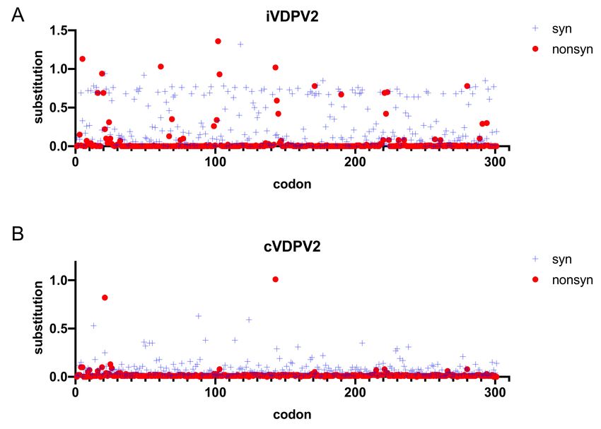
Viruses 2021, 13, 1407                                                                                             2 of 12

                         in chronic infections can lead to the emergence of unique genetic variants with distinct
                         phenotypes from the parental viruses, even for those that are commonly associated with
                         acute viral infections [14–17]. Thus, chronic infections of RNA viruses are a potential source
                         for future epidemics. In this study, we will examine the prevalence of iVDPV and molecular
                         analysis of iVDPV isolates, especially for type 2 iVDPV (iVDPV2). We then discuss the
                         future challenges in elucidating the molecular evolution and characterization of iVDPV2
                         and the remaining risk of iVDPV2 in the post-OPV era.

                         2. Definition and Classification of Vaccine-Derived Polioviruses
                              VDPVs are diverse OPV-derived variants with more than 1% nucleotide divergence
                         in the capsid VP1 region from the corresponding type 1 and type 3 Sabin strains and
                         more than 0.6% nucleotide divergence from the Sabin 2 strain [12]. VDPVs are categorized
                         by the source of samples and epidemiological information, including cVDPVs identified
                         in person-to-person transmission in the community, iVDPVs isolated from persons with
                         primary immunodeficiency (PID), and ambiguous VDPVs (aVDPVs) indicating neither
                         cVDPV nor iVDPV [12,18–20]. Some of the PID individuals may excrete iVDPV for a
                         prolonged period. In this study, we classified iVDPV excretors as long-term (6 months to
                         5 years) or chronic (more than 5 years) [12].

                         3. Status of Polio Outbreaks Due to cVDPV2
                              As previously mentioned, type 2 OPV strain (OPV2) was mainly associated with
                         cVDPV outbreaks. Recently, the World Health Organization (WHO) implemented a revised
                         polio immunization policy in the Global Polio Eradication Initiative Strategic Plan 2013–
                         2018 [21] that eliminates the type 2 component from tOPV in April 2016 [22], thereby
                         switching from the tOPV to the bivalent OPV (bOPV) including only types 1 and 3. At the
                         same time, WHO encouraged at least one dose of IPV for routine immunization [22,23]
                         to maintain the population immunity against type 2 poliovirus. In addition, a global
                         stockpile of monovalent type 2 OPV (mOPV2) was prepared and maintained for response
                         to cVDPV2 outbreaks.
                              As expected, immediately after the switch to bOPV, few cVDPV2 epidemics were
                         identified, only in the Democratic Republic of the Congo and Syria, in 2017. However, the
                         number of cVDPV2 outbreaks has been growing since 2018. There were 71 AFP cases in
                         5 countries in 2018, 366 cases in 16 countries in 2019, and 1054 cases in 24 countries in 2020 (a
                         WHO weekly report as of 1 June 2021) [24]. Molecular epidemiological analysis of cVDPV2
                         isolates revealed that most of the cVDPV2 outbreaks were derived from supplemental
                         immunization with mOPV2 after switching to bOPV in 2016, presumably in areas with
                         suboptimal intestinal immunity against type 2 poliovirus [8,25].
                              There is a critical dilemma in the creation of new cVDPV2 outbreaks by mOPV2
                         immunization in high-risk areas. Novel type 2 OPVs (nOPV2s), type 2 OPV candidates
                         more genetically stable than the conventional Sabin 2 strain, were developed and intro-
                         duced in some countries with endemic cVDPV2 transmissions as emergency responses
                         to the ongoing cVDPV2 outbreaks [26–28]. The coronavirus disease 2019 (COVID-19)
                         pandemic disrupted the surveillance and immunization activities for vaccine-preventable
                         diseases, including polio, since 2020 [11,29]. However, nOPV2 was already introduced to
                         two cVDPV2-affected countries, Nigeria and Liberia in Africa, by the end of May 2021 [30].
                         The risk of transmission of type 2 poliovirus will be significantly reduced after controlling
                         the cVDPV2 outbreaks. However, the presence of chronic iVDPV2 excretors will remain a
                         potential source of cVDPV2 transmission in communities with lowered intestinal immunity
                         against type 2 poliovirus.

                         4. Prevalence of iVDPV2-Positive Cases
                              According to Macklin et al. [12], 149 cases of iVDPV (with or without AFP) were
                         reported to the WHO from 1961 to 2019. About 60% of the iVDPV cases involved the
                         type 2 OPV strain, including 83 (56%) single iVDPV2 infections, 3 (2%) infections with a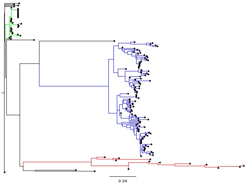
Viruses 2021, 13, 1407                                                                                                                      3 of 12

                                    mixed serotype of types 1 and 2, and 3 (2%) infections with a mixed serotype of types 2
                                    and 3. After the switch from tOPV to bOPV in 2016, the risk of iVDPV2 emergence was
                                    expected to decrease substantially. In fact, only two iVDPV2 cases were identified during
                                    2017–2019 [12,31], including a chronic iVDPV2 excretor in the Philippines (see Section 6).
                                         Among the different types of PID, patients with common variable immunodeficiency
                                    (CVID) or severe combined immunodeficiency (SCID) are associated with higher risks for
                                    long-term iVDPV infections; thus, they are less likely to clear iVDPVs spontaneously before
                                    death. However, only a limited number of chronic iVDPV excretors were reported, partly
                                    due to the low probability of survival of the PID patients and insufficient surveillance to
                                    monitor the iVDPV cases with or without AFP, especially in low-income countries. Among
                                    the 10 reported cases of chronic iVDPV excretors (Table 1), 8 were reported to be CVID,
                                    and 5 were associated with iVDPV2 [12,32,33]. Among the patients with predominantly
                                    antibody deficiencies, CVID patients are more associated with asymptomatic and prolonged
                                    iVDPV infection than those with agammaglobulinemia and hypogammaglobulinemia [32].

                                                      Table 1. Chronic iVDPV excretors.

                                                                                                                   Estimated
                     Year                                                                  Maximum VP1
    Case No.                     Localization           PID Type            Serotype                              Replication        Reference
                    Detected                                                               Divergence (%)
                                                                                                                 Period (years)
        1                1981        USA                  CVID                  1                10 †                 7.6 †             [32]
           §             1986                                                   1                5.4 †                4.7 ‡
       2                             USA                  CVID                                                                         [32,33]
                         1992                                                   2               11.8 †                9.6 †
        3                1990      Germany                CVID                  1                8.3 †                9.5 †             [32]
        4                1995        UK                   CVID                  2               17.9 †               27.83 †            [32]
        5                2000      Germany                CVID                  1                8.5 †                8.5 †             [32]
        6                2002        UK                   CVID                  2                6.3 †                6.3 †             [32]
        7                2009       India                 CVID                  1                5.2 †                 5†               [32]
        8                2009        USA                  CVID                  2               12.3 †               11.9 †             [32]
        9                2015       India                 SCID                  3               10.2 †                 6†               [32]
                                                    Hypokalemia and
       10                2019     Philippines                                   2               7.6 ||               5.0 ||             [12]
                                                   infectious diarrhea
      † Referenced from Supplementary Table 4 of Reference [32]. ‡ Referenced from Table 2 of Reference [33]. § Two iVDPVs were isolated from

      the same patient at different times. || Referenced from Table 2 of Reference [12]. CVID: common variable immunodeficiency. SCID: severe
      combined immunodeficiency.

                                    5. Genetic Analysis and Molecular Characterization of iVDPV2 Isolates
                                    5.1. Available Sequence Dataset of iVDPV2 Isolates
                                          The sequence information on cVDPV isolates is expanding due to the current cVDPV
                                    outbreaks [34]. Accordingly, there is increasing information on the molecular evolution of
                                    cVDPV [18,25,35]. However, it remains difficult to characterize iVDPVs genetically and
                                    elucidate the trends in the molecular evolution of iVDPV because of the limited availability
                                    of the sequences and clinical information of iVDPV and the lack of appropriate annotation
                                    of each iVDPV case [36,37]. The histories of the patients with long-term iVDPV infections
                                    vary greatly in terms of the duration of infection, immunological status, treatment and
                                    formulation of intravenous immunoglobulin products, and the kinds of clinical speci-
                                    mens. There are also lingering concerns about the reliability of molecular analyses using
                                    limited and potentially biased sequence datasets of the iVDPV isolates. Moreover, very
                                    little information on the capsid or full-genome sequences of the iVDPVs is available for
                                    investigating the evolutionary dynamics of iVDPV in the microenvironment in a long-term
                                    iVDPV excretor.
                                          Therefore, in this study, we have updated the iVDPV2 sequence dataset in reference
                                    to the previous reports [36–47] (Table 2). Our dataset is based on the sequence information
                                    in GenBank; thus, some of the clinical and epidemiological information is incomplete. In
                                    addition, it should be noted that there is no direct evidence of long-term excretion in the
                                    suspected iVDPV isolates from the environmental samples, although the involvement of
                                    iVDPV excretion has been highly suggested [43,44]. According to the phylogenetic analysis
                                    of the VP1 regions in our iVDPV2 dataset, the iVDPV2 isolates derived from a certain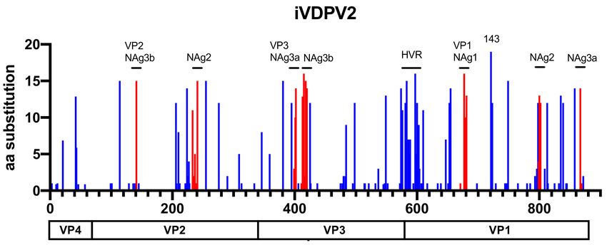
Viruses 2021, 13, 1407                                                                                                                    4 of 12

                                    long-term iVDPV excretor are genetically closely related to each other and form a unique
                                    genetic cluster, and are not related to those from other iVDPV cases (Figure 1). In addition,
                                    these iVDPV2 sequences are not genetically related to those of the cVDPV2 or aVDPV2
                                    available in the GenBank database (data not shown).

                                         Table 2. Sequence dataset of probable type 2 iVDPV strains.

                                                                                                                      Estimated
  GenBank Accession                                                                                VP1 Divergence
                         Genome Region      Year Detected   Localization    Source of Samples                        Replication     Reference
        No.                                                                                             (%)
                                                                                                                    Period (years)
      GU390704                 P1               1992           USA                  NA                  10.4             NA            [39]
    KR817050-817060            P1                                             Chronic excretor
                                              1995-2013         UK                                      17.9             28            [45]
       AJ544513           Full genome                                        (Case 4 in Table 1)
                                                                               Environmental
       AJ288062               P1                1998           Israel                                   9.4              NA            [44]
                                                                                  samples
       AY177685           Full genome           2000           Italy            iVDPV2 case             0.88             1.42          [42]
       DQ890387           Full genome           2002          Nigeria           iVDPV2 case              2.5              1.5          [41]
                                                                               Environmental
    JX913541-913647          VP1              2003-2005      Slovakia                                   3.4              NA            [43]
                                                                                  samples
       FJ517648               P1                2007          Belarus       iVDPV2 case (AFP)           1.88            1.6            [40]
                                                                                                                    NA (no OPV
       FJ517649               P1                2007          Russia        iVDPV2 case (AFP)           1.44                           [40]
                                                                                                                     history)
                                                                             Chronic excretor
       GU390707           Full genome           2009           USA                                     11.83             11.9          [39]
                                                                            (Case 8 in Table 1)
     KR709241                VP1                2013         Germany          iVDPV2 case               1.0            0.5–0.9          [38]
     KR709242                VP1                2013         Germany          iVDPV2 case               4.4            2.4–2.8          [38]
   MK660464-660492           VP1                2015          Israel          iVDPV2 case               >0.8             NA           [46,47]

                                                              NA: not available.

      Figure 1. The phylogenetic tree of VP1 sequences in 157 iVDPV2 isolates. The sequence alignment was constructed using
      Clustal Omega, and the tree was inferred using the FastTree 2.1.10 [48] with the GTR model and gamma distribution and
      visualized with FigTree. Large genetic clusters of each case study are indicated by different colors, red for a chronic excretor
      in the United Kingdom 1995–2013 [45], blue for the environmental samples from Slovakia in 2003–2005 [43], and green for
      the stool and oropharyngeal samples from Israel in 2018 [46,47].

                                    5.2. Molecular Evolution of iVDPV2
                                         During the community transmission of type 1 wild polioviruses, the rate of nu-
                                    cleotide substitution at all the sites in the entire capsid P1 region was estimated to be
Viruses 2021, 13, 1407 5 of 12
1.03 × 10⁻² substitutions/site/year [49]. Similarly, the average estimated substitution rate
was 1.14 × 10⁻² substitutions/site/year for the major genetic lineages of cVDPV2 isolates
in Nigeria [18]. In the case of iVDPV2, the rate of nucleotide substitution in the VP1 region
was 1.51 × 10⁻² substitutions/site/year for the most long-term iVDPV2 excretor in the
United Kingdom (for approximately 28 years at the time) [45]. Shaghaghi et al. observed
rapid molecular evolution in some iVDPV cases at the initial stages of virus replication
after OPV administration [32]. After the initial replication period of the OPV strains, the
subsequent mutations would accumulate at a nearly uniform rate of 1 to 2% nucleotide
changes per year [32,50,51]. Due to a strong positive selection in vaccinees immunized
with type 2 OPV, the so-called gatekeeper mutations from the attenuated Sabin 2 strain,
including three mutations at A481G, U2909C (VP1-I143T), and U398C, are rapidly selected
and fixed in the first several weeks post-vaccination [35,52,53]. Those gatekeeper mutations
may contribute to the virus replication fitness in the human intestine and higher initial
evolution rates of iVDPV and cVDPV immediately after the OPV administration.
5.3. Amino Acid Substitutions in Phenotypic Determinants
Consistent with Zhao et al. [37], it is difficult to distinguish between the iVDPV and
cVDPV strains in our iVDPV2 dataset by only comparing the nucleotide and amino acid
sequences in the VP1 region (data not shown). However, a codon-by-codon comparison
throughout the VP1 region using SNAP [54] showed that the rates of non-synonymous
substitutions in the iVDPV strains were higher than those in cVDPV2 (Figure 2). These data
suggest that the molecular evolution of the capsid proteins associated with phenotypic
determinants, including antigenic sites, is more likely to occur in the iVDPV strains than in
the cVDPV strains, especially for long-term iVDPV infections. Among the 19 iVDPV2 cases,
amino acid substitutions are frequently identified in some of the neutralizing antigenic
sites (NAgs) and a hypervariable region at the N-terminus of VP1 and VP1–143 in the
non-NAg sites (Figure 3). The N-terminus of VP1 forms an amphipathic helix and is
presumably involved in the interaction with the host cell membrane. Regardless of the high
variability of this domain, the predicted amphipathic helix is maintained [55]. McDonald
et al. reported a decline in neutralizing capacity of human sera immunized with IPV and
OPV against the highly evolved iVDPV2 from long-term excretors due to the amino acid
substitutions in the NAgs [36]. On the other hand, there was no significant change in the
neutralization titers of the human sera against a series of iVDPV2 variants from a chronic
iVDPV2 excretor in the United Kingdom, compared with those against the parental Sabin
2 strain, regardless of amino acid substitutions in the NAgs in the Sabin 2 strain [46]. No
significant changes in neutralizing capacity were observed in the highly evolved cVDPV2
isolates in Nigeria compared to those in the Sabin 2 strain, suggesting that the changes
in the NAgs of the cVDPV2 were lower than those in the iVDPV2 [36,37,55] (Figure 4).
Non-synonymous substitutions frequently occurred not only in the NAg sites but also in
the non-NAg sites located on the surface of the capsid (Figure 4), potentially affecting the
antigenicity or in vivo fitness of the VDPVs.Viruses 2021, 13, 1407 6 of 12
Figure 2. The frequency of synonymous and non-synonymous substitutions of the VP1 amino acid sequences from
157 iVDPV2 and 567 cVDPV2 isolates. Sequence divergence was analyzed using SNAP (Synonymous Non-synonymous
Analysis Program, https://www.hiv.lanl.gov/content/sequence/SNAP/SNAP.html (accessed on 28 May 2021)) [54]. The
synonymous (blue crosses) and non-synonymous substitutions (red circles) of the VDPV sequence relative to the Sabin 2
strain are depicted. (A) iVDPV2. (B) cVDPV2.
Figure 3. The number of amino acid substitutions in each position of the P1 capsid region of 19 iVDPV2 isolates. The amino
acid substitutions in the neutralizing antigenic (NAg) sites are shown in red. The positions of hypervariable residues (HVRs)
at the N-terminal and a gatekeeper substitution at VP1–143 of VP1 are indicated.Viruses 2021, 13, 1407 7 of 12
Figure 4. The locations of amino acid substitutions in selected iVDPV2 isolates. The three-dimensional structure of type 2
wild poliovirus strain MEF-1 was obtained from PDB 1EAH and represented as a pentameric unit ((A): outside; (B): inside
of capsid). Each protomer contains a single copy of VP1, VP2, VP3, and VP4 distinguished by gray colors. The location
of frequent amino acid substitutions (more than 4 out of 19 iVDPV2 isolates) relative to the Sabin 2 strain is colored. The
neutralizing antigenic sites are red. The hypervariable residues and the gatekeeper substitution of VP1-143 are green. Other
amino acid changes are blue. The image was generated using PyMOL 2.4.2 (Schrödinger).
G481 and VP1-I143T were identified as the major attenuation determinants for the
Sabin 2 strain [9,19]. As previously mentioned, these two sites are gatekeeper mutations
that are rapidly selected and fixed immediately after the administration of OPV2 [35]; most
of the iVDPV2 isolates very likely have those mutations in the attenuation sites. In fact, a
series of highly evolved iVDPV2 isolates from a chronic excretor in the United Kingdom
are more neurovirulent than the parental Sabin 2 strain in a human poliovirus receptor
transgenic mouse model [45].
5.4. Genomic Recombination
Most of the cVDPV isolates associated with polio outbreaks are recombinants of OPV-
derived capsid sequences and non-capsid sequences derived from non-polio enteroviruses
(NPEVs), particularly those belonging to Enterovirus C [9,18–20,56–61]. To our knowledge,
there is no concrete evidence of long-term excretion or local transmission of iVDPV strains
recombinant with NPEV in part due to limited information on the full-genome sequences
of iVDPV isolates. The recombinant viruses derived from the Sabin 2 capsid sequences
with the non-capsid sequences from Sabin 3 or Sabin 1 are frequently detected in healthy
vaccinees and VAPP cases in the early stages of virus replication after tOPV immuniza-
tion [55,62,63]. The recombination between different genetic lineages among genetically
divergent iVDPV variants was identified in a long-term type 1 iVDPV excretor in Tai-
wan [64]; therefore, recombinations, as well as mutations, may contribute to the molecular
evolution and viral fitness in the microenvironment of iVDPV infection. However, the
virological significance of viral recombination in the host is still uncertain because only a
few reports based on the full-genome sequences of iVDPV are available.
5.5. In Vivo Fitness and Quasi-Species
In individuals with long-term iVDPV infections, genetically diverse iVDPV variants
may co-exist as viral quasi-species. The role of quasi-species in the molecular evolution of
iVDPV remains unclear; however, in general, the genetic diversity of polioviruses within
infected individuals is known to be involved in the in vivo molecular evolution and the
rise of certain viral phenotypes, including neurovirulence [65–67]. Historically, poliovirusViruses 2021, 13, 1407 8 of 12
quasi-species were analyzed by the conventional plaque cloning and Sanger sequencing of
virus isolates. Recently, next-generation sequencing (NGS) technologies were applied to
study viral quasi-species and the molecular evolution of various RNA viruses, including
those of the OPV strains and cVDPVs, but not for those from iVDPV cases [25,52,53]. In
the future, more comprehensive and quantitative analyses on the genetic diversity of the
quasi-species of iVDPV variants in infected individuals, compared to those in cVDPV and
OPV strains, using NGS will be required.
6. Current and Future Risk of iVDPV2
Although there is no substantial evidence of polio outbreaks caused by cVDPV2
derived from iVDPV2 excretors, the relative risk of long-term iVDPV2 infection will rise as
the population with lower intestinal immunity to type 2 poliovirus among the individuals
immunized with bOPV and IPV increase [10,13,68,69]. The WHO Western Pacific Region
(WPR) was certified to be wild polio-free since 2000, except for several importations of wild
polioviruses from endemic countries [70–72]. Since 2001, several cVDPV outbreaks were
reported in the WPR; however, all of them were effectively controlled using supplemental
OPV immunization [60,73,74]. In September 2019, type 1 VDPV isolates (more than 3%
nucleotide divergence from the Sabin 1 strain in the VP1 region) were identified from
environmental sewage samples collected in the National Capital Region (NCR) in the
Philippines. Highly evolved type 2 VDPV isolates (more than 7% nucleotide divergence
from the Sabin 2 strain in the VP1 region) were concomitantly detected in some of the same
sewage samples [8,25].
Meanwhile, more extensive AFP and environmental surveillance identified many
VDPV1 and VDPV2 isolates from AFP cases and environmental samples from different geo-
graphical areas, confirming that the simultaneous and widespread transmission of cVDPV1
and cVDPV2 was associated with paralytic polio outbreaks in the Philippines and Malaysia
in 2019–2020. At the same time, intensified surveillance identified highly divergent VDPV2
isolates (more than 7% nucleotide difference in the VP1) from a 5-year-old male with re-
duced antibody levels who had received three doses of tOPV from August 2014 to February
2015 in Laguna province, close to the NCR in the Philippines [12]. Follow-up samplings
demonstrated that genetically related VDPV2 variants were continuously detected in stool
samples of the patient from August 2019 to now (as of May 2021), indicating that the patient
is a chronic iVDPV2 excretor.
At least in the VP1 region, the cVDPV2 isolates in the Philippines in 2019–2020 are not
genetically related to the iVDPV2 isolates from the chronic excretor in Laguna. Macklin et al.
estimated that the cVDPV2 isolates in the Philippines were seeded by tOPV immunization
around 2014 before the switch to bOPV in 2016 [25]. According to the genetic diversity of
the iVDPV2 isolates from the Sabin 2 strain and the history of OPV immunization of the
patient, the chronic iVDPV2 infection might have been initiated in 2014–2015 at almost the
same time with the emergence of cVDPV2. The origin and evolution of both cVDPV2 and
iVDPV2 remain uncertain epidemiologically and genetically, partly due to the lack of any
iVDPV2 and cVDPV2 cases for nearly 5 years in the Philippines.
Recently, Valesano et al. found that most OPV2-derived variants with the gatekeeper
mutations that appeared in OPV recipients rapidly could not survive during virus trans-
mission to the close contacts of the recipients due to the tight transmission bottleneck. This
finding suggests distinct mechanisms of molecular evolution between viral replication in
the hosts and during community transmission [52]. More detailed and comprehensive
genetic characterization, taking the previously mentioned points on the molecular charac-
teristics of iVDPV2 into account, is in progress to elucidate the relationship and molecular
evolution of cVDPV2 and iVDPV2 in the Philippines.
7. Conclusions
The last cVDPV2 isolate in the Philippines was detected from an environmental sample
collected in January 2020. No cVDPV2 was identified from AFP and environmental samplesViruses 2021, 13, 1407 9 of 12
after that. However, the incidences of cVDPV2 and iVDPV2 in the Philippines and Malaysia
in 2019–2020 highlight the risk of inapparent infections or silent VDPV transmission
or both, even in areas with a long-standing polio-free status. Further research on the
genetic characterization and molecular evolution of iVDPV2 will enable us to mitigate the
remaining risk of widespread transmission of iVDPV2 during the post-OPV era.
Author Contributions: Writing—initial draft preparation, H.S.; writing—review and editing, K.K. and
H.S.; visualization, K.K. All authors have read and agreed to the published version of the manuscript.
Funding: This research was funded by the Research Program on Emerging and Re-emerging In-
fectious Diseases from the Japan Agency for Medical Research and Development, Grant Number
JP21fk0108084 (to K.K. and H.S.).
Institutional Review Board Statement: Not applicable.
Informed Consent Statement: Not applicable.
Data Availability Statement: All data of this study are available within this manuscript (Table 2).
Acknowledgments: The authors thank Enago for the scientific editing of the manuscript. The authors
also thank Minetaro Arita and Yorihiro Nishimura for helpful comments on the manuscript.
Conflicts of Interest: The authors declare no conflict of interest.
References
1. Adams, A.; Salisbury, D.M. Eradicating polio. Science 2015, 350, 609. [CrossRef]
2. Makoni, M. Africa eradicates wild polio. Lancet Microbe 2020, 1, e243. [CrossRef]
3. Tuma, J.N.; Wilkinson, A.L.; Diop, O.M.; Jorba, J.; Gardner, T.; Snider, C.J.; Anand, A.; Ahmed, J. Surveillance to track progress
toward polio eradication—Worldwide, 2019–2020. MMWR Morb. Mortal. Wkly. Rep. 2021, 70, 667–673. [CrossRef]
4. Global Polio Eradication Initiative. Wild Poliovirus List. Available online: https://polioeradication.org/wp-content/uploads/20
21/06/weekly-polio-analyses-WPV-20210615.pdf (accessed on 2 July 2021).
5. Cochi, S.L.; Pallansch, M.A. The long and winding road to eradicate vaccine-related polioviruses. J. Infect. Dis. 2021, 223, 7–9.
[CrossRef]
6. Chumakov, K.; Ehrenfeld, E.; Agol, V.I.; Wimmer, E. Polio eradication at the crossroads. Lancet Glob. Health 2021. [CrossRef]
7. Global Polio Eradication Initiative. Circulating Vaccine-Derived Poliovirus. Available online: http://polioeradication.org/polio-
today/polio-now/this-week/circulating-vaccine-derived-poliovirus/ (accessed on 2 July 2021).
8. Alleman, M.M.; Jorba, J.; Greene, S.A.; Diop, O.M.; Iber, J.; Tallis, G.; Goel, A.; Wiesen, E.; Wassilak, S.G.F.; Burns, C.C. Update on
vaccine-derived poliovirus outbreaks—Worldwide, July 2019–February 2020. MMWR Morb. Mortal. Wkly. Rep. 2020, 69, 489–495.
[CrossRef]
9. Burns, C.C.; Diop, O.M.; Sutter, R.W.; Kew, O.M. Vaccine-derived polioviruses. J. Infect. Dis. 2014, 210 (Suppl. S1), S283–S293.
[CrossRef]
10. Yan, D.; Wang, D.; Zhang, Y.; Li, X.; Tang, H.; Guan, J.; Song, Y.; Zhu, S.; Xu, W. Implication of a High Risk for Type 2 Vaccine-
Derived Poliovirus Emergence and Transmission After the Switch From Trivalent to Bivalent Oral Poliovirus Vaccine. J. Infect.
Dis. 2021, 223, 113–118. [CrossRef]
11. Meyer, E.; Sikka, N.; Durry, E.; Datta, D. Notes from the field: CDC polio surge response to expanding outbreaks of Type 2
circulating vaccine-derived poliovirus—Africa and Philippines, September 2019–March 2020. MMWR Morb. Mortal. Wkly. Rep.
2020, 69, 1182–1183. [CrossRef]
12. Macklin, G.; Diop, O.M.; Humayun, A.; Shahmahmoodi, S.; El-Sayed, Z.A.; Triki, H.; Rey, G.; Avagyan, T.; Grabovac, V.; Jorba, J.;
et al. Update on immunodeficiency-associated vaccine-derived polioviruses—Worldwide, July 2018–December 2019. MMWR
Morb. Mortal. Wkly. Rep. 2020, 69, 913–917. [CrossRef]
13. Aghamohammadi, A.; Abolhassani, H.; Kutukculer, N.; Wassilak, S.G.; Pallansch, M.A.; Kluglein, S.; Quinn, J.; Sutter, R.W.; Wang,
X.; Sanal, O.; et al. Patients with primary immunodeficiencies are a reservoir of poliovirus and a risk to polio eradication. Front.
Immunol. 2017, 8, 685. [CrossRef] [PubMed]
14. Mbala-Kingebeni, P.; Pratt, C.; Mutafali-Ruffin, M.; Pauthner, M.G.; Bile, F.; Nkuba-Ndaye, A.; Black, A.; Kinganda-Lusamaki, E.;
Faye, M.; Aziza, A.; et al. Ebola virus transmission initiated by relapse of systemic Ebola virus disease. N. Engl. J. Med. 2021, 384,
1240–1247. [CrossRef]
15. Kemp, S.A.; Collier, D.A.; Datir, R.P.; Ferreira, I.; Gayed, S.; Jahun, A.; Hosmillo, M.; Rees-Spear, C.; Mlcochova, P.; Lumb, I.U.;
et al. SARS-CoV-2 evolution during treatment of chronic infection. Nature 2021, 592, 277–282. [CrossRef] [PubMed]
16. Perelygina, L.; Chen, M.H.; Suppiah, S.; Adebayo, A.; Abernathy, E.; Dorsey, M.; Bercovitch, L.; Paris, K.; White, K.P.; Krol, A.;
et al. Infectious vaccine-derived rubella viruses emerge, persist, and evolve in cutaneous granulomas of children with primary
immunodeficiencies. PLoS Pathog. 2019, 15, e1008080. [CrossRef] [PubMed]Viruses 2021, 13, 1407 10 of 12
17. Xue, K.S.; Stevens-Ayers, T.; Campbell, A.P.; Englund, J.A.; Pergam, S.A.; Boeckh, M.; Bloom, J.D. Parallel evolution of influenza
across multiple spatiotemporal scales. Elife 2017, 6. [CrossRef]
18. Burns, C.C.; Shaw, J.; Jorba, J.; Bukbuk, D.; Adu, F.; Gumede, N.; Pate, M.A.; Abanida, E.A.; Gasasira, A.; Iber, J.; et al. Multiple
independent emergences of Type 2 vaccine-derived polioviruses during a large outbreak in northern Nigeria. J. Virol. 2013, 87,
4907–4922. [CrossRef]
19. Kew, O.M.; Sutter, R.W.; de Gourville, E.M.; Dowdle, W.R.; Pallansch, M.A. Vaccine-derived polioviruses and the endgame
strategy for global polio eradication. Annu. Rev. Microbiol. 2005, 59, 587–635. [CrossRef]
20. Kew, O.M.; Wright, P.F.; Agol, V.I.; Delpeyroux, F.; Shimizu, H.; Nathanson, N.; Pallansch, M.A. Circulating vaccine-derived
polioviruses: Current state of knowledge. Bull. World Health Organ. 2004, 82, 16–23.
21. Global Polio Eradication Initiative. Polio Eradication and Endgame Strategic Plan 2013–2018. Available online: https://
polioeradication.org/wp-content/uploads/2016/07/PEESP_EN_A4.pdf (accessed on 2 July 2021).
22. Garon, J.; Seib, K.; Orenstein, W.A.; Ramirez Gonzalez, A.; Chang Blanc, D.; Zaffran, M.; Patel, M. Polio endgame: The global
switch from tOPV to bOPV. Expert Rev. Vaccines 2016, 15, 693–708. [CrossRef]
23. Global Polio Eradication Initiative. Introduction of inactivated poliovirus vaccine and switch from trivalent to bivalent oral
poliovirus vaccine—Worldwide, 2013–2016. MMWR Morb. Mortal. Wkly. Rep. 2015, 64, 699–702.
24. Global Polio Eradication Initiative. Global Circulating Vaccine-Derived Poliovirus (cVDPV) as of 01 June 2021. Available
online: https://polioeradication.org/wp-content/uploads/2021/06/weekly-polio-analyses-cVDPV-20210601.pdf (accessed on 2
July 2021).
25. Macklin, G.R.; O’Reilly, K.M.; Grassly, N.C.; Edmunds, W.J.; Mach, O.; Santhana Gopala Krishnan, R.; Voorman, A.; Vertefeuille,
J.F.; Abdelwahab, J.; Gumede, N.; et al. Evolving epidemiology of poliovirus serotype 2 following withdrawal of the serotype 2
oral poliovirus vaccine. Science 2020, 368, 401–405. [CrossRef] [PubMed]
26. Sáez-Llorens, X.; Bandyopadhyay, A.S.; Gast, C.; Leon, T.; DeAntonio, R.; Jimeno, J.; Caballero, M.I.; Aguirre, G.; Oberste,
M.S.; Weldon, W.C.; et al. Safety and immunogenicity of two novel type 2 oral poliovirus vaccine candidates compared with a
monovalent type 2 oral poliovirus vaccine in children and infants: Two clinical trials. Lancet 2021, 397, 27–38. [CrossRef]
27. Yeh, M.T.; Bujaki, E.; Dolan, P.T.; Smith, M.; Wahid, R.; Konz, J.; Weiner, A.J.; Bandyopadhyay, A.S.; Van Damme, P.; De Coster,
I.; et al. Engineering the live-attenuated polio vaccine to prevent reversion to virulence. Cell Host Microbe 2020, 27, 736–751.e8.
[CrossRef]
28. Van Damme, P.; De Coster, I.; Bandyopadhyay, A.S.; Revets, H.; Withanage, K.; De Smedt, P.; Suykens, L.; Oberste, M.S.; Weldon,
W.C.; Costa-Clemens, S.A.; et al. The safety and immunogenicity of two novel live attenuated monovalent (serotype 2) oral
poliovirus vaccines in healthy adults: A double-blind, single-centre phase 1 study. Lancet 2019, 394, 148–158. [CrossRef]
29. Zomahoun, D.J.; Burman, A.L.; Snider, C.J.; Chauvin, C.; Gardner, T.; Lickness, J.S.; Ahmed, J.A.; Diop, O.; Gerber, S.; Anand,
A. Impact of COVID-19 pandemic on global poliovirus surveillance. MMWR Morb. Mortal. Wkly. Rep. 2021, 69, 1648–1652.
[CrossRef] [PubMed]
30. WHO. Statement Following the Twenty-Eighth IHR Emergency Committee for Polio. Available online: https://www.who.int/
news/item/21-05-2021-statement-following-the-twenty-eighth-ihr-emergency-committee-for-polio (accessed on 2 July 2021).
31. Jorba, J.; Diop, O.M.; Iber, J.; Henderson, E.; Zhao, K.; Sutter, R.W.; Wassilak, S.G.F.; Burns, C.C. Update on vaccine-derived
polioviruses—Worldwide, January 2017–June 2018. MMWR Morb. Mortal. Wkly. Rep. 2018, 67, 1189–1194. [CrossRef] [PubMed]
32. Shaghaghi, M.; Soleyman-Jahi, S.; Abolhassani, H.; Yazdani, R.; Azizi, G.; Rezaei, N.; Barbouche, M.R.; McKinlay, M.A.;
Aghamohammadi, A. New insights into physiopathology of immunodeficiency-associated vaccine-derived poliovirus infection;
systematic review of over 5 decades of data. Vaccine 2018, 36, 1711–1719. [CrossRef] [PubMed]
33. Centers for Disease Control and Prevention. Update on vaccine-derived polioviruses. MMWR Morb. Mortal. Wkly. Rep. 2006, 55,
1093–1097.
34. Jorgensen, D.; Pons-Salort, M.; Shaw, A.G.; Grassly, N.C. The role of genetic sequencing and analysis in the polio eradication
programme. Virus Evol. 2020, 6, veaa040. [CrossRef] [PubMed]
35. Stern, A.; Yeh, M.T.; Zinger, T.; Smith, M.; Wright, C.; Ling, G.; Nielsen, R.; Macadam, A.; Andino, R. The evolutionary pathway
to virulence of an RNA virus. Cell 2017, 169, 35–46.e19. [CrossRef] [PubMed]
36. McDonald, S.L.; Weldon, W.C.; Wei, L.; Chen, Q.; Shaw, J.; Zhao, K.; Jorba, J.; Kew, O.M.; Pallansch, M.A.; Burns, C.C.; et al.
Neutralization capacity of highly divergent type 2 vaccine-derived polioviruses from immunodeficient patients. Vaccine 2020, 38,
3042–3049. [CrossRef] [PubMed]
37. Zhao, K.; Jorba, J.; Shaw, J.; Iber, J.; Chen, Q.; Bullard, K.; Kew, O.M.; Burns, C.C. Are circulating type 2 vaccine-derived
polioviruses (VDPVs) genetically distinguishable from immunodeficiency-associated VDPVs? Comput. Struct. Biotechnol. J. 2017,
15, 456–462. [CrossRef] [PubMed]
38. Schubert, A.; Böttcher, S.; Eis-Hübinger, A.M. Two cases of vaccine-derived poliovirus infection in an Oncology Ward. N. Engl. J.
Med. 2016, 374, 1296–1298. [CrossRef] [PubMed]
39. DeVries, A.S.; Harper, J.; Murray, A.; Lexau, C.; Bahta, L.; Christensen, J.; Cebelinski, E.; Fuller, S.; Kline, S.; Wallace, G.S.; et al.
Vaccine-derived poliomyelitis 12 years after infection in Minnesota. N. Engl. J. Med. 2011, 364, 2316–2323. [CrossRef] [PubMed]
40. Yakovenko, M.L.; Korotkova, E.A.; Ivanova, O.E.; Eremeeva, T.P.; Samoilovich, E.; Uhova, I.; Gavrilin, G.V.; Agol, V.I. Evolution
of the Sabin vaccine into pathogenic derivatives without appreciable changes in antigenic properties: Need for improvement of
current poliovirus surveillance. J. Virol. 2009, 83, 3402–3406. [CrossRef]Viruses 2021, 13, 1407 11 of 12
41. Adu, F.; Iber, J.; Bukbuk, D.; Gumede, N.; Yang, S.J.; Jorba, J.; Campagnoli, R.; Sule, W.F.; Yang, C.F.; Burns, C.; et al. Isolation of
recombinant type 2 vaccine-derived poliovirus (VDPV) from a Nigerian child. Virus Res. 2007, 127, 17–25. [CrossRef]
42. Buttinelli, G.; Donati, V.; Fiore, S.; Marturano, J.; Plebani, A.; Balestri, P.; Soresina, A.R.; Vivarelli, R.; Delpeyroux, F.; Martin, J.;
et al. Nucleotide variation in Sabin type 2 poliovirus from an immunodeficient patient with poliomyelitis. J. Gen. Virol. 2003, 84,
1215–1221. [CrossRef]
43. Hovi, T.; Paananen, A.; Blomqvist, S.; Savolainen-Kopra, C.; Al-Hello, H.; Smura, T.; Shimizu, H.; Nadova, K.; Sobotova, Z.;
Gavrilin, E.; et al. Characteristics of an environmentally monitored prolonged Type 2 vaccine derived poliovirus shedding
episode that stopped without intervention. PLoS ONE 2013, 8, e66849. [CrossRef]
44. Shulman, L.M.; Manor, Y.; Handsher, R.; Delpeyroux, F.; McDonough, M.J.; Halmut, T.; Silberstein, I.; Alfandari, J.; Quay, J.;
Fisher, T.; et al. Molecular and antigenic characterization of a highly evolved derivative of the type 2 oral poliovaccine strain
isolated from sewage in Israel. J. Clin. Microbiol. 2000, 38, 3729–3734. [CrossRef]
45. Dunn, G.; Klapsa, D.; Wilton, T.; Stone, L.; Minor, P.D.; Martin, J. Twenty-eight years of poliovirus replication in an immunodefi-
cient individual: Impact on the global polio eradication initiative. PLoS Pathog. 2015, 11, e1005114. [CrossRef]
46. Weil, M.; Rahav, G.; Somech, R.; Stauber, T.; Alfandari, J.; Weiss, L.; Silberstein, I.; Indenbaum, V.; Or, I.B.; Mendelson, E.; et al.
First report of a persistent oropharyngeal infection of type 2 vaccine-derived poliovirus (iVDPV2) in a primary immune deficient
(PID) patient after eradication of wild type 2 poliovirus. Int. J. Infect. Dis. 2019, 83, 40–43. [CrossRef] [PubMed]
47. Weil, M.; Shulman, L.M.; Heiman, S.; Stauber, T.; Alfandari, J.; Weiss, L.; Silberstein, I.; Indenbaum, V.; Mendelson, E.; Sofer, D.
Prolonged excretion of type-2 poliovirus from a primary immune deficient patient during the transition to a type-2 poliovirus-free
world, Israel, 2016. Euro Surveill. 2016, 21. [CrossRef] [PubMed]
48. Price, M.N.; Dehal, P.S.; Arkin, A.P. FastTree 2–Approximately maximum-likelihood trees for large alignments. PLoS ONE 2010, 5,
e9490. [CrossRef] [PubMed]
49. Jorba, J.; Campagnoli, R.; De, L.; Kew, O. Calibration of multiple poliovirus molecular clocks covering an extended evolutionary
range. J. Virol. 2008, 82, 4429–4440. [CrossRef]
50. Shaghaghi, M.; Irannejad, M.; Abolhassani, H.; Shahmahmoodi, S.; Hamidieh, A.A.; Soleyman-Jahi, S.; Yazdani, R.; Azizi, G.;
Aghamohammadi, A. Clearing vaccine-derived poliovirus infection following hematopoietic stem cell transplantation: A case
report and review of literature. J. Clin. Immunol. 2018, 38, 610–616. [CrossRef]
51. Odoom, J.K.; Yunus, Z.; Dunn, G.; Minor, P.D.; Martin, J. Changes in population dynamics during long-term evolution of Sabin
type 1 poliovirus in an immunodeficient patient. J. Virol. 2008, 82, 9179–9190. [CrossRef]
52. Valesano, A.L.; Taniuchi, M.; Fitzsimmons, W.J.; Islam, M.O.; Ahmed, T.; Zaman, K.; Haque, R.; Wong, W.; Famulare, M.; Lauring,
A.S. The early evolution of oral poliovirus vaccine is shaped by strong positive selection and tight transmission bottlenecks. Cell
Host Microbe 2021, 29, 32–43.e4. [CrossRef]
53. Famulare, M.; Chang, S.; Iber, J.; Zhao, K.; Adeniji, J.A.; Bukbuk, D.; Baba, M.; Behrend, M.; Burns, C.C.; Oberste, M.S. Sabin
vaccine reversion in the field: A comprehensive analysis of Sabin-like poliovirus isolates in Nigeria. J. Virol. 2016, 90, 317–331.
[CrossRef] [PubMed]
54. Korber, B. HIV signature and sequence variation analysis. In Computational Analysis of HIV Molecular Sequences; Rodrigo, A.G.,
Learn, G.H., Eds.; Kluwer Academic Publishers: Dordrecht, The Netherlands, 2000; pp. 55–72.
55. Shaw, J.; Jorba, J.; Zhao, K.; Iber, J.; Chen, Q.; Adu, F.; Adeniji, A.; Bukbuk, D.; Baba, M.; Henderson, E.; et al. Dynamics of
evolution of poliovirus neutralizing antigenic sites and other capsid functional domains during a large and prolonged outbreak.
J. Virol. 2018, 92. [CrossRef]
56. Muslin, C.; Joffret, M.L.; Pelletier, I.; Blondel, B.; Delpeyroux, F. Evolution and emergence of enteroviruses through intra- and
inter-species recombination: Plasticity and phenotypic impact of modular genetic exchanges in the 50 untranslated region. PLoS
Pathog. 2015, 11, e1005266. [CrossRef]
57. Jegouic, S.; Joffret, M.L.; Blanchard, C.; Riquet, F.B.; Perret, C.; Pelletier, I.; Colbere-Garapin, F.; Rakoto-Andrianarivelo, M.;
Delpeyroux, F. Recombination between polioviruses and co-circulating coxsackie A viruses: Role in the emergence of pathogenic
vaccine-derived polioviruses. PLoS Pathog. 2009, 5, e1000412. [CrossRef]
58. Rakoto-Andrianarivelo, M.; Guillot, S.; Iber, J.; Balanant, J.; Blondel, B.; Riquet, F.; Martin, J.; Kew, O.; Randriamanalina, B.;
Razafinimpiasa, L.; et al. Co-circulation and evolution of polioviruses and species C enteroviruses in a district of Madagascar.
PLoS Pathog. 2007, 3, e1911. [CrossRef]
59. Arita, M.; Zhu, S.L.; Yoshida, H.; Yoneyama, T.; Miyamura, T.; Shimizu, H. A Sabin 3-derived poliovirus recombinant contained a
sequence homologous with indigenous human enterovirus species C in the viral polymerase coding region. J. Virol. 2005, 79,
12650–12657. [CrossRef]
60. Shimizu, H.; Thorley, B.; Paladin, F.J.; Brussen, K.A.; Stambos, V.; Yuen, L.; Utama, A.; Tano, Y.; Arita, M.; Yoshida, H.; et al.
Circulation of type 1 vaccine-derived poliovirus in the Philippines in 2001. J. Virol. 2004, 78, 13512–13521. [CrossRef]
61. Kew, O.; Morris-Glasgow, V.; Landaverde, M.; Burns, C.; Shaw, J.; Garib, Z.; Andre, J.; Blackman, E.; Freeman, C.J.; Jorba, J.; et al.
Outbreak of poliomyelitis in Hispaniola associated with circulating type 1 vaccine-derived poliovirus. Science 2002, 296, 356–359.
[CrossRef] [PubMed]
62. Cuervo, N.S.; Guillot, S.; Romanenkova, N.; Combiescu, M.; Aubert-Combiescu, A.; Seghier, M.; Caro, V.; Crainic, R.; Delpeyroux,
F. Genomic features of intertypic recombinant Sabin poliovirus strains excreted by primary vaccinees. J. Virol. 2001, 75, 5740–5751.
[CrossRef]Viruses 2021, 13, 1407 12 of 12
63. Gavrilin, G.V.; Cherkasova, E.A.; Lipskaya, G.Y.; Kew, O.M.; Agol, V.I. Evolution of circulating wild poliovirus and of vaccine-
derived poliovirus in an immunodeficient patient: A unifying model. J. Virol. 2000, 74, 7381–7390. [CrossRef]
64. Yang, C.F.; Chen, H.Y.; Jorba, J.; Sun, H.C.; Yang, S.J.; Lee, H.C.; Huang, Y.C.; Lin, T.Y.; Chen, P.J.; Shimizu, H.; et al. Intratypic
recombination among lineages of type 1 vaccine-derived poliovirus emerging during chronic infection of an immunodeficient
patient. J. Virol. 2005, 79, 12623–12634. [CrossRef]
65. Acevedo, A.; Brodsky, L.; Andino, R. Mutational and fitness landscapes of an RNA virus revealed through population sequencing.
Nature 2014, 505, 686–690. [CrossRef]
66. Vignuzzi, M.; Wendt, E.; Andino, R. Engineering attenuated virus vaccines by controlling replication fidelity. Nat. Med. 2008, 14,
154–161. [CrossRef]
67. Vignuzzi, M.; Stone, J.K.; Arnold, J.J.; Cameron, C.E.; Andino, R. Quasispecies diversity determines pathogenesis through
cooperative interactions in a viral population. Nature 2006, 439, 344–348. [CrossRef]
68. Kalkowska, D.A.; Pallansch, M.A.; Cochi, S.L.; Kovacs, S.D.; Wassilak, S.G.F.; Thompson, K.M. Updated characterization of
post-OPV cessation risks: Lessons from 2019 Serotype 2 outbreaks and implications for the probability of OPV restart. Risk Anal.
2021, 41, 320–328. [CrossRef]
69. Duintjer Tebbens, R.J.; Thompson, K.M. Comprehensive screening for immunodeficiency-associated vaccine-derived poliovirus:
An essential oral poliovirus vaccine cessation risk management strategy. Epidemiol. Infect. 2017, 145, 217–226. [CrossRef]
70. Gurung, S.; Harris, J.B.; Eltayeb, A.O.; Hampton, L.M.; Diorditsa, S.; Avagyan, T.; Schluter, W.W. Experience with inactivated
polio vaccine introduction and the “switch” from trivalent to bivalent oral polio vaccine in the World Health Organization’s
western Pacific region. J. Infect. Dis. 2017, 216 (Suppl. S1), S101–S108. [CrossRef]
71. Adams, A.; Boualam, L.; Diorditsa, S.; Gregory, C.; Jee, Y.; Mendoza-Aldana, J.; Roesel, S. Maintaining polio-free certification
in the World Health Organization western Pacific Region for over a decade. J. Infect. Dis. 2014, 210 (Suppl. S1), S259–S267.
[CrossRef] [PubMed]
72. Luo, H.M.; Zhang, Y.; Wang, X.Q.; Yu, W.Z.; Wen, N.; Yan, D.M.; Wang, H.Q.; Wushouer, F.; Wang, H.B.; Xu, A.Q.; et al.
Identification and control of a poliomyelitis outbreak in Xinjiang, China. N. Engl. J. Med. 2013, 369, 1981–1990. [CrossRef]
73. Bauri, M.; Wilkinson, A.L.; Ropa, B.; Feldon, K.; Snider, C.J.; Anand, A.; Tallis, G.; Boualam, L.; Grabovac, V.; Avagyan, T.; et al.
Notes from the field: Circulating vaccine-derived poliovirus Type 1 and outbreak response—Papua New Guinea, 2018. MMWR
Morb. Mortal. Wkly. Rep. 2019, 68, 119–120. [CrossRef]
74. Zhang, Y.; Yan, D.; Zhu, S.; Nishimura, Y.; Ye, X.; Wang, D.; Jorba, J.; Zhu, H.; An, H.; Shimizu, H.; et al. An insight into
recombination with Enterovirus species C and Nucleotide G-480 reversion from the viewpoint of neurovirulence of vaccine-
derived polioviruses. Sci. Rep. 2015, 5, 17291. [CrossRef]You can also read