The loss of DHX15 impairs endothelial energy metabolism, lymphatic drainage and tumor metastasis in mice
←
→
Page content transcription
If your browser does not render page correctly, please read the page content below
ARTICLE
                  https://doi.org/10.1038/s42003-021-02722-w                OPEN

                  The loss of DHX15 impairs endothelial energy
                  metabolism, lymphatic drainage and tumor
                  metastasis in mice
                  Jordi Ribera 1, Irene Portolés1, Bernat Córdoba-Jover1, Juan Rodríguez-Vita 1,2, Gregori Casals 1,
                  Bernardino González-de la Presa1, Mariona Graupera 3, Estel Solsona-Vilarrasa4,5, Carmen Garcia-Ruiz4,5,6,
                  José C. Fernández-Checa4,5,6, Guadalupe Soria7,8, Raúl Tudela7,8, Anna Esteve-Codina 9,
                  Guadalupe Espadas10, Eduard Sabidó 10, Wladimiro Jiménez 1,11, William C. Sessa 12 &
                  Manuel Morales-Ruiz 1,11 ✉
1234567890():,;

                  DHX15 is a downstream substrate for Akt1, which is involved in key cellular processes
                  affecting vascular biology. Here, we explored the vascular regulatory function of DHX15.
                  Homozygous DHX15 gene deficiency was lethal in mouse and zebrafish embryos. DHX15—/—
                  zebrafish also showed downregulation of VEGF-C and reduced formation of lymphatic
                  structures during development. DHX15+/− mice depicted lower vascular density and impaired
                  lymphatic function postnatally. RNAseq and proteome analysis of DHX15 silenced endothelial
                  cells revealed differential expression of genes involved in the metabolism of ATP biosynth-
                  esis. The validation of these results demonstrated a lower activity of the Complex I in the
                  mitochondrial membrane of endothelial cells, resulting in lower intracellular ATP production
                  and lower oxygen consumption. After injection of syngeneic LLC1 tumor cells, DHX15+/−
                  mice showed partially inhibited primary tumor growth and reduced lung metastasis. Our
                  results revealed an important role of DHX15 in vascular physiology and pave a new way to
                  explore its potential use as a therapeutical target for metastasis treatment.

                  1 Biochemistry  and Molecular Genetics Department, Hospital Clínic of Barcelona, Institut d’Investigacions Biomèdiques August Pi i Sunyer (IDIBAPS), Centro
                  de Investigación Biomédica en Red de Enfermedades Hepáticas y Digestivas (CIBERehd), Barcelona, Spain. 2 German Cancer Research Center,
                  Heidelberg, Germany. 3 Vascular Signalling Laboratory, Program Against Cancer Therapeutic Resistance (ProCURE), Institut d’Investigació Biomèdica de
                  Bellvitge (IDIBELL). CIBERonc, Barcelona, Spain. 4 Cell Death and Proliferation, Institute of Biomedical Research of Barcelona (IIBB), Consejo Superior
                  Investigaciones Científicas (CSIC), Liver Unit, Hospital Clínic, IDIBAPS, Universitat de Barcelona, Barcelona 08036, Spain. 5 CIBERehd, Instituto de Salud
                  Carlos III, Madrid 28029, Spain. 6 USC Research Center for ALPD, Keck School of Medicine, Los Angeles, CA 90033, USA. 7 Experimental 7T-MRI Unit,
                  IDIBAPS, Barcelona, Spain. 8 CIBERbbn, University of Barcelona, Barcelona, Spain. 9 CNAG-CRG, Centre for Genomic Regulation (CRG), Barcelona Institute of
                  Science and Technology (BIST), Universitat Pompeu Fabra (UPF), Barcelona, Spain. 10 Proteomics Unit, Centre for Genomic Regulation (CRG), The Barcelona
                  Institute for Science and Technology, Universitat Pompeu Fabra, Barcelona, Spain. 11 Department of Biomedicine-Biochemistry Unit, School of Medicine
                  University of Barcelona, Barcelona, Spain. 12 Department of Pharmacology, Department of Cardiology, Vascular Biology and Therapeutics Program, Yale
                  University School of Medicine, New Haven, CT, USA. ✉email: morales@clinic.cat

                  COMMUNICATIONS BIOLOGY | (2021)4:1192 | https://doi.org/10.1038/s42003-021-02722-w | www.nature.com/commsbio                                             1
ARTICLE                                                              COMMUNICATIONS BIOLOGY | https://doi.org/10.1038/s42003-021-02722-w

R
        NA helicases are highly conserved and widespread enzymes               The results of our study revealed a vascular regulatory role for
        that play a fundamental role in RNA metabolism through              DHX15 that has an impact in pathological processes such as
        the control of basic RNA processes such as ribosome for-            impaired lymphatic drainage and tumor growth. Also, RNAseq and
mation, pre-mRNA maturation, nuclear transportation, RNA                    proteome analysis of DHX15 silenced endothelial cells revealed
translation, transcription initiation, degradation, and folding             differential expression of genes involved in the metabolism of ATP
of RNA1. They also play an essential role in the detection of               biosynthesis. The validation of these results demonstrated lower
viral RNAs2. RNA helicases are classified into two different                 activity of the Complex I in the mitochondrial membrane of
subfamilies: the DEAD-box family (DDX), and the DEAH-box                    endothelial cells, resulting in lower intracellular ATP production
(DHX) family2,3. Specifically, the DHX family consists of 16                 and lower oxygen consumption.
members, which have been identified based on their homology
within the amino acid sequences of the helicase domain3,4.
                                                                            Results
    RNA helicases act by remodeling RNA structures through ATP
                                                                            Akt and DHX15 crosstalk in endothelial cells. Akt activity is
hydrolysis, which exerts a mechanical force resulting in the
                                                                            necessary for major endothelial cell functions, including cell
alteration of the RNA configuration that is fundamental for many
                                                                            growth, chemotaxis, survival, vascular tone, and angiogenesis. We
cellular processes. This RNA reconfiguration is due to the
                                                                            previously demonstrated that DHX15 is a downstream phos-
translocation of the helicase along the RNA which unwinds RNA
                                                                            phorylation target of Akt135. Here we investigated further the
duplexes and dissociates bound proteins5–7. It has been recently
                                                                            regulatory crosstalk between these two proteins in endothelial
discovered that since DHX helicases lack target selectivity they
                                                                            cells, using a LEC cell line engineered to express the Tet-On®
require the action of adapter proteins, such as G-patch proteins,
                                                                            induction system for silencing DHX15 (siL-DHX15-LEC) (Sup-
to aid in the recruitment of RNA targets to the functional site of
                                                                            plementary Fig. 1a). LEC displayed endothelial cell markers
the helicase. These kinds of DHX activators stabilize a functional
                                                                            (CD31, eNOS, and uptake of oxidized low-density lipoprotein;
conformation with high RNA affinity enhancing the catalytic
                                                                            Supplementary Fig. 1b and d) but also classical lymphatic cell
activity of the helicase8.
                                                                            markers (LYVE-1 and podoplanin; Supplementary Fig. 1c and d).
    In recent years, RNA helicases have gained notoriety due to
                                                                            As shown in Fig. 1a, a three-fold upregulation of Akt1 expression
their role in cell maintenance, controlling many biological pro-
                                                                            was observed in LEC with silenced DHX15 gene, compared with
cesses, including cell differentiation and apoptosis9. Also, several
                                                                            control LEC (3.26 ± 0.39 vs. 1.00 ± 0.14 relative units, respectively;
groups have linked the defects in helicase functioning with can-
                                                                            p < 0.01). Akt1 overexpression mediated by DHX15 silencing is
cer, infectious diseases, immune response, and neurodegenerative
                                                                            isoform specific since we did not observe a variation in Akt2
disorders2,10. For instance, recent studies have shown that the
                                                                            expression under the same experimental conditions. Further-
deregulated expression of an increasing number of these enzymes
                                                                            more, we determined AKT activity in siL-DHX15-LEC cells
usually appears in many types of tumors11,12, hence being related
                                                                            measuring GSK-3α/β phosphorylation. We found two-fold
to carcinogenesis and cancer progression13–17. Despite these
                                                                            increased levels of AKT activity in siL-DHX15-LEC cells com-
initial studies, the understanding of the pathological mechanisms
                                                                            pared with WT cells (Fig. 1b).
driven by the RNA helicases and their individual specificity or
redundancy over their molecular targets is still limited.
    DHX15 is a newly identified member of the DEAH-box RNA                   Homozygous loss of DHX15 gene is associated with embryonic
helicase family, located in the cell nucleus that regulates pre-mRNA        lethality in mice. The role of DHX15 in vascular function and
maturation18,19. DHX15 is known to contribute to ribosome bio-              vascular development is unknown. Therefore, we generated a
genesis by participating in some steps of the small subunit                 global knockout mouse for DHX15 as described under the
maturation, and in splicing by dissociating the spliceosome mod-            Methods section. Exon 2 of the DHX15 gene was selected as
ules after completion of its function20–23. DHX15 has ubiquitous            TALEN target site for knockout mouse production, resulting in
variable expression in healthy tissues and organs24. This helicase is       two different DHX15-deficient mouse clones: Mouse-ID#35, and
also present in retinal endothelial cells that line the arborizing          Mouse-ID#39. Both clones were used interchangeably in the
microvasculature in the human retina25. In pathological situations,         subsequent experiments without detecting significant differences
some studies have shown that DHX15 expression can be dysre-                 in the phenotypes or the results obtained (Supplementary Fig. 2a).
gulated due to exacerbated autoimmune response and promotes                    DHX15 expression was analyzed in wild-type mouse embryos
antiviral defense against RNA viruses as a coreceptor for Nlrp6 or          at embryonic day (E) stage 9.5 and E10.5. The expression of
Rig-I26–28. Also, DHX15 is downregulated in ulcerative colitis              DHX15 in wild-type embryos was mainly located at the
patients, promoting intestinal antibacterial response through Wnt/          prosencephalon (PRO), rhombencephalon (RHO), and somitic
β-catenin signaling29 and its expression is altered in several types of     mesoderm (SM) at stage E9.5. At stage E10.5, DHX15 expression
cancer30–33.                                                                was still present at the somatic mesoderm, and also was located
    Among all these physiological and pathophysiological mechanisms         on the incipient telencephalon (T), diencephalon (D), pharyngeal
found to be associated with DHX15, its role in vascular function is         arches (PA) and forelimbs (FL) (Fig. 2a).
still unknown. In this context, the serine/threonine kinase Akt1 plays         Heterozygous DHX15 (DHX15+/−) mice were viable without
an essential role in vascular biology as a central signaling node that      any apparent phenotypic abnormalities. We confirmed DHX15
coordinates major cellular processes34–36. Its primary signaling            heterozygous deficiency in selected organs and vascular beds by
function relays on the fact that Akt1-dependent phosphorylation             RT-qPCR. As seen in Supplementary Fig. 2b we observed a
leads to the regulation of critical mediators that control different        significant reduction of DHX15 expression in kidney, spleen,
cellular processes, including cell death, cell growth, and chemotaxis.      heart, liver and primary liver endothelial cells from DHX15+/−
In a previous study, we demonstrated that only the Akt1 isoform can         mice, compared with WT mice. In contrast, intercrosses between
phosphorylate DHX15 in mouse lung endothelial cells35. This                 DHX15+/− showed no viable homozygous mice (DHX15−/−). To
observation led us to hypothesize that DHX15 contributes to some            establish the embryonic lethality period, timed pregnancies of
extent to the vascular functions of Akt. Therefore, the goal of the         DHX15+/− breeding were examined at E8.5. No DHX15−/−
present study was to characterize the vascular phenotypes and the           embryos were obtained at this time point suggesting post-
pathological mechanism associated with the DHX15 gene deficiency             implantation embryonic lethality before E8.5 (Supplementary
generated by gene editing of this enzyme in mice and zebrafish.              Fig. 2c).

2                         COMMUNICATIONS BIOLOGY | (2021)4:1192 | https://doi.org/10.1038/s42003-021-02722-w | www.nature.com/commsbio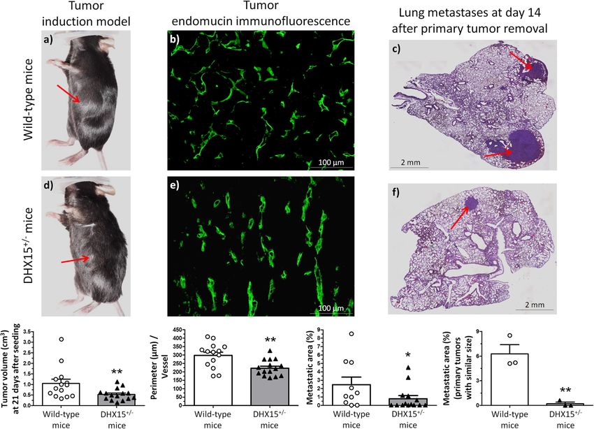
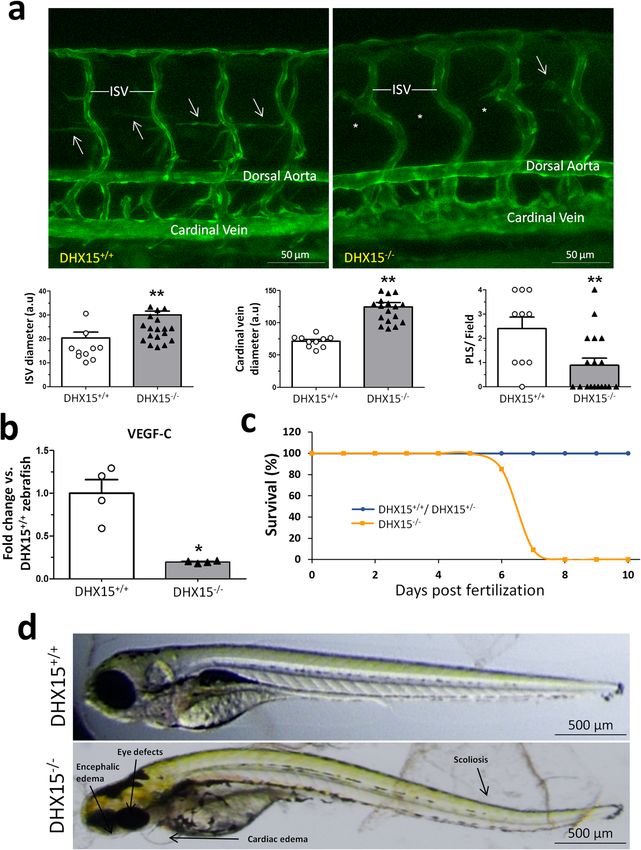
COMMUNICATIONS BIOLOGY | https://doi.org/10.1038/s42003-021-02722-w                                                                         ARTICLE

Fig. 1 Characterization of the Akt and DHX15 signaling crosstalk in endothelial cells. a The expression of DHX15, Akt1, and Akt2 proteins was evaluated
by western blot using cell lysates from wild-type and siL-DHX15-LECs. β-actin was used as a loading control. The densitometric analysis of the protein
expression is shown on the bar graph. **p < 0.01 vs. wild-type LEC (n = 3 biologically independent samples for each condition). b The activity of Akt was
determined in non-silenced and DHX15-silenced endothelial cells in a kinase reaction using recombinant GSK-3 as substrate. The levels of GSK-3α/β
phosphorylation were analyzed by western blot using a phospho-GSK-3 specific antibody. β-actin was used as a loading control. The densitometric analysis
of the protein expression is shown on the bar graph. **p < 0.01 vs. wild-type LEC (n = 3 biologically independent samples for each condition). All statistical
analyses were performed using unpaired two-tailed Student’s t-test. All bar graphs are presented as mean ± SEM.

DHX15 gene deficiency causes blood and lymphatic vascular                           To characterize further the role of DHX15 in vascular
defects during embryonic stages. At stage E10.5, the yolk sac of                definition, we studied whether there was a preferential expression
control mice presented a well-organized vascular network consist-               of DHX15 among different vascular beds of wild-type mice. The
ing of both capillaries and large vitelline vessels (Fig. 2b). However,         mRNA quantification of DHX15 in thoracic aorta and portal vein
DHX15+/− littermates exhibited yolk sacs with reduced vascular                  showed comparable results without significant differences in both
density and fewer vascular branches in vitelline vessels, compared              vascular territories. Next, we wanted to compare the preferential
with WT (0.54 ± 0.05 vs. 1.36 ± 0.07 percentage of vascular area,               expression of DHX15 between blood vessels and lymphatic
respectively; p < 0.01, and 10.67 ± 1.17 vs. 24.00 ± 1.73 total bran-           vessels. However, and due to the technical difficulty of isolating
ches, respectively; p < 0.01) (Fig. 2b). Next, we performed whole-              entire lymphatic vessels, we quantified DHX15 expression in
mount blood vessel staining in embryos at the same stage, E10.5.                isolated-primary cells. We found ~3-fold higher expression of
DHX15+/− embryos did not exhibit significant defects in seg-                     DHX15 in primary liver sinusoidal endothelial cells compared
mentation. However, heterozygous embryos showed lower vascular                  with primary mesenteric lymphatic endothelial cells, which
density compared with wild-type (Fig. 2c). This deficiency was                   revealed a differential expression of DHX15 in the blood vessel
mainly evident in the heart region and the intersomitic arteries that           endothelium compared with the lymphatic endothelium (Supple-
sprout out or were located between the somites.                                 mentary Fig. 2d). Both cell types, were isolated with high purity

COMMUNICATIONS BIOLOGY | (2021)4:1192 | https://doi.org/10.1038/s42003-021-02722-w | www.nature.com/commsbio                                                3
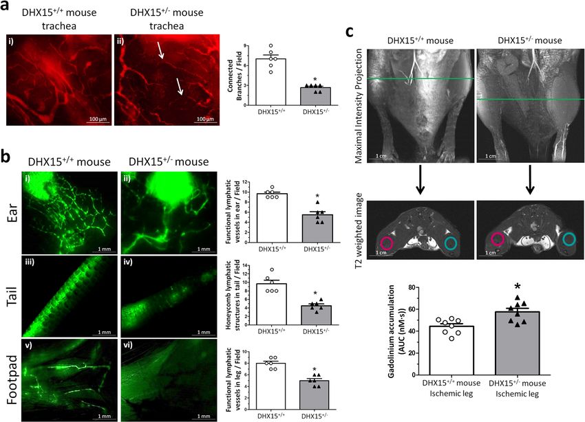
ARTICLE                                                                COMMUNICATIONS BIOLOGY | https://doi.org/10.1038/s42003-021-02722-w

Fig. 2 Embryonic characterization of DHX15 gene deficiency and expression in the gene-edited mouse and zebrafish models. a Representative
immunostaining of the DHX15 expression (green) from mouse embryos at E9.5 and E10.5 of embryonic development (n = 3 animals for each embryonic
day). Maximal projection is shown. Original magnification: ×20 for E9.5 and ×10 for E10.5. b Representative bright-field images of the yolk sac vasculature
from mouse embryos at E10.5 of embryonic development. Maximal projection is shown for each genotype. Original magnification: ×8. The vascular density
quantification is shown on the histograms expressed as the percentage of vascular area (upper graph) and total branches (lower graph). Bars represent the
mean ± SEM, **p < 0.01 vs. wild-type, unpaired two-tailed Student’s t-test (n = 3 animals for each condition). c Representative immunostaining of the
vasculature with endomucin (green) from mouse embryos at the stage E10.5 of embryonic development. The white arrows denote areas of decreased
vascular density (n = 6 animals for each condition). Maximal projection and 3D rendering from the microscope are shown for each genotype. Original
magnification: ×20 and ×40.

and maintained their blood vessel and lymphatic cell markers                     Similar to mice, global DHX15 deficiency was lethal in
after the isolation (Supplementary Fig. 3).                                   zebrafish. To establish the embryonic lethality period, we
   Early embryonic lethality caused in mice by DHX15 deficiency                monitored the larvae mortality during the first 10 dpf (Fig. 3c).
limits the characterization of embryonic vascular anomalies                   The DHX15−/− larvae started to die at 6 dpf and reached a 100%
motivated by this genetic disturbance in homozygosis. In order                mortality at the stage 8 dpf. These animals developed morpho-
to overcome this limitation, we generated a DHX15 gene-deficient               logical defects including encephalic and cardiac edema, scoliosis,
zebrafish mutant by Crispr/Cas9 editing in a Tg(flk1:EGFP)                      and impaired neural/eye growth; compared to wild-type embryos
background, as described in the “Methods” section. Wild-type                  (Fig. 3d), as previously described by McElderry et al.38, and the
zebrafish embryos showed DHX15 mRNA expressed broadly                          onset of these morphological defects was at 3 dpf. In contrast,
across the larvae during the period of 3 days post fertilization (dpf)        DHX15+/+ and DHX15+/− did not show significant differences
(Supplementary Fig. 4, panels b, f, and j) with an enriched                   in survival throughout embryonic development or postnatally.
expression in the vascular system, specifically in the dorsal aorta
and the intersegmental vessels (ISV) (Supplementary Fig. 4, panels            Vascular density and lymphatic functionality are impaired
d, h, and l), as post-natal development progresses (24, 48, and               postnatally in DHX15+/− mice. Due to the mortality associated
72 h). DHX15−/− zebrafish embryos at stage 5 dpf were also                     with the lack of DHX15 in homozygotes, it was only possible to
screened for the expression of GFP in the vasculature. DHX15                  characterize the vascular role of DHX15 in viable adult hetero-
deficiency caused vascular development impairment in primary                   zygous mice (DHX15+/−). DHX15+/− mice showed significant
arteries and veins, compared with the wild-type embryos. This                 vascular impairment compared with wild-type mice. Whole-
impairment was characterized by generalized dilatation of the                 mount preparations of trachea tissue for the quantification of
vasculature, especially in the cardinal vein and the ISV (Fig. 3a).           vessel pruning demonstrated that DHX15+/− mice exhibited
These defects were extended to developing lymphatic structures,               reduced vascular densities and an impaired connectivity between
such as the parachordal line37. In control embryos, the                       large vessels (arrows), compared with littermates WT mice
parachordal line was detected in nearly every somite segment                  (2.67 ± 0.33 vs. 7.00 ± 1.16 connected branches/field, respectively;
(Fig. 3a, arrows). By contrast, the number of parachordal lines was           p < 0.05) (Fig. 4a).
63% lower in the DHX15−/− embryos. In agreement with the                         We also evaluated lymphatic function in adult DHX15+/−
impairment in the formation of embryonic lymphatic structures,                mice by two different methodologies. First, fluorescent lymphan-
DHX15−/− embryos showed downregulation of VEGF-C com-                         giographies were performed in peripheral regions using FITC-
pared to wild-type embryos (Fig. 3b). These results further support           dextran. The lymphatic vasculature of DHX15+/− mice depicted
the role of DHX15 as a regulator of the lymphatic endothelium.                diminished fluid drainage, compared with WT mice in three

4                           COMMUNICATIONS BIOLOGY | (2021)4:1192 | https://doi.org/10.1038/s42003-021-02722-w | www.nature.com/commsbio
COMMUNICATIONS BIOLOGY | https://doi.org/10.1038/s42003-021-02722-w                                                                      ARTICLE

Fig. 3 Characterization of embryonic vascular anomalies associated with DHX15 gene deficient in zebrafish. a Representative vascular images of
DHX15+/+ and DHX15−/− larvae at 5 day post fertilization (dpf) revealing a reduced formation of the parachordal line (arrows). Asterisks denote the
absence of these vascular structures in DHX15−/− animals. Quantifications of cardinal vein diameter, intersegmental vessels (ISV), and number of
parachordal lymphangioblast strings (PLS) are shown in the graphs. **p < 0.01 vs. wild-type zebrafish, unpaired two-tailed Student’s t-test (n = 10 and
n = 18 for the DHX15+/+ and DHX15−/− conditions, respectively). Arbitrary units: a.u. b RNA extraction of zebrafish embryos at 4 dpf from either wild-
type or DHX15 knockout larvae was performed. mRNA expression was analyzed by RT-qPCR. The graph shows the different expression levels of the VEGF-
C gene in the DHX15+/+ and DHX15−/− conditions. mRNA levels are shown as fold change relative to HPRT mRNA levels. *p < 0.05 vs. wild-type unpaired
two-tailed Student’s t-test (n = 4 biologically independent samples for each condition). c Survival assessment assay. The graph shows the larvae survival
rate through the first 10 dpf according to their different genotype (n = 15 zebrafish larvae for each condition). d Representative images comparing wild-type
and DHX15−/− larvae at 7 dpf where morphological defects including encephalic and cardiac edema, scoliosis, and impaired neural/eye growth are evident.
All bar graphs are presented as mean ± SEM.

different peripheral tissues: the ear (5.67 ± 1.20 vs. 9.67 ± 0.33             (red circles Fig. 4c), all the DHX15+/− mice showed impaired
functional lymphatic vessels/field, respectively; p < 0.05), the tail           lymphatic drainage with the consequent significant increase in
(4.33 ± 0.88 vs. 9.67 ± 1.67 honeycomb lymphatic structures/field,              gadolinium accumulation, as observed by the increased area
respectively; p < 0.05) and the foodpad (5.00 ± 0.63 vs. 8.00 ± 0.63           under the concentration curve calculated in DHX15+/− mice,
functional lymphatic vessels/field, respectively; p < 0.05) (Fig. 4b).          compared to control animals (57.72 ± 3.19 vs. 44.45 ± 2.35 nM s,
Second, we quantified lymphatic drainage of the contrast agent                  respectively; p < 0.05).
gadolinium in the hindlimbs after femoral artery ligation. This
model is associated with increased vascular permeability and the               Mechanistic insights of DHX15 deficiency in mouse endothe-
consequent accumulation of fluid in interstitial spaces. For this               lial cells. Role of DHX15 in carbohydrate metabolism. The
purpose, we performed MRI in WT and DHX15+/− mice,                             in vivo experiments showed us that reduced levels of DHX15
4 weeks after the femoral artery ligation. In the ischemic legs                caused cardiovascular and lymphatic abnormalities. To obtain

COMMUNICATIONS BIOLOGY | (2021)4:1192 | https://doi.org/10.1038/s42003-021-02722-w | www.nature.com/commsbio                                             5
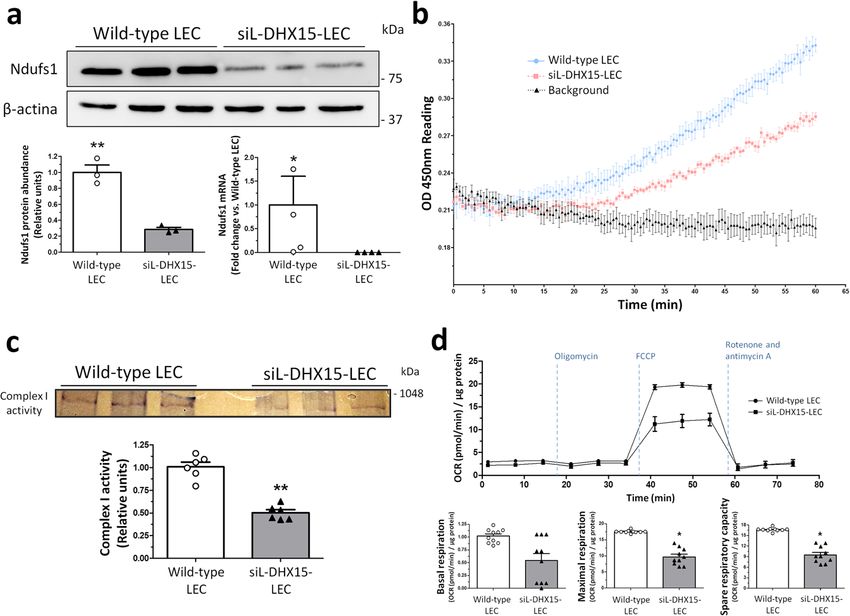
ARTICLE                                                                     COMMUNICATIONS BIOLOGY | https://doi.org/10.1038/s42003-021-02722-w

Fig. 4 DHX15+/- mice showed cardiovascular and lymphatic vasculature alterations. a Representative immunofluorescent images (red CD31 staining) of
mouse trachea vessels. White arrowhead evidences lack of connectivity between large vessels. Vascular density quantification is shown on the histogram.
*p < 0.05 vs. wild-type (n = 5 animals for each contiditon). b Lymphatic drainage of 2000 KDa FITC-dextran analyzed by lymphangiography. Fluorescent
dye was injected intradermally in the ear (panels i and ii), in the interstitium of the tail-tip (panels iii and iv) and in the footpad (panels v and vi) to assess
lymphatic uptake. Lymphatic uptake quantification is shown on the histograms. *p < 0.05 vs. wild-type (n = 5 animals for each contidion). c Representative
magnetic resonance images (MRI) for both strains of mice. First row shows the maximal intensity projection of the time of flight (TOF) angiography. The
green line indicates the position of the coronal image (second row: T2-weighted image) where the regions of interest (ROIs) for the analysis of the dynamic
contrast enhanced-MRI experiment were positioned. In blue, ROIs for the control leg, in red, ROIs for the ischemic leg. The lower graph shows the area
under the concentration curve (AUC) calculated for the ischemic leg in WT and DHX15+/− mice. *p < 0.05 vs. wild-type mouse (n = 8 animals for each
condition). All statistical analyses were performed using unpaired two-tailed Student’s t-test. All bar graphs are presented as mean ± SEM.

information on the mechanisms responsible for these phenotypes                     Enrichment based on Gene Ontology Analysis (GSEA-GO) and
in both vascular systems, we performed RNAseq and proteomics                       pathway analysis (IPA; Ingenuity). First, we used the annotations
in endothelial cells with or without DHX15 gene silencing. The                     of our differential expressed isoforms to find out which GO terms
RNA-seq experiment allowed us to generate 122,968 paired-end                       are changed in response to DHX15 knockdown in endothelial
reads that were successfully mapped to the mouse reference                         cells (Supplementary Data 3). As seen in Supplementary Fig. 5b,
GRCm38 genome assembly. After quality control analysis, four                       major functions related to vascular growth were underrepre-
samples per experimental condition were included in the analysis.                  sented, such as regulation of chemotaxis, angiogenesis, and
A total of 5408 isoforms were differentially expressed when                        wound healing. In contrast, metabolic processes such as ATP
considering a stringent threshold of FDR < 0.001 (Supplementary                    biosynthetic process and carbohydrate catabolic process were
Data 1). Unsupervised hierarchical clustering analysis of the                      overrepresented, pointing to these mechanisms as direct targets of
RNAseq results showed that all the samples from the DHX15-                         DHX15 in endothelial cells. Next, the RNAseq results were
silenced cells clustered together and formed a statistically differ-               compared against global molecular networks to identify asso-
ent group from the wild-type condition, supporting the DHX15                       ciated diseases and canonical pathways affected by the perturba-
role in the regulation of the endothelial expression program.                      tion of DHX15. The DHX15 silencing significantly affects two
The heatmap clustering showed in Supplementary Fig. 5a por-                        networks: (1) endocrine system disorders, organismal injury and
trays the DHX15-sensitive genes that significantly contribute to                    abnormalities and cancer, and (2) gastrointestinal disease,
the clustering of both experimental conditions.                                    organismal injury and abnormalities and carbohydrate metabo-
   For the proteomics experiment, protein extracts (n = 5 for each                 lism (Supplementary Fig. 6a and b). To validate RNAseq results
experimental condition) were digested into peptides and they                       within the glycolytic pathway, we evaluated RNA expression of
were quantified by mass spectrometry analysis, as described in the                  several participating enzymes such as pyruvate kinase, G3PDH,
“Methods” section. We prioritized those protein variations that                    Aldo A and UGGT1 (Supplementary Fig. 7). In accordance to
showed a p-value lower than 0.05 or when the protein was present                   RNAseq results, in DHX15-silenced cells, pyruvate kinase and
in one condition and undetectable in all the cases of the other                    G3PDH presented a significantly higher expression, whilst
experimental condition. The list of proteins with relevant changes                 analysis in Aldo A and UGGT1 depicted a non-significant
in their abundance due to the DHX15 silencing are detailed                         tendency towards an increased RNA expression in silenced cells
in Supplementary Data 2.                                                           (p = 0.49 and p = 0.69, respectively).
   In an effort to understand the biological relevance of the                         Next, we performed a bioinformatic analysis to determine
-omics results, we combined the results of the RNAseq and                          alternative splicing in genes targeted by DHX15. The rMATS
proteomic experiments and subjected the final list to Gene Set                      analysis of alternative 5′ splice site from the RNA-seq data

6                             COMMUNICATIONS BIOLOGY | (2021)4:1192 | https://doi.org/10.1038/s42003-021-02722-w | www.nature.com/commsbio
COMMUNICATIONS BIOLOGY | https://doi.org/10.1038/s42003-021-02722-w                                                                        ARTICLE

Fig. 5 siL-DHX15-LEC showed impaired mitochondrial respiratory chain activity. a Protein and gene expression of NDUFS1 was analyzed. Protein was
evaluated by western blot using cell lysates from wild-type and siL-DHX15-LEC. β-actin was used as a loading control. The densitometric analysis of the
protein expression are shown on the bottom left bar graph. **p < 0.01 (n = 3 biologically independent samples for each condition). mRNA expression was
analyzed by RT-qPCR. mRNA levels are illustrated as fold change relative to HPRT mRNA levels. Bottom right graph. *p < 0.05 (n = 4 biologically
independent samples for each condition). b Complex I activity was assessed using a commercial colorimetric enzymatic reaction reagent for the
mitochondrial respiratory complex I in wild-type and siL-DHX15-LEC. Seven hundred µg of total protein was used in each experimental condition (n = 3
biologically independent samples for each condition). Wild-type-LEC: upper line, siL-DHX15-LEC: middle line, background: bottom line. c In-gel activity
staining on clear-native page (CN-PAGE) of the respiratory complex I from wild-type and siL-DHX15-LEC’s mitochondria. The quantification of the relative
band intensities of complex I activity is shown in the graph below. **p < 0.01 vs. wild-type LEC (n = 6 biologically independent samples for each condition).
d Wild-type and siL-DHX15-LEC were evaluated for basal, coupled and maximal respiration in a mito-stress assay using a Seahorse XFe24 analyzer (n = 10
independent Seahorse wells for each condition). OCR is normalized to µg of total protein. *p < 0.05 vs. wild-type LEC. All statistical analyses were
performed using unpaired two-tailed Student’s t-test. All data are presented as mean ± SEM.

identified 220 significant splicing event modifications (with                      (0.59 ± 0.33 vs. 1.02 ± 0.11 pmol O2/min/μg protein, respectively;
FDR < 5%, absolute difference >5% and >70 number of reads)                      p = 0.29) and maximal respiration (10.04 ± 2.14 vs. 17.55 ± 0.11
caused by DHX15 silencing (Supplementary Data 4). Among                         pmol O2/min/μg protein, respectively; p < 0.05) compared with
these genes, NADH ubiquinone oxidoreductase core subunit S1                     non-silenced LEC cells. These differences were reflected in a
(NDUFS1) showed a differential alternative splicing characterized               lower spare respiratory capacity of the DHX15 silenced cells
by the presence of an alternative 5’ splice site, giving rise to a              (Fig. 5d). Next, we wanted to evaluate the specificity of DHX15 in
longer exon 1 for NDUFS1, when DHX15 was silenced in LECs                       the regulation of mitochondrial oxygen consumption in endothe-
(Supplementary Fig. 5c). This gene encodes a subunit of the                     lial cells by measuring this parameter in hepatocytes. The mito-
mitochondrial respiratory chain complex I. To validate these                    stress experiments performed in an immortalized hepatocyte cell
RNA-seq results, NDUFS1 protein expression in LEC cells was                     line under the wild-type and the DHX15 silenced conditions
analyzed. A significant 70% reduction of NDUFS1 was found in                     showed no differences in the values of OCR (Supplementary
siL-DHX15-LEC (Fig. 5a, bottom graph left). In addition, a                      Fig. 8), thus ruling out that the effect of DHX15 on mitochondrial
reduced NDUFS1 RNA expression was found in siL-DHX15-LEC                        oxygen consumption is an universal mechanism.
(Fig. 5a, bottom graph right). To obtain a functional validation of                To further confirm the mitochondrial impairment observed in
these results, we studied the activity of the mitochondria electron             siL-DHX15-LEC, we measured ATP generation in LEC and
transport chain in endothelial cells. In agreement with the                     primary liver endothelial cells. The alterations in mitochondrial
reduced NDUFS1 expression, we found a significant reduction                      activity were linked to lower ATP production compared to WT
of the complex I activity in siL-DHX15-LEC cells compared                       LEC (417.7 ± 31.37 vs. 524.2 ± 20.40 nM ATP, respectively;
with non-silenced LEC cells (Fig. 5b). Such results were                        p < 0.05) (Fig. 6a). Such results were reproduced in primary liver
confirmed by the in-gel activity measurement, were the activity                  endothelial cells where a lower ATP production was also observed
of complex I was reduced significantly by ~50% in the siL-                       compared to WT cells (24.97 ± 3.41 vs. 38.05 ± 3.23 nM ATP,
DHX15-LEC condition, compared with non-silenced LEC cells                       respectively; p < 0.05) (Fig. 6b).
(0.50 ± 0.04 vs. 1.00 ± 0.03 relative units, respectively; p < 0.01)               Next, we quantified cell proliferation and migration to assess
(Fig. 5c).                                                                      the impact of reduced energy biosynthesis in siL-DHX15-LEC.
   Mitochondrial function was further evaluated with the Sea-                   Silencing of DHX15 reduced BrdU uptake compared to control
horse mito-stress test kit, which allows the analysis of                        LEC (55.53 ± 0.49 vs. 66.43 ± 0.46% of cells BrdU positives,
mitochondrial function through the measurement of the cellular                  respectively; p < 0.01) (Supplementary Fig. 9a). Accordingly, the
oxygen consumption rate (OCR). siL-DHX15-LEC depicted a                         reduction of DHX15 expression also resulted in an impaired cell
decrease in real-time OCR, leading to reduced rates of basal                    migration. In this context, the siL-DHX15-LEC cell line showed

COMMUNICATIONS BIOLOGY | (2021)4:1192 | https://doi.org/10.1038/s42003-021-02722-w | www.nature.com/commsbio                                               7
ARTICLE                                                                COMMUNICATIONS BIOLOGY | https://doi.org/10.1038/s42003-021-02722-w

Fig. 6 siL-DHX15-LEC presented less cell migration. a ATP production was evaluated by a luminescence assay in wild-type and siL-DHX15-LEC. Bars
represent the mean ± SEM, *p < 0.05 vs. wild-type LEC (n = 6 biologically independent samples for each condition). b ATP production was evaluated by a
luminescence assay in MLiEC isolated from wild-type and DHX15+/− mice. Bars represent the mean ± SEM, *p < 0.05 vs. wild-type LEC (n = 3 biologically
independent samples for each condition). c Cell migration was quantified after performing a scratch wound in confluent non-silenced and silenced LECs
cells that were cultured in six-well plates. Then images of wound healing were acquired after 0, 7, and 24 h (n = 6 independent experiments). Graph shows
the quantification of the wound closure after 24 h as percentage of migration. Bars represent the mean ± SEM, *p < 0.05 vs. respective wild-types and
#p < 0.05 vs. siL-DHX15-LECs without ATP and the pyruvate condition (Pyr). For a and b statistical analyses were performed using unpaired two-tailed

Student’s t-test; for c statistical analysis was performed using one-way ANOVA with Tukey’s post hoc test for multiple comparisons. All bar graphs are
presented as mean ± SEM.

delayed wound healing capability after 24 h compared with                     lymphatic vessels constitute one of the main routes of metastasis
control LEC (Fig. 6c, panels i, ii, ix, and x).                               in most cancers39–41. Considering also that one of the pathways
   Next, we evaluated the effect of ATP supplementation over the              modified by the reduction of DHX15 was “endocrine system
impaired wound healing observed in siL-DHX15-LEC. With this                   disorders, organismal injury and abnormalities and cancer”, we
purpose, we added two additional experimental conditions to the               aimed at evaluating the role of DHX15 in tumor development.
wound healing cell migration experiment: 1/cell culture medium                   First, we explored how the loss of DHX15 affected the growth
supplemented with 5 μM ATP to equilibrate the lower ATP                       of tumors implanted into WT and DHX15+/− mice by injecting
production of siL-DHX15-LEC, and 2/cell culture medium                        syngeneic LLC1 cells (1 × 10⁵) into the flanks of both strains. We
supplemented with 2 mM pyruvate. The purpose of the last                      measured the volume of the primary tumors implanted in these
condition was to discard a deficient production of pyruvate that               mice 21 days post-injection of the LLC1 cells. Primary tumors
may compromise the levels of acetyl-CoA, and as a result                      were significantly smaller in DHX15+/− mice compared with
endogenous ATP production, in the mitochondria. The results of                controls (0.54 ± 0.07 vs. 1.06 ± 0.17 cm3, respectively; p < 0.01)
this experimental setting showed that ATP (Fig. 6c, panels iii, vii,          (Fig. 7, panels a and d). Tumors from DHX15+/− mice also
and xi) but not pyruvate supplementation (Fig. 6c, panels iv, viii,           showed an impaired vascular network characterized by smaller
and xii) was able to restore the wound healing capability of siL-             vascular perimeter, compared with WT mice (199.1 ± 9.81 vs.
DHX15-LEC after 24 h of incubation.                                           312.5 ± 17.16 μm perimeter, respectively; p < 0.01) (Fig. 7, panels
   In order to check if some of the results obtained in LEC cells             b and e).
could be extrapolated to the lymphatic vasculature, we performed                 Since growth of the primary tumor is rate-limiting and
another scratch-induced wound-healing assay in primary lym-                   precludes analyses of metastasis in this model, we established a
phatic endothelial cells. The results were similar that those                 postsurgical metastasis model. primary tumors were completely
obtained in LEC cells, LyECs from DHX15+/− presented delayed                  resected after three weeks of subcutaneous LLC1 cells implan-
wound-healing after 24 h compared with control LyECs from                     tation and postsurgical lung metastasis were evaluated 2 weeks
DHX15+/+ (Supplementary Fig. 9b), indicating that some                        later. Fewer primary tumors from DHX15+/− mice metasta-
deficiencies observed in the blood vasculature extrapolated to                 sized the lungs compared with wild-type mice (p < 0.01)
those of the lymphatic vasculature.                                           (Supplementary Fig. 10a). Noteworthy, and considering the
                                                                              mice with lung metastasis, the overall area of metastases was
                                                                              strongly decreased in DHX15+/− mice, compared with the
Heterozygous DHX15 gene deficiency reduced tumor growth                        wild-type group (0.88 ± 0.32 vs. 2.53 ± 0.69% lung metastases,
and metastases. Abnormal function of blood and lymphatic                      respectively; p < 0.05) (Fig. 7, panels c and f).
vessels plays a pathological role in multiple pathological condi-                To investigate further whether the differences in the size of
tions including inflammation and cancer. Also, there is a strong               lung metastasis were due to a potential lower LLC1 cell seeding in
link between the lymphatic vasculature and tumor spread, as                   the primary tumors, we followed over time a group of mice with

8                           COMMUNICATIONS BIOLOGY | (2021)4:1192 | https://doi.org/10.1038/s42003-021-02722-w | www.nature.com/commsbio
COMMUNICATIONS BIOLOGY | https://doi.org/10.1038/s42003-021-02722-w ARTICLE Fig. 7 Tumor growth and metastases in DHX15+/− mouse. Macroscopic images of tumor size in wild-type (a) and DHX15+/- mice (d) three weeks after mouse Lewis lung cancer cells (LLC1) implantation. The arrows indicate the primary tumor. The quantification of tumor volume (cm3) is shown on the lower graph. **p < 0.01 vs. wild-type mice (n = 15 animals for each condition). Middle panels show endomucin immunostaining of intratumoral blood vessels in wild-type (b) and DHX15+/− mice (e). The quantification of the total vascular perimeter of all the intratumoral blood vessels that were positive for endomucin immunostaining was performed with the software Image J. Then, the total vascular perimeter per field was divided by the total number of endomucin+ vessels per field. The statistical comparison between experimental groups was made considering the result of this index (Perimeter/Vessel). **p < 0.01 vs. wild-type mice (n = 15 biologically independent samples for each condition; original magnification: ×200). Right panels show representative lung sections of lung metastatic area after haematoxylin-eosin staining (H&E) in wild-type (c) and DHX15+/− mice (f). The arrows indicate the metastatic areas. Quantifications of the percentage of lung metastases are shown in the graphs below. In the graph on the left: all tumors. *p < 0.05 vs. wild-type mice (n = 15 biologically independent samples for each condition). In the graph on the right: primary tumors with similar size. **p < 0.01 vs. wild-type mice (n = 3 biologically independent samples for each condition). Original magnification: ×10. All statistical analyses were performed using unpaired two-tailed Student’s t-test. All bar graphs are presented as mean ± SEM. similar primary large-size tumors. In this experimental group expression in DHX15+/− mice was also decreased compared with (size of primary tumors ranging from 0.81 to 1.13 cm3), we also the wild-type condition, although this difference did not reach found significant differences when we compared the areas of statistical significance (Supplementary Fig. 10c). In addition, lung metastasis between wild-type and DHX15+/− mice, being VEGF-C downregulation was associated with a significant significantly lower in the DHX15+/− mice (6.27 ± 1.12 vs. decrease in intratumoral Lyve-1 immunostaining, which is a 0.20 ± 0.20% lung metastases, respectively; p < 0.01) (Fig. 7, lower specific marker for the presence of lymphatic endothelium right graph). Mice survival was monitored during the experi- (Supplementary Fig. 10d). mental procedure. Supplementary Fig. 10b shows that DHX15+/− mice presented a tendency for a higher survival-rate compared with wild-type mice, without reaching statistical significance Discussion (83.33 vs. 66.67 percentage of survival at 5 weeks after LLC1 cells In previous studies, we identified DHX15 as a downstream target implantation, respectively). of Akt135, which has the greatest influence on vascular regulation To understand the mechanisms that drive the changes in the compared with the other Akt isoforms. For instance, Akt1-/- intratumoral vasculature and the metastatic risk, we next studied mouse shows a reduction in postnatal vascular density in different the differential expression of molecular regulators in the primary areas such as the myocardium42 and the retina35. Also, Akt1 tumors induced by LLC1 allotransplantation. With this purpose, activity is critically required during lymphatic development in we quantified the intratumoral expression of several angiogenic, mice43. In the current study, we demonstrated increased AKT1 lymphangiogenic, and metastasis-related chemokines by RT- expression in DHX15 silenced endothelial cells, pointing out the qPCR: VEGF-A, VEGF-C, VEGF-D, angiopoietin-1, angiopoie- existence of isoform-specific crosstalk between AKT1 and tin-2, and SDF-1. Among them, only VEGF-C and stromal cell- DHX15. All together, let us hypothesize that DHX15 may play a derived factor-1 (SDF-1) showed significant changes in the relevant role in vascular function and growth. DHX15+/− mice being reduced in this experimental condition Homozygous DHX15 deficiency was associated with embryo- compared with wild-type mice (Supplementary Fig. 10c). It is nic mortality in mice. In contrast, the loss of just one DHX15 noteworthy that VEGF-C was also downregulated in the allele resulted in viable embryos. However, DHX15+/− mice DHX15−/− zebrafish experimental model (Fig. 3b). This showed impaired lymphatic drainage and decreased vascular concordant result points out a conserved regulation of this density in the yolk sac and different territories of embryos and lymphangiogenic factor by DHX15 across species. Angiopoietin-2 adult mice. Importantly, DHX15 protein expression was detected COMMUNICATIONS BIOLOGY | (2021)4:1192 | https://doi.org/10.1038/s42003-021-02722-w | www.nature.com/commsbio 9
ARTICLE                                                             COMMUNICATIONS BIOLOGY | https://doi.org/10.1038/s42003-021-02722-w

in wild-type mice at the embryonic stages E9.5 and E10.5, which            tricarboxylic acid cycle and the oxidative phosphorylation50,51.
is a key timing for vasculature development and definition44.               However, some studies support the notion that mitochondria
   These in vivo results are in agreement with the reduced cell            electron transport chain activity is also playing a role in driving
proliferation and migration that we observed in LEC after silen-           endothelium angiogenesis and function. For example, low con-
cing the expression of DHX15. Why vasculature is affected by the           centration of oxygen results in the generation of mitochondrial
partial loss of DHX15 in adult mice remains unclear if we only             ROS through the Complex III of the electron transport chain52.
consider the results of this experimental model. Under this                This increase in mitochondrial ROS regulates angiogenic
experimental setting, we cannot discard the possibility that adult         factors such as HIF-1a, promoting its stabilization and allowing
mice developed these vascular defects because of accumulated               the transcription of the VEGF-A gene53,54. Also, HUVEC and
damage occurring during the postnatal stage in the DHX15                   HDMEC depend on mitochondrial oxidative phosphorylation to
deficiency background. However, and by studying the role of this            maintain energy supplies for proliferation and growth, as
RNA helicase in DHX15 gene-deficient zebrafish, we showed                    demonstrated by measuring oxygen consumption and ATP pro-
that the DHX15-related vascular defects are also occurring during          duction in the presence of the mitochondrial uncoupler
development. In this experimental model, we found that                     embelin55. Another transport electron chain inhibitor, rotenone
DHX15−/− larvae were not viable. Also, these unviable larvae               that blocks Complex I activity, reduced the angiogenic capacity of
presented blood vascular alterations and reduced formation of              vasa vasorum endothelial cells56. Our study is in line with these
developing lymphatic structures, which were associated with                observations and supports the idea that for optimal angiogenic
cardiac and encephalic edema in the embryos.                               response, endothelial cells require dynamic crosstalk between
   Vascular growth and lymphangiogenesis are crucial in tumor              glycolysis and mitochondria activity, driven likely by regulators
growth and metastasis45,46. We found that heterozygous DHX15               that promote a metabolic switch, as we have seen when we
deficiency partially inhibits primary tumor growth and reduced              modified the levels of DHX15. Interestingly, DHX15 deficiency
lung metastases. This effect was associated with an intratumoral           impaired mitochondrial oxygen consumption in endothelial cells
reduction in the length of the blood vessel capillaries. Our results       but not in hepatocytes, thus ruling out that the effect of DHX15
agree with several clinical studies that underline a role of DHX15         on mitochondrial oxygen consumption is an universal mechan-
in tumor progression in several types of cancer, such as acute             ism. We also described other DHX15-related mechanisms inde-
myeloid leukemia33, hepatocellular carcinoma47, malignant per-             pendent of the mitochondrial respiratory chain that target the
ipheral nerve sheath tumors31, prostate cancer48 and non-small-            vasculature. In this context, we observed a consistent down-
cell lung cancer49. In all these situations, modifications of DHX15         regulation of VEGF-C in mouse and zebrafish DHX15-deficient
activity and/or its overexpression favors tumor growth. Several            animals, and reduced intratumoral levels of SDF-1 in DHX15+/−
mechanisms have been proposed in these studies to explain the              mice. VEGF-C and SDF-1 play relevant roles in lymphangio-
tumorigenicity effect of DHX15, such as the co-activation of the           genesis, metastasis and recruitment of endothelial progenitor
androgen receptor in prostate cancer and the transcriptional               cells57–60. Therefore, several factors targeted by DHX15 appear to
activation of NF-kB in leukemia cells. In our study, we provided           be involved in the control of vascular homeostasis.
an additional mechanism of DHX15 that is relevant for tumor                   The fact that our study described many alterations in the
progression: the regulation of vascular growth and function.               vasculature does not mean that we attribute exclusively the
   The use of RNAseq and proteomics, combined with bioinfor-               pathophysiological role of DHX15 to endothelial cells. For
matics down-stream analysis, is one of the most potent approa-             instance, McElderry et al.38 generated the first vertebrate
ches to investigate the role played by RNA helicases, as changes in        knockout model for DHX15 in zebrafish and described head and
differential splicing of a gene may or may not be associated with          embryonic development defects in stages previous to the forma-
changes in its protein abundance; therefore, we need the com-              tion of the cardiovascular system, leading to mortality at a similar
bined result of both high-throughput approaches. Adopting this             embryonic stage as the one described in our DHX15−/− zebrafish
strategy, we were able to identify in the DHX15-silenced                   model. Similar results have recently been shown using the same
LEC significant changes in key pathways that metabolize                     experimental model by Cai et al.61, introducing an additional role
carbohydrates. Some of the gene products that varied were                  for DHX15 in the hematopoiesis process through the unfolded
glyceraldehyde-3-phosphate dehydrogenase (G3PDH) and pyr-                  protein response pathway. We also showed that E9.5-10.5 mice
uvate kinase (PKM). These differential expressions were also               embryos robustly expressed DHX15 in the brain. Therefore, we
associated with a reduction in the intracellular concentration of          cannot exclude the possibility that defects in the vasculature of
ATP. The upregulation of glycolytic enzymes suggests a com-                DHX15+/− mouse embryos or DHX15−/− zebrafish embryos
pensatory mechanism to counteract the impaired energy pro-                 may be secondary to the global DHX15 deficiency background.
duction in DHX15 deficient endothelial cells. Supporting this               However, we demonstrated in our study the existence of a direct
possibility, we observed by RNAseq significant expression chan-             pathological mechanism of DHX15 deficiency on blood endo-
ges in members of the family of the NADH dehydrogenase                     thelial and lymphatic endothelial cells, supporting the direct role
[ubiquinone] 1 alpha subcomplex assembly factor (members                   of DHX15 in vascular regulation.
from NDUFS1, NDUFS5 and NDUFS7). These members are
accessory subunits of the mitochondrial membrane respiratory
                                                                           Methods
chain NADH dehydrogenase (Complex I), that transfer electrons              DHX15 transgenic mice. DHX15 gene-deficient C57BL/6 mice were generated by
from NADH to ubiquinone in the mitochondrial respiratory                   genomic editing by microinjecting TALEN (transcription activator-like effector
chain. Among them, we observed a significant alteration in the              nuclease technology) RNA in pronucleated oocytes (Cyagen). The mouse DHX15
splicing of the NDUFS1 gene. This alteration in gene splicing              gene (GenBank accession number: NM_007839.3; Ensembl:
                                                                           ENSMUSG00000029169) is located on chromosome 5. Fourteen exons have been
was linked with a 50% reduction of the activity of the complex I           identified for this gene, with the ATG start codon in exon 1 and the TGA stop codon
in the mitochondria of DHX15 silenced cells that may explain the           in exon 14. Exon 2 was selected as TALEN target sites. TALENs were constructed
decrease of ATP intracellular levels and, as a consequence, the            using the Golden Gate Assembly method62 and confirmed by sequencing. The
impairment in endothelial cell proliferation and vascular growth.          amplicons were then purified and sent for DNA sequencing analysis. TALEN
                                                                           mRNAs generated by in vitro transcription were injected into fertilized eggs for
   Endothelial cells are mainly considered a glycolytic cell type          knockout mouse production (cDNA sequence: TGTTGGTGAGACTGGGTC). The
that maintains their energy demands by exploiting the glycolytic           pups were genotyped by PCR followed by sequence analysis. The positive founders
pathway preferentially without the need of coupling to the                 were breeding to the next generation, which was genotyped by PCR and DNA

10                       COMMUNICATIONS BIOLOGY | (2021)4:1192 | https://doi.org/10.1038/s42003-021-02722-w | www.nature.com/commsbio
COMMUNICATIONS BIOLOGY | https://doi.org/10.1038/s42003-021-02722-w ARTICLE
sequencing analysis. DNA sequencing revealed two different DHX15-deficient femoral artery was isolated, and 5-0 suture was tied tightly around artery at a
mouse clones: Mouse-ID#35 that was missing eight bases in one strand, and Mouse- ∼3 mm distance to the inguinal ligament. Mice were allowed 4 weeks to recover
ID#39 that was missing one base in one strand. Wild-type DNA was used as a following the surgical procedure.
negative control for sequencing in parallel. The mRNA transcribed from the targeted MRI experiments were conducted on a 7T BioSpec 70/30 horizontal animal
allele with frameshift undergoes nonsense-mediated decay (NMD). scanner (Bruker BioSpin, Ettlingen, Germany), equipped with a 12 cm inner
DHX15-deficient and WT male and female mice were used at 8–12 weeks of age diameter actively shielded gradient system (400 mT/m) using a surface coil
for all the experiments. All animals were kept under constant temperature and dedicated to abdominal imaging. Animals were first anesthetized (1.5% isoflurane
humidity in a 12 h controlled dark/light cycle, and they were fed ad libitum on a in a mixture of 30% O2 and 70% CO2) and the tail vein was cannulated for
standard pellet diet. All experimental procedures performed in the models of administration of contrast agent. Then, animals were transferred under the same
mouse and zebrafish were approved by the Investigation and Ethics Committees of anesthesia regime to a Plexiglas holder in supine position with a nose cone for
the Hospital Clínic and the University of Barcelona. administering anesthetic gases and fixed by a tooth bar, ear bars, and adhesive tape.
3D-localizer scans were used to ensure accurate position of the animal’s midline at
the level of the posterior limbs in the isocenter of the magnet. T2-weighted images
Mouse genotyping. Mouse genomic DNA was isolated from tail biopsies using a
were acquired by a RARE (rapid acquisition with relaxation enhancement)
specific kit (Extract-N-Amp™ Tissue PCR Kit; Sigma-Aldrich, Darmstadt, Ger-
sequence with an effective echo time (TE) of 24 ms, repetition time (TR) 1201 ms
many). PCR was performed using the primer pairs to amplify the DHX15 gene
and RARE factor 8. Matrix size was 256 × 256 with an in-plane voxel size of
(primer forward: 5′-CACCAACCTGCCCCATACTCCT-3′ and primer reverse:
0.156 × 0.156 mm2, 15 slices, slice thickness 1 mm, resulting in a field of view
5′-TGTATTGTCCCAGGGTAAAATGTGTTG-3′). PCR conditions were as fol-
(FOV) of 40 × 40 × 15 mm3. Time of flight 3D angiography was acquired a FLASH
lows: 35 cycles at 94 °C for 30 s, 59.3 °C for 30 s, and 72 °C for 60 s. PCR product
(Fast Low Angle Shot) protocol with TE = 2.4 ms, TR = 14,000 ms, flip angle 20°, 3
was sequenced by sanger sequencing to distinguish the DHX15 wild-type mice and
averages, matrix size: 448 × 256 × 128, voxel size 0.078 × 0.078 × 0.234 mm³,
DHX15 transgenic mice.
resulting in a FOV of 35 × 20 × 30 mm³. The shortening of the T1-relaxation time
by the contrast agent enhanced the tissue signal. T1 map was acquired using
Immunological staining of mouse embryo whole-mounts. Embryos were har- RARE-VTR (rapid acquisition with relaxation enhancement and variable repetition
vested at different points between E8.5 to E10.5. Embryos at E10.5 were fixed in 4% time) sequence, with TE = 7 ms, 6 TR = 200, 400, 800, 1500, 3000, 5500 ms, matrix
paraformaldehyde overnight at 4 °C. For immunostaining of whole-mount size: 96 × 96 × 3, voxel size 0.417 × 0.417 × 1 mm³, resulting in a FOV of
embryos, after paraformaldehyde fixation, the embryos were sequentially dehy- 40 × 40 × 3 mm³.
drated in methanol and then incubated in the permeabilization buffer (PBlec) (PBS To estimate the T1-relaxation rate and to measure the contrast agent-relative
pH6.8, 1% Tween 20, 1 mM CaCl2, 1 mM MgCl2, 0.1 mM MnCl2) for 20 min at concentration DCE (dynamic contrast enhanced)-MR imaging was used with a T1-
room temperature. After permeabilization, the embryos were incubated with pri- weighted gradient-echo sequence. FLASH protocol was used with TE = 1.5 ms,
mary antibody rat anti-endomucin or rabbit anti-DHX15 (Abcam, Cambridge, TR = 12500 ms, flip angle 15°, 600 repetitions and identical matrix, resolution and
UK) (1:20 dilution) in PBlec buffer overnight at 4 °C. To remove residual primary FOV than T1 map. 0.025 mM/kg of gadoteridol was administered after the first 100
antibody, embryos were washed with PBT (PBS pH 6.8, 0.1% Tween) for repetitions were acquired as baseline. Altogether, the MRI session lasted for 50 min
5 × 10 min. Next, the embryos were incubated with secondary antibody Alexa Fluor approximately. After that, mice were returned to their home cage under close
488-conjugated goat anti-rat or Alexa Fluor 488-conjugated goat anti-rabbit supervision until they were recovered from the anesthesia.
(Thermo Fisher, Waltham, MA, USA) (1∶500 dilution) in the dark overnight at T1 maps were calculated in Paravision 6.0 software (Bruker BioSpin, Ettlingen,
4 °C, and then washed with PBT for 3 × 10 min and postfix in 4% paraformalde- Germany). Later these maps were processed with custom-made algorithms
hyde. Negative controls for endomucin and DHX15 detection were performed by programmed in Matlab (The MathWorks, Inc, Natick, MA, USA). A binary mask
omitting the primary antibodies in the immunofluorescence reactions. Images were was manually drawn over the T1 map in order to segment the muscle in both legs.
acquired using fluorescence stereomicroscope (Leica Microsystems, Heerbrugg, The first 40 volumes of the DCE acquistion were removed to assure the signal
Switzerland) and immunofluorescence microscope (Nikon Eclipse E600, Kana- stabilization. Also, the last 50 volumes were discarded to avoid second pass effects.
gawa, Japan) systems. The slices of the temporal acquisition were spatially smoothed with a Gaussian
filter (standard deviation = 0.5) and temporal smoothed with a moving average of
25 neighbors. The baseline of the signal was considered using the 20 volumes after
Generation of the Zebrafish animal model. Adults wild-type zebrafish (Danio the 10th. The signal intensities of the temporal acquisitions were converted to
rerio), in a Tg(flk1:EGFP);Tg(fabp10:RFP) background, purchased from KIT gadolinium concentrations using the method described in63–65. The T1 map
-European Zebrafish Resource Center (EZRC), were maintained at 28–29 °C on a acquired before the DCE was used as reference values for the magnetization and
light cycle of 14 h light/10 h dark. The Crispr/cas9 design for gene knock-out was the Gadoteridol relaxivity was considered to be 3.35 s−1mM−1 65. Finally, the
performed as follows: Gene sequences were retrieved from http://www.ncbi.nlm. obtained concentration curves were also smoothed with a moving average of nine
nih.gov/gene and http://www.ensembl.org/Danio_rerio/Info/Index. The sgRNA neighbors. From the concentration curves, different parameters were estimated,
was designed using the online tool http://crispor.tefor.net, based on exon site and such as the time to peak (TTP), the bolus arrival time (BAT) (using the method
high efficacy and off-target published algorithms. Microinjection was performed at described in Cheong et al.66, relative time to peak (rTTP) (considering the BAT as
1-cell stage embryos. Fertilized zebrafish embryos were collected in E3 medium. starting point), the wash in and wash out slopes, and the area under the gadolinium
Then, embryos were grown at 28.5 °C. concentration curve (AUC).
Zebrafish whole-mount in situ hybridization (ISH). cDNAs were amplified by
PCR from a custom zebrafish cDNAs library obtained by RT-PCR from an mRNA Mouse-induced tumor model. LLC1 (Mouse Lewis lung cancer cells) (ATCC,
pool coming from 5 days post fertilization zebrafish larvae. We included a Manassas, VA, USA) were cultured in Dulbecco’s Modified Eagle Medium
SP6 sequence linker in reverse primers to directly use the synthesized PCR pro- (DMEM) supplemented with 10% fetal calf serum, 50 U/ml penicillin and 50 μg/ml
ducts as templates to amplify the reverse riboprobe to be used for ISH. For the ISH, streptomycin in humified atmosphere at 37 °C and 5% CO2. Syngeneic LLC1 tumor
embryos were fixed overnight with 4% PFA and washed twice with PBT. Then, cells (1 × 10⁵) were subcutaneously injected into the flank of DHX15+/− and wild-
embryos were incubated with hybridation buffer (50% formamide, 5× SSC buffer type mice. Primary tumor growth was controlled during the first 3 weeks. Primary
pH 6 (0.75 M NaCl, 0.075 M sodium citrate), 0.1% triton, 50 μg/mL yeast RNA, tumors were surgically removed 21 days after seeding. Tumor volume was calcu-
50 μg/mL heparin) at least 1 h. Next, embryos were incubated with hybridation lated by following formula: V = 4/3 × π × [length × depth × width]. Primary tumors
buffer containing the reverse riboprobe overnight. Finally, embryos were washed were fixed in 4% PFA and cryoconserved in tissue-tek O.C.T. compound (Sakura,
with washing solution (50% formamide, 1× SSC buffer, 0.1% Tween) 30 min twice Flemingweg, Netherlands). The Post-surgical metastasis model was performed as
and with MABT (100 mM maleic acid pH 7.5, 150 mM NaCl, 0.1% Tween) 10 min follows: Two weeks after primary tumor removal, LLC1 injected mice showed
five times. Stained embryos were processed for imaging with bright field stereo- distant metastasis formed in the lungs. Tile scan images of haematoxylin-eosin
scope to determine the overall expression pattern. (H&E) stained paraffin lung sections were visualized using a microscope system
(Nikon Eclipse E600, Kanagawa, Japan) and the percentage of the pulmonary
metastatic area as percent of total lung area was measured with Image J software
Zebrafish vascular characterization. Five-day-old larvae obtained by pairwise (ImageJ version 1.52b; National Institutes of Health, Bethesda, MD, USA).
mating of adult Tg(flk1:EGFP;fab10:RFP; DHX15+/−) were sorted in two groups
depending on two criteria: (a) curly larvae with abnormal development or (b)
normal developed larvae. Larvae were flat-mounted and analyzed by confocal
DHX15 silencing in liver endothelial cells and hepatocytes. The silencing of
imaging (Zeiss AxioObserver Z1) to evaluate putative phenotypical defects in the
DHX15 was carried out in mouse primary hepatic endothelial cells and mouse
trunk angiogenesis caused by the gene knockout. Genomic DNA extracted from
primary hepatocytes both immortalized with the SV40 virus (LEC and Hep;
the whole embryos (using Extract-N-Amp™ Tissue PCR Kit, Sigma) was used for
abmGood, Richmond, Canada), through shRNA by lentiviral infection (Dharma-
the genotyping after vasculature imaging.
con, Lafayette, Colorado, USA). The SMARTvector incorporated the bipartite 3G
Tet-On® induction system, an inducible system with minimal basal expression and
Mouse femoral artery ligation model and magnetic resonance imaging (MRI). potent activation after induction with doxycycline. Endothelial cells were cultured
Mice were anesthetized with a mixture of 4% isoflurane and 100% oxygen. The in Microvascular Endothelial Cell Growth Complete Medium and hepatocytes in
COMMUNICATIONS BIOLOGY | (2021)4:1192 | https://doi.org/10.1038/s42003-021-02722-w | www.nature.com/commsbio 11You can also read