The Correction of Facial Morphea Lesions by Hyaluronic Acid: A Case Series and Literature Review
←
→
Page content transcription
If your browser does not render page correctly, please read the page content below
Dermatol Ther (Heidelb)
https://doi.org/10.1007/s13555-020-00438-z

 CASE SERIES

The Correction of Facial Morphea Lesions
by Hyaluronic Acid: A Case Series and Literature
Review
Agnieszka Owczarczyk-Saczonek . Marta Kasprowicz-Furmańczyk .
Anna Kruszewska . Magdalena Krajewska-Włodarczyk .
Agata Bechtold        . Paulina Klimek . Waldemar Placek

Received: July 10, 2020
 The Author(s) 2020

ABSTRACT                                                     life and self-esteem; thus, there is a demand for
                                                             safe and effective methods of treatment.
Introduction: The aim of the study is to assess              Case Presentation: The paper presents three
the long-term safety and efficacy of hyaluronic              female patients aged 16, 17 and 70 with facial
acid (HA) administration in correction of facial             morphea lesions who had HA preparation
morphea lesions and to review the literature on              Juvéderm Voluma or Volux, Vycross tech-
the subject. Morphea is a chronic inflammatory               nology, Allergan, injected. One of the patients
disease of the connective tissue which may lead              had additionally fractional ablative CO2 laser
to serious deformations. The lesions located on              (FAL) therapy.
the face particularly affect patients’ quality of            Discussion: The literature provides reports on
                                                             successful use of HA, polymethylmethacrylate
                                                             and poly-L-lactic acid for the correction of facial
                                                             defects in localized scleroderma. HA is a natural
Digital Features To view digital features for this article   component of the extracellular matrix and it
go to https://doi.org/10.6084/m9.figshare.12807083.          therefore minimizes the probability of
                                                             immunogenicity. The application technique
A. Owczarczyk-Saczonek  M. Kasprowicz-                      also plays an important role. On the other
Furmańczyk  A. Kruszewska  A. Bechtold (&)
P. Klimek  W. Placek                                        hand, FAL therapy leads to the degradation of
The Chair and Clinic of Dermatology, Sexually                the abnormal collagen and the induction of
Transmitted Diseases and Clinical Immunology,                normal collagen synthesis.
The Municipal Polyclinical Hospital in Olsztyn,              Conclusions: HA injection and combination of
Olsztyn, Poland
e-mail: aganek@wp.pl;                                        HA application with FAL are minimally inva-
agata.bechtold@umed.lodz.pl                                  sive, effective and safe therapeutic options for
                                                             patients suffering from morphea.
M. Krajewska-Włodarczyk
Department of Rheumatology, The Municipal
Polyclinical Hospital in Olsztyn, Olsztyn, Poland            Keywords: Correction; FAL; Fractional ablative
                                                             laser; Hyaluronic acid; Morphea; Scleroderma
M. Krajewska-Włodarczyk
Department of Internal Medicine, School of
Medicine, Collegium Medicum, University of
Warmia and Mazury, Olsztyn, Poland

A. Bechtold
Psychodermatology Department, Medical
University of Lodz, Łódź, Poland

       Pobrano z https://publicum.umed.lodz.pl / Downloaded from Repository of Medical University of Lodz 2022-03-02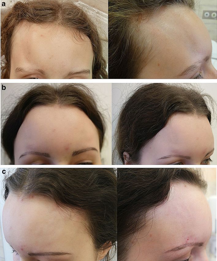
Dermatol Ther (Heidelb)

                                                         INTRODUCTION
 Key Summary Points
                                                         Morphea (localized scleroderma, LoSc) is a
 Why carry out this study?                               chronic inflammatory disease of connective
                                                         tissue which presents with a wide spectrum of
 Morphea is a chronic inflammatory
                                                         clinical symptoms depending on the activity of
 disease of connective tissue leading to
                                                         the disease, involved region and the depth of
 deformations of the involved region,
                                                         the lesions. The lesions most often affect dermis
 including face, thus causing significant
                                                         and subcutaneous tissue and show tendency for
 lowering of patients’ quality of life
                                                         spontaneous remission, yet causing skin atro-
 Existing therapeutic methods focus on                   phy and dyspigmentation [1]. Less often the
 silencing active process, ignoring the                  process affects deeper tissues (fascia, muscles
 deformities the patients’ are left with                 and bones), leading to deep atrophy and
                                                         deformations. The excessive synthesis of colla-
 The study aimed at assessment of long-                  gen type I and III and extracellular components
 term safety and efficacy of hyaluronic acid             is regarded to play the key role in the formation
 (HA) administration in correction of facial             of characteristic skin lesions [2].
 morphea lesions                                             The etiopathogenesis of this condition is still
 What was learned from the study?                        not clear. The genetic and environmental fac-
                                                         tors, as well as microchimerism, are considered
 The combination of HA injection and FAL                 to activate keratinocytes to release inflamma-
 is a minimally invasive, effective and safe             tory mediators (interleukin-4 (IL-4), IL-6, IL-10,
 therapeutic option for patients suffering               IL-17A, IL-27, interferon-c), which subsequently
 from morphea                                            stimulate lymphocytes, endothelial cells and
 In some cases it may be beneficial to                   fibroblasts. Of particular significance, the acti-
 complement the therapy with fractional                  vation of endothelial cells and lymphocytes
 ablative CO2 laser                                      provokes fibroblasts to increase collagen syn-
                                                         thesis, which clinically manifests as focal skin
 If promising results are further confirmed,             hardening and active inflammatory border (lilac
 the method should be widely                             ring) [1–4].
 recommended for correction of the                           Despite the suggestions of developmental
 defects produced by morphea                             origins of morphea (due to frequent pattern of
                                                         lesions alongside Blaschko’s lines), numerous
                                                         evidence confirms its autoimmune background
                                                         (the correlation with personal and family his-
                                                         tory of autoimmune diseases, the presence of
DIGITAL FEATURES                                         human leukocyte antigen (HLA) class II mole-
                                                         cules found also in other autoimmune disor-
This article is published with digital features to       ders) [1].
facilitate understanding of the article. You can             The facial lesions lead to the deformation of
access the digital features on the article’s asso-       the face, lowering patients’ quality of life,
ciated Figshare page. To view digital features for       especially in adults [5, 6]. It is most pronounced
this article go to https://doi.org/10.6084/m9.           in en coup de sabre type, which is characterized
figshare.12807083.                                       by linear atrophy and hardening of the skin,
                                                         subcutaneous tissue, muscles and bones, usually
                                                         extending from the upper limit of the eyebrow
                                                         to the scalp, often with concomitant alopecia
                                                         and eyebrow loss [1]. The therapeutic options
                                                         proposed by consensuses aim at silencing active

   Pobrano z https://publicum.umed.lodz.pl / Downloaded from Repository of Medical University of Lodz 2022-03-02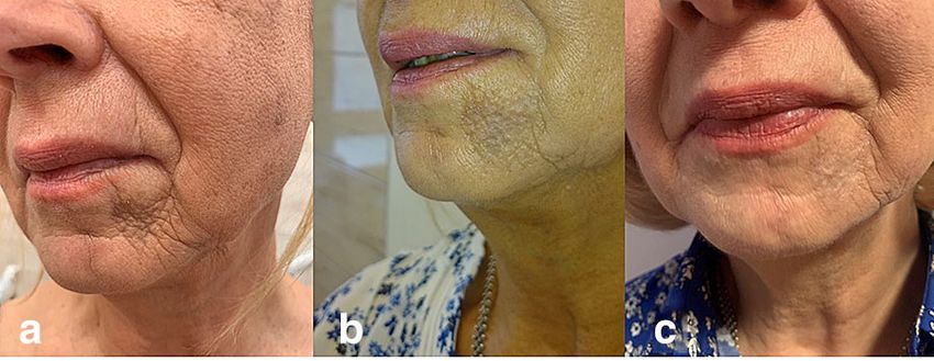
Dermatol Ther (Heidelb)

forms, hindering the progression of the disease,          eyebrows to the scalp and accompanying scar-
but usually they do not discuss methods of                ring (cicatricial) alopecia and partial hair loss
correction of the defects generated by the                within the eyebrows and the eyelashes were
lesions [1, 7–9].                                         observed. After a few months of the disease a
    The literature provides reports on correction         similar linear atrophic lesion was noticed in the
of facial defects in localized scleroderma (mor-          left frontoparietal region. The histopathologic
phea, en coup de sabre, hemiatrophia faciei               examination revealed preserved skin appen-
Parry-Romberg) using hyaluronic acid (HA)                 dages, the epidermis with no tangible abnor-
[10–12], polymethylmethacrylate [13], poly-L-             malities,    the     homogenization       of    the
lactic acid [14] and autologous fat [15–17] per-          subepidermal layer of the dermis and noticeable
formed successfully and with no delayed type              concentration of elastic fibres on orcein stain-
hypersensitivity.                                         ing. On the basis of the clinical presentation
    The aim of the study is to assess the long-           and histopathological examination, limited
term safety and efficacy of HA administration in          scleroderma en coup de sabre was diagnosed.
correction of facial morphea lesions. This article        The patient had no personal history of chronic
presents our experience on the subject. In this           diseases and no family history of autoimmune
paper we present case series of three female              disorders. The laboratory results showed an
patients with morphea, who had their lesions              increased level of anti-thyroid peroxidase anti-
corrected by HA. In one of the patients, the              bodies (286 IU/ml) with normal level of thyroid
aforementioned procedure was combined with                stimulating hormone and also elevated ANA-
fractional carbon dioxide laser (FAL) therapy.            Hep2 titre 1:1280 (negative immunoblotting).
All the patients have had contraindications to            Antibodies IgG and IgM against Borrelia
fillers excluded (active infection, allergy/hyper-        burgdorferi were negative. The patient was con-
sensitivity to the filler and lignocaine, unsta-          sulted by a neurologist and ophthalmologist—
ble autoimmune disease) [18]. After informed              no notable abnormalities were discovered.
consent was obtained, HA preparation Juvé-               Magnetic resonance imaging (MRI) was per-
derm Voluma or Volux, Vycross technology,               formed to exclude anomalies of the central
manufactured by Allergan, was administered.               nervous system and did not shown any signifi-
The patients gave informed consent to partici-            cant changes (in the white matter of the right
pate in the study and for the publication of the          cerebellar hemisphere at the posterior-lateral
results. The patients were examined every sec-            outline of IV ventricle a small band with
ond week, without observing any adverse reac-             increased signal in FLAIR, T2, PD-gliosis? 5 mm;
tions (observation time ranged from 6 months              other brain structures, ventricular system and
to 3 years) and achieving patients complete               basal cisterns were normal). The X-ray of the
satisfaction with the correction of the defect.           skull presented normal structure. Treatment
The result was described with the Localized               with methylprednisolone 4 mg a day was
Scleroderma Damage Index (LoSDI), part of the             applied, yet as a result of the progression of skin
Localized Scleroderma Assessment Tool (LoS-               lesions the dose was increased to 16 mg daily
CAT). LoSDI assesses dermal atrophy (DAT),                and additionally mofetil mycophenolate 1 g a
subcutaneous atrophy (SAT) and dyspigmenta-               day was introduced.
tion (DP) ranging from 0 to 3 in each domain,                 Six months after a stabilization of the dis-
with 9 points in total indicating the most severe         ease, the patient had HA agent injected into
skin damage.                                              morphea lesions. The initial LoSDI score was 7
                                                          (DAT 3, SAT 3, DP 1). To avoid potential com-
                                                          plications of administering excessive amounts
CASE 1                                                    of the medication and following compression of
                                                          arterial vessels in the frontal region, where the
In this 17-year-old female patient a linear               amount of subcutaneous tissue is limited, ini-
induration of the skin with subcutaneous tissue           tially only 0.5 ml was injected into each side of
atrophy located on the forehead from the                  the induration and skin depression from above

       Pobrano z https://publicum.umed.lodz.pl / Downloaded from Repository of Medical University of Lodz 2022-03-02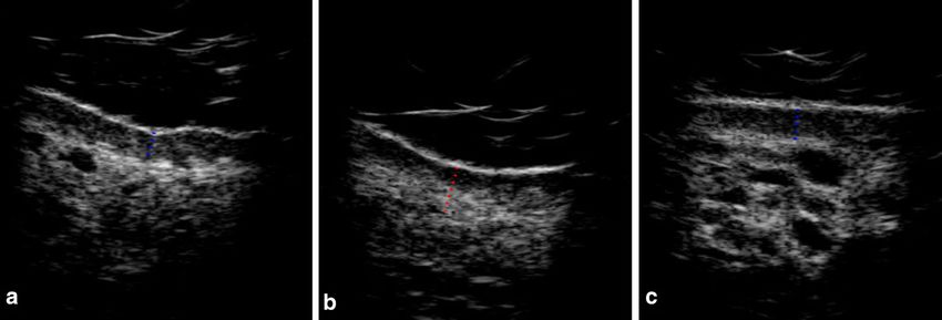
Dermatol Ther (Heidelb)
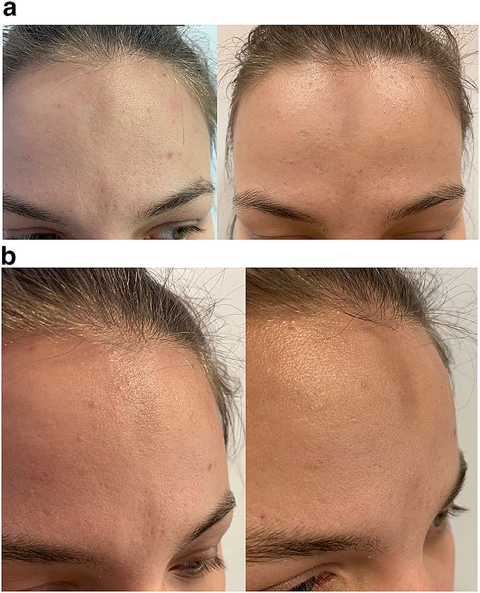
Fig.1 The effect before the treatment (a), after the first
intervention (b) and the final result (c) Fig. 2 The patient before and a week after the injection of
Voluma 1 ml, via 27G cannula—front view (a) and side
view (b)
the eyebrow up to the hairline. The injection
was performed without anaesthesia using a 25G
follow-up lasted for 8 months after the reduc-
cannula inserted into subcutaneous tissue with
tion of the dose of mofetil mycophenolate up to
the needle deep on the bone surface. Partial
500 mg per day and 6 months after discontin-
shallowing of the lesions was achieved. After a
uation of the drug—a desirable aesthetic result
week, the procedure was repeated, also using
and no progression of morphea lesions were
0.5-ml portions of HA, but with a 30G needle
observed.
and in the form of boluses (each injection pre-
ceded by an aspiration). A very good aesthetic
effect was obtained which is reflected by the CASE 2
LoSDI 3 (DAT 1, SAT 1, DP 1) and can be seen in
the photographs (Fig. 1a–c). A 16-year-old female reported to the hospital
Within 1.5 years after the intervention no because of numerous skin lesions located on the
progression has occurred. The patient was on trunk (occurred approximately 1 year before
mofetil mycophenolate 1 g a day and methyl- admission) and on the face (appeared 2 months
prednisolone in gradually reduced doses up to before hospitalization). The patient had no
2 mg every second day. After 1.5 years slight record of chronic diseases and no family history
reduction in volume of administered HA has led of autoimmune disorders. Physical examination
to the decision to repeat the procedure. The revealed flat, oval, silvery, porcelain lesions a
filling within both morphea lesions with 0.5 ml few centimetres in size with a discrete viola-
of HA per side (25G cannula) produced an out- ceous erythema ‘‘lilac ring’’ on the circumfer-
come satisfactory for the patient. After the ence of the lesion and palpable induration
procedure, a gradual reduction of the dose of located on the trunk, mainly on the back. On
immunosuppressants has taken place. The the forehead along the medial line from the
Pobrano z https://publicum.umed.lodz.pl / Downloaded from Repository of Medical University of Lodz 2022-03-02Dermatol Ther (Heidelb)
glabella up to the hairline the lesion presented treatment with doxycycline and cefuroxime as a
as linear, bifocal dyspigmentation with a dis- result of borreliosis (positive IgM and IgG anti-
crete depression alongside (Fig. 2a, b). bodies against B. burgdorferi). By this time her
Both laboratory results and medical imaging skin lesions were treated only topically without
have not revealed any significant abnormalities. success.
ANA-Hep2—1:160, antibodies against Physical examination revealed diffused
B. burgdorferi IgM and IgG were negative. On the cohesive, well-demarcated, pale red papules
basis of the clinical manifestation and with circular palpable borders on the upper
histopathological examination limited sclero- limbs, lower abdomen and hips. Additionally,
derma was diagnosed (morphea). on the left side of the chin the patient had an
During hospitalization procaine penicillin extensive atrophic scar 3 9 3 cm in size, a
1.2 million IU, PUVA therapy, Piascledine and remnant of prior deep morphea (Fig. 3a). The
tacrolimus 0.1% were applied, which led to an LoSDI was assessed to be 5 (DAT 2, SAT 2, DP 1).
improvement—the disappearance of ‘‘lilac ring’’ On the basis of histopathological examina-
and the discontinuation of the emergence of tion granuloma annulare was diagnosed (the
new lesions. presence of minor infiltration consisting of
Six months after the stabilization of the dis- CD68? histiocytes and CD3? lymphocytes,
ease, on obtaining informed consent, the CD1a? and S-100? Langerhans cells in small
patient underwent a correction of morphea clusters). The laboratory results and imaging did
lesion on the forehead with the use of HA. Thus, not show any significant abnormalities.
0.5 ml of the filler was injected without anaes- During the hospitalization the patient was
thesia into each region of induration and skin treated topically with mometasone and also by
depression from the glabella up to the hairline UVB 311 nm phototherapy, obtaining substan-
with a 27G cannula inserted into subcutaneous tial improvement regarding skin lesions associ-
tissue with the needle deep on the bone surface. ated with granuloma annulare.
The LoSDI was lowered from 5 (DAT 2, SAT 2, The correction of the morphea lesion was
DP 1) to 3 (DAT 1, SAT 1, DP 1). performed with a 1.0-ml bolus of HA agent,
injected into subcutaneous tissue without
anesthesia with a 30G needle, obtaining signif-
CASE 3 icant shallowing of the lesion (Fig. 3b). The
image of the filling was captured by skin ultra-
A 70-year-old female patient reported to our sonography (Fig. 4a, b). The patient was dis-
dermatology clinic as a result of disseminated charged from the hospital with
granuloma annulare lesions for 6 months. recommendations to continue previous
Besides, she suffered from hypertension and as a treatment.
teenager she developed morphea of the face. On physical examination during the follow-
This year in June the patient underwent up, it was decided to incorporate FAL treatment.
The procedure was applied monthly, three
times in total (power 23 W, microbeam spacing
1400 lm, energy density 28 mJ/cm2; Metrum,
Cryoflex, Poland). As a result of the partial
resorption of HA, half a year after the first
injection, 1 ml of a more cross-linked HA agent
(Volux) was applied. After 2 weeks it was fol-
lowed by an application of 1 ml of less cross-
linked HA, observing a very good cosmetic
Fig. 3 Patient no. 3: a month after 1 ml of Voluma effect (Figs. 3c, 4c). It resulted in lowering the
injection (a); after a series of three FAL therapies and LoSDI score to 1 (DAT 1, SAT 0, DP 0).
2 weeks after Volux injection (b); 6 months after correc-
tion (c)
Pobrano z https://publicum.umed.lodz.pl / Downloaded from Repository of Medical University of Lodz 2022-03-02Dermatol Ther (Heidelb)
Fig. 4 Ultrasound examination of the lesion. The lesion compared to before the treatment (0.79 mm) and notice-
before the treatment: local atrophy of subcutaneous tissue able skin resmoothing (filling of the defect) (c). Morpho-
and skin thinning (a); 1 week after first FAL treatment: logical skin lesions were examined with DermaMed
oedema of the dermis, no significant alteration in equipment and software (DRAMIŃSKI, Olsztyn, Poland)
subcutaneous tissue thickness (b); the lesion after two with a linear head of a frequency 48 MHz. In order to
FAL treatments and HA injection: visible HA deposits in avoid exerting pressure on surface tissues, an appropriate
subcutaneous tissue, an increase in skin thickness amount of gel (without gel pads) was used
DISCUSSION purified HA biosynthesized via bacterial fer-
mentation [15]. The widespread use of HA in
The application of fillers in correction of mor- orthopaedics for intra-articular injections for
phea, an autoimmune disease, raises many many years serves as evidence of its safety pro-
doubts and causes the fear of provocation of file [23].
inflammation and recurrence of the disease. The remodelling of skin lesions in our
Therefore, surgical procedures should be per- patients was performed with Voluma, which is a
formed during the inactive stage of the disease composition of low molecular weight
to minimize the risk of relapse [9, 19]. The (\ 1 MDa) and high molecular weight HA
current consensus of world experts has not ([ 1 MDa), producing a mobile network with
named an absolute contraindication to the use advantageous structural properties. It possesses
HA in the case of scleroderma and other also unique rheological properties: high elastic
autoimmune diseases [20]. modulus G’ and low swelling ratio, determining
A perfect agent for the injection should be its ability to return to the initial shape with a
non-immunogenic, biocompatible and stable in slight change in implant volume after its
the site of an implantation with minimal risk of application. The durability of the filling may be
complications. The cross-linked HA is the clos- up to 24 months [24]. In one of the cases Volux
est to this ideal with its decreased rate of was used to prolong the effect of the procedure.
degradation and high water absorption, and Even though the usage of HA in morphea
therefore can be a useful substance to correct appears to be safe, the procedure is performed
even substantial skin defects [21, 22]. with great caution and the number of available
HA is a natural glycosaminoglycan compo- case reports in the literature is still very limited
nent of the extracellular matrix. Its main (Table 1). The longest observation of the highest
advantage is the identical structure of the number of patients was reported by Sharquie
molecule in any living being, which minimizes et al. They performed the procedure twice in
the probability of immunogenicity [21, 22]. So each of the 16 patients with morphea using
far, no single case of reactivation or stimulation Neauvia Organic gel. The study lasted from
of morphea was reported after administration of 2008 until 2019 and none of the subjects suf-
fered from a relapse of the disease or any other
Pobrano z https://publicum.umed.lodz.pl / Downloaded from Repository of Medical University of Lodz 2022-03-02Dermatol Ther (Heidelb)
Table 1 Usage of HA in morphea
Author Subtype of HA Technique Additional Follow-up
morphea product methods
Kerdel and En coup de Juvederm 0.7 ml injected above the No 0.5 ml injected after
Skopit sabre Ultra Plus periosteum and throughout 6 weeks; 6 months after a
[25] XC the linear defect with a 27G good effect was still
needle observed
Choksi and En coup de 1 ml A linear threading technique, No Effect sustained for at least
Orringer sabre Perlane with an 75% improvement 5 months, after repeat
[11] injection of 1 ml of HA
was given with 90%
improvement
Thareja En coup de 2 ml HA ? Intradermal No Further injections 6 months
et al. [12] sabre after with improvement
Mashiko En coup de 1.0 ml Injection below the periosteum No Effect up to 12 months
et al. [26] sabre Restylane for osteoinduction from the
underlying bone—a sharp
needle (30 to 34G) with
overcorrection 10–20%
Sivek and En coup de 0.8 ml A blunt-tipped microcannula No Effect maintained for more
Emer sabre Juvederm (25G) than 9 months
[27] Ultra Plus
Arsiwala Morphea 1 ml A bolus technique with a 30G No After 9 months the
[10] involving the Perlane Q needle correction is still present
chin Med
Ponzo et al. Facial 3 ml, ? Partial improvement was 3 9 IPL 3.5 years without adverse
[28] hemiatrophy Juvederm observed (50%) after effects
of the left Voluma
chin and lip
Sharquie 16 patients Neauvia Two injections, 1 month apart No Marked response after
et al. [23] morphea (1 Organic in 10 patients with active 2 months; one patient
with en coup lesion and in 6 with maintained full correction
de sabre) nonactive; HA injected deep after 4 years
into the dermis, including
the scarred area, using a
cannula and a thin needle for
inflammatory lesions
adverse effects. The outcome in one of the Of importance, the application of HA pro-
patients persisted for 4 years after the procedure duces a temporary, reversible effect; the gel is
[23]. usually absorbed for 6 months up to 1 year after
filling of facial wrinkles. However, ‘‘bolus’’
Pobrano z https://publicum.umed.lodz.pl / Downloaded from Repository of Medical University of Lodz 2022-03-02Dermatol Ther (Heidelb)
technique may decrease the degradation and autoimmune/inflammatory syndrome induced
prolong the preservation of the filler, which was by adjuvants). HA, as an adjuvant, enhances
implemented in patient no. 1, apart from more antigen-specific immune response, induces
superficial administration of the gel with a inflammatory cytokines release and interacts
cannula [29]. Additionally, HA establishes a with Toll-like receptors and inflammasome.
scaffold for newly formed tissue via migration This reaction leads to innate and adaptive
and proliferation of fibroblasts, collagen stimu- immune response (polyclonal activation of
lation and angiogenesis as well as subsequent B cells, effect on cellular immunity) and can
adipocyte proliferation [29]. What is more, HA cause symptoms of autoimmunization and
acts as an antioxidant and indirectly stimulates autoimmune disease [37–40]. Solutions of an
neocollagenogenesis (collagen type I and II) unknown origin, poorly purified and contain-
together with the production of elastin and ing bacterial DNA pose a particular threat [35].
extracellular matrix proteins via mechanical Therefore, the long-term observation and
stress exerted on the tissue [30, 31]. Wang et al. monitoring of the patient as well as the choice
have observed fibroblasts to take stretched of a solution approved by the US Food and Drug
morphological form and more active phenotype Administration (FDA) or European Medicines
in presence of HA [32]. Agency (EMA) are of the utmost importance.
Moreover, Japanese authors, based on their One of the patients additionally had FAL
own experience, encourage the administration therapy. As a result of massive furrowing and
of HA with a needle (30 to 34G) to stimulate the hyperpigmentation of the lesion in patient no.
periosteum. The destruction and the lasting 3, fractional ablative CO2 laser therapy was
inflammation around the injected HA mole- implemented before the administration of HA
cules may activate periosteal stem cells to to improve the coloration and the tension of
undergo neo-osteogenesis while HA is being the skin after first Voluma injection.
absorbed [25]. It is necessary to repeat the pro- FAL therapy significantly improved the final
cedure depending on patient’s individual needs; effect in both cases. The isolated columns of
however, with time the demand for correction ablated skin do not disrupt proper healing and
may become less frequent. they stimulate collagen remodelling and
Despite encouraging results, it should be decrease the level of fibrosis marker transform-
noted that delayed hypersensitivity reactions ing growth factor beta-1 (TGFb1). This therapy
may occur weeks, months or even years after leads to the degradation of the abnormal colla-
the procedure. In the course of foreign-body gen in morphea lesions and the induction of
reactions, monocytes and macrophages release normal collagen synthesis [15, 41]. The superi-
inflammatory cytokines, producing multinu- ority of this method in comparison with low
cleated giant cells, characteristic for granulo- dose UVA-1 was observed in 17 patients in the
mas. The reaction aims to isolate and disrupt study by Shalaby et al. [41]. FAL showed statis-
the migration of an introduced substance, tically significant advantage over UVA-1 in
which cannot be removed by enzymatic degra- terms of clinical assessment of the lesions
dation or phagocytosis [22, 33, 34]. There are a (LoSCAT). There was an improvement in colla-
growing number of another types of reaction— gen homogenization and inflammatory infil-
delayed T cell reaction, stimulated by super- tration (histopathological assessment), yet the
antigens. In rare cases HA can act as a super- difference between the groups was not statisti-
antigen [33–35]. Furthermore, Beleznay et al. cally significant. Both methods produced sta-
suggested that the product, subject to natural tistically significant decrease in TGFb1 and
hyaluronidases, releases low molecular weight increase in matrix metalloproteinase-1 (MMP1)
HA, which becomes an antigen, thus provoking expression (immunohistochemical evaluation),
delayed reactions [36]. as well as the reduction of the skin thickness
Some authors suggest that general and local (ultrasound assessment) [41].
signs due to reaction to HA are an example of The beneficial effects of FAL in morphea may
still vaguely characterised ASIA reaction (the be due to several factors:
Pobrano z https://publicum.umed.lodz.pl / Downloaded from Repository of Medical University of Lodz 2022-03-02Dermatol Ther (Heidelb)
• Induction of production of MMP1, 3, 9 and CONCLUSIONS
13.
• Increase in the expression of heat shock Hyaluronic acid fillers are an efficient, mini-
protein 72 around microscopic treatment mally invasive therapeutic option for correction
zones (MTZs) (up to 3 months), which of volumetric defects in localized scleroderma
contributes to the activation of epidermal in the facial region (hemiatrophia faciei Parry-
stem cells and dermal cells. Romberg and en coup de sabre). Moreover, they
• Ablation of MTZs not only removes part of act via stimulation of production of new, nor-
the homogenized tissue but also relaxes the mal connective tissue. The procedure appears to
skin within morphea lesions, improving its be safe, neither leading to an exacerbation nor
texture. reactivation of the disease, and benefits out-
• Hyperpigmentation removal [41–44]. weigh the risk for the patients. However, the
An additional advantage of FAL treatment is quality of the filler used for the injection is
the short healing time: epidermis repair occurs required to be high; therefore, we recommend
within 24 to 48 h. On the other hand, tem- the use of HA solution approved by the FDA or
porarily opened epidermal barrier (TOR) pro- EMA. Moreover, the procedure needs to be
motes absorption of, among others, medicinal performed by an experienced professional,
substances [42]. Together with repair processes, aware of the risk of adverse effects.
approximately 72 h after the procedure an The hyaluronic acid injection can be an
inflammatory response develops. What is more, effective and safe therapeutic option for
it facilitates the removal of coagulated residuals patients suffering from morphea, considerably
of homogenized collagen, melanin and lowering skin damage reflected by reduction in
destroyed keratinocytes, allowing for its the LoSDI score and enhancing patients’ quality
replacement with newly synthesized molecules of life. In the case of an insufficient effect,
[42]. fractional ablative CO2 laser may be considered.
From our point of view, the combination of Because the results are very promising, the
HA and FAL therapy produced significantly method should be further explored in order to
better aesthetic results than use of one method confirm its safety and efficacy on a larger
only. This was confirmed by our patients’ sat- experimental group.
isfaction and clinical assessment (LoSDI—re-
duction from 5 to 1). The combined methods
for correction of morphea were also applied by ACKNOWLEDGEMENTS
Yeager and Ozog, who used FAL with poly-L-
lactic acid with very satisfying outcome [14]. We thank the participants of the study.
In the latest analysis of the literature assess-
ing the current procedure regarding the correc- Funding. No funding or sponsorship was
tion of en coup de sabre and hemiatrophia faciei received for this study or publication of this
Parry-Romberg defects, surgical procedures were article.
the most frequent (59%). Autologous fat graft-
ing was most commonly used as a cosmetic Authorship. All named authors meet the
procedure (50% of procedures), followed by free International Committee of Medical Journal
flap transfers (24%) [45]. Because the method Editors (ICMJE) criteria for authorship for this
we applied can be a fast and comparatively article, take responsibility for the integrity of
painless means for the improvement of the the work as a whole, and have given their
patient’s appearance, its popularisation should approval for this version to be published.
be given due consideration.
Disclosures. Agnieszka Owczarczyk-Sac-
zonek, Marta Kasprowicz-Furmańczyk, Anna
Kruszewska, Magdalena Krajewska-Włodarczyk,
Pobrano z https://publicum.umed.lodz.pl / Downloaded from Repository of Medical University of Lodz 2022-03-02Dermatol Ther (Heidelb)
Agata Bechtold, Paulina Klimek and Waldemar 4. Zulian F. New developments in localized sclero-
Placek have nothing to disclose. derma. Curr Opin Rheumatol. 2008;20:601.
5. Das S, Bernstein I, Jacobe H. Correlates of self-re-
Compliance with Ethics Guidelines. All ported quality of life in adults and children with
subjects provided informed consent before the morphea. J Am Acad Dermatol. 2014;70:905.
procedure and agreed to the publication of the
6. Szramka-Pawlak B, Dańczak-Pazdrowska A, Rzepa T,
results. _
Szewczyk A, Sadowska-Przytocka A, Zaba R. Quality
of life and optimism in patients with morphea.
Data Availability. Data sharing is not Appl Res Qual Life. 2014;9:863.
applicable to this article as no datasets were
generated or analyzed during the current study. 7. Zulian F, Culpo R, Sperotto F, et al. Consensus-
based recommendations for the management of
juvenile localised scleroderma. Ann Rheum Dis.
Open Access. This article is licensed under a 2019;78:1019.
Creative Commons Attribution-NonCommer-
cial 4.0 International License, which permits 8. Fett N. Morphea: evidence-based recommendations
any non-commercial use, sharing, adaptation, for treatment. Indian J Dermatol Venereol Leprol.
2012;78:135.
distribution and reproduction in any medium
or format, as long as you give appropriate credit 9. Knobler R, Moinzadeh P, Hunzelmann N, et al.
to the original author(s) and the source, provide European dermatology forum S1-guideline on the
a link to the Creative Commons licence, and diagnosis and treatment of sclerosing diseases of
the skin, Part 1: localized scleroderma, systemic
indicate if changes were made. The images or
sclerosis and overlap syndromes. J Eur Acad Der-
other third party material in this article are matol Venereol. 2017;31:1401.
included in the article’s Creative Commons
licence, unless indicated otherwise in a credit 10. Arsiwala S. Persistence of hyaluronic acid filler for
subcutaneous atrophy in a case of circumscribed
line to the material. If material is not included
scleroderma. J Cutan Aesthet Surg. 2015;8:69.
in the article’s Creative Commons licence and
your intended use is not permitted by statutory 11. Choksi AN, Orringer JS. Linear morphea-induced
regulation or exceeds the permitted use, you atrophy treated with hyaluronic acid filler injec-
tions. Dermatol Surg. 2011;37:880.
will need to obtain permission directly from the
copyright holder. To view a copy of this licence, 12. Thareja SK, Sadhwani D, Alan Fenske N. En coup de
visit http://creativecommons.org/licenses/by- sabre morphea treated with hyaluronic acid filler.
nc/4.0/. Report of a case and review of the literature. Int J
Dermatol. 2015;54:823.
13. de Araujo Franco JP, Serra MS, Lima RB, D’Acri AM,
Martins CJ. Scleroderma en coup de sabre treated
REFERENCE with polymethylmethacrylate—case report. An Bras
Dermatol. 2016;91:209.
1. Krasowska D, Rudnicka L, Dańczak-Pazdrowska A, 14. Yeager D, Ozog DM. Persistent improvement at
et al. Localized scleroderma (morphea). Diagnostic three year follow-up in a patient with localized deep
and therapeutic recommendations of the Polish morphea treated with both injected and laser-as-
Dermatological Society. Przegl Dermatol. 2019;106: sisted topical poly-l-lactic acid. Lasers Surg Med.
4. 2019;1:S11.
2. Budzynska-Wlodarczyk J, Michalska-Jakubus MM, 15. Aksu Arica D. Cosmetical treatments of connective
Kowal M, Krasowska D. Evaluation of serum con- tissue disorders. Dermatol Ther. 2019;32:e12935.
centrations of the selected cytokines in patients
with localized scleroderma. Postep Dermatol Aler- 16. Del Papa N, Sambataro D, Zaccara E, et al. SAT0446
gol. 2016;33:47. autologous fat transplantation has a long term
efficacy on scleroderma skin fibrosis: results from a
3. Torok KS, Li SC, Jacobe HM, et al. Immunopatho- controlled study versus hyaluronic acid filler. Ann
genesis of pediatric localized scleroderma. Front Rheum Dis. 2015;1:822.
Immunol. 2019;10:908.
Pobrano z https://publicum.umed.lodz.pl / Downloaded from Repository of Medical University of Lodz 2022-03-02Dermatol Ther (Heidelb)
17. Onesti MG, Fioramonti P, Carella S, Fino P, 28. Ponzo MG, Carruthers A, Humphrey S. Corrective
Marchese C, Scuderi N. Improvement of mouth hyaluronic acid fillers and combination cosmetic
functional disability in systemic sclerosis patients treatments for facial cutaneous defects due to
over one year in a trial of fat transplantation versus autoimmune connective tissue diseases: a retro-
adipose-derived stromal cells. Stem Cells Int. 2016. spective review. Dermatol Surg. 2017;43:1510.
https://doi.org/10.1155/2016/2416192.
29. Mochizuki M, Aoi N, Gonda K, Hirabayashi S,
18. Lafaille P, Benedetto A. Fillers: contraindications, Komuro Y. Evaluation of the in vivo kinetics and
side effects and precautions. J Cutan Aesthet Surg. biostimulatory effects of subcutaneously injected
2010;3(1):16–9. hyaluronic acid filler. Plast Reconstr Surg. 2018;142:
112.
19. Kreuter A, Krieg T, Worm M, et al. German guide-
lines for the diagnosis and therapy of localized 30. Paliwal S, Fagien S, Sun X, et al. Skin extracellular
scleroderma. J Dtsch Dermatol Ges. 2016;14(2): matrix stimulation following injection of a hya-
199–216. luronic acid-based dermal filler in a rat model. Plast
Reconstr Surg. 2014;134:1224.
20. De Boulle K, Heydenrych I. Patient factors influ-
encing dermal filler complications: prevention, 31. Sisti A, Boczar D, Restrepo DJ, Nisi G, Forte AJ.
assessment, and treatment. Clin Cosmet Investig Evaluation of the in vivo kinetics and biostimula-
Dermatol. 2015;8:205. tory effects of subcutaneously injected hyaluronic
acid filler. Plastic Reconstr Surg. 2019;143:659e.
21. Kogan G, Šoltés L, Stern R, Mendichi R. Hyaluronic
acid: a biopolymer with versatile physico-chemical 32. Wang F, Garza LA, Kang S, et al. In vivo stimulation
and biological properties. In: Pethrick RA, Ballada of de novo collagen production caused by cross-
A, Zaikov GE, editors. Handbook of polymer linked hyaluronic acid dermal filler injections in
research: monomers, oligomers, polymers and photodamaged human skin. Arch Dermatol.
composites. New York: Nova Science; 2007. 2007;143:155.
22. Owczarczyk-Saczonek A, Znajewska-Pander A, Kra- 33. Alijotas-Reig J, Fernández-Figueras MT, Puig L.
jewska-Włodarczyk MPW. Lip rejuvenation for a Inflammatory, immune-mediated adverse reactions
patient with systemic scleroderma. PRIME J. related to soft tissue dermal fillers. Sem Arthr
2017;24–7. Rheum. 2013;43(2):241–58.
23. Sharquie KE, Fatema A, Al-Jaralla IKS. Intralesional 34. Seok J, Hong JY, Park KY, et al. Delayed immuno-
injection of hyaluronic acid as a long lasting ther- logic complications due to injectable fillers by
apy of morphea sclerosis. Am J Dermatol Venereol. unlicensed practitioners: our experiences and a
2019;8(3):45–8. review of the literature. Dermatol Ther. 2016;29:41.
24. Callan P, Goodman GJ, Carlisle I, et al. Efficacy and 35. Bitterman-Deutsch O, Kogan L, Nasser F. Delayed
safety of a hyaluronic acid filler in subjects treated immune mediated adverse effects to hyaluronic
for correction of midface volume deficiency: a 24 acid fillers: report of five cases and review of the
month study. Clin Cosmet Investig Dermatol. literature. Dermatol Rep 2015;7(1).
2013;6:81.
36. Beleznay K, Carruthers JDA, Carruthers A, Mum-
25. Kerdel F, Skopit S. En coup de sabre morphea trea- mert ME, Humphrey S. Delayed-onset nodules sec-
ted with hyaluronic acid filler while maintained on ondary to a smooth cohesive 20 mg/mL hyaluronic
systemic therapy: a case presentation and discus- acid filler: cause and management. Dermatol Surg.
sion. https://cdn.ymaws.com/www.aocd.org/ 2015;41:929.
resource/resmgr/jaocd/contents/volume39/39-09.
pdf. 37. Owczarczyk-Saczonek A, Wygonowska E, Bud-
kiewicz M, Placek W. Serum sickness disease in a
26. Mashiko T, Mori H, Kato H, et al. Semipermanent patient with alopecia areata and Meniere’ disease
volumization by an absorbable filler: onlay injec- after PRP procedure. Dermatol Ther. 2019;32:
tion technique to the bone. Plast Reconstr Surg e12798.
Glob Open. 2013;1:1.
38. Vera-Lastra O, Medina G, Cruz-Dominguez Mdel P,
27. Sivek R, Emer J. Use of a blunt-tipped microcannula Jara LJ, Shoenfeld Y. Autoimmune/inflammatory
for soft tissue filler injection in the treatment of syndrome induced by adjuvants (Shoenfeld’s syn-
linear scleroderma (en coup de sabre). Dermatol drome): clinical and immunological spectrum.
Surg. 2014;40:1439. Expert Rev Clin Immunol. 2013;9:361.
Pobrano z https://publicum.umed.lodz.pl / Downloaded from Repository of Medical University of Lodz 2022-03-02Dermatol Ther (Heidelb)
39. Watad A, Quaresma M, Bragazzi NL, et al. The 42. Helbig D, Paasch U. Molecular changes during skin
autoimmune/inflammatory syndrome induced by aging and wound healing after fractional ablative
adjuvants (ASIA)/Shoenfeld’s syndrome: descriptive photothermolysis. Ski Res Technol. 2011;17:119.
analysis of 300 patients from the international ASIA
syndrome registry. Clin Rheumatol. 2018;37:483. 43. Kineston D, Kwan JM, Uebelhoer NS, Shumaker PR.
Use of a fractional ablative 10.6-lm carbon dioxide
40. Cohen Tervaert JW. Autoinflammatory/autoimmu- laser in the treatment of a morphea-related con-
nity syndrome induced by adjuvants (ASIA; tracture. Arch Dermatol. 2011;147:1148.
Shoenfeld’s syndrome): a new flame. Autoimmun
Rev. 2018;17(12):1259–64 44. Paasch U. The future of fractional lasers. Facial Plast
Surg. 2016;32:261.
41. Shalaby SM, Bosseila M, Fawzy MM, Abdel Halim
DM, Sayed SS, Allam RSHM. Fractional carbon 45. Glaser DH, Schutt C, Schollaert-Fitch K, Torok K.
dioxide laser versus low-dose UVA-1 phototherapy Linear scleroderma of the head—updates in man-
for treatment of localized scleroderma: a clinical agement of parry romberg syndrome and en coup
and immunohistochemical randomized controlled de sabre: a rapid scoping review across subspecial-
study. Lasers Med Sci. 2016;31:1707. ties. Eur J Rheumatol. 2020;7:S48.
Pobrano z https://publicum.umed.lodz.pl / Downloaded from Repository of Medical University of Lodz 2022-03-02You can also read


