The benzene hematotoxic and reactive metabolite 1,4-benzoquinone impairs the activity of the histone methyltransferase SETD2 and causes aberrant ...
←
→
Page content transcription
If your browser does not render page correctly, please read the page content below
Molecular Pharmacology Fast Forward. Published on July 15, 2021 as DOI: 10.1124/molpharm.121.000303
          This article has not been copyedited and formatted. The final version may differ from this version.

The benzene hematotoxic and reactive metabolite 1,4-benzoquinone impairs
the activity of the histone methyltransferase SETD2 and causes aberrant
H3K36 trimethylation (H3K36me3)

Jérémy Berthelet1,#, Christina Michail1, Linh-Chi Bui1, Louise Le Coadou1, Valentina Sirri1, Li
Wang2, Nicolas Dulphy3, Jean-Marie Dupret1, Christine Chomienne4,5, Fabien Guidez4 and
Fernando Rodrigues-Lima1
1
  Université de Paris, Unité de Biologie Fonctionnelle et Adaptative (BFA), UMR 8251, CNRS,
75013 Paris, France
2
  The First Affiliated Hospital of Chongqing Medical University, Department of Hematology,
Chongqing, China
3
  Université de Paris, Institut de Recherche Saint-Louis (IRSL), UMRS 1160, INSERM, 75010
Paris, France

                                                                                                                    Downloaded from molpharm.aspetjournals.org at ASPET Journals on June 4, 2022
4
  Université de Paris, Institut de Recherche Saint-Louis (IRSL), UMRS 1131, INSERM, 75010
Paris, France
5
  Service de Biologie Cellulaire, Assistance Publique des Hôpitaux de Paris (AP-HP), Hôpital
Saint Louis, Paris, France
#
 Present address: Université de Paris, Centre Epigénétique et Destin Cellulaire (CEDC), UMR
7216, CNRS, 75013, Paris, France.

                                                                                                                1
Molecular Pharmacology Fast Forward. Published on July 15, 2021 as DOI: 10.1124/molpharm.121.000303
This article has not been copyedited and formatted. The final version may differ from this version.
Running Title : BQ inhibits the H3K36me3 histone methyltransferase SETD2
Corresponding author: Fernando Rodrigues-Lima, Université de Paris, BFA, UMR 8251, CNRS,
F-75013, Paris, France. E-mail: fernando.rodrigues-lima@univ-paris-diderot.
Downloaded from molpharm.aspetjournals.org at ASPET Journals on June 4, 2022
2Molecular Pharmacology Fast Forward. Published on July 15, 2021 as DOI: 10.1124/molpharm.121.000303
This article has not been copyedited and formatted. The final version may differ from this version.
Text Pages: 28
Tables: 0
Figures: 4
References: 57
Abstract: 246
Introduction: 647
Discussion: 684
Downloaded from molpharm.aspetjournals.org at ASPET Journals on June 4, 2022
Abbreviations: SETD2, SET domain containing 2; H3K36me3, trimethylated Histone H3 Lysine
36; BZ, benzene ; BQ, 1,4-benzoquinone; NBT, nitroblue tetrazolium; IAF, iodoacetamide-
fluorescein;
3Molecular Pharmacology Fast Forward. Published on July 15, 2021 as DOI: 10.1124/molpharm.121.000303
This article has not been copyedited and formatted. The final version may differ from this version.
ABSTRACT
Human SETD2 is the unique histone methyltransferase that generates H3K36me3, an
epigenetic mark that plays a key role in normal hematopoiesis. Interestingly, recurrent-
inactivating mutations of SETD2 and aberrant H3K36 trimethylation (H3K36me3) are
increasingly reported to be involved in hematopoietic malignancies.
Benzene (BZ) is an ubiquitous environmental pollutant and carcinogen that causes leukemia.
The leukemogenic properties of BZ depend on its biotransformation in the bone marrow into
oxidative metabolites in particular 1,4-benzoquinone (BQ). This hematotoxic metabolite can
form DNA and protein adducts that result in the damage and the alteration of cellular
processes. Recent studies suggest that BZ-depend leukemogenesis could depend on
Downloaded from molpharm.aspetjournals.org at ASPET Journals on June 4, 2022
epigenetic perturbations notably aberrant histone methylation. We investigated whether
H3K36 trimethylation by SETD2 could be impacted by BZ and its hematotoxic metabolites.
Herein, we show that BQ, the major leukemogenic metabolite of BZ, inhibits irreversibly the
human histone methyltransferase SETD2 resulting in decreased H3K36 trimethylation
(H3K36me3). Our mechanistic studies further indicate that the BQ-dependent inactivation of
SETD2 is due to covalent binding of BQ to reactive Zn-finger cysteines within the catalytic
domain of the enzyme. The formation of these quinoprotein adducts results in loss of
enzyme activity and protein cross-links/oligomers. Experiments conducted in hematopoietic
cells confirm that exposure to BQ results in the formation of SETD2 cross-links/oligomers
and concomitant loss of H3K36me3 in cells. Taken together, our data indicate that BQ, a
major hematotoxic metabolite of BZ could contribute to BZ-dependent leukemogenesis by
perturbing the functions of SETD2, an histone lysine methyltransferase of hematopoietic
relevance.
Significance statement
Benzoquinone is a major leukemogenic metabolite of benzene. Dysregulation of histone
methyltransferase is involved in hematopoietic malignancies. We found that benzoquinone
irreversibly impairs SETD2, a histone H3K36 methyltransferase that plays a key role in
hematopoiesis. Benzoquinone forms covalent adducts on Zn-finger cysteines within the
catalytic site leading to loss of activity, protein cross-links/oligomers and concomitant
decrease of H3K36me3 histone mark. Our data provide evidence that a leukemogenic
metabolite of benzene can impair a key epigenetic enzyme.
4Molecular Pharmacology Fast Forward. Published on July 15, 2021 as DOI: 10.1124/molpharm.121.000303
This article has not been copyedited and formatted. The final version may differ from this version.
INTRODUCTION
Benzene (BZ) is an aromatic compound of industrial importance which is predominantly used
as a solvent or as a primary material for the chemical synthesis. BZ is also a carcinogen
considered as an ubiquitous environmental pollutant (Smith, 2010). Exposure to BZ is indeed
a well-established cause of hematopoietic malignancies (Smith et al., 2011; McHale et al.,
2012; Snyder, 2012; Eastmond et al., 2014). The leukemogenic properties of BZ are known to
rely on its metabolization in bone marrow cells into oxidative species, notably 1,4-
benzoquinone (BQ) (Smith, 2010). This oxidative phenolic species is thought to be one of the
major hematotoxic metabolites of BZ (Frantz et al., 1996; Whysner et al., 2004; Smith, 2010;
Holmes and Winn, 2019; North et al., 2020). BQ is known to be particularly reactive and
Downloaded from molpharm.aspetjournals.org at ASPET Journals on June 4, 2022
capable of binding proteins through Michael addition on certain reactive cysteine residues
(Wang et al., 2006; Bolton and Dunlap, 2017). Although the mechanisms by which BZ
induces hematologic malignancies remains poorly understood, different studies indicate that
BZ metabolites act through multiple modes of action (McHale et al., 2012; Sauer et al., 2018;
North et al., 2020). In particular, inhibition of topoisomerases II by BZ metabolites and
subsequent chromosomal damage, alteration of cell signaling pathways and immune
mediated bone marrow dysfunctions have been reported (Frantz et al., 1996; R. Hunter
Lindsey et al., 2004; Sauer et al., 2018; Duval et al., 2019; Lu et al., 2020; North et al., 2020).
Increasing evidence indicate that BZ induces epigenetic changes leading to aberrant DNA
and histone methylation and that epigenetic alterations contributes to BZ-dependent
leukemogenesis (Smith, 2010; Chappell et al., 2016; Fenga et al., 2016; Yu et al., 2019; Chung
and Herceg, 2020). Interestingly, normal and malignant hematopoiesis is known to rely on
epigenetic processes such as histone modifications (Butler and Dent, 2013; Chopra and
Bohlander, 2015). As such, epigenetic enzymes, notably histone methyltransferases, are
recurrently mutated and dysregulated in hematological malignancies (Butler and Dent, 2013;
Husmann and Gozani, 2019).
The histone mark H3K36me3 is a key epigenetic modification that is linked to transcription,
alternative splicing and DNA repair (Wagner and Carpenter, 2012; Li et al., 2016; Fahey and
Davis, 2017; Husmann and Gozani, 2019). The non-redundant histone methyltransferase
SETD2 is the sole methyltransferase that mediates trimethylation of H3K36 to generate
H3K36me3 (Edmunds et al., 2008; Husmann and Gozani, 2019). Recurrent SETD2-
inactivating mutations and altered H3K36me3 levels are found in cancer at high frequency
5Molecular Pharmacology Fast Forward. Published on July 15, 2021 as DOI: 10.1124/molpharm.121.000303
This article has not been copyedited and formatted. The final version may differ from this version.
and several studies suggest that SETD2 acts as tumor suppressor (Kudithipudi and Jeltsch,
2014; Fahey and Davis, 2017; Husmann and Gozani, 2019). Importantly, SETD2 and the
H3K36me3 mark play a crucial role in hematopoiesis (Wang et al., 2018; Zhang et al., 2018;
Zhou et al., 2018; Chen et al., 2020). Accordingly, loss of function mutations in SETD2 and
subsequent aberrant H3K36 trimethylation are among the commonest alterations found in
blood malignancies supporting that disruption of the SETD2-dependent H3K36me3 mark
contributes to malignant hematopoiesis (Huether et al., 2014; Mar et al., 2014, 2017; Zhu et
al., 2014; Dong et al., 2019). Using molecular and cellular approaches, we provide here
mechanistic evidence that human SETD2 is inactivated by BQ leading to reduced levels of
H3K36me3. More specifically, we found that loss of SETD2 methyltransferase activity is due
Downloaded from molpharm.aspetjournals.org at ASPET Journals on June 4, 2022
to covalent binding of BQ to cysteine residues within the catalytic domain of SETD2, in
particular Zn2+-bound cysteines located in the AWS zinc-finger. We also observed that
generation of BQ-SETD2 quinoproteins also resulted in the formation of SETD2
crosslinks/oligomers in agreement with the aggregation prone properties of SETD2 and
previous studies with other enzymes (Shu et al., 2019; Shu, Cheng, et al., 2020). Taken
together, our data suggest that the hematotoxic metabolite of BZ, 1,4-benzoquinone, may
contribute to BZ-dependent leukemogenesis by altering the activity of the H3K36
methyltransferase SETD2. More broadly our work provides molecular and cellular evidences
that certain hematotoxic metabolites of benzene could directly impact epigenetic processes
of hematopoietic relevance.
MATERIALS AND METHODS
Chemicals and reagents
Antibodies used in this paper are: H3K36me3 (ab9050, Abcam), SETD2 (PA5-34934,
ThermoFisher), 6xHistidine-tag (H1029, Sigma-Aldrich, France), GFP-tag (B2, SantaCruz),
plumbagin (kind gift of Dr. Hiroyuki Tanaka (Kyushu University)), H3 (3638S, Cell Signaling,
France), γ-H2AX (CR55T33, eBioscience), secondary anti-rabbit (A1949, Sigma-Aldrich) and
secondary anti-mouse (31430, ThermoFisher). 1,4-benzoquinone, hydroquinone, phenol,
benzene, plumbagin, nitro blue tetrazolium, iodoacetamide-fluorescein, S-
adenosylmethionine, 4-(2-pyridylazo)resorcinol, N,N,N′,N′-tetrakis(2-pyridinylmethyl)-1,2-
ethanediamine, N-Ethylmaleimide, dithiothreitol, dimethylsulfoxide, β-mercaptoethanol,
6Molecular Pharmacology Fast Forward. Published on July 15, 2021 as DOI: 10.1124/molpharm.121.000303
This article has not been copyedited and formatted. The final version may differ from this version.
His-select nickel resin, protease inhibitors, lysozyme, isopropyl β-D-1-thiogalactopyranoside,
formaldehyde, Triton X-100, horseradish peroxidase were from Sigma-Aldrich (France).
pET28-MHL SETD2 (1435-1711) plasmid was from Addgene (#40741), GFP-SETD2 (500-2564)
plasmid was a kind gift of Dr. Sérgio De Almeida (Faculdade de Medicina da Universidade de
Lisboa). PD-10 desalting column was from GE Healthcare (52-1308-00), protein A-agarose
and G-agarose were from Santa-Cruz (sc2001 and sc-2002), Metafectene was from Biontex
and Bradford reagent from Biorad.H3K36 peptide (5-FAM-TGGVKRPHR-NH2) and its
methylated form H3K36me (5-FAM-TGGVKmeRPHR-NH2) were from Proteogenix. 1,4-
benzoquinone, hydroquinone, phenol, benzene and plumbagin were diluted in DMSO at a
stock concentration of 150 mM.
Downloaded from molpharm.aspetjournals.org at ASPET Journals on June 4, 2022
Recombinant protein expression and purification
cDNA sequence from human SETD2 encoding for the AWS domain (residues 1494-1550) was
cloned into pET28a expression vector. pET28-MHL SETD2 and pET28a AWS domain were
transformed into HI-Control™ BL21 (DE3) E. coli and bacteria were grown in Luria-Broth (LB)
medium at 37°C under agitation until reaching an OD of 0.6. Protein expression was then
induced by adding 750 µM isopropyl β-D-1-thiogalactopyranoside (IPTG) and lowering the
temperature to 16°C overnight, under agitation. Cells were pelleted and resuspended in lysis
buffer (phosphate buffer saline (PBS) pH 8, 300 mM NaCl, 1% Triton X-100, 1 mg/ml
lysozyme and protease inhibitors) for 30 min at 4°C. The resulting lysate was sonicated (10
min, 10 sec ON, 20 sec OFF, 20 % power) and clarified by centrifugation (15000 g, 30 min,
4°C). The supernatant was then incubated 2 h on ice in the presence of 10 mM imidazole and
1 ml His-select nickel resin under agitation. Beads were loaded on a column equilibrated
with PBS pH 8, 300 mM NaCl, and washed with washing buffer (PBS pH 8, 300 mM NaCl,
0,1% Triton X-100) followed by a second wash with washing buffer without Triton X-100.
Protein was eluted using elution buffer (PBS pH 8, 300 mM NaCl and 300 mM imidazole) and
highly concentrated fractions were pooled and incubated on ice 30 min in the presence of 10
mM DTT. Purified proteins were buffer-exchanged using PD-10 desalting columns to
methyltransferase buffer (50 mM Tris-HCl, 50 mM NaCl, pH 8) and its purity was determined
by SDS-PAGE and Coomassie-staining. Proteins were kept at –80 °C. Before use, recombinant
SETD2 or AWS domain were incubated in the presence of 2 mM DTT for 15 min on ice.
Proteins were then buffer exchanged using PD-10 desalting columns to methyltransferase
7Molecular Pharmacology Fast Forward. Published on July 15, 2021 as DOI: 10.1124/molpharm.121.000303
This article has not been copyedited and formatted. The final version may differ from this version.
buffer and protein concentration was assessed by Bradford assay.
UFLC-mediated SETD2 methyltransferase activity assay
The H3K36 methyltransferase activity of SETD2 was determined as previously described
(Duval et al., 2015). Recombinant SETD2 was incubated with 75 µM H3K36 peptide and 75
µM S-adenosylmethionine (SAM) in 50 µl of methyltransferase buffer overnight at room
temperature. Reaction was then stopped with the addition of 50 µl perchloric acid (HClO4)
(15% v/v in water) and 10 μl aliquots of the mixture were injected in a reverse phase ultra-
fast liquid chromatography system (RP-UFLC, Shimadzu, France). The mobile phase used for
the separation consisted of 2 solvents: A was water with 0.1% HClO4 and B was acetonitrile
Downloaded from molpharm.aspetjournals.org at ASPET Journals on June 4, 2022
with 0.1% trifluoacetic acid (TFA). Separation was performed by an isocratic flow (80 % A/20
% B) rate of 1 ml/min on a Kromasil column 100-5-C18 4.6×250 mm at 40°C. H3K36 peptide
(substrate) and its methylated form (H3K36me, product) were monitored by fluorescence
emission (λ= 530 nm) after excitation at λ= 485 nm and quantified by integration of the peak
fluorescence area.
Effects of benzene and benzene metabolites on SETD2 activity
Recombinant SETD2 (3 µM) was incubated with increasing concentrations of benzene (BZ),
phenol (PH), hydroquinone (HQ) (25, 50, 100 µM), 1,4-benzoquinone (BQ; 3, 6, 12 µM) or
vehicle (Ctrl) for 10 min at room temperature. 10 µl of the mixture was then diluted with 40
µl methyltransferase buffer containing 75 µM H3K36 peptide and 75 µM SAM (final
concentrations). SETD2 H3K36 methyltransferase activity was assayed over night at room
temperature and was assessed by UFLC as described above.
Determination of SETD2 H3K36 methyltransferase activity on core histones substrates
Recombinant SETD2 (3 µM) was incubated with 30 µM benzene (BZ), phenol (PH),
hydroquinone (HQ), 1,4-benzoquinone (BQ) or vehicle (Ctrl) for 10 min at room temperature
in methyltransferase buffer. 10 µl of the mixture was then diluted with 10 µl
methyltransferase buffer containing 1 µg purified human core histones (from SETD2-KO
HEK293 cells (Hacker et al., 2016)) and 100 µM SAM (final concentrations) and incubated
overnight at room temperature. Samples were then separated by SDS-PAGE (18 % gel) and
transferred onto a nitrocellulose membrane. SETD2-dependent methylation of H3K36 was
8Molecular Pharmacology Fast Forward. Published on July 15, 2021 as DOI: 10.1124/molpharm.121.000303
This article has not been copyedited and formatted. The final version may differ from this version.
detected using an anti H3K36me3 antibody. Ponceau staining was used to ensure equal
protein loading.
Effects of buffer exchange and DTT reducing agent on BQ-inactivated SETD2
To test the effect of buffer-exchange on BQ-inactivated SETD2, recombinant SETD2 (3 µM)
was incubated with 30 µM BQ or vehicle (Ctrl) for 10 min at room temperature in
methyltransferase buffer. Samples were then buffer exchanged using a PD-10 column to
methyltransferase buffer. SETD2 H3K36 methyltransferase activity was assessed by UFLC as
described above.
To test the effect of dithiothreitol (DTT) on BQ-inactivated SETD2, recombinant SETD2 (3
Downloaded from molpharm.aspetjournals.org at ASPET Journals on June 4, 2022
µM) was incubated with 30 µM BQ or vehicle (Ctrl) for 10 min at room temperature,
followed by a second incubation in the presence of 10 mM DTT for 10 min at room
temperature in methyltransferase buffer. SETD2 H3K36 methyltransferase activity was
assessed by UFLC as described above.
Detection of quinoprotein adducts by NBT
Detection of quinone-protein adducts were carried out using the NBT method as reported
previously (Shu et al., 2019). Briefly, recombinant SETD2 (5 µg) or recombinant AWS domain
(5 µg) were incubated with increasing concentration of BQ (7, 15, 30 µM), plumbagin (PBG;
30 µM) or vehicle (Ctrl) for 10 min (BQ) or 15 min (PBG) at room temperature in
methyltransferase buffer. Reactions were stopped with the addition of Laemmli sample
buffer containing 400 mM β-mercaptoethanol and samples were separated by SDS-PAGE
and transferred onto a nitrocellulose membrane. Membrane was then incubated with a
solution of 2 M potassium glycinate, 0.6 mg/ml nitroblue tetrazolium (NBT), pH 10.0 at room
temperature for 45 min in the dark. Upon purple precipitate apparition, membrane was
further blocked with 5 % non-fat milk in phosphate buffer containing 1 % Tween-20 (PBST)
for 1 h at room temperature and later used for immunodetection of SETD2 protein or
ponceau staining.
Labelling of free cysteine residues using IAF
Labelling of free cysteine residues with IAF was carried out as reported previously (Duval et
al., 2019). Recombinant SETD2 (5 µg) or recombinant AWS domain (5 µg) were incubated in
9Molecular Pharmacology Fast Forward. Published on July 15, 2021 as DOI: 10.1124/molpharm.121.000303
This article has not been copyedited and formatted. The final version may differ from this version.
the increasing concentration of BQ (0, 7, 15, 30 µM), plumbagin (PBG, 30 µM) or vehicle
(Ctrl) for 10 min (BQ) or 15 min (PBG) at room temperature in methyltransferase buffer. 20
µM iodoacetamide-fluorescein (IAF) was added to the mixture and samples were further
incubated for 15 min at 37°C. Reactions were stopped by the addition of Laemmli sample
buffer containing 400 mM β-mercaptoethanol and samples were separated by SDS-PAGE
and transferred onto a nitrocellulose membrane. IAF fluorescence on labelled cysteine
residues was detected using a Fujifilm LAS 4000 detection system (λexc = 485 nm, λem = 530
nm). A ponceau staining of the membrane was done in order to ensure equal protein
loadings.
Downloaded from molpharm.aspetjournals.org at ASPET Journals on June 4, 2022
Kinetics of SETD2 inactivation by 1,4-benzoquinone
Recombinant SETD2 (5 µM) was incubated with increasing concentration of BQ (0, 20, 30, 40
µM) on ice in methyltransferase buffer. Every 30 sec, an aliquot of the mixture was taken,
diluted 10 times and SETD2 residual activity was measured by UFLC as described above. Data
were analyzed as described previously for irreversible inhibitors under pseudo first under
conditions using OriginPro software (Version 2021, OriginLab Corporation, Northampton,
MA, USA) (Copeland, 2005). The equation rate of inhibition of SETD2 by BQ can be
represented as follows: Ln (% residual SETD2 activity)=-kobs x t, where kobs is apparent first-
order inhibition rate constant, and t is time. For each BQ concentration, the kobs values were
extracted from the slopes of the natural logarithm of the percentage of SETD2 activity as a
function of time. The second order rate constant constant (kinact) was obtained from the
slope of kobs values plotted as a function of BQ concentration (kinact=kobs/[BQ]).
PAR-mediated Zn2+ release analysis
Recombinant SETD2 (5 µg) or recombinant AWS domain (5 µg) were incubated with 30 µM
benzene (BZ), phenol (PH), hydroquinone (HQ), 1,4-benzoquinone (BQ), plumbagin (PBG), 1
mM N-ethylmaleimide (NEM) or vehicle (Ctrl) in the presence of 100 µM 4-(2-
pyridylazo)resorcinol (PAR) for 20 min at room temperature in methyltransferase buffer. PAR
reacts with free Zn2+ in solution according to the equation:
2PAR+Zn2+ --> PAR(2)-Zn2+
The orange-colored complex PAR(2)-Zn2+ has an absorption peak at 490 nm. The reaction
was started by the addition of benzene, benzene metabolites, PBG or NEM and monitored
10Molecular Pharmacology Fast Forward. Published on July 15, 2021 as DOI: 10.1124/molpharm.121.000303
This article has not been copyedited and formatted. The final version may differ from this version.
by reading the absorbance of the solution every minute at 490 nm using a microplate reader
(Biotek Instruments, France).
Mass spectrometry analysis of plumbagin adducts
Recombinant SETD2 catalytic domain or AWS Zn-finger domain (3 µM) were incubated with
60 µM plumbagin (PBG) for 30 min at room temperature in assay buffer. The reaction was
stopped with 10 mM DTT, diluted 10 times in methyltransferase buffer and unmodified
cysteines were blocked by addition of 10 mM N-ethylmaleimide for 10 minutes. Samples
were then incubated overnight at 37 °C with trypsin (Promega, France) at 12.5 ng/μl in 25
mM ammonium bicarbonate pH 8.0. The supernatant containing peptides was acidified with
Downloaded from molpharm.aspetjournals.org at ASPET Journals on June 4, 2022
formic acid (FA), desalted on C18 tips (Pierce C18 tips, Thermo Scientific, France), and eluted
in 10 µl 70% ACN, 0.1% FA. Desalted samples were evaporated using a SpeedVac then taken
up in 10 µl of buffer A (water, 0.1% FA) and 5 µl were injected on a nanoLC HPLC system
(Thermo Scientific, France) coupled to a hybrid quadrupole-Orbitrap mass spectrometer
(Thermo Scientific, France). Peptides were loaded on a reverse phase C18 µ-precolumn (C18
PepMap100, 5µm, 100A, 300 µm i.d.x5 mm) and separated on a C18 column (EASY-spray
C18 column, 75 µm i.d.x50 cm) at a constant flow rate of 300 nl/min, with a 120 min
gradient of 2 to 40% buffer B (buffer B: 20% water, 80% ACN, 0.1% FA). MS analyses were
performed by the Orbitrap cell with a resolution of 120.000 (at m/z 200). MS/MS fragments
were obtained by HCD activation (collisional energy of 28%) and acquired in the ion trap in
top-speed mode with a total cycle of 3 seconds. The database search was performed against
the Swissprot database and the Homo sapiens taxonomy with Mascot v2.5.1 software with
the following parameters: tryptic peptides only with up to 2 missed cleavages, variable
modifications: cysteine plumbagin and methionine oxidation. MS and MS/MS error
tolerances were set respectively to 7 ppm and 0.5 Da. Peptide identifications were validated
using a 1% False Discovery Rate (FDR) threshold obtained by Proteome Discoverer (version
2.2, Thermo Scientific, France) and the percolator algorithm. The candidate sequences
modified by plumbagin were manually inspected for de novo sequencing.
Bioactivation of HQ into BQ using peroxidase (PO) hydroquinone-bioactivation system
Bioactivation of hydroquinone (HQ) into 1,4-benzoquinone (BQ) by PO was carried out as
previously described (Eastmond et al., 2005). Horseradish PO (0.03 mg/ml) was incubated in
11Molecular Pharmacology Fast Forward. Published on July 15, 2021 as DOI: 10.1124/molpharm.121.000303
This article has not been copyedited and formatted. The final version may differ from this version.
the presence of hydroquinone (300 μM) and H2O2 (50 μM) for 30 min at room temperature
in methyltransferase buffer. Heat-inactivated PO (100°C, 30 min) was used as a negative
bioactivation control. Conversion of HQ to BQ was assessed by spectrophotometry (UV-
1650PC, Shimadzu, France) by reading the absorbance spectrum of the solution between
220 and 340 nm. Recombinant SETD2 (3 µM) was then incubated with a 1/10 dilution of the
previous bioactivation mixture for 10 min at room temperature in methyltransferase buffer.
Residual SETD2 H3K36 methyltransferase activity was assessed by UFLC as described above.
Western blotting
Samples were separated by SDS-PAGE followed by a transfer onto a nitrocellulose
Downloaded from molpharm.aspetjournals.org at ASPET Journals on June 4, 2022
membrane (0.22 µm, GE Healthcare) for 1 h at 4°C. Membranes were blocked in 5% non-fat
milk in PBS 0.1 % Tween-20 (PBST) for 1 h and incubated overnight with primary antibody in
1% non-fat milk PBST at 4 °C. The next day, membranes were washed 3 times with PBST
prior to incubation with secondary antibody for 1 h at room temperature. Membranes were
then washed 3 more times and the signal was detected by chemiluminescence using ECL
Prime reagent (GE Healthcare) on an Amersham Imager 600 detection system (GE
Healthcare, France).
Cell culture, transfection of HEK293T cells and cell treatment
HEK293T, K562 and HeLa cells were grown in RPMI 1640 medium with 10 % heat-inactivated
fetal bovine serum (FBS) and 1 mM L-glutamine at 37°C under 5 % CO2. Human CD 34+ cells
(from a healthy human donor) were maintained in RPMI 1640 medium with 20 % heat-
inactivated fetal bovine serum (FBS), penicillin (100 U/ml), streptomycin (100 µg/ml) and 1
mM L-glutamine at 37°C under 5 % CO2. Cells were routinely tested for mycoplasma using
DAPI staining. For transfection, HEK293T cells were seeded at 30 000 cells/cm² in a 100 cm²
Petri dish and directly transfected using a solution containing 9 µg of GFP-SETD2 plasmid and
18 µl metafectene. Cells were then put back in the incubator at 37°C and 5 % CO2 for 48 h.
For 1,4-benzoquinone treatment, K562, CD34+ and HeLa cells (10 x 106) were exposed to BQ
or Vehicle (Ctrl) for 30 min at 37°C, 5% CO2 in RPMI 1640 medium, then put back in the
incubator for 3 more hours (6 hours for HeLa) at 37°C, 5% CO2 in RPMI 1640 medium. BQ
concentrations used were 0, 1, 2, 5, 10 µM for K562 cells, 10 µM for CD34+ cells and 20 µM
for HeLa cells.
12Molecular Pharmacology Fast Forward. Published on July 15, 2021 as DOI: 10.1124/molpharm.121.000303
This article has not been copyedited and formatted. The final version may differ from this version.
Immunofluorescence detection of SETD2 in BQ-treated HeLa cells
HeLa cells (10 x 106) were fixed on slides using methanol at -20°C for 20 min. Methanol was
then evaporated and the slides were dried for 10 min. Cells were permeabilized with a PBS
solution containing 0.1 % Triton X-100 for 10 min at room temperature and washed 3 times
with PBS before being incubated overnight with SETD2 (1/1000) and γ-H2AX (1/2000)
antibodies in PBST at 4°C. After 3 washes in PBS, the slides were incubated for 45 min at
room temperature in PBST containing a suitable secondary antibody α-rabbit (Alexa Fluor
488, 1/500) or α-mouse (Alexa Fluor 594, 1/500). The slides were then washed with PBS,
mounted and marked with DAPI (Flourshield).
Downloaded from molpharm.aspetjournals.org at ASPET Journals on June 4, 2022
Acid extraction of endogenous histones
Treated K562 cells were lysed using cell lysis buffer (PBS 150 mM NaCl, pH 7,5, 1 % Triton X-
100, protease inhibitors) for 30 min at 4°C under agitation. The lysate was sonicated for 2
sec (10 % power) and then centrifuged for 15 min at 15,000 g (4°C). The soluble fraction
corresponds to total protein cell extract, and the pellet to insoluble chromatin and
membranes. Total protein cell extracts were kept to be further used in western blot analysis,
and pellets were further incubated overnight in the presence of 0.2 N HCl at 4°C under
agitation. The next day, the acid extracted histones were centrifugated for 15 min at 15 000
g (4°C) and the supernatant containing the extracted histones was kept. Histones
concentrations were determined using Bradford assay. 2.5 µg of extracted histones were
separated by SDS-PAGE (18% gel) and transferred onto a nitrocellulose membrane. SETD2-
dependent methylation of H3K36 was detected using an anti-H3K36me3 antibody. A
ponceau staining of the membrane was done to ensure equal protein loading.
Treatment of cell extracts and immunoprecipitation of GFP-SETD2
GFP-SETD2 transfected HEK293T cells were lysed using cell lysis buffer (PBS 150 mM NaCl,
pH 7,5, 1 % Triton X-100, protease inhibitors) for 30 min at 4°C under agitation. The lysate
was sonicated for 2 sec (10 % power) and then centrifuged for 15 min at 15,000 g (4°C). One
mg of total protein extract (soluble fraction) was incubated with increasing concentrations of
BQ (0, 25, 50 µM) or vehicle (Ctrl) for 30 min on ice, and further incubated overnight at 4°C
with 1.5 µg anti-GFP antibody and protease inhibitors under agitation. The following day,
13Molecular Pharmacology Fast Forward. Published on July 15, 2021 as DOI: 10.1124/molpharm.121.000303
This article has not been copyedited and formatted. The final version may differ from this version.
samples were rocked for 2 more hours at 4°C with 30 μl protein G agarose. Beads were then
washed 3 times with cell lysis buffer, 2 times with methyltransferase buffer and splitted in
two: 1/5 of the beads was mixed with 20 µl Laemmli sample buffer containing 400 mM β-
mercaptoethanol to be further used in western blot analysis, and the rest was incubated
with 30 µl methyltransferase buffer containing 75 µM H3K36 peptide, 75 µM SAM, 1 mM
DTT and protease inhibitors overnight at room temperature. Reaction was then stopped
with the addition of 50 µl perchloric acid (HClO4) (15% v/v in water) and SETD2 H3K36
methyltransferase activity was assessed by UFLC as described above.
Chromatin immunoprecipitation (ChIP) of BQ-treated K562 cells.
Downloaded from molpharm.aspetjournals.org at ASPET Journals on June 4, 2022
K562 cells (10 x 106) were fixed with 1% formaldehyde for 10 min at 37 °C. The reaction was
stopped with the addition of 0.125 M glycine for 5 min at 4°C. Cells were then proceeded for
ChIP using True Microchip kit (Diagenode SA) according to manufacturer instructions. DNA
was immunoprecipitated using H3K36me3 and H3 (positive control) antibodies. Rabbit IgG
were also used as a negative control. Immunocomplexes were isolated with protein-A-
agarose beads. DNA was isolated using the MicroChIP DiaPure columns (Diagenode SA). The
primers used for qPCR were from Hacker et al. (2016) and correspond to MYC exon 2 and
CDK2 exon 6 (Hacker et al., 2016).
Human MYC_F : TGCCCCTCAACGTTAGCTTC
Human MYC_R : GGCTGCACCGAGTCGTAGTC
Human CDK2_F : CCCTATTCCCTGGAGATTCTG
Human CDK2_R : CTCCGTCCATCTTCATCCAG
Fold enrichments of ChIP experiments were calculated by comparing the H3K36me3 ChIP
with the IgG control and then normalized to the H3 antibody internal control of ChIP quality.
Statistical analysis
The experiments carried out in this manuscript have not been designed to address a specific
quantifiable statistical null hypothesis and have thus an exploratory character. The
computed p-values are therefore only descriptive. Values are presented as mean ± standard
deviation (SD) of three independent experiments and analyzed by either t-test or one-way
analysis of variance (ANOVA) using Rstudio (R Core Team (2017), R Foundation for Statistical
Computing, Vienna, Austria) or Prism GraphPad 5 (GraphPad Prism version 5.0.0 for
14Molecular Pharmacology Fast Forward. Published on July 15, 2021 as DOI: 10.1124/molpharm.121.000303
This article has not been copyedited and formatted. The final version may differ from this version.
Windows, GraphPad Software, San Diego, California USA) followed by two-tailed Dunnet
test. A p.value < 0.05 was used to consider differences as statistically significant.
RESULTS
BQ inhibits SETD2 H3K36 methyltransferase activity
BZ is known to be readily biotransformed into oxidative metabolites in bone marrow cells
(Fig. 1A) (Smith, 2010). We tested the effects of BZ and its major oxidative metabolites on
SETD2 catalytic domain (residues 1435-1711). As shown in Fig. 1B-E, the H3K36
methyltransferase activity of SETD2 was significantly inactivated by low micromolar
Downloaded from molpharm.aspetjournals.org at ASPET Journals on June 4, 2022
concentrations of BQ (IC50 6 M). Conversely, no or very modest effects of phenol or
hydroquinone were observed at concentrations up to 100 M. Moreover, BQ generated
from hydroquinone by an in vitro peroxidase/H2O2 system that mimics the formation of BQ
in bone marrow was able to completely inactivate SETD2 thus further underlining the
reactivity of this BZ metabolite towards SETD2 (Frantz et al., 1996) (Supp. Fig. 1). The levels
of BQ in bone marrow tissues upon BZ exposure have not been reported. However, different
studies indicate that metabolites of BZ (including PH and HQ) could reach high M
concentrations (180 M/ppm of BZ) (S. Kim et al., 2006; Rappaport et al., 2010). Levels of
HQ (the precursor of BQ) close to 50 M have been measured in urine of workers exposed to
BZ (Sungkyoon Kim et al., 2006). Histone methyltransferase assays conducted with purified
core histones confirmed that BQ was the sole BZ metabolite able to impair the
trimethylation of H3K36 by SETD2 (Fig. 1F).
BQ forms Michael adducts on active site zinc-finger cysteines and impairs irreversibly
SETD2 histone methyltransferase activity
BQ is known to be capable of reacting with certain redox-sensitive cysteines forming
covalent quinone-thiol Michael adducts (Shu et al., 2019). As shown in Fig. 2A and 2B, NBT
redox staining and free cysteine labeling with IAF supported the formation of BQ-protein
adducts on cysteines within the catalytic domain of SETD2. We also found that BQ led to the
formation of non-reducible SETD2 protein cross-links/oligomers as previously observed for
other enzymes inhibited by BQ (Fig. 2A and 2C) (Shu, Cheng, et al., 2020; Shu, Hägglund, et
al., 2020). Interestingly, SETD2 has been shown to be inherently prone to protein
15Molecular Pharmacology Fast Forward. Published on July 15, 2021 as DOI: 10.1124/molpharm.121.000303
This article has not been copyedited and formatted. The final version may differ from this version.
crosslink/aggregation which can be detected by SDS-PAGE/western blot or
immunofluorescence (Bhattacharya and Workman, 2020). We found that the activity of
SETD2 could not be restored by buffer-exchange nor reduction by dithiothreitol (DTT) which
is consistent with the formation of irreversible cysteine-BQ adducts and protein
crosslinks/oligomers (Fig. 2D and 2E). Moreover, the inactivation of SETD2 by BQ was
concentration- and time-dependent with a second-order inhibition rate constant (ki) equal to
4. 102 M-1. s-1, thus further supporting the irreversible nature of the inactivation (Fig. 2F).
Cysteine residues are chemically and kinetically favored sites for BQ adduction and, as such,
certain enzymes can be impaired by formation of BQ adducts on reactive cysteines (Bender
et al., 2007; Mbiya et al., 2013; Duval et al., 2019; Shu, Hägglund, et al., 2020). SETD2
Downloaded from molpharm.aspetjournals.org at ASPET Journals on June 4, 2022
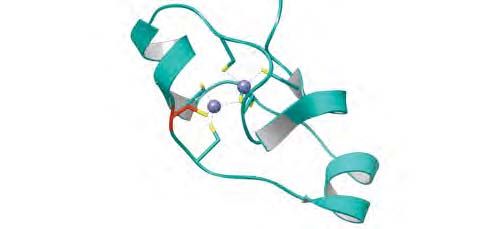
catalytic domain possesses two Zn-finger regions (AWS and SET/post-SET) containing three
Zn atoms chelated by cysteines that contribute to the folding of the domain and insure
enzyme function (Yang et al., 2016; Zhang et al., 2017) (supplementary Fig. 2A). Moreover,
electrophilic agents such as BQ can react with certain reactive Zn-bound cysteines in Zn-
fingers causing Zn ejection and protein unfolding (Lee et al., 2013). In agreement with this,
we observed that BQ (but not BZ nor the other BZ-metabolites) was able to cause Zn release
from SETD2 catalytic domain (Fig. 3A). Mass spectrometry experiments were carried out to
identify the cysteines adducted by BQ. However, in initial attempts, no individual amino acid
residues were identified. This is likely due to the ability of BQ to cross-link multiple residues
in SETD2 as observed previously for topoisomerase II (Bender et al., 2007). To circumvent
this technical difficulty, plumbagin (a quinone that has one single reactive site for adduction)
was used as a surrogate of BQ as previously described by Bender et al. (2007) (Bender et al.,
2007) (Fig. 3B). As observed with BQ, we confirmed that plumbagin (PBG) formed covalent
adducts with cysteine residues within SETD2 catalytic domain with concomitant loss of
enzyme activity and Zn release (Fig. 3C and 3D). Mass spectrometry analysis confirmed the
presence of a PBG adduct on Cys1499 of the AWS Zn-finger (Fig. 3E). Accordingly, NBT and
IAF staining, confirmed that PBG could form quinoprotein adducts on AWS Zn-finger
cysteines and induce Zn release (Supplementary Fig. 2B-2D). Interestingly, the 1499 cysteine
residue chelates a Zn atom present in the AWS Zn-finger (Yang et al., 2016). However, as the
peptide coverage was rather low (30%), we cannot rule out that other cysteines could be
adducted. Further experiments carried out with the purified AWS Zn-finger domain
confirmed that BQ could form covalent adducts with AWS Zn-finger cysteines, induce Zn
16Molecular Pharmacology Fast Forward. Published on July 15, 2021 as DOI: 10.1124/molpharm.121.000303
This article has not been copyedited and formatted. The final version may differ from this version.
release and protein crosslinks/oligomers (Supplementary Fig. 2D and 2E). Consistent with
our results, we found that TPEN, a well-known Zn chelator, could inactivate SETD2 catalytic
domain by depleting Zn atoms (Supplementary Fig. 3). Altogether, our results suggest that
BQ binds covalently to AWS domain Zn-finger cysteines within SETD2 catalytic domain,
resulting in Zn release, loss of histone methyltransferase activity and protein
crosslinks/oligomers formation. This is in agreement with the key structural and functional
role of the AWS Zn-finger domain of SETD2 (Yang et al., 2016; Zhang et al., 2017).
Impairment of SETD2 histone methyltransferase activity and decreased H3K36me3 levels
in cells exposed to BQ
Downloaded from molpharm.aspetjournals.org at ASPET Journals on June 4, 2022
We further tested the potential relevance of BQ-dependent inactivation of SETD2 by
conducting experiments in cells. To this end, the effects of BQ were first analyzed in
HEK293T cells expressing GFP-tagged SETD2. This cellular approach has notably been used to
analyze the formation of SETD2 aggregates/oligomers by western-blot (Bhattacharya and
Workman, 2020). As shown in Fig. 4.A, exposure of transfected cells to BQ led to the
formation of inactive SETD2 cross-links/oligomers as previously observed for other enzymes
inhibited by BQ (Shu, Cheng, et al., 2020; Shu, Hägglund, et al., 2020). SETD2 is inherently
prone to protein crosslink/aggregation and SETD2 aggregates/oligomers have been observed
by immunofluorescence as puncta in cells (Bhattacharya and Workman, 2020). As shown in
Fig. 4B, immunofluorescence studies showed the presence of puncta of SETD2 upon
exposure of cells to BQ thus supporting the formation of SETD2 cross-links/oligomers as
previously observed by Bhattacharya and Workman, 2020) (Bhattacharya and Workman,
2020). Further experiments were conducted in human hematopoietic K562 cells which
express functional SETD2. Consistent with the results described above, we found that
exposure of K562 cells to BQ led to the formation of endogenous SETD2 cross-
links/oligomers (Fig. 4C). Concomitantly and in agreement with the data reported above, we
observed significantly decreased levels of the H3K36me3 mark on histones extracted from
BQ-treated K562 cells (Fig. 4D). Congruent with the results obtained with the K562 cell line,
exposure of human primary hematopoietic CD34+ stem cells to BQ led also to decrease of
the H3K36me3 mark which is consistent with inactivation of SETD2 by BQ (Fig. 4E).
H3K36me3 levels have been shown to be present along the gene body with preference for
exons, notably in MYC and CDK2 genes (Eastmond et al., 2005). Using ChIP-qPCR, we
17Molecular Pharmacology Fast Forward. Published on July 15, 2021 as DOI: 10.1124/molpharm.121.000303
This article has not been copyedited and formatted. The final version may differ from this version.
analyzed the H3K36me3 levels at exons of MYC and CDK2 in K562 cells exposed to BQ as
described previously by Hacker et al. (2016) (Hacker et al., 2016). Congruent with the above
results, significantly decreased levels of H3K36me3 were observed for the exons studied
further supporting that BQ alters SETD2-dependent H3K36me3 epigenetic marks (Fig. 4F).
DISCUSSION
BQ is considered as the major hematotoxic metabolite of BZ (Smith, 2010; North et al.,
2020). The leukemogenic properties of BQ are thought to rely, at least in part, on the
formation of covalent Michael adducts with cysteine residues that may affect the structure
Downloaded from molpharm.aspetjournals.org at ASPET Journals on June 4, 2022
and the function of proteins involved in the regulation of hematopoietic cells (Smith et al.,
2011; Bolton and Dunlap, 2017; North et al., 2020; Shu, Hägglund, et al., 2020). So far,
inhibition of topoisomerase II enzymes by BQ is considered as one of the key mechanisms
contributing to BZ hematotoxicity (Bender et al., 2007; Eastmond et al., 2014; Holmes and
Winn, 2019). However, recent studies suggest that epigenetic mechanisms, notably DNA and
histone methylation, could be altered in BZ-induced leukemogenesis (Yu et al., 2019; Chung
and Herceg, 2020). We show here that the histone H3K36-specific trimethyltransferase
SETD2 is inactivated by BQ. SETD2 is the sole lysine methyltransferase which catalyzes the
formation of H3K36me3, a key epigenetic mark involved in transcriptional activation and
DNA repair (Husmann and Gozani, 2019). SETD2 is considered as a tumor suppressor and
recurrent-inactivating mutations of SETD2 and aberrant H3K36me3 levels have been
reported in hematopoietic malignancies (Mar et al., 2014; Zhu et al., 2014; Husmann and
Gozani, 2019). We found that BQ-dependent irreversible inactivation of SETD2 activity
occurs through the formation of covalent BQ adducts on Zn-fingers cysteines present in the
catalytic domain of the enzyme, notably within the AWS subdomain (Fig. 3 and
supplementary Fig. 3). The formation of these quinoprotein adducts on SETD2 result in Zn
release from the enzyme. It has been shown that Zn-bound cysteines in Zn-fingers are redox-
sensitive and react readily with electrophilic compounds to form covalent adducts that are
accompanied by Zn ejection and alteration of protein structure and function (Lee et al.,
2013). Interestingly, the histone methyltransferases G9a and GLP are inhibited by
electrophilic compounds able to eject Zn from Zn-fingers from their catalytic domain
(Lenstra et al., 2018). Moreover, quinone containing compounds have been reported to
18Molecular Pharmacology Fast Forward. Published on July 15, 2021 as DOI: 10.1124/molpharm.121.000303
This article has not been copyedited and formatted. The final version may differ from this version.
inhibit the histone demethylase KDM4A and the CREBBP/p300 acetyltransferase at least in
part through Zn ejection from their catalytic Zn-finger domains (Jayatunga et al., 2015;
Zhang et al., 2021). We also found that generation of BQ adducts on cysteine Zn-fingers
within SETD2 catalytic domain led to the formation of protein cross-links/oligomers that may
contribute to enzyme inactivation (Figure 2 and Figure 4). This is consistent with
observations indicating that disruption of Zn-bound cysteines in Zn-fingers, notably through
oxidation or reaction with electrophiles, can lead to Zn release and subsequent protein
unfolding (Quintal et al., 2011; Lee et al., 2013; Kluska et al., 2018). Interestingly, SETD2 has
been shown to be a rather unstable protein and prone to aggregation (Bhattacharya and
Workman, 2020). More broadly, protein cross-linking upon covalent binding of BQ to
Downloaded from molpharm.aspetjournals.org at ASPET Journals on June 4, 2022
cysteines have been shown to occur with enzymes such as glyceraldehyde-3-phosphate
dehydrogenase, creatine kinase or thioredoxin 1 and are thought to be responsible, at least
in part, for their inhibition (Shu, Cheng, et al., 2020; Shu, Hägglund, et al., 2020). Moreover,
the findings obtained with the purified SETD2 catalytic domain were further supported by
experiments carried out in cells. Indeed, we found that exposure to BQ of cells expressing
ectopically GFP-SETD2 or endogenous SETD2 (resulted in protein cross-links/oligomers and
loss of SETD2 activity as observed with the purified SETD2 catalytic domain (Fig. 2 and Fig. 4).
Consistent with this, we found that exposure to BQ caused a marked decrease of the
H3K36me3 mark on histones from human hematopoietic K562 or primary bone marrow
CD34+ cells. SETD2 activity and trimethylation of H3K36 are involved in normal
hematopoiesis and alteration of SETD2 and H3K36me3 levels are recurrently observed in
hematopoietic malignancies (Mar et al., 2017; Chen et al., 2020). Of note, dysregulation of
epigenetic processes such as DNA and histone methylation by BZ has been reported over the
last years (Smith, 2010; Fenga et al., 2016; Chung and Herceg, 2020). Our data support these
observations and provide mechanistic evidence that BQ, an oxidative and hematotoxic
metabolite of BZ, is able to react with and perturb the activity of a key epigenetic enzyme.
ACKNOWLEDGEMENTS
We thank the technical platform “BioProfiler” (BFA, CNRS UMR 8251 Unit, Université de
Paris) for provision of UFLC facilities.
19Molecular Pharmacology Fast Forward. Published on July 15, 2021 as DOI: 10.1124/molpharm.121.000303
This article has not been copyedited and formatted. The final version may differ from this version.
AUTHORS CONTRIBUTIONS
Participated in research design: JB, FG and FRL
Conducted experiments: JB, CM, LCB, LLC, VS and FG performed research and analyzed the
Performed data analysis: JB, CM, LCB, LLC, VS, LW, ND, JMD, CC, FG and FRL
Wrote or contributed to the writing of the manuscript: JB and FRL
Downloaded from molpharm.aspetjournals.org at ASPET Journals on June 4, 2022
20Molecular Pharmacology Fast Forward. Published on July 15, 2021 as DOI: 10.1124/molpharm.121.000303
This article has not been copyedited and formatted. The final version may differ from this version.
REFERENCES
Bender RP, Ham A-JJL, and Osheroff N (2007) Quinone-induced enhancement of DNA
cleavage by human topoisomerase IIalpha: adduction of cysteine residues 392 and 405.
Biochemistry 46:2856–64.
Bhattacharya S, and Workman JL (2020) Regulation of SETD2 stability is important for the
fidelity of H3K36me3 deposition. Epigenetics and Chromatin 13:40.
Bolton JL, and Dunlap T (2017) Formation and biological targets of quinones: Cytotoxic
versus cytoprotective effects. Chem Res Toxicol 30:13-37.
Butler JS, and Dent SYR (2013) The role of chromatin modifiers in normal and malignant
hematopoiesis. Blood 121:3076-3084.
Chappell G, Pogribny IP, Guyton KZ, and Rusyn I (2016) Epigenetic alterations induced by
genotoxic occupational and environmental human chemical carcinogens: A systematic
literature review. Mutat Res 768:27-45.
Downloaded from molpharm.aspetjournals.org at ASPET Journals on June 4, 2022
Chen BY, Song J, Hu CL, Chen SB, Zhang Q, Xu CH, Wu JC, Hou D, Sun M, Zhang YL, Liu N, Yu
PC, Liu P, Zong LJ, Zhang JY, Dai RF, Lan F, Huang QH, Zhang SJ, Nimer SD, Chen Z, Chen
SJ, Sun XJ, and Wang L (2020) SETD2 deficiency accelerates MDS-associated
leukemogenesis via S100a9 in NHD13 mice and predicts poor prognosis in MDS. Blood
135:2271–2285.
Chopra M, and Bohlander SK (2015) Disturbing the histone code in leukemia: translocations
and mutations affecting histone methyl transferases. Cancer Genet 208:192–205.
Chung FFL, and Herceg Z (2020) The promises and challenges of toxico-epigenomics:
Environmental chemicals and their impacts on the epigenome. Environ Health Perspect
128:15001.
Copeland RA (2005) Evaluation of enzyme inhibitors in drug discovery. A guide for medicinal
chemists and pharmacologists. Methods Biochem Anal 46:1–265.
Dong Y, Zhao X, Feng X, Zhou Y, Yan X, Zhang YY, Bu J, Zhan D, Hayashi Y, Zhang YY, Xu Z,
Huang R, Wang J, Zhao T, Xiao Z, Ju Z, Andreassen PR, Wang Q fei, Chen W, and Huang
G (2019) SETD2 mutations confer chemoresistance in acute myeloid leukemia partly
through altered cell cycle checkpoints. Leukemia 33:2585–2598.
Duval R, Bui LC, Mathieu C, Nian Q, Berthelet J, Xu X, Haddad I, Vinh J, Dupret JM, Busi F,
Guidez F, Chomienne C, and Rodrigues-Lima F (2019) Benzoquinone, a leukemogenic
metabolite of benzene, catalytically inhibits the protein tyrosine phosphatase PTPN2
and alters STAT1 signaling. J Biol Chem 294:12483–12494.
Duval R, Fritsch L, Bui L-C, Berthelet J, Guidez F, Mathieu C, Dupret J-M, Chomienne C, Ait-Si-
Ali S, and Rodrigues-Lima F (2015) An acetyltransferase assay for CBP based on RP-UFLC
of fluorescent histone H3 peptides. Anal Biochem 486:35-37.
Eastmond DA, Keshava N, and Sonawane B (2014) Lymphohematopoietic cancers induced by
chemicals and other agents and their implications for risk evaluation: An overview.
Mutat Res 761:40-64.
Eastmond DA, Mondrala ST, and Hasegawa L (2005) Topoisomerase II inhibition by
myeloperoxidase-activated hydroquinone: A potential mechanism underlying the
genotoxic and carcinogenic effects of benzene. Chem Biol Interact 153–154:207–216.
Edmunds JW, Mahadevan LC, and Clayton AL (2008) Dynamic histone H3 methylation during
gene induction: HYPB/Setd2 mediates all H3K36 trimethylation. EMBO J 27:406–420.
Fahey CC, and Davis IJ (2017) SETting the Stage for Cancer Development: SETD2 and the
Consequences of Lost Methylation. Cold Spring Harb Perspect Med 7:a026468.
21Molecular Pharmacology Fast Forward. Published on July 15, 2021 as DOI: 10.1124/molpharm.121.000303
This article has not been copyedited and formatted. The final version may differ from this version.
Fenga C, Gangemi S, and Costa C (2016) Benzene exposure is associated with epigenetic
changes (Review). Mol Med Rep 13:3401–3405.
Frantz CE, Chen H, and Eastmond DA (1996) Inhibition of human topoisomerase II in vitro by
bioactive benzene metabolites. Environ Health Perspect 104:1319–1323.
Hacker KE, Fahey CC, Shinsky SA, Chiang Y-CJCJ, DiFiore J V., Jha DK, Vo AH, Shavit JA, Davis
IJ, Strahl BD, and Rathmell WK (2016) Structure/Function Analysis of Recurrent
Mutations in SETD2 Protein Reveals a Critical and Conserved Role for a SET Domain
Residue in Maintaining Protein Stability and Histone H3 Lys-36 Trimethylation. J Biol
Chem 291:21283–21295.
Holmes TH, and Winn LM (2019) DNA Damage and Perturbed Topoisomerase IIα as a Target
of 1,4-Benzoquinone Toxicity in Murine Fetal Liver Cells. Toxicol Sci 171:339–346.
Huether R, Dong L, Chen X, Wu G, Parker M, Wei L, Ma J, Edmonson MN, Hedlund EK, Rusch
MC, Shurtleff SA, Mulder HL, Boggs K, Vadordaria B, Cheng J, Yergeau D, Song G,
Becksfort J, Lemmon G, Weber C, Cai Z, Dang J, Walsh M, Gedman AL, Faber Z, Easton J,
Downloaded from molpharm.aspetjournals.org at ASPET Journals on June 4, 2022
Gruber T, Kriwacki RW, Partridge JF, Ding L, Wilson RK, Mardis ER, Mullighan CG,
Gilbertson RJ, Baker SJ, Zambetti G, Ellison DW, Zhang J, and Downing JR (2014) The
landscape of somatic mutations in epigenetic regulators across 1,000 paediatric cancer
genomes. Nat Commun 5:3630.
Husmann D, and Gozani O (2019) Histone lysine methyltransferases in biology and disease.
Nat Struc Mol Biol 26:880-889.
Jayatunga MKP, Thompson S, McKee TC, Chan MC, Reece KM, Hardy AP, Sekirnik R, Seden
PT, Cook KM, McMahon JB, Figg WD, Schofield CJ, and Hamilton AD (2015) Inhibition of
the HIF1α-p300 interaction by quinone- and indandione-mediated ejection of structural
Zn(II). Eur J Med Chem 94:509–516.
Kim Sungkyoon, Vermeulen R, Waidyanatha S, Johnson BA, Lan Q, Rothman N, Smith MT,
Zhang L, Li G, Shen M, Yin S, and Rappaport SM (2006) Using urinary biomarkers to
elucidate dose-related patterns of human benzene metabolism. Carcinogenesis 27:772–
781.
Kim S., Vermeulen R, Waidyanatha S, Johnson BA, Lan Q, Smith MT, Zhang L, Li G, Shen M,
Yin S, Rothman N, and Rappaport SM (2006) Modeling Human Metabolism of Benzene
Following Occupational and Environmental Exposures. Cancer Epidemiol Biomarkers
Prev 15:2246–2252.
Kluska K, Adamczyk J, and Krężel A (2018) Metal binding properties, stability and reactivity of
zinc fingers. Coord Chem Rev 367:18–64.
Kudithipudi S, and Jeltsch A (2014) Role of somatic cancer mutations in human protein lysine
methyltransferases. Biochim Biophys Acta 1846:366–79.
Lee YM, Wang YT, Duh Y, Yuan HS, and Lim C (2013) Identification of labile zn sites in drug-
target proteins. J Am Chem Soc 135:14028–14031.
Lenstra DC, Al Temimi AHK, and Mecinović J (2018) Inhibition of histone lysine
methyltransferases G9a and GLP by ejection of structural Zn(II). Bioorganic Med Chem
Lett 28:1234–1238.
Li J, Duns G, Westers H, Sijmons R, van den Berg A, and Kok K (2016) SETD2: an epigenetic
modifier with tumor suppressor functionality. Oncotarget 7:50719–50734.
Lu PCW, Shahbaz S, and Winn LM (2020) Benzene and its effects on cell signaling pathways
related to hematopoiesis and leukemia. J Appl Toxicol 40:1018–1032.
Mar BG, Bullinger LB, McLean KM, Grauman P V, Harris MH, Stevenson K, Neuberg DS, Sinha
AU, Sallan SE, Silverman LB, Kung AL, Lo Nigro L, Ebert BL, and Armstrong SA (2014)
22Molecular Pharmacology Fast Forward. Published on July 15, 2021 as DOI: 10.1124/molpharm.121.000303
This article has not been copyedited and formatted. The final version may differ from this version.
Mutations in epigenetic regulators including SETD2 are gained during relapse in
paediatric acute lymphoblastic leukaemia. Nat Commun 5:3469.
Mar BG, Chu SH, Kahn JD, Krivtsov A V., Koche R, Castellano CA, Kotlier JL, Zon RL, McConkey
ME, Chabon J, Chappell R, Grauman P V., Hsieh JJ, Armstrong SA, and Ebert BL (2017)
SETD2 alterations impair DNA damage recognition and lead to resistance to
chemotherapy in leukemia. Blood 130:2631–2641.
Mbiya W, Chipinda I, Siegel PD, Mhike M, and Simoyi RH (2013) Substituent effects on the
reactivity of benzoquinone derivatives with thiols. Chem Res Toxicol 26:112–123.
McHale CM, Zhang L, and Smith MT (2012) Current understanding of the mechanism of
benzene-induced leukemia in humans: implications for risk assessment. Carcinogenesis
33:240–252.
North CM, Rooseboom M, Kocabas NA, Schnatter AR, Faulhammer F, and Williams SD (2020)
Modes of action considerations in threshold expectations for health effects of benzene.
Toxicol Lett 334:78–86.
Downloaded from molpharm.aspetjournals.org at ASPET Journals on June 4, 2022
Quintal SM, Depaula QA, and Farrell NP (2011) Zinc finger proteins as templates for metal
ion exchange and ligand reactivity. Chemical and biological consequences. Metallomics
3:121–139.
R. Hunter Lindsey J, Kenneth D. Bromberg, Carolyn A. Felix and, and Neil Osheroff (2004) 1,4-
Benzoquinone Is a Topoisomerase II Poison. Biochemistry 43:7563-7574.
Rappaport SM, Kim S, Lan Q, Li G, Vermeulen R, Waidyanatha S, Zhang L, Yin S, Smith MT,
and Rothman N (2010) Human benzene metabolism following occupational and
environmental exposures. Chem Biol Interact 184:189–195.
Sauer E, Gauer B, Nascimento S, Nardi J, Göethel G, Costa B, Correia D, Matte U, Charão M,
Arbo M, Duschl A, Moro A, and Garcia SC (2018) The role of B7 costimulation in
benzene immunotoxicity and its potential association with cancer risk. Environ Res
166:91–99.
Shu N, Cheng Q, Arnér ESJ, and Davies MJ (2020) Inhibition and crosslinking of the
selenoprotein thioredoxin reductase-1 by p-benzoquinone. Redox Biol 28:101335.
Shu N, Hägglund P, Cai H, Hawkins CL, and Davies MJ (2020) Modification of Cys residues in
human thioredoxin-1 by p-benzoquinone causes inhibition of its catalytic activity and
activation of the ASK1/p38-MAPK signalling pathway. Redox Biol 29:101400.
Shu N, Lorentzen LG, and Davies MJ (2019) Kinetics and biological consequences of quinone-
induced protein adduction. Free Radic Biol Med 120:S55.
Smith MT (2010) Advances in understanding benzene health effects and susceptibility. Annu
Rev Public Health 31:133–148.
Smith MT, Zhang L, McHale CM, Skibola CF, and Rappaport SM (2011) Benzene, the
exposome and future investigations of leukemia etiology. Chem Biol Interact 192:155-
159.
Snyder R (2012) Leukemia and benzene. Int J Environ Res Public Health 9:2875-2893.
Wagner EJ, and Carpenter PB (2012) Understanding the language of Lys36 methylation at
histone H3. Nat Rev Mol Cell Biol 13:115–26, Nat Rev Mol Cell Biol.
Wang L, Niu N, Li L, Shao R, Ouyang H, and Zou W (2018) H3K36 trimethylation mediated by
SETD2 regulates the fate of bone marrow mesenchymal stem cells. PLoS Biol 16, Public
Library of Science.
Wang X, Thomas B, Sachdeva R, Arterburn L, Frye L, Hatcher PG, Cornwell DG, and Ma J
(2006) Mechanism of arylating quinone toxicity involving Michael adduct formation and
induction of endoplasmic reticulum stress. Proc Natl Acad Sci U S A 103:3604–3609,
23You can also read