Systemic lupus Erythematous as an Unusual Cause of Intussusception in Sudanese Woman: Case report
←
→
Page content transcription
If your browser does not render page correctly, please read the page content below
Posted on Authorea 25 Sep 2021 — The copyright holder is the author/funder. All rights reserved. No reuse without permission. — https://doi.org/10.22541/au.163257004.48666749/v1 — This a preprint and has not been peer reviewed. Data may be preliminary.
Systemic lupus Erythematous as an Unusual Cause of
Intussusception in Sudanese Woman: Case report
Salwa Dafa Allah Salih Mohammadeen1 , Amar F.Eldow 2 , Rania Eisa Abdelmutalib1 , Sara
galal osman hamza1 , Elnour Mohammed Elagib1 , huyam H.Awadalla1 , Abubkar Hassan3 ,
Abdelkareem A.Ahmed4 , and Mohammed Elmujtba Adam Essa5
1
Sudan Medical Specialization Board
2
Wayne State University School of Medicine
3
MCR
4
MCRI
5
Al Fashir University Faculty of Medicine
September 25, 2021
Abstract
A middle-aged Sudanese woman has been presented complained about multiple joint pain, skin rash, chest pain, hair loss,
severe abdominal pain associated with abdominal distension, bloody diarrhoea and vomiting. Lab investigation and computed
tomography (CT) abdomen revealed the patient have an intussusception on top of SLE. The patient was treated
Introduction
SLE is a complex autoimmune chronic inflammatory disorder with an extensive cascade of symptoms, the
disease can involve the kidney, lung, nervous system and heart (1). Abdominal pain does have a wide
differential diagnosis, however, gastrointestinal involvement is not unusual in SLE (2, 3). A lot of causes of
abdominal pain have been described in lupus patients such as lupus enteritis drug reactions and infections
due to steroids (4, 5). In addition, the likelihood of SLE as a contributing factor in jejunal diverticulosis has
been reported (6).
An intussusception itself as a complication or presenting symptom of SLE is rare, until now there are no
more than seven cases have been reported in SLE (4, 6). Two of those cases it was secondary to mesenteric
vacuities and five cases had an intussusception as an initial presentation of SLE (7). In this case, we are
reporting a case of SLE with intussusception as the initial presentation.
Case report
A 40-year old Sudanese female, presented with complaints of facial skin rash, bilateral small & large joints
pain, swelling, Palpitations, Shortness of breath, Pleuritic chest pain, Hair loss and Muscle pain.
Soon after she has been admitted to our rheumatology unite at Omdurman military hospital, Khartoum Su-
dan, the patient developed acute severe cramping periumbilical abdominal pain associated with abdominal
distension, vomiting & infrequent bloody diarrhoea. Her examination showed high blood pressure (150/100),
pulse rate of 100, temperature 37.5, she has malar rash sparing the nasolabial folds with hyperpigmented
patches, no rash in other sites, broken hair in the frontal and temporal areas and no nail changed. Cardiovas-
cular examination revealed short localized Systolic murmur in the mitral area, chest examination indicated
1
decreased air entry bilaterally with dull percussion notes, her abdomen was distended, tender left periumbil-
Posted on Authorea 25 Sep 2021 — The copyright holder is the author/funder. All rights reserved. No reuse without permission. — https://doi.org/10.22541/au.163257004.48666749/v1 — This a preprint and has not been peer reviewed. Data may be preliminary.
ical mass, Joints examination showed active synovitis involve meta-carbophalengial joints bilaterally, Knees
& shoulders pain with passive and active movements. General Investigations were done (Table 1) suggestive
of SLE and the patient abdominal CT showed rounded lumber mass with a double layer concluded intestinal
intussusception.
The patient was seen by the surgical team, underwent laparotomy with bowel resection, the biopsy
examination report multiple fragments measuring 16 cm with attached binding 12.5 cm showed congested
vascular channels and hypertrophied muscular fibers with no granulomas or malignancy saw, all these features
consistent with a diagnosis of intussusception, no features of granuloma or neoplastic cells.
The patient was diagnosed with a case of systemic lupus erythematosus complicated with intussusception
and the possibility of lupus nephritis. post-operatively the patient received prednisone 30 mg with tapering
Hydroxychloroquine 200 mg per day Azathioprine 50 mg twice per day Lisinopril 20 mg per day, tonics
Bone and gastric protection, although, the patient therapy showed good response yet, she is still on regular
hemodialysis because renal biopsy done to the patient showed grade 4 lupus nephritis.
Discussion
SLE is a multisystem autoimmune disease in which deposition of immune complexes and pathogenic autoan-
tibodies lead to a wide variety of symptoms (8). Abdominal pain in SLE patients can occur for multiple
reasons including severe lupus vasculitis presenting as gastrointestinal perforation or mesenteric thrombosis,
acute pancreatitis, cholecystitis, lupus mesenteric vasculitis and hepatitis (9). Intussusception is a condition
in which the proximal segment of the intestine telescoping into the distal part of it (10). Although, intus-
susception I uncommon in adults and more likely to occur during childhood but adult have an underlying
pathology in approximately 90% of cases. An intussusception in the small intestine mainly due to secondary
causes, either due to extraluminal or intraluminal lesions ( Meckel’s diverticulum, lymphoma, lipoma, post-
operative, adhesions and metastases), however, intussusception in the large bowel is mostly due to malignant
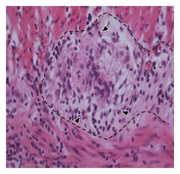
aetiology (11). The surgical specimen can shows macroscopically aspects of intussusception in the intestine
(Figure 1) as well as the microscopically histopathology features (Figure 2)
At usually computer tomography (CT) Scan for the abdomen can identify the potential cause. The presence
of bowel with bowel configuration with or without the existence of adipose tissue and mesenteric vessels is
pathognomonic for intussusception (5).
The pathogenesis of the intussusception in our patient related to SLE could be mostly the initial manifestation
of the disease. Only seven cases of intussusception in Patients with SLE have been described in the literature.
In four cases were secondary to lupus mesenteric vasculitis (LMV) (7), one of them lymphadenopathy was
the main cause, one was related to Burkitt’s lymphoma, one was secondary to changes in the peristalsis of
oedematous small intestine (4, 7, 12, 13).
The first report has been published by Hermann (11), on a five years old child with known who had an
intussusception as a complication to LMV. The exact pathology and mechanism of LMV causing intussus-
ception is not fully understood (4). A potential explanation is that vascular necrosis occurred by diffuse
vasculitis with partial devitalisation of the intestinal segment, this can cause the interruption of the normal
neuromuscular function, with concomitant intussusception and possibility of venous infarction necessitating
bowel resection (4).
Intussusception is a very serious complication carrying a high mortality rate (14), CT abdomen has a central
role in early detection of LMV and delivers a precise imaging of the lesions. However, the detection of the
underlying causative factor of the intussusception can be challenging due to differentiation from bowel wall
edema may not be possible (figure 3). In our case the CT abdomen of the patient Showed bilaterally pleural
effusion, moderate free peritoneal fluid collection (Ascites) and rounded lumber mass with double layers
suggestive of intussusception.
2Another report described a known case of lupus with fungal infection with intussusception. It is not fully
Posted on Authorea 25 Sep 2021 — The copyright holder is the author/funder. All rights reserved. No reuse without permission. — https://doi.org/10.22541/au.163257004.48666749/v1 — This a preprint and has not been peer reviewed. Data may be preliminary.
clear whether there is an association between these conditions (15). However, in our case intussusception
occur few days after the patient present complaining from the rheumatological features.
In conclusion, a 40-year-old female presented with multiple joints pain, skin rash, chest pain, hair loss and
intussusception, bowel resection was done and they found no evidence of malignancy, infection causes or
mesenteric vasculitis. Therefore, it is more likely that the intussusception was a secondary cause to SLE in
this patient.
Acknowledgments
The case was diagnosed and treated in Dr Elnour Mohammed Elagib rheumatology unite at Omdurman
Military hospital, Khartoum, Sudan.
Ethical approval and consent to publish
Obtained
Conflict of Interest
Non
Written consent from the patient
Obtained
Authors contributions
All authors contributed equally
Funding
No fund have been received
Authors’ contribution
All authors contributed equally
Availability of data and materials
All the data used in the study are available from the first and corresponding author on reasonable request.
Reference
1. Farshad S, Kanaan C, Savedchuk S, Karmo DS, Halalau A, Swami A. Systemic Lupus Erythematosus
(SLE) with Acute Nephritis, Antineutrophil Cytoplasmic Antibody- (ANCA-) Associated Vasculitis, and
Thrombotic Thrombocytopenic Purpura (TTP): A Rare Case Report with Literature Review. Case Rep
Rheumatol. 2019;2019:8750306.
2. Chu YC, Hsu BB, Tseng KC. Lupus mesenteric vasculitis with GI and genitourinary tract involvement.
Clin Gastroenterol Hepatol. 2014;12(8):e69-70; quiz e1-2, e3.
3. de Carvalho JF. Mesenteric vasculitis in a systemic lupus erythematosus patient with a low sledai: an
uncommon presentation. Clinics (Sao Paulo). 2010;65(3):337-40.
4. Lin YJ, Chen PC, Chen HA. Mesenteric vasculitis causing ileocecal intussusception as the initial presen-
tation of systemic lupus erythematosus: a case report. Clin Rheumatol. 2013;32 Suppl 1:S37-40.
5. Zhang J, Fang M, Wang Y, Mao J, Sun X. Intestinal pseudo-obstruction syndrome in systemic lupus
erythematosus. Lupus. 2011;20(12):1324-8.
36. Fukaya S, Yasuda S, Hashimoto T, Oku K, Kataoka H, Horita T, et al. Clinical features of haemophago-
Posted on Authorea 25 Sep 2021 — The copyright holder is the author/funder. All rights reserved. No reuse without permission. — https://doi.org/10.22541/au.163257004.48666749/v1 — This a preprint and has not been peer reviewed. Data may be preliminary.
cytic syndrome in patients with systemic autoimmune diseases: analysis of 30 cases. Rheumatology (Oxford).
2008;47(11):1686-91.
7. Glijn N, Korswagen LA, Lam-Tse WK. Systemic lupus erythematosus (SLE): an unusual cause of ileocolic
intussusception. BMJ Case Rep. 2017;2017.
8. Albuquerque-Netto AF, Cavalcante EG, Sallum AM, Aikawa NE, Tannuri U, Silva CA. Mesenteric
vasculitis in a juvenile systemic lupus erythematosus patient. Rev Bras Reumatol. 2013;53(2):219-22.
9. Malaviya AN, Sharma A, Agarwal D, Kapoor S, Garg S, Singh S, et al. Acute abdomen in SLE. Int J
Rheum Dis. 2011;14(1):98-104.
10. Bergmann KR, Arroyo AC, Tessaro MO, Nielson J, Whitcomb V, Madhok M, et al. Diagnostic Accuracy
of Point-of-Care Ultrasound for Intussusception: A Multicenter, Noninferiority Study of Paired Diagnostic
Tests. Ann Emerg Med. 2021.
11. Hermann G. Intussusception secondary to mesenteric arteries. Complication of systemic lupus erythe-
matosus in a 5-year-old child. JAMA. 1967;200(1):74-5.
12. Yagmur Y, Aldemir M, Buyukbayram H, Tacyildiz I. Multiple jejunal diverticulitis with perforation in
a patient with systemic lupus erythematosus: report of a case. Surg Today. 2004;34(2):163-6.
13. Chang D-K, Yoo D-H, Kim T-H, Kim IS, Jun KY, Park MH, et al. Burkitt’s lymphoma presenting as
ileocaecal intussusception in systemic lupus erythematosus. Clinical rheumatology. 1999;18(3):253-6.
14. Yagmur Y, Gumus S. Burkitt’s lymphoma causing intussusception in adults: report of two cases and
review of the literature. Journal of Gastroenterology and Hepatology Research. 2015;4(7):1702-6.
15. Wei CC, Chen JH, Cheng HH. Systemic lupus erythematosus with intussusception: a case report.
Zhonghua Yi Xue Za Zhi (Taipei). 1996;58(1):58-61.
16. Kaemmerer E, Tischendorf JJW, Steinau G, Wagner N, Gassler N. Ileocecal Intussusception with His-
tomorphological Features of Inflammatory Neuropathy in Adenovirus Infection. Gastroenterology Research
and Practice. 2009;2009:579501.
17. Kim YH, Blake MA, Harisinghani MG, Archer-Arroyo K, Hahn PF, Pitman MB, et al. Adult intestinal
intussusception: CT appearances and identification of a causative lead point. Radiographics. 2006;26(3):733-
44.
Table 1: Shows the lab results performed for the patient
Investigations Results
WBC 6.8 cells/mcl
HB 7.3 g/dl
Platelet 127 cells/mcl
ESR 60 mm/hour
Serum Urea 32 mg/dl
S.Creatinine 1.1 mg/dl
CRP 20
S.Albumin 2.9 g/dl
Total Protein 5.9 G/dl
Total Bilirubin 0.4 mg/dl
ALT 33 U/L
AST 27 U/L
ALP 105 U/L (24-147 UL)
Urine General Albumin, no casts
4Investigations Results
Posted on Authorea 25 Sep 2021 — The copyright holder is the author/funder. All rights reserved. No reuse without permission. — https://doi.org/10.22541/au.163257004.48666749/v1 — This a preprint and has not been peer reviewed. Data may be preliminary.

7-9 pus
Direct Coomb Test Negative
Antinuclear antibodies positive Antidouble stranded DNA Antihistone positive AntiRibosomal p protein positive Anti nuc
Figure 1: Shows the morphological features of ileocecal surgical specimens with intussusception (16).
Figure 2: Tissue section of the terminal ileum demonstrates the plexus myentericus (dotted line) and several
infiltrating lymphocytes and eosinophiles (arrowheads) (16).
5Posted on Authorea 25 Sep 2021 — The copyright holder is the author/funder. All rights reserved. No reuse without permission. — https://doi.org/10.22541/au.163257004.48666749/v1 — This a preprint and has not been peer reviewed. Data may be preliminary.
wall edema, making differentiation difficult (17)
6
Figure (3): CT scans shows an intussusception in the abdomen, the mass is isoattenuating relative to bowelPosted on Authorea 25 Sep 2021 — The copyright holder is the author/funder. All rights reserved. No reuse without permission. — https://doi.org/10.22541/au.163257004.48666749/v1 — This a preprint and has not been peer reviewed. Data may be preliminary. 7
You can also read