Primary Pulmonary Lymphatic Flow Disorder Associated Plastic Bronchitis

Page created by Derrick Cox
 
CONTINUE READING
Primary Pulmonary Lymphatic Flow Disorder Associated Plastic Bronchitis
Posted on Authorea 10 Jan 2022 — The copyright holder is the author/funder. All rights reserved. No reuse without permission. — https://doi.org/10.22541/au.164183219.98138934/v1 — This a preprint and has not been peer reviewed. Data may be preliminary.

                                                                                                                                                                                                                                                               Primary Pulmonary Lymphatic Flow Disorder Associated Plastic
                                                                                                                                                                                                                                                               Bronchitis
                                                                                                                                                                                                                                                               Xiaoyan Zhang1 , Jinrong Liu2 , hui xu3 , haiming yang3 , and Shunying Zhao3
                                                                                                                                                                                                                                                               1
                                                                                                                                                                                                                                                                 Beijing Children’s Hospital Capital Medical University
                                                                                                                                                                                                                                                               2
                                                                                                                                                                                                                                                                 Beijing Children’s Hospital Affiliated to Capital Medical University
                                                                                                                                                                                                                                                               3
                                                                                                                                                                                                                                                                 Beijing Children’s Hospital

                                                                                                                                                                                                                                                               January 10, 2022

                                                                                                                                                                                                                                                               Abstract
                                                                                                                                                                                                                                                               Lymphatic plastic bronchitis (PB) most commonly occurs in children with congenital heart disease as a result of secondary
                                                                                                                                                                                                                                                               pulmonary lymphatic flow disorder (PLFD). However, PB caused by primary PLFD is rare. The clinical symptoms of lym-
                                                                                                                                                                                                                                                               phocytic PB are nonspecific; thus, the diagnosis may be long delayed, especially in the absence of chylothorax. We made a
                                                                                                                                                                                                                                                               retrospective analysis of two children diagnosed with PB due to primary PLFD, in order to contribute to further understanding
                                                                                                                                                                                                                                                               of these disorders. Patient 1, an eight-year-old boy, presented with chronic productive cough and expectorated milky-white
                                                                                                                                                                                                                                                               mucous plugs accompanied by intermitted wheezing for one year. Patient 2, a nine-month-old girl, presented with episodes of
                                                                                                                                                                                                                                                               acute respiratory distress with expectoration of milky-white bronchial casts for four months. There was no obvious evidence
                                                                                                                                                                                                                                                               of infection in either child. Bilateral thickening of bronchovascular bundles and interlobular septal, as well as multiple patchy
                                                                                                                                                                                                                                                               ground-glass opacities were seen on chest computed tomography (CT) in both patients. Lymphangioscintigraphy demonstrated
                                                                                                                                                                                                                                                               pulmonary lymph reflux in both patients and slowed lymphatic drainage of the lower limbs in patient 1. Primary PLFD was
                                                                                                                                                                                                                                                               considered for both patients, and a diagnosis of yellow nail syndrome was made in patient 1. Both patients received lymphatic
                                                                                                                                                                                                                                                               interventional treatment, but all experienced recurrence following the procedure.

                                                                                                                                                                                                                                                               Primary Pulmonary Lymphatic Flow Disorder Associated Plastic Bronchitis
                                                                                                                                                                                                                                                               To the Editor,
                                                                                                                                                                                                                                                               Plastic bronchitis (PB) is a rare disease characterized by the expectoration of rubbery, cohesive, and branched
                                                                                                                                                                                                                                                               bronchial casts that obstruct the airway partially or totally. BP is associated with respiratory disorders
                                                                                                                                                                                                                                                               such as bronchopulmonary infection, asthma, allergic bronchopulmonary aspergillosis (ABPA), and cystic
                                                                                                                                                                                                                                                               fibrosis. Besides, BP commonly occurs in children with congenital heart disease, especially those with single
                                                                                                                                                                                                                                                               ventricle physiology following the Fontan procedure. Elevation of central venous pressure after the procedure
                                                                                                                                                                                                                                                               and secondary pulmonary lymphatic flow disorder (PLFD) leading to abnormal perfusion of the bronchial
                                                                                                                                                                                                                                                               submucosa with lymph and formation of the airway cast 1-2 . However, PB caused by congenital lymphatic
                                                                                                                                                                                                                                                               dysplasia and primary PLFD is rare, the clinical manifestation and severity vary among patients.
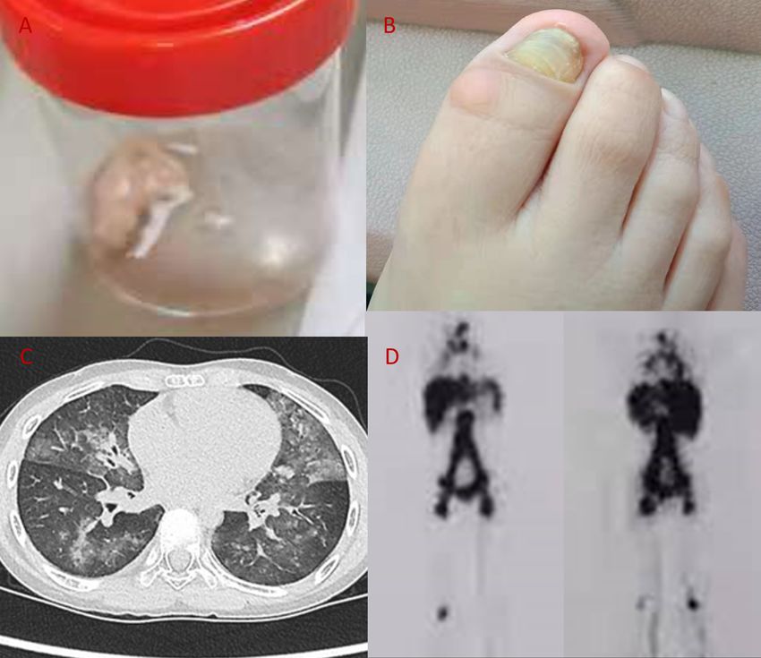
                                                                                                                                                                                                                                                               Patient 1 an eight-year-old boy admitted with a complain of chronic productive cough, expectorated milky-
                                                                                                                                                                                                                                                               white mucous plugs every 4-5 days (Figure. 1A), accompanied by intermitted wheezing for 1 year. Treatment
                                                                                                                                                                                                                                                               of antibiotics, inhaled corticosteroids and brinchoidilaters fail to control the symptoms. His medical history
                                                                                                                                                                                                                                                               revealed recurrent pneumonia as well as intermitted wheezing from two years of age, and he was diagnosed
                                                                                                                                                                                                                                                               with asthma. His mother noticed the slow yellowing and thickening of the first toenail on his right foot
                                                                                                                                                                                                                                                               about three years ago. The nail changes had been interpreted as onychomycosis and the first toenail had been
                                                                                                                                                                                                                                                               removed before presentation to the hospital and a small and yellowish new toenail was observed (Figure. 1B).
                                                                                                                                                                                                                                                               He was normally developed, there was no peripheral edema and the physical examination was unremarkable.

                                                                                                                                                                                                                                                                                                                              1
The patient’s white blood cell (WBC) count, eosinophil count, and C-reactive protein (CRP) were normal.
Posted on Authorea 10 Jan 2022 — The copyright holder is the author/funder. All rights reserved. No reuse without permission. — https://doi.org/10.22541/au.164183219.98138934/v1 — This a preprint and has not been peer reviewed. Data may be preliminary.

                                                                                                                                                                                                                                                               The serum levels of aspergillus specific IgE, 1,3-β-glucan, and galactomannan were normal. Autoantibodies
                                                                                                                                                                                                                                                               were all negative. Sputum cultures were positive forCandida albicans , while bacterial and mycobacterium
                                                                                                                                                                                                                                                               cultures were negative. Fungal microscopic examination and culture of the first toenail were negative.
                                                                                                                                                                                                                                                               Serial chest computed tomography (CT) revealed migratory patchy ground-glass opacities and focal mild
                                                                                                                                                                                                                                                               bronchiectasis with mucus impactions. The bronchial lavage fluid (BALF) was slightly milky-white and
                                                                                                                                                                                                                                                               turbid, but no casts were observed. Fluid cultures showed no pathogens. BALF cell composition analysis
                                                                                                                                                                                                                                                               was as follows: small lymphocytes 60%, macrophages 20%, exfoliated epithelial cells 20%. The pulmonary
                                                                                                                                                                                                                                                               function test showed mild obstructive ventilatory disturbance and the bronchodilation test was negative. An
                                                                                                                                                                                                                                                               echocardiogram showed no abnormalities.
                                                                                                                                                                                                                                                               Suspicion of allergic bronchopulmonary mycosis (ABPM) was arised after the patient’s admission. He was
                                                                                                                                                                                                                                                               treated with fluconazole (8 mg/kg.d) and prednisone (1 mg/kg.d, gradually decreased to 0.5 mg/kg.d)
                                                                                                                                                                                                                                                               for three weeks. However, his cough and expectoration of mucous plugs worsened. Repeated chest CT
                                                                                                                                                                                                                                                               showed the progression of lung lesions with bilateral thickening of bronchovascular bundles, multiple patchy
                                                                                                                                                                                                                                                               ground-glass opacities, and interlobular septal thickening, mainly in the middle-inferior lung, indicating
                                                                                                                                                                                                                                                               pulmonary lymphatic stasis (Figure. 1C). Evaluation of the lymphatic system by lymphangioscintigray was
                                                                                                                                                                                                                                                               performed subsequently and the results demonstrated slow lymphatic drainage in both lower limbs, abnormal
                                                                                                                                                                                                                                                               distribution of the radiotracer in the bilateral hilar and lungs, suggesting pulmonary chylous reflux, as well
                                                                                                                                                                                                                                                               as thoracic duct obstruction (Fig. 1D).
                                                                                                                                                                                                                                                               Based on the patient’s clinical picture, lymphangioscintigraphy findings, and the yellow toenail on his right
                                                                                                                                                                                                                                                               foot, the patient was finally diagnosed with yellow nail syndrome (YNS) and lymphatic PB. He was treated
                                                                                                                                                                                                                                                               with a low-fat diet and percutaneous embolization of the thoracic duct was performed three weeks later. His
                                                                                                                                                                                                                                                               respiratory symptoms were relieved. However, three months after the surgery, he experienced recurrence of
                                                                                                                                                                                                                                                               bronchial cast expectoration occasionally. His parents refused further evaluation and treatment as he had
                                                                                                                                                                                                                                                               developed no other symptoms.
                                                                                                                                                                                                                                                               Patient 2 a nine-month-old girl was referred to our department for episodes of acute respiratory distress with
                                                                                                                                                                                                                                                               cough and expectoration of milky-white bronchial casts requiring bronchoscopy treatment for 4 months.
                                                                                                                                                                                                                                                               During the course of the disease, there was no obvious fever. She had been admitted to the local intensive
                                                                                                                                                                                                                                                               care unit and required mechanical ventilation two times because of severe bouts of dyspnea. Bronchoscopy
                                                                                                                                                                                                                                                               demonstrated many milky-white casts blocking the airway (Figure. 2A); these were extracted by aspiration
                                                                                                                                                                                                                                                               and her dyspnea and hypoxemia can be temporarily relieved. Chest CT demonstrated diffuse thickening
                                                                                                                                                                                                                                                               of the interlobular septa and bronchovascular bundles, left partial atelectasis and consolidations, as well
                                                                                                                                                                                                                                                               as enlargement of the mediastinum and hilar lymph nodes (Figure. 2B). Diagnoses of severe pneumonia
                                                                                                                                                                                                                                                               and PB were made. However, despite vigorous therapy with antimicrobials (including antifungal drugs),
                                                                                                                                                                                                                                                               intermittent systemic and inhaled glucocorticoids, and intravenous immunoglobulin, she continued to exhibit
                                                                                                                                                                                                                                                               chronic expectoration of bronchial casts and episodes of acute respiratory distress requiring bronchoscopy to
                                                                                                                                                                                                                                                               clear the airways about every two weeks. She had normal development and no history of trauma or surgery.
                                                                                                                                                                                                                                                               Physical examination revealed tachypnea, a respiratory rate of 42 breaths/min, and oxygen saturation of
                                                                                                                                                                                                                                                               90% on room air. Chest auscultation showed bilateral moist rales and wheezing. The remainder of her
                                                                                                                                                                                                                                                               examination was unremarkable.
                                                                                                                                                                                                                                                               The patient’s WBC count, eosinophil count, hemoglobin, and CRP were normal. BALF cultures showed a
                                                                                                                                                                                                                                                               small amount of Klebsiella pneumoniae. Tests for influenza virus, adenovirus, and mycoplasma detection were
                                                                                                                                                                                                                                                               all negative. The echocardiogram showed no abnormalities. Abdominal ultrasound showed multiple cysts
                                                                                                                                                                                                                                                               of the spleen. Lymphangioscintigraphy was performed subsequently and revealed thoracic duct obstruction,
                                                                                                                                                                                                                                                               abnormal distribution of the radiotracer in the bilateral hilar and lungs, suggesting pulmonary chylous reflux
                                                                                                                                                                                                                                                               (Figure. 2C).
                                                                                                                                                                                                                                                               Given her early age of onset, congenital lymphatic dysplasia was considered. Diagnostic pulmonary biopsy
                                                                                                                                                                                                                                                               was not performed because of the risk of massive chyloptysis or refractory chylothorax3 . Splenic cystic lesions
                                                                                                                                                                                                                                                               were observed on abdominal ultrasound, suggesting extrathoracic involvement. The child was treated with

                                                                                                                                                                                                                                                                                                                      2
high medium-chain triglycerides (MCT) milk powder, and oral rapamycin was administered (0.8-1 mg/m2 .d).
Posted on Authorea 10 Jan 2022 — The copyright holder is the author/funder. All rights reserved. No reuse without permission. — https://doi.org/10.22541/au.164183219.98138934/v1 — This a preprint and has not been peer reviewed. Data may be preliminary.

                                                                                                                                                                                                                                                               Her symptoms had gradually improved and there were no further episodes of acute dyspnea for one month.
                                                                                                                                                                                                                                                               Serum sirolimus levels were maintained at 8-13 ng/mL without any adverse reactions. Because the patient
                                                                                                                                                                                                                                                               was still coughing up casts intermittently, percutaneous lymphatic intervention was performed (thoracic duct
                                                                                                                                                                                                                                                               and right lymph duct outlet compression band loosening and stenotic adventitia stripping). Unfortunately,
                                                                                                                                                                                                                                                               on postoperative day ten, she was readmitted with acute respiratory distress requiring emergent intubation.
                                                                                                                                                                                                                                                               Bronchoscopy demonstrated that tenacious bronchial casts were obstructing the airways. After a series
                                                                                                                                                                                                                                                               of bronchoscopies, her acute respiratory distress could not be alleviated, and thus, surgical ligation of the
                                                                                                                                                                                                                                                               thoracic duct was performed. Her respiratory symptomsimproved. Currently, six months post-surgery, she
                                                                                                                                                                                                                                                               is asymptomatic.
                                                                                                                                                                                                                                                               Here we described two cases of PB caused by primary PLFD with different clinical manifestations. Patient
                                                                                                                                                                                                                                                               1 was finally diagnosed as YNS. YNS is a rare disorder characterized by thickened yellow nails, primary
                                                                                                                                                                                                                                                               lymphedema, and respiratory manifestations. The etiology of YNS remains unclear; however, a unifying
                                                                                                                                                                                                                                                               lymphatic mechanism characterized by anatomical and/or functional lymphatic drainage abnormalities has
                                                                                                                                                                                                                                                               been proposed4 . The main respiratory system manifestations are bronchiectasis and pleural effusion, and
                                                                                                                                                                                                                                                               about 20% of the pleural effusion cases are chylothorax4-5 . In our patient 1, there are no pleural effusion and
                                                                                                                                                                                                                                                               lymphedema, the nail changes on the first toe had been interpreted as onychomycosis. thus, the consideration
                                                                                                                                                                                                                                                               of chyloptysis and YNS are challenging, resulting in a long diagnosis delay. To our knowledge, this is the first
                                                                                                                                                                                                                                                               report of YNS presenting as lymphatic PB which further supports a lymphatic mechanism of YNS. There
                                                                                                                                                                                                                                                               was also diagnosis delay in patient 2 and unnecessary antimicrobial therapies were administered.
                                                                                                                                                                                                                                                               The chest CT findings, even if non-specific, were highly suggestive of PLFD. The typical findings include
                                                                                                                                                                                                                                                               smooth thickening of the interlobular septa and bronchovascular bundles, patchy ground-glass opacities,
                                                                                                                                                                                                                                                               mediastinal and hilar masses, as well as pleural effusions6 . Our two cases both had features suggestive of
                                                                                                                                                                                                                                                               PLFD. Pulmonary lymphatic flow disorder was demonstrated by lymphoscintigraphy in both children.
                                                                                                                                                                                                                                                               Due to the rarity of primary PLFD, there are no standardized treatment protocols. Recently, treatments with
                                                                                                                                                                                                                                                               the mTOR inhibitor sirolimus have been shown to be effective in patients with lymphatic malformations,
                                                                                                                                                                                                                                                               with good tolerability and few side effects 7 . In the current study, patient 2 received diet and sirolimus
                                                                                                                                                                                                                                                               treatment only for one month and exhibited gradual improvement in her symptoms, thus the efficacy of
                                                                                                                                                                                                                                                               these treatments could not be fully evaluated. It has been reported that percutaneous embolization of
                                                                                                                                                                                                                                                               abnormal pulmonary lymphatic vessels in pediatric patients with a single-ventricle and in adult patients
                                                                                                                                                                                                                                                               with PB results in alleviation of the symptoms 2,8 . However, the two children in the current study both
                                                                                                                                                                                                                                                               had recurrence of casts after the lymphatic interventional procedure. Reasons for the recurrence might be
                                                                                                                                                                                                                                                               associated with congenital lymphodysplasia, abnormal generation and proliferation of lymphatic vessels, as
                                                                                                                                                                                                                                                               well as collateral development.
                                                                                                                                                                                                                                                               In conclusion, primary PLFD is a rare but important cause of PB in children. The clinical symptoms of
                                                                                                                                                                                                                                                               lymphocytic PB are nonspecific; thus, the diagnosis may be long delayed, especially in the absence of chy-
                                                                                                                                                                                                                                                               lothorax. Chest CT findings have highly suggestive significance for the diagnosis. Lymphatic interventional
                                                                                                                                                                                                                                                               procedure is effective for short-term resolution of the symptoms, but it has a tendency to relapse.
                                                                                                                                                                                                                                                               Xiaoyan Zhang, MD
                                                                                                                                                                                                                                                               Jinrong Liu, MD*
                                                                                                                                                                                                                                                               Hui Xu, MD
                                                                                                                                                                                                                                                               Haiming Yang, MD,
                                                                                                                                                                                                                                                               Shunying Zhao, MD
                                                                                                                                                                                                                                                               Department of No. 2 Respiratory Medicine, Beijing Children’s Hospital, Capital Medical University; National
                                                                                                                                                                                                                                                               Center for Children’s Health; China National Clinical Research Center of Respiratory Disease. Beijing, China
                                                                                                                                                                                                                                                               * Correspondence authors

                                                                                                                                                                                                                                                                                                                      3
Jinrong Liu, Beijing Children’s Hospital, No 56, Nan Li Shi Lu, Xicheng District, Beijing 100045, P.R. China
Posted on Authorea 10 Jan 2022 — The copyright holder is the author/funder. All rights reserved. No reuse without permission. — https://doi.org/10.22541/au.164183219.98138934/v1 — This a preprint and has not been peer reviewed. Data may be preliminary.

                                                                                                                                                                                                                                                               (email:liujinrong2006@163.com ; Tel:010-59616314)
                                                                                                                                                                                                                                                               Conflicts of interest
                                                                                                                                                                                                                                                               All authors declare that there are no conflicts of interests.
                                                                                                                                                                                                                                                               Authors’ contributions
                                                                                                                                                                                                                                                               XYZ was involved in diagnosis and treatment of both patients and drafted the initial manuscript. HX
                                                                                                                                                                                                                                                               and HMY were involved in the diagnostic process of the patients. JRL and SYZ were responsible for the
                                                                                                                                                                                                                                                               diagnosis and treatment of both patients, and revised the manuscript. All authors read and approved the
                                                                                                                                                                                                                                                               final manuscript.
                                                                                                                                                                                                                                                               References
                                                                                                                                                                                                                                                               [1] Rubin BK. Plastic Bronchitis. Clin Chest Med. 2016;37(3):405-408.
                                                                                                                                                                                                                                                               [2] Dori Y, Keller MS, Rome JJ, et al. Percutaneous Lymphatic Embolization of Abnormal Pulmonary
                                                                                                                                                                                                                                                               Lymphatic Flow as Treatment of Plastic Bronchitis in Patients with Congenital Heart Disease. Circulation.
                                                                                                                                                                                                                                                               2016; 133(12):1160-1170.
                                                                                                                                                                                                                                                               [3] Harnisch E, Sukhai R, Oudesluys-Murphy AM. Serious complications of pulmonary biopsy in a boy
                                                                                                                                                                                                                                                               with chylopericardium and suspected pulmonary lymphangiomatosis. BMJ Case Rep 2010; 6; 2010:
                                                                                                                                                                                                                                                               bcr08.2009.2206.
                                                                                                                                                                                                                                                               [4] Vignes S, Baran R. Yellow nail syndrome: a review. Orphanet J Rare Dis. 2017;12(1):42.
                                                                                                                                                                                                                                                               [5] Valdés L, Huggins JT, Gude F, et al. Characteristics of patients with yellow nail syndrome and pleural
                                                                                                                                                                                                                                                               effusion. Respirology. 2014;19(7) :985-992.
                                                                                                                                                                                                                                                               [6] Swensen SJ, Hartman TE, Mayo JR, et al. Diffuse pulmonary lymphangiomatosis: CT findings. J Comput
                                                                                                                                                                                                                                                               Assist Tomogr. 1995;19(3):348-52.
                                                                                                                                                                                                                                                               [7] Wiegand S, Wichmann G, Dietz A. Treatment of Lymphatic Malformations with the mTOR Inhibitor
                                                                                                                                                                                                                                                               Sirolimus: A Systematic Review. Lymphat Res Biol. 2018;16(4):330-339.
                                                                                                                                                                                                                                                               [8] Itkin MG, McCormack FX, Dori Y. Diagnosis and Treatment of Lymphatic Plastic Bronchitis in
                                                                                                                                                                                                                                                               Adults Using Advanced Lymphatic Imaging and Percutaneous Embolization. Ann Am Thorac Soc.
                                                                                                                                                                                                                                                               2016;13(10):1689-1696.
                                                                                                                                                                                                                                                               Figure legends
                                                                                                                                                                                                                                                               Figure 1. Patient 1. A, Expectorated milky-white mucous plugs. B, Yellow toenail on the right foot. C,
                                                                                                                                                                                                                                                               Chest CT showing interlobular septal thickening and multiple patchy ground-glass opacities. D, Lymphos-
                                                                                                                                                                                                                                                               cintigraphy obtained 3 hours after injection reveals excessive accumulation of the radiotracer in the bilateral
                                                                                                                                                                                                                                                               hilar and lungs.
                                                                                                                                                                                                                                                               Figure 2. Patient 2. A, Extracted bronchial cast with preservation of subsegmental airway divisions. B,
                                                                                                                                                                                                                                                               Chest CT demonstrates diffuse thickening of the interlobular septa and bronchovascular bundles, left partial
                                                                                                                                                                                                                                                               atelectasis and consolidations, enlargement of the mediastinum and hilar lymph nodes. C, Lymphoscintigra-
                                                                                                                                                                                                                                                               phy obtained 3 hours after injection reveals excessive accumulation of the radiotracer in the bilateral hilar
                                                                                                                                                                                                                                                               and lungs.

                                                                                                                                                                                                                                                                                                                      4
Posted on Authorea 10 Jan 2022 — The copyright holder is the author/funder. All rights reserved. No reuse without permission. — https://doi.org/10.22541/au.164183219.98138934/v1 — This a preprint and has not been peer reviewed. Data may be preliminary.

5
You can also read