STRUCTURE BASED VIRTUAL SCREENING OF INDOLE DERIVATIVES AS ANTI NIPAH VIRAL AGENTS
←
→
Page content transcription
If your browser does not render page correctly, please read the page content below
© 2021 JETIR January 2021, Volume 8, Issue 1                                  www.jetir.org (ISSN-2349-5162)

 STRUCTURE BASED VIRTUAL SCREENING
 OF INDOLE DERIVATIVES AS ANTI NIPAH
            VIRAL AGENTS
                         1
                        Sruthi.K,2Meher Unnisa*,3Mysa Maneesha,4Sumakanth.M
                            1
                              Associate Professor,2Student,3Student, Professor
                                1
                                  Department of Pharmaceutical Chemistry,
                  1
                    RBVRR Women’s College Of Pharmacy, Hyderabad, Telangana, India.

 Abstract: Nipah virus (NiV), a newly emergent zoonotic paramyxovirus, has reported to cause several
outbreaks in humans and is mainly associated with severe encephalitic diseases. Till these days, neither
vaccines nor drugs with best possible accession against the virus are available. To identify novel
inhibitors of NiV-G using structure based virtual screening of indole derivatives was performed using
CHEMBL database and PyRx Software. The Screened molecules were subjected to structure based
molecular docking studies using the crystal structure of the NiV-G to virtually screen for novel
inhibitors of NiV-G and 4 potential compounds with highest potential ability to inhibit the NiV-G were
found.

 IndexTerms - Nipah virus, indole derivatives, virtual screening, molecular docking.

 I.       Introduction to Nipah virus infections
 Nipah virus infection is rising zoonotic disease which is associated with high mortality rate in humans which
ranges from 40% to 100%. It is associated with predominant respiratory and neurologic features. Not long
ago there was an outbreak in Perambra, Calicut district of Kerala, India.
 Epidemiology: The first human outbreak of Nipah virus was reported in 1998 from Malaysia among pig
farmers which was associated with 40% fatality rate. The virus was named nipah following the name of the
village of “Sungai Nipah”, in Malaysia, were it was first recognized. The outbreak in Singapore in the year
1991 was associated with 9% mortality, whereas the outbreak in Siliguri district of West Bengal in the year
2001 was associated with 74% mortality. There were number of outbreaks in Bangladesh and the first
reported outbreak was in the year 2001 and it was an epidemic in Bangladesh. Most of the cases were in the
northern- central districts of Bangladesh where date palm sap collection is very common and the area is
referred as “Nipah belt”. 2007 outbreak of nipah in Nadia district, West Bengal, India was associated with
100% case fatality. Nipah virus Infection, which is fatal zoonotic infection, has got many features which
made it a potential agent for Bioterrorism and is categorized as Biosafety level organisms. In the recent
outbreak that is 2018 may in Perambra, Kerala, India patients developed both neurological as well as
respiratory symptoms and there was high human to human transmission. Most appreciable fact about the
Kerala outbreak was that the team of doctors were able to identify the nipah virus infection in the second
patient itself, compared to other outbreaks where it took months to identify causative organism. Speedy
diagnosis of Nipah outbreak aided to effectively implement preventive measures (wahed 2011).
 Nipah Virus: Nipah virus belongs to genius – henipavirus, family - paramyxovirus family and closely
related to Hendra virus and Cedar virus. The virus in an envelope virus having negative sense single stranded
non segmented RNA genome. It is inactivated by 600c for 1 hour. It is sensitive to common soaps,
disinfectants and lipid solvents like alcohol, ether and sodium.
 Natural host: Fruit bats of Pteropus genus, Pteropodidae family are the natural host for Nipah virus and
they are migratory. The bats that are carrying virus are asymptomatic. In recent past African fruit bats that
belongs to family pteropodidae, genus Eidolon is also found positive for Nipah virus. It is hypothesized that
geographic distribution of henipavirus overlaps with that of pteropus bats. Nipah outbreak in pigs and else
domestic animals like horse, goats, sheep, cat and dogs were first described during the Malaysian outbreak
in 1999. Pigs can be asymptomatic or symptomatic with neurological and respiratory features. Pigs gets
infected during incubation period which ranges from 4 days to 14 days.
 Human infection: Human gets infection upon exposure to infected domestic animals like pig, cow or by
consumption of fresh date palm soup contaminated with infected bat saliva, urine or fecal matters. Person to
person transmission through droplet was also common during the outbreak. Disclosure to patients’ secretion
JETIR2101064       Journal of Emerging Technologies and Innovative Research (JETIR) www.jetir.org        480
© 2021 JETIR January 2021, Volume 8, Issue 1                                     www.jetir.org (ISSN-2349-5162)
is a high risk factor for the occurrence of disease. Incubation period varies from 4 to 45 days. In person-to-
person transmission the average incubation period is 6 days to 11 days (Hsu 2004). Transmission in
Malaysian outbreak, it transferred from natural host (fruit bat) to amplification host (pigs) and then to human
being, by direct contact with sick pigs or their contaminated tissue. Transmission through respiratory droplet,
contact with body secretions or tissues of sick animals (Figure 1). But in Bangladesh outbreak transmission
directly from fruit bats without transmission into the amplifying host. Consumption of fruits, fruit products
and raw date palm sap contaminated with urine or saliva of infected bats ended in the outbreak in Bangladesh.
People working under the trees also got infected with virus (Montgomery 2008). Virus transmit directly from
person to person through close contact, preferentially with the patient’s secretions. In Siliguri, India outbreak,
75% of infected persons were among the hospital staff or visitors indicating the high risk of transmission
through close contact (Stachowaik 2012).
  Clinical Features: Nipah virus infected individual mostly complains about high fever, headache, myalgia
and sore throat. The respiratory and neurological symptoms start 4 days after the onset of fever. The
neurological symptoms include dizziness, vomiting, impairment in spatial perception, myoclonus, altered
consciousness, drowsiness, seizure and abnormal plantar response which can be further rapidly progressed to
comma within 24-48 hours suggesting acute encephalitis. Symptoms related to brain stem dysfunction like
hypertension and tachycardia were also seen. Patients also acquired respiratory symptoms like cough,
breathlessness and features of atypical pneumonia and acute respiratory distress. Seriously infected patients
develop septicemia, renal impairment and gastro-intestinal bleeding. In Malaysian outbreak it was
predominantly neurological signs (encephalitis presentation) where as in Bangladesh, both respiratory and
neurological symptoms were common. This may explain the high risk of human- to-human transmission
noticed in Bangladesh outbreak. The variation in the clinical presentation may be because of genetic difference
in the nipah virus strain. The outbreak in Malaysia was associated with single strain, where as in Bangladesh
it is because of diverse strain (Bellini 2005). Case fatality rate ranges from 40-100% in different outbreaks.
The patients who were infected with Nipah virus have high fever, respiratory symptoms and absence of plantar
reflex and it is associated with high risk of mortality. About 20% of infected patients who got cured were
having residual neurological consequence like persistent convulsions and personality changes. Recovered
patient may experience relapse in the coming years and in sub clinically infected individual there may be
development of neurological features years later.

  Fig 1: Overview on Nipah Virus infections
  Treatment: There are no specific antiviral agents effective against Nipah presently. The utmost treatment
is intensive support care for ARDS and encephalitis and treatment of symptoms. Ribavirin, a broad spectrum
antiviral agent active against both RNA and DNA virus was given to Nipah virus infected patients tried in
patients. It can penetrate blood brain barrier following oral administration of drug making it useful in the
treatment of viral encephalitis.
  Initial trial with ribavirin in Malaysia showed 36% reduction in mortality and increased survival rate
without neurological deficits. Ribavirin also subdued the duration of ventilator support and full hospital stay
in patients with nipah virus infection (Chong 2001). Neutralizing human mechanical antibody the m102.4,
that identified the receptor binding domain of the nipah virus G glycoprotein is successfully tested in animal
model (Bossart 2009). Favipiravir (T-705; 6-flouro-3-hydroxy-2- pyrazinecarboxamine) a purine analogue
that inhibit viral RNA-dependent RNA polymerase (RdRp), got potent anti-influenza activity which also act

JETIR2101064        Journal of Emerging Technologies and Innovative Research (JETIR) www.jetir.org           481
© 2021 JETIR January 2021, Volume 8, Issue 1                                    www.jetir.org (ISSN-2349-5162)
against different RNA viruses including bunyaviruses, arenaviruses, filoviruses, norovirus, flaviviruses,
alphaviruses, enteroviruses, paramyxoviruses, Ebola virus and rhabdoviruses.
 Various trails with Favipiravir elicit promising results in nipah virus infection (Dawes 2018). Adenosine
nucleoside analogue GS-441524, its monophosphate prodrug GS-5734 and other nucleoside analogue,
R1479 (balapiravir), was also found to be effective against NiV and HeV in various studies (Lo 2017, Hotard
2017).
                                                              CH3                    NH2
                                                                        O
                                                     H3C                                  N

                          O                                       O
                                                                                     N         O
                                                         -                      O
     F         N                                       N      N
                                                                +
                                                                        N

                               NH2                                                  O
                                                                    O       O                  O

               N          OH                                    H3C
                                                                            CH3 H3C           CH3

                 Favipiravir                                                Balapiravir

Computer-aided drug design (CADD) has become utmost part of the rational drug design process now a day.
This CADD involves extensive computer modeling methods to reduce the costs and speed up the drug
developing process (Douglass 2011). Designing a drug is not a talk of overnight it is a very sensitive, time-
consuming, costly, sophisticated, and inefficient process. Collier et al. estimated that, the cost for the new
drug development is about US$ 1.3-1.7 billion and it takes about eight - ten years (Robinson 1983). CADD
helps to identify potential drug by its speed and efficiency. High-throughput robotic screening methods
accelerate this process, but still, this is a time-taking process as a large number of compounds must be trialed
(Lehninger 1993). By making use structure-based drug designing process, it is hoped that at least a smaller
number of compounds will be identified which are active against the target and that very small number of
compounds are then can be taken for trial. The availability of the 3D structure of the targeted macromolecule,
usually by X-ray crystallography or nuclear magnetic resonance (NMR) or in few instances, homology
models, enhances the procedure (Cornforth 1938). In general, the more specific the 3D structure and their
information are the more accurate predictive results shall be identified in the context of drug discovery,
significane of high-throughput docking has getting elevation day by day (Stanley 1967, Yamamoto 1982).
Though there are many technical challenges in predicting the mode of binding of a molecule with the target
and making comparison among the binding affinities to other compounds, molecular docking campaigns have
produced important upliftment of hit rate when compared to random screening in a commencing number of
cases (Bergman 1989, Dalian 1991, Watson 2000, Murakami 1995). In this particular study, we have
incorporated structure based virtual screening method to search out the potential drug like compounds for the
treatment against NiV-encephalitis targeting the NiV attachment glycoproteins (Niv-G).

Preparation of G-glycoprotein structure: The 3D crystal structure of G-glycoprotein of NiV (PDB ID:
6jb3) in complex with N-glycosylation site was retrieved from the Protein Data Bank (Yu 2013). All the water
molecules were evacuated to make the structure of G-glycoprotein of NiV ready for structure based virtual
screening and molecular docking processes.
Ligand preparation for virtual screening: Compound input libraries of 2000 drug like small compounds
from about 3500 drug-like compounds, based on Lipinski rule of five from CHEMBL database and 50 Indole
derivatives compounds with a rich structural as well as pharmacophore diversity, were chosen as the molecules
in demand for virtual screening. The pharmacophores for the current structure were weighted to all other
pharmacophores identified in structures already.
Virtual screening Using PYRx: For this present study, we have used PYRx as the primary program for
performing virtual screening (Amira). We prepared the 6jb3 pdbqt file. SBVS was performed on the common
gene in all nine pathogens, MetK, and the metabolite that it produces, SAM which is next utilized in
methylation reactions. Before performing molecular docking, the proteins as well as ligands were prepared
for efficient and accurate docking results. Protein preparation was done by removing water molecules, adding

JETIR2101064       Journal of Emerging Technologies and Innovative Research (JETIR) www.jetir.org          482
© 2021 JETIR January 2021, Volume 8, Issue 1 www.jetir.org (ISSN-2349-5162)
hydrogen atoms, merging non-polar bonds, and computing Gasteiger charges in Auto Dock-Tools
(http://mgltools.scripps.edu/). Similarly, ligand preparation was done in Openbabel GUI (Prathipati 2007)
available in PyRx interface by adding hydrogens, energy minimization and converted to pdbqt file format, a
use-able file format for docking afterwards.
Molecular Docking using Autodock Vina: The conformation which is having the lowest docked energy was
selected after the docking interactions since, the greater the negative binding energy value, the stronger is the
binding of the ligand with the target [determined grid box sizes using Auto dock Tools version 4.2 (The
Scripps Research Institute, La Jolla, USA). Auto dock Vina is reported for its accurate results and speedy,
which is far faster than its ancestor program, Auto dock4. we prepared the input pdbqt file of 3D11 and have
set the size and also the center of the grid box by employing Auto dock Tools. The 3D11 structure was
incorporated with the Kollman charges and polar hydrogen atoms. Over and above, we have set the grid box
sizes at 60, 60, and 60 Å and have given space between grid points at 0.375 angstroms. The predicted binding
energy (kcal/mol), which questioned at how strongly a ligand binds to the receptor, is computed depending
on the scoring function used in Auto Dock Vina. The more the negative value of binding affinity is, the
stronger the affinity is. Pursuant that, the scoring function of the software Auto Dock Vina is divided into 2
portions: i) Conformation-dependent portion, which can be regarded as a sum of intramolecular and
intermolecular contributions, and ii) Conformation-independent portion, which relies on the number of
rotatory bonds between heavy atoms in the ligand. Contributions of each of the portions are given an
unidentical weighted value in the scoring function of the Auto Dock Vina (Xiaoqian 2014). The contributions
include the steric, hydrophobic, hydrogen bonding and number of rotatory bonds
ADMET properties prediction: The toxic profiles and drug-likeness based on the binding energies were
predicted using the OSIRIS program (Gul 2005). OSIRIS calculates different drug relevant properties like
molecular weight, cLogP, cLogS, Druglikeness and toxicities like mutagenicity, tumorigenicity, reproductive
effects and irritant effects in the lead molecules based on functional groups present in their structures
Table :1 Physico-chemical Parameters used for screening compounds using CHEMBL database
HBD 0-5
HBA 0-10
Log P 0-5
Molecular weight 160-500
(M. wt)
TSPA 20-140A2
Number of rotatable bond 0-10
IV. RESULTS AND DISCUSSION
4.1 Virtual screening results of for Antiviral activity with Indoles.
S.NO CHEMBL ID ZINC ID ENERGY PYRX VALUE (B.E)
1 1094073 ZINC000049070965 714.95 -8.5
2 3040484 ZINC000096285114 438.81 -7.1
3 1533368 ZINC000003503757 267.8 -6.9
4 2008069 ZINC000001653330 574.46 -7.0
5 1254204 ZINC00010617027 403.64 -7.9
JETIR2101064 Journal of Emerging Technologies and Innovative Research (JETIR) www.jetir.org 483© 2021 JETIR January 2021, Volume 8, Issue 1 www.jetir.org (ISSN-2349-5162)
6 1622046 ZINC000003460446 245.65 -6.6
7 1205589 ZINC0000027104491 451.01 -7.7
8 2159625 ZINC 0000095574934 611.83 -7.8
9 163916 ZINC000013492827 393.51 -7.4
10 234715 ZINC000028817993 452.17 -6.8
11 3086118 ZINC000103285452 359.51 -7.4
12 417713 ZINC00013779438 289.3 -7.3
13 1933103 ZINC0003938300 636.61 -8.7
14 180067 ZINC0001890596 732.04 -6.7
15 1288259 ZINC0003816716 398.64 -6.3
16 1766320 ZINC00071315826 808.33 -8.0
17 2442586 ZINC00038189739 598.08 -6.1
18 499067 ZINC00040973170 631.71 -9.5
19 221765 ZINC000013585526 445.57 -6.9
20 1907348 ZINC000004114773 382.01 -7.6
21 603081 ZINC0000045375185 716.09 -7.7
22 295135 ZINC000001484671 542.96 -7.6
23 1619347 ZINC000005137889 505.19 -8.6
24 94773 ZINC000003831916 493.74 -9.4
25 1650867 ZINC0000066104757 536.25 -8.0
26 16424 ZINC0000043176090 973.16 -7.5
27 1099245 ZINC0000049792576 619.95 -7.9
28 381035 ZINC0000028524705 531.65 -8.1
29 163034 ZINC000001489440 626.36 -7.8
30 3133833 ZINC00000103297578 424.48 -7.6
31 2313790 ZINC000095595626 621.74 -7.1
32 3809343 ZINC000653819851 513.97 -7.7
JETIR2101064 Journal of Emerging Technologies and Innovative Research (JETIR) www.jetir.org 484© 2021 JETIR January 2021, Volume 8, Issue 1 www.jetir.org (ISSN-2349-5162)
33 1213854 ZINC000100752548 410.03 -8.0
34 562978 ZINC000042920242 398.52 -6.5
35 347133 ZINC000027716098 550.11 -7.6
36 1181735 ZINC000028468542 968.17 -8.2
37 3304393 ZINC0000139093762 684.8 -9.1
38 388176 ZINC000028651317 408.44 -6.7
39 329441 ZINC000026647126 642.27 -7.9
40 1195589 ZINC000043013568 250.61 -9.1
41 40149 ZINC000029326534 503.26 -6.8
42 2365277 ZINC000095597064 469.56 -8.8
43 2079144 ZINC000084687177 799.71 -8.7
44 2079184 ZINC000084652418 1212.73 -9.6
45 1188953 ZINC000027873967 400.6 -8.1
46 1740663 ZINC00003244555 572.5 -9.1
47 529859 ZINC000045303149 743.83 -8.7
48 1179679 ZINC000013555507 411.23 -7.7
49 1187710 ZINC000038457296 450.22 -7.0
50 2021691 ZINC000084587149 673.6 -9.6
That potent molecules having highest anti-Nipah viral activity are shown below:
O
H
N CH3
N
NH
Cl O
O NH2
Cl NH
CHEMBL ID – 1650867 E=536.25 CHEMBL-ID – 2365277 E= 469.56
JETIR2101064 Journal of Emerging Technologies and Innovative Research (JETIR) www.jetir.org 485© 2021 JETIR January 2021, Volume 8, Issue 1 www.jetir.org (ISSN-2349-5162)
OH CH3
O O
O
HN
NH
N
HO Cl
N
O
N
HN
O NH2
CH3 HN
CHEMBL ID – 1094073 E= 714.95 CHEMBL ID – 94773 E=493.74
O
NH
O
NH
OH
NH
O
CHEMBL ID – 1933103 E=636.61
MOLECULAR DOCKING
In the field of molecular modelling, docking is a method which predicts the preferred orientation of ligand
in the binding pocket of the target protein. As the binding affinity studies between ligands and their receptors
form the basis of physiological activity and pharmacological effects of chemical compounds. We carried out
docking studies to investigate the correct binding pose of potent & derived (novel) compounds in the active
site pocket of the G-Glycoprotein to evaluate the affinity of the title compounds towards the protein in
order to assess their potency in anti-Nipah viral activity.
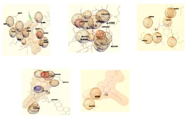
Table :2 Molecular Docking Studies of Potent Molecules (Screened by using Virtual Screening)
S.NO STRUCTURE OF LEAD MOLECULES Scores
Interactions With G-Glycoprotein
Hydrog Other interactions
en
bonding
O
H
N CH3
N Hydrophobic
NH interactions VAL431,
1 Cl O ASP503 LEU405. -8.7
Hydrophilic
interactions LYS432,
LYS466, GLU345.
Cl
JETIR2101064 Journal of Emerging Technologies and Innovative Research (JETIR) www.jetir.org 486© 2021 JETIR January 2021, Volume 8, Issue 1 www.jetir.org (ISSN-2349-5162)
Hydrophobic
interactions ILE484,
2 ASP503 LEU481, GLN482, -7.9
LEU493, LEU504.
Hydrophilic
O interactions TYR501,
NH2
ASP503.
NH
Hydrophobic
interactions ILE484,
3 OH LYS- VAL475. -8.7
O O 483 Hydrophilic
interactions
LYS485, GLN482,
HN
LYS502.
HO Cl
N
O
HN
O
CH3
Hydrophobic
CH3 interactions LEU481,
4 O
ASP503 ILE484, LEU493, -7.5
VAL478.
N
NH Hydrophilic
interactions GLN479,
ASP503, GLN482,
TYR501.
N
NH2
HN
Hydrophobic
interactions PRO394.
5 LYS- Hydrophilic -7.5
O 483 interactions GLN479,
GLU476, ASP390.
NH
O
NH
OH
NH
O
JETIR2101064 Journal of Emerging Technologies and Innovative Research (JETIR) www.jetir.org 487© 2021 JETIR January 2021, Volume 8, Issue 1 www.jetir.org (ISSN-2349-5162)
Molecular Docking Results with Potent Molecules:
MOLECULAR PROPERTY CALCULATION AND TOXICITY PREDICTION
Bioactivity prediction of indole derivatives using Molinspiration software.
The molecular property of the newly synthesized compounds were calculated values of some basic
molecular description such as logp, logs, molecular weight, Polar Surface Area, number of hydrogen bonds
donor and number of hydrogen bonds acceptor in molecule membrane hydrophobicity and bioavailability
were predicted.
Table-4 the Lipinski rule of five (Lipinski rule et al 1997) was adopted to sort out the drug likeness of
synthesized compounds. The results are presented in the following table:
Table 4: Molecular Property Calculation of the title compounds:
s.no CHEMBL ZINC ID M I TPSA N Mol. no No. N N Volume
ID logp atoms Wt N HN violati rotb
H on
1 1650867 66104757 4.13 56.41 29 428.32 5 1 0 2 353.97
2 94773 3831316 3.65 96.8 34 447.54 6 4 0 6 411.19
3 1094073 49070965 3.91 107.66 32 453.88 3 0 5 376.39
4 19933103 3938300 2.53 111.29 33 411.49 7 4 0 8 396.04
5 2365277 95597064 4.49 51.05 26 342.44 3 3 0 6 3260.4
Discussion
From the observations of virtual screening, Molecular studies following observations could be made:
2,3-Disubstituted derivatives are found reported to be more potent than other substituted derivatives.
The extent of binding interactions also confirmed the significance of these substitutions.
JETIR2101064 Journal of Emerging Technologies and Innovative Research (JETIR) www.jetir.org 488© 2021 JETIR January 2021, Volume 8, Issue 1 www.jetir.org (ISSN-2349-5162)
Eg: The Indole derivative with CHEMBL ID 1650867 which is a 2,3-Disubstituted derivative of indole was
found be more potent in terms of binding affinity rather than other derivatives.
O
H
N CH3
N
NH
Cl O
Cl
Indole –NH at position plays a significant role in mediating the interactions of all the molecules with
various amino acid residues like Asp-503, Lys-483 of G-Glycoprotein of Nipah Virus enzyme.
Therefore free –N-H is essential for mediating there interactions with the protein and substitution of
position-1 decreases the antiviral activity.
Incorporation of –CO group/-CONH at 2- position of Indole ring also displayed a significant role in
mediating its interactions with G-Glycoprotein of Nipah virus.
CH3
O
O
NH
N
NH
O
NH
N OH
NH
NH2 O
HN
CHEMBL ID – 94773 CHEMBL ID - 1933103
Introduction of Aryl substitution at position-5 also contributed to the antiviral activity of these
derivatives.
O NH2
NH
CHEMBL ID - 2365277
Hydrophobic interactions of the molecule with ILE 484, LEU 481, GLN 482, LEU 493, LEU 504 is
attributed because of the aryl substitution at position-5 of indole ring.
Conclusion
As there are no specific antiviral drugs effective against Nipah currently and the available drugs are
obtained through drug repurposing approaches, there is a strong necessity for the development of a
potent anti-Nipah viral agent.
With the observations of the SAR studies on the substituted indole derivatives screened by structure
based virtual screening, a lead molecule could be designed and could be developed as a potent anti-
Nipah viral agents
JETIR2101064 Journal of Emerging Technologies and Innovative Research (JETIR) www.jetir.org 489© 2021 JETIR January 2021, Volume 8, Issue 1 www.jetir.org (ISSN-2349-5162)
ACKNOWLEDGMENT
We are really grateful to our beloved guide Dr. Sruthi.K Associate professor, Department of Pharmaceutical
chemistry for her enormous support, this work would not have been possible without her guidance. I am
especially indebted to Prof. M. Sumakanth Principal RBVRR Women’s College of Pharmacy.
References:
1. Wahed F., Kader SA., Akhtarunnessa. & Mahamud MM. (2011), Nipah Virus: An Emergent Deadly
Paramyxovirus Infection in Bangladesh, J Bangladesh Soc Physiol vol.6, 134-139.
2. Hsu VP., Hossain MJ., Parashar UD., Ali MM., Ksiajek TG. & Kuzmin. (2004), Nipah Virus
Encephalitis Re-emergence, Bangladesh, Emerging Infectious Diseases vol.10, 2082-2087.
3. Montgomery JM., Hossain MJ., Gurley E., Carroll GD. & Croisier A. (2008), Risk factors for Nipah
virus encephalitis in Bangladesh, Emerg Infect Dis, vol.14, 1526-1532.
4. Stachowiak B. & Weingartl HM, (2012), Nipah virus infects specific subsets of porcine peripheral blood
mononuclear cells, PLoS One, vol.7, e30855.
5. Bellini WJ., Harcourt BH., Bowden N. & Rota PA. (2005), Nipah virus: an emergent paramyxovirus
causing severe encephalitis in humans, J Neuro- virol, vol.11, 481–7.6.
6. Chong HT., Kamarulzaman A., Tan CT., Goh KJ., Thayaparan T., Kunjapan SR., Chew NK., Chua KB.
& Lam SK. (2001), Treatment of acute Nipah virus encephalitis with ribavirin, Ann Neurol, vol.49,
810–813.
7. Bossart KN., Zhu Z., Middleton D., Klippel J. & Crameri G. (2009), A Neutralizing Human Monoclonal
Antibody Protects against Lethal Disease in a New Ferret Model of Acute Nipah Virus Infection, PLoS
Pathog, vol.5, e1000642.
8. Dawes BE., Kalveram B. & Ikegami T. (2018), Favipiravir protects against Nipah virus infection in the
hamster model, Sci Rep, vol.8, 7604.
9. Lo MK. (2017), GS-5734 and its parent nucleoside analog inhibit Filo-, Pneumo-, and Paramyxoviruses,
Sci Rep, vol.7, 43395, https://doi. org/10.1038/srep43395.
10. Hotard AL., He B., Nichol ST., Spiropoulou CF. & Lo MK. (2017), 4’-Azidocytidine (R1479) inhibits
henipaviruses and other paramyxoviruses with high potency, Antiviral Res, vol.144, 147–152.
11. Douglass FT. & Pavan KT. (2011), Indole synthesis: a review and proposed classification Tetrahedron,
67, 7195-7210.
12. Robinson B. (1982), The Fischer indole synthesis; John Wiley & Sons: Chichester, 48-59.
13. Lehninger A., Nelson D. & Cox M. (1993), Principles of Biochemistry 2nd ed, Worth Publishers, New
York.
14. Cornforth JW., Hughes GK., Lions F., Harradence RH., J Proc Roy Soc. (1938), N S Wales, vol.71,
486.
15. Stanley W. & Marvin MM. (1967), 1,2,5,6-Tetrahydro-12H-pyrrolo[1',2':1,2]azepino-[3,4-b]indoles
and 8,9-dihydro-5H,7H,14H,-isoindolo[2',1':1,2]-azepino[3,4-b]indoles, J. Org. Chem, vol.32, 3618-
3622.
16. Yamamoto K. & Watanabe H. (1982), Composition of "polyphosphoric acid trimethylsilyl ester (ppse)"
and its use as a condensation reagent, Chem. Lett, 1225-1228.
17. Bergman J. & Pelcman B. (1989), Synthesis of indolo[2,3-a]pyrrolo[3,4-c]carbazoles by double Fischer
indolizations , J Org Chem, 54(4), 824-828.
18. Dalian Z., David L., Hughes., Dean RB., Anthony MD. & Paul JR. (1991), Regioselective Fischer indole
route to 3-unsubstituted indoles, J Org Chem, vol.56, 3001-3006.
19. Watson TJN., Horgan SW., Shah RS., Farr RA., Schnettler RA., Nevill CR., Weiberth FJ., Huber EW.,
Baron BM., Webster ME., Mishra RJ., Harrison BL., Nyce PL., Rand CL. & Gorlaski CT. (2000),
Chemical development of MDL 103371:An N-methyl-D-aspartate-type glycine receptor antagonist for
the treatment of stroke, Org Process Res Dev, vol.4, 477-487.
20. Murakami T., Watanabe T., Hagiwara Y., Akiyama. & H Ishii. (1995), Chem. Pharm. Bull, vol.43,
1281.
21. Yu H., Jin H., Gong W., Wang Z. & Liang H. (2013), Pharmacological actions of multi-target-directed
evodiamine, Molecules, vol.18, 1826-1843.
22. Amira A Sadawe., Omran Fhid., Inass A Sadawe1., Nisreen H Meiqal., Abdulathim A A Alshoushan.,
Salah M Bensaber., Anton Hermann. & Abdul M Gbaj. Virtual Screening Of N’-Methylene-1H-Indole-
2-Carbohydrazide Schiff ‘s Base Derivatives as Cyclooxygenase, Lupine Online Journal of Medical
Sciences Research Article, 376-379.
JETIR2101064 Journal of Emerging Technologies and Innovative Research (JETIR) www.jetir.org 490© 2021 JETIR January 2021, Volume 8, Issue 1 www.jetir.org (ISSN-2349-5162)
23. Prathipati P., Dixit A. & Saxena A K. (2007), Computer-aided drug design: Integration of structure-
based and ligand-based approaches in drug design, Curr. Comp. Aided Drug Design vol.3, 341-352.
24. Xiaoqian Xue., Xiangyu Jin., YuSong., Jing Li., Xiaoyu Luo., Ming Song., Weiqun Yan., Hongrui
Song. & Yong Xu. (2014), Discovery of 2-oxo-1,2-dihydrobenzo[cd]indole-6-sulfonamide derivatives
as new RORγ inhibitors using virtual screening, synthesis and biological evaluation, European Journal
of Medicinal Chemistry, Vol.78, 431-441.
25. Gul W. & Hamann MT. (2005), Indole alkaloid marine natural products: an established source of cancer
drug leads with considerable promise for the control of parasitic, neurological and other diseases, Life
Science, vol.78, 442–453.
JETIR2101064 Journal of Emerging Technologies and Innovative Research (JETIR) www.jetir.org 491You can also read


