Stellar Neuroretinitis Revealing a Lyme Disease: Case Report
←
→
Page content transcription
If your browser does not render page correctly, please read the page content below
Open Access
Austin Journal of Clinical Ophthalmology
Case Report
Stellar Neuroretinitis Revealing a Lyme Disease: Case Report
Kawtar Bouirig*, Taha Baiz, El moubarik Najoua, Abstract
Nourdine Boutimzine and Lalla Ouafae Cherkaoui
Lyme disease is a zoonosis, caused by a spirochete of the Bor-
Ophthalmology Department “A”, Ibn Sina University relia family (Borrelia burgdorferi), often underdiagnosed. Because
Hospital (Hôpital des Spécialités), Mohammed V of its severity, Lyme disease must hold the attention of all doctors
University, Rabat, Morocco. and especially ophthalmologists in front of any ocular manifesta-
*Corresponding author: Kawtar Bouirig tion. All tunics of the eye can be affected, the sometimes confus-
ing clinical picture makes it difficult to establish the diagnosis of
Ophthalmology Department “A”, Ibn Sina University
this fact borreliosis must be systematically evoked and its research
Hospital (Hôpital des Spécialités), Mohammed V
must be an integral part of a standard uveitis assessment. Oph-
University, Rabat, Morocco. thalmological damage assimilated to neuro Lyme disease will be
Received: November 26, 2022; Accepted: January 10, treated as such,early treatment with antibiotic therapy makes it
2023; Published: January 16, 2023 possible to cut short its chronic course and avoid irreversible com-
plications.
We report the case of a patient in whom the diagnosis of neu-
roretinitis secondary to lyme disease was evoked and treated early
allowing a significant visual recovery.
Keywords: case reportuveitis; Lyme disease; Borrelia burgdor-
feri; Neuroborreliosis
Introduction Anterior segment examination revealed an incipient cataract
without inflammatory signs. The right fundus revealed a cellular
Lyme disease is a zoonosis caused by a spirochete of the vitreous Tyndall with 1 cross, a stage 1 papilledema associated
Borrelia family (Borrelia burgdorferi). This disease, of worldwide with inter-papillomacular and macular exudates arranged
distribution, is transmitted by ticks which are hematophagous in a stellate pattern, and the appearance of a small macular
at all stages of their development. First described for its acute serous detachment bulla with no chorioretinal focus or sign
dermatological manifestations [1] and then neurological of associated vasculitis (Figure 1). The left fundus examination
manifestations [2], Lyme disease remains under-diagnosed, was unremarkable, in particular with no signs of diabetic or
especially when the picture is not classical [3]. The establishment hypertensive retinopathy.
of the diagnosis is based on a combination of several arguments:
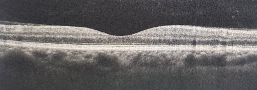
the positivity of the serology (Elisa confirmed by Western-Blot), Diagnostic assessment: Fluorescein angiography shows
the description of the clinic and the absence of another etiology papillary hyperfluorescence on the right without evidence
that could explain the symptoms and the favorable evolution of vasculitis (Figure 2). OCT showed a macular retinal serous
under antibiotic therapy [4]. We report our diagnostic and detachment (Figure 3). A first-line workup for uveitis was
therapeutic approach in a case allowing an early and adapted ordered, supplemented by Bartonella henselae serology.
treatment to avoid sometimes irreversible complications. Borrelia burgdorferi serology came back positive in ELISA, a
confirmation in Western-Blot was carried out. Based on all these
Patient et observation clinical and paraclinical arguments, we retained the diagnosis of
Patient information: The patient was 56 years old and had a neuroretinin secondary to Lyme disease.
history of arterial hypertension under ARB2 and well-balanced Therapeutic intervention: The patient received antibiotic
diabetes under insulin. She presented with a rapidly progressive therapy based on intravenous ceftriaxone for 3 weeks
decrease in visual acuity in her right eye for 15 days. The patient associated with oral corticosteroid therapy 0.5 mg/kg/d, with
also reported close contact with dogs. In addition, questioning rapid degression.
revealed the appearance of erythema migrans one month ago.
Follow up and outcomes: The evolution was favorable with
Clinical findings: On ophthalmologic examination, visual almost complete recovery of visual acuity of 8/10 in the right
acuity was “finger count” on the right and 10/10 on the left. eye, regression of papilledema and disappearance of exudates
Austin Journal of Clinical Ophthalmology - Volume 10
Citation: Bouirig K, Baiz T, Najoua E, Boutimzine N, Cherkaoui LO. Stellar Neuroretinitis
Issue 2 - 2023 www.austinpublishinggroup.com
Revealing a Lyme Disease: Case Report. Austin J Clin Ophthalmol. 2023; 10(2): 1140.
Bouirig K © All rights are reserved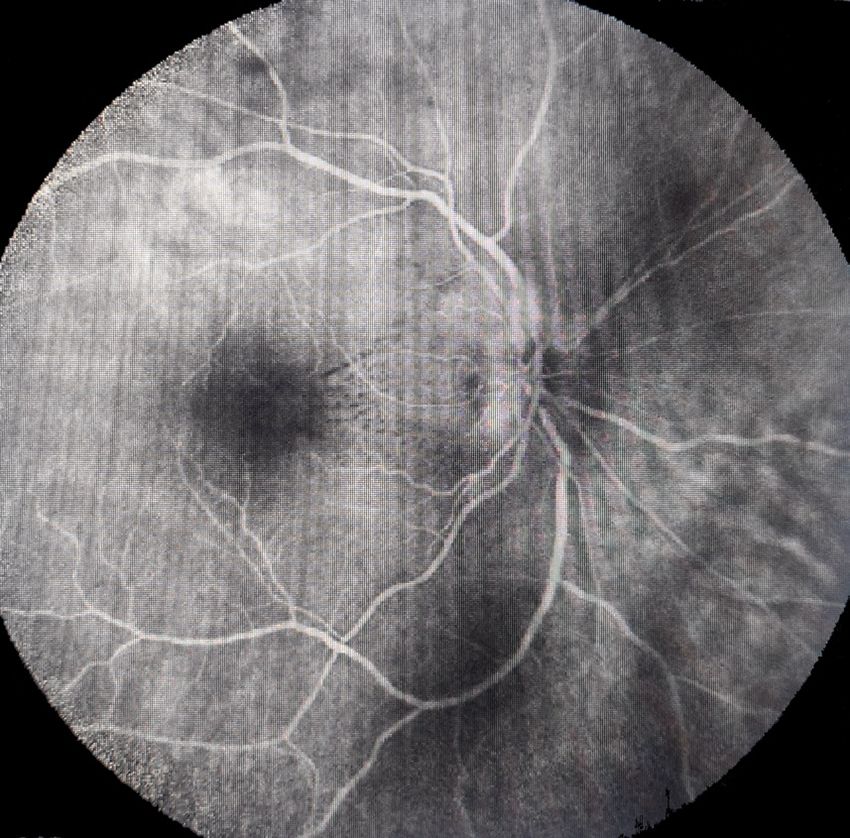
Bouirig K Austin Publishing Group
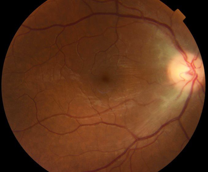
on the fundus (Figure 4) and complete resorption of the retinal
serous detachment (Figure 5). Regular follow-up over a period
of 14 months did not reveal any recurrence or contralateral or
extraocular involvement.
Figure 1: Right fundus image: papilledema (black arrow), fine stel-
late exudates in the inter-papillomacular area (white arrow) and
Figure 2: Fluorescein angiography: papillary hyperfluorescence.
macular serous detachment (red arrow).
Figure 3: Macular OCT of the right eye : macular serous detachment.
evocative manifestation is erythema migrans, which, although
typical, is not found in 1 out of 3 cases [4]. Thus, the diagnosis of
Lyme disease is essentially based on epidemiological and clini-
cal data, with serological data being used only to confirm the
disease. However, the importance of these tests appears pri-
mordial in atypical forms. [4]. The diagnosis is serological, based
on the ELISA technique and supported by the Western blot to
avoid false positives and cross-reactions. [6]
In typical forms with erythema migrans, short courses of oral
antibiotics usually prevent the development of extracutane-
ous complications [6,7]. However, experts have not been able
to establish consensus recommendations for ophthalmologic
involvement, which is considered neuro Lyme and should be
treated as such. Treatment with ceftriaxone 2 g/d IV for 2 to 4
weeks is recommended [6].
Figure 4: Retinophotography showing a normal aspect of the fun- Conclusion
dus after treatment.
Lyme disease is a multisystemic infection caused by the spi-
rochete Borrelia burgdorferi which is usually transmitted by the
Discussion
Ixodes tick. For any unexplained ocular symptom, even in chil-
Neuro-ophthalmologic involvement in Lyme disease is un- dren, Lyme disease should be considered, especially in endemic
common. It classically occurs during the early phase of the dis- areas. The diagnosis of neuroretinitis is based on the association
ease, probably in connection with a privileged passage of the of papilledema and a macular star. Its clinical diagnosis is easy
blood-brain barrier by the spirochete [5]. In Europe, the notion but the multiplicity of causes makes the etiological investigation
of a bite is found in only about 1 out of 2 patients, and the most delicate. Treatment and prophylaxis is based on doxycycline.
Submit your Manuscript | www.austinpublishinggroup.com Austin Journal of Clinical Ophthalmology 10(2): id1140 (2023) - Page - 02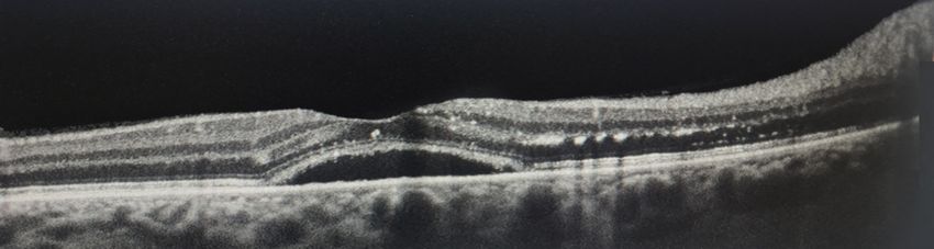
Austin Publishing Group

     Figure 5

      References
1.     Afzelius A. Acta Derm Venereol. (Stockh.) 1921; 2: 120- 125.

A.       Garin Ch, Bujadoux A. Paralysis by ticks. 1922. Clin Infect Dis.
         1993; 16: 168-169.

B.       Klaeger AJ, Herbort CP. Cotton wool spots as possible indicators
         of retinal vascular pathology in ocular lyme borreliosis. Int Oph-
         thalmol. 2010; 30: 599-602.

C.       Kadz B, Putteman A, Verougstraete C, Caspers L. Lyme disease
         from the ophthalmologist’s perspective. J Fr Ophthalmol. 2005;
         28; 218-223.

2.       Chabouni Chaker N, Turkey F, Bouraoui R, Hasnaoui W, Mrabet I,
         et al. Neuroretinitis revealing Lyme disease in a child. J Fr Oph-
         thalmol. 2008; 31.

3.       Wormser G, Nadelman R, Dattwyler R. Practice Guidelines for
         the treatment of Lyme Disease. Clin Infect Dis. 1999; 31: S1-14.

4.       Steeres C. Duration of antibiotic therapy for Lyme disease. Ann
         Intern Med. 2003; 138: 761-762.

Submit your Manuscript | www.austinpublishinggroup.com                        Austin Journal of Clinical Ophthalmology 10(2): id1140 (2023) - Page - 03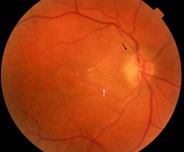
You can also read