Severe Crohn's Disease With Intra-abdominal Fistula: First Reported Case From Tanzania - Cureus
←
→
Page content transcription
If your browser does not render page correctly, please read the page content below
Open Access Case
                               Report                                                  DOI: 10.7759/cureus.21277

                                                Severe Crohn’s Disease With Intra-abdominal
                                                Fistula: First Reported Case From Tanzania
Review began 01/11/2022
                                                Nadeem Kassam 1 , Omar Aziz 2, 3 , Eric Aghan 4 , Melchisedeck Mandwa 4 , Caroline Ngimba 4 , Hanifa
Review ended 01/13/2022                         Mbithe 5, 6 , Salim Surani 7, 8 , Casmir Wambura 9
Published 01/15/2022

© Copyright 2022                                1. Internal Medicine, Aga Khan University Medical College, East Africa, Dar es Salaam, TZA 2. Medicine, Aga Khan
Kassam et al. This is an open access article    University Medical College, East Africa, Dar es Salaam, TZA 3. Medicine, Aga Khan Hospital, Dar es Salaam, TZA 4.
distributed under the terms of the Creative     Family Medicine, Aga Khan Hospital, Dar Es Salaam, TZA 5. Internal Medicine, Aga Khan University Medical College,
Commons Attribution License CC-BY 4.0.,         East Africa, Dar-es-Salaam, TZA 6. Internal Medicine, Aga Khan Hospital, Dar Es Salaam, TZA 7. Anesthesiology, Mayo
which permits unrestricted use, distribution,
                                                Clinic, Rochester, USA 8. Medicine, Texas A&M University, College Station, USA 9. Gastroenterology, Aga Khan
and reproduction in any medium, provided
                                                University Hospital, Dar Es Salaam, TZA
the original author and source are credited.

                                                Corresponding author: Salim Surani, srsurani@hotmail.com

                                                Abstract
                                                We describe a case of Crohn's disease occurring in a young Tanzanian female. Crohn’s disease is rare in
                                                Africa and not encountered normally. The presentation of Crohn’s disease overlaps with many other
                                                abdominal disorders that are common in an African setting, such as tuberculosis and schistosomiasis. The
                                                disease is probably underdiagnosed in Africa due to limitations in diagnostic testing and rarity.

                                                Categories: Family/General Practice, Internal Medicine, Gastroenterology
                                                Keywords: crohn’s disease (cd), diarrhea, gi fistula formation, abdominal pain, enterocolitis, irritable bowel
                                                syndrome

                                                Introduction
                                                Crohn’s disease (CD) is a chronic relapsing inflammatory bowel disease (IBD). It is chiefly characterized by a
                                                transmural granulomatous inflammation affecting any part of the gastrointestinal tract, most frequently the
                                                terminal ileum [1]. CD arises from a complex interaction between genetic and environmental factors [2]. The
                                                incidence and prevalence of CD appear to be growing globally but vary by geographic location and are
                                                considered uncommon in an African setting. The prevalence of tuberculous enterocolitis and
                                                schistosomiasis in Africa is considered an impediment to effectively diagnosing CD since both have clinical
                                                and histological similarities [3-5]. The distinction of CD from tuberculosis is relatively even more
                                                challenging if the lesion is granulomatous [5]. A case of histopathologically confirmed CD is presented here
                                                because of the paucity and clinical curiosity of this entity.

                                                Case Presentation
                                                A 24-year-old female of African origin was admitted to our hospital with a six-month history of fresh
                                                persistent bloody diarrhea and joint pain. She reported that the diarrhea was foul-smelling, mucoid, and
                                                infrequently watery. Her bowel frequency was progressively increasing over time to around 8 to 10 bowel
                                                movements per day associated with crampy abdominal pain especially on defecation. Her symptoms were
                                                associated with unintentional weight loss of around 10 kg over the past three months. The joint pains were
                                                localized to both the ankles and the metacarpophalangeal (MCP) joints, which were worse in the morning.
                                                She reported no fevers throughout her illness but developed a few episodes of non-bilious vomiting one day
                                                prior to the admission. The patient was a primary school teacher with no history of alcohol consumption or
                                                cigarette smoking and denied use of any chronic medication. She had multiple outpatient visits to several
                                                facilities for which she was treated for infectious diarrhea with no improvement. She tested negative for HIV,
                                                was given steroids for her joint pain, and was asked to adjust her diet. Her past medical history was
                                                unremarkable for chronic disease or previous surgeries; she was nulliparous and had normal menstrual
                                                cycles.

                                                On examination, the patient was ill-looking, afebrile, mildly pale, and dehydrated with three pustular
                                                lesions noted on her face. At the time of admission, the patient had normal vitals, apart from mild
                                                tachycardia (heart rate of 110 beats/minute). Abdominal examination revealed mild epigastric tenderness,
                                                and a digital rectal examination was normal. On local examination of the perineum, she had multiple
                                                excoriations and three well-demarcated shallow ulcers with regular margins having a diameter of less than 1
                                                cm. A musculoskeletal examination revealed evident swelling of both her ankle joints and MCPs. There was
                                                no back deformity noted but was tender on both sacroiliac joints. The patient’s initial investigation revealed
                                                the following: hemoglobin (Hb) of 9 g/dL (normal: 12.3-15.2 g/dL), an elevated erythrocyte sedimentation
                                                rate (ESR) at 73 mm/hour (normal: 0-20 mm/hour), and mildly decreased albumin level of 31.05 g/L (normal:
                                                34-54 g/L). The patient underwent esophagogastroduodenoscopy and colonoscopy, which revealed severe
                                                pancolitis, as shown in Figure 1.

                               How to cite this article
                               Kassam N, Aziz O, Aghan E, et al. (January 15, 2022) Severe Crohn’s Disease With Intra-abdominal Fistula: First Reported Case From Tanzania.
                               Cureus 14(1): e21277. DOI 10.7759/cureus.21277
FIGURE 1: (A) Mucosa of the colon revealing presence of skip lesions
and linear ulcerative lesions. (B) Erythema in the gastric mucosa and
cobblestone appearance.
A biopsy of the colon was taken, histology of which revealed transmural inflammation with granulomatous
inflammation suggestive of CD (Figure 2). Esophageal and gastric tissue histology showed features
suggestive of active chronic gastritis (Figure 3). The patient was diagnosed with severe CD with IBD
associated with arthritis.
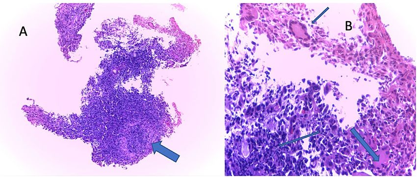
FIGURE 2: (A, B) Hematoxylin and eosin staining (x10) showing a
section of the colon with transmural inflammation with granulomatous
inflammation in the submucosa.
FIGURE 3: (A) Hematoxylin and eosin staining (x20) of the above
section of the colon showing a non-caseating granuloma. (B)
Hematoxylin and eosin staining (x40) showing epithelioid cells,
multinucleated giant cells, and chronic inflammatory cell infiltrates
2022 Kassam et al. Cureus 14(1): e21277. DOI 10.7759/cureus.21277 2 of 4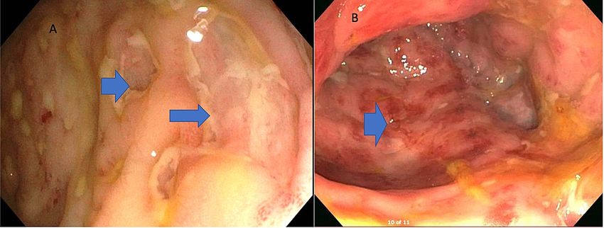
The patient was started on sulfasalazine 500 mg every 12 hours and prednisolone 40 mg daily. On return to
                                                 the clinic, she reported persistent but mildly improved symptoms. Abdominal computed tomography (CT)
                                                 was performed, as seen in Figure 4, which revealed bowel-wall thickening, strictures at the distal ileum and
                                                 rectosigmoid junction, and ileocolic and ileoileal fistula.

                                                   FIGURE 4: (A, B) Axial and sagittal CT images showing fistula in the
                                                   abdomen.

                                                 Discussion
                                                 CD causes inflammation of the digestive tract, mostly affecting the ileum [1]. CD is considered a disease with
                                                 bimodal distribution having two peaks of onset, the first peak between age 20 and 30 years, as seen in our
                                                 patient, and the second peak at age 50 years with a median age of 30 years [6]. Despite its changing
                                                 epidemiology and with the incidence rising in Asia and Eastern Europe [7,8], CD is still considered a rare
                                                 entity in the developing world, especially among black African patients [9,10]. We hypothesize that this
                                                 disease is likely underdiagnosed due to the lack of endoscopic resources and the overlap of symptomology
                                                 with schistosomiasis and tuberculosis of the bowel [4]. Review of risk factors attributable to CD remains
                                                 incompletely understood despite several theories stating the interplay between infective agents,
                                                 environmental, and genetic factors [11,12]. Our case cannot truly ascertain the risk factor in our patient;
                                                 nevertheless, low vitamin D level is a well-studied phenomenon and associated with disease activity in CD
                                                 [13]. Lifestyle factors such as stress, sleep, obesity, and exercise are poorly studied in an African context.

                                                 The presentation of CD may vary and can be insidious and nonspecific [14], and alarming symptoms as seen
                                                 in our patients such as weight loss, bloody diarrhea, and other systemic manifestations should require
                                                 further evaluation. We postulate that limitations of diagnostic capabilities serve as a hindrance to an
                                                 established diagnosis of CD. These limitations force clinicians in low-resource settings to provide empirical
                                                 care for common diseases that present in a similar fashion such as malaria and many waterborne diseases. It
                                                 is likely that this patient would have benefitted from induction therapy with an anti-tumor necrosis factor
                                                 (TNF) agent such as infliximab in combination with an immunomodulator such as azathioprine, which is
                                                 considered first-line therapy for patients presenting with fistulizing disease. She would also need close
                                                 follow-up with a dietician and surgical expertise in IBD. All of these are, sadly, hard to come by in our
                                                 general setting.

                                                 Conclusions
                                                 We presented the case as a wake-up call to the health care providers to start considering CD as a possibility
                                                 in patients presenting with GI symptoms in an African context. Our case had an almost hallmark
                                                 presentation of CD with both gastrointestinal and extragastrointestinal manifestations, which,
                                                 unfortunately, was not picked up at her index visit. CD is considered by many as a very rare disease in Africa.
                                                 The symptoms of CD may mimic many other abdominal conditions for which medical attention is required.
                                                 However, it should be kept in mind as one of the causes of acute abdomen, especially in those patients who
                                                 have a long history of intestinal complaints whose treatments greatly differ. A histopathologically confirmed
                                                 diagnosis is necessary for the medical field especially of the emerging evidence with an increased risk of
                                                 adenocarcinoma in patients with CD.

                                                 Additional Information
                                                 Disclosures
                                                 Human subjects: Consent was obtained or waived by all participants in this study. Conflicts of interest: In
                                                 compliance with the ICMJE uniform disclosure form, all authors declare the following: Payment/services

2022 Kassam et al. Cureus 14(1): e21277. DOI 10.7759/cureus.21277                                                                                                  3 of 4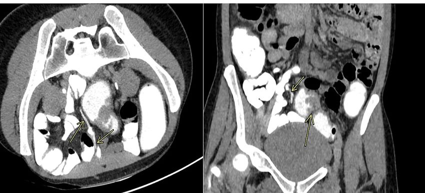
info: All authors have declared that no financial support was received from any organization for the
                                                 submitted work. Financial relationships: All authors have declared that they have no financial
                                                 relationships at present or within the previous three years with any organizations that might have an
                                                 interest in the submitted work. Other relationships: All authors have declared that there are no other
                                                 relationships or activities that could appear to have influenced the submitted work.

                                                 References
                                                     1.   Thia KT, Sandborn WJ, Harmsen WS, Zinsmeister AR, Loftus EV Jr: Risk factors associated with progression
                                                          to intestinal complications of Crohn's disease in a population-based cohort. Gastroenterology. 2010,
                                                          139:1147-55. 10.1053/j.gastro.2010.06.070
                                                     2.   Ha F, Khalil H: Crohn's disease: a clinical update . Therap Adv Gastroenterol. 2015, 8:352-9.
                                                          10.1177/1756283X15592585
                                                     3.   Ribeiro MB, Greenstein AJ, Sachar DB, et al.: Colorectal adenocarcinoma in Crohn's disease . Ann Surg. 1996,
                                                          223:186-93. 10.1097/00000658-199602000-00011
                                                     4.   Cho KJ, Suh YL, Kim CW, Chi JG: Crohn's disease--a case report . J Korean Med Sci. 1987, 2:133-6.
                                                          10.3346/jkms.1987.2.2.133
                                                     5.   Shivashankar R, Tremaine WJ, Harmsen WS, Loftus EV Jr: Incidence and prevalence of Crohn's disease and
                                                          ulcerative colitis in Olmsted County, Minnesota from 1970 through 2010. Clin Gastroenterol Hepatol. 2017,
                                                          15:857-63. 10.1016/j.cgh.2016.10.039
                                                     6.   Feuerstein JD, Cheifetz AS: Crohn disease: epidemiology, diagnosis, and management. Mayo Clin Proc.
                                                          2017, 92:1088-103. 10.1016/j.mayocp.2017.04.010
                                                     7.   Ouyang Q, Tandon R, Goh KL, Ooi CJ, Ogata H, Fiocchi C: The emergence of inflammatory bowel disease in
                                                          the Asian Pacific region. Curr Opin Gastroenterol. 2005, 21:408-13.
                                                     8.   Wright JP, Froggatt J, O'Keefe EA, et al.: The epidemiology of inflammatory bowel disease in Cape Town
                                                          1980-1984. S Afr Med J. 1986, 70:10-5.
                                                     9.   Steury EM, Templeton AC: Crohn's disease in Africa. A case report and review . Trop Geogr Med. 1980,
                                                          32:172-3.
                                                    10.   Nguyen GC, Torres EA, Regueiro M, et al.: Inflammatory bowel disease characteristics among African
                                                          Americans, Hispanics, and non-Hispanic Whites: characterization of a large North American cohort. Am J
                                                          Gastroenterol. 2006, 101:1012-23. 10.1111/j.1572-0241.2006.00504.x
                                                    11.   Logan I, Bowlus CL: The geoepidemiology of autoimmune intestinal diseases . Autoimmun Rev. 2010,
                                                          9:A372-8. 10.1016/j.autrev.2009.11.008
                                                    12.   Monstad I, Hovde Ø, Solberg IC, A. Moum B: Clinical course and prognosis in ulcerative colitis: results from
                                                          population-based and observational studies. Ann Gastroenterol. 2014, 27:95-104.
                                                    13.   Ham M, Longhi MS, Lahiff C, Cheifetz A, Robson S, Moss AC: Vitamin D levels in adults with Crohn's
                                                          disease are responsive to disease activity and treatment. Inflamm Bowel Dis. 2014, 20:856-60.
                                                          10.1097/MIB.0000000000000016
                                                    14.   Cheifetz AS: Management of active Crohn disease . JAMA. 2013, 309:2150-8. 10.1001/jama.2013.4466

2022 Kassam et al. Cureus 14(1): e21277. DOI 10.7759/cureus.21277                                                                                                        4 of 4
You can also read