Serum amyloid A, ferritin and carcinoembryonic antigen as biomarkers of severity in patients with COVID 19
←
→
Page content transcription
If your browser does not render page correctly, please read the page content below
BIOMEDICAL REPORTS 16: 13, 2022

          Serum amyloid A, ferritin and carcinoembryonic antigen
            as biomarkers of severity in patients with COVID‑19
         DINA A. ABDELHAKAM1, FATMA MOHAMMED BADR2, MOHAMMED ABD EL MONEM TEAMA2,
                     NOURAN M. BAHIG ELMIHI1 and MARWA ADHAM EL‑MOHAMDY1

                           Departments of 1Clinical Pathology and 2Internal Medicine and Rheumatology,
                                 Faculty of Medicine, Ain Shams University, Cairo 11566, Egypt

                                        Received August 18, 2021; Accepted November 18, 2021

                                                       DOI: 10.3892/br.2021.1496

Abstract. In view of the rapid spread of COVID‑19 and the high          value of 0.928, with a specificity of 93.1%, and a sensitivity
mortality rate of severe cases, reliable risk stratifying indicators    of 98.5% at a cut‑off of 16 mg/l. The multi‑ROC curve for
of prognosis are necessary to decrease morbidity and mortality.         SAA and ferritin showed 100% specificity, 100% sensitivity
The aim of the present study was to evaluate the value of serum         and 100% efficiency, with an AUC of 1.000. Thus, combining
amyloid A (SAA) and carcinoembryonic antigen (CEA) as                   SAA and ferritin may have guiding significance for predicting
prognostic biomarkers in comparison to other predictors, inclu‑         COVID‑19 severity. SAA alone showed the highest prognostic
ding C‑reactive protein (CRP) and ferritin levels. This study           significance. Both SAA and CEA were positively correlated
included 124 patients diagnosed with COVID‑19, and they                 with the CT‑SS. Early monitoring of these laboratory markers
were assigned to one of two groups: Mild and severe, based on           may thus provide significant input for halting disease progres‑
the severity of the infection. Radiological and laboratory inves‑       sion and reducing mortality rates.
tigations were performed, including evaluation of CRP, ferritin,
D‑Dimer, SAA and CEA levels. Significantly higher levels of             Introduction
CRP, ferritin, D‑Dimer, SAA and CEA were observed in severe
cases. SAA was significantly correlated with CRP (r= 0.422,             Coronavirus disease 2019 (COVID‑19) is caused by severe acute
P
2 ABDELHAKAM et al: SAA, FER AND CEA AS BIOMARKERS OF SEVERITY IN COVID-19
organs, thus contributing to organ failure and death during the Methods. The recruited cohort consisted of 124 patients
course of secondary amyloidosis (9). diagnosed with COVID‑19 at the Ain Shams University
Another important potential biomarker is carcinoembry‑ Hospital. A detailed history was obtained for all patients,
onic antigen (CEA); a glycoprotein formed in the respiratory with a particular emphasis on age, sex, duration of disease
and colonic epithelium during embryogenesis, and it has been and clinical symptoms. Data from routine investigations
widely utilized as a tumor marker to monitor tumor progres‑ were retrieved, including liver enzyme levels [alanine trans‑
sion (10). CEA is related to respiratory or digestive cancers aminase (ALT) and aspartate transaminase (AST)], kidney
and infectious diseases, such as gonorrhea, or chronic inflam‑ function tests [blood urea nitrogen (BUN), creatinine], inflam‑
matory diseases such as interstitial lung diseases (ILD) (11). matory parameters (CRP, ferritin) and D‑Dimer levels, in
Immunohistochemical staining of lung specimens from addition to complete blood count (CBC), hemoglobin, white
patients with pulmonary fibrosis demonstrated strong expres‑ blood count (WBC), neutrophil count, lymphocyte count and
sion of CEA in the metaplastic bronchiolar and type II alveolar platelet (PLT) count (15).
epithelia (12). Significant hyperplasia of type II alveolar epithe‑ Detection of viral RNA was performed using the
lial cells and interstitial fibrosis have been described in several CerTest ViasureVR SARS‑CoV‑2 RT‑qPCR Detection kit
reports of COVID‑19 autopsies and biopsies, similar to the (CerTest, Biotec) according to the manufacturer's instruc‑
pathological changes observed in ILD (13). tions. The detection was performed in a one‑step real‑time
In the present study, SAA and CEA were evaluated as reverse‑transcription format, where the reverse transcription
potential prognostic biomarkers in comparison to other and subsequent amplification of a specific target sequence
commonly used inflammatory predictors, including C‑reactive occurred in the same reaction well. The isolated RNA target
protein (CRP) and ferritin, and their association with the was transcribed to generate cDNA using the included reverse
severity of COVID‑19 and CT scan findings, and whether they transcriptase, followed by the amplification of a conserved
may be a beneficial tool for patient stratification was assessed. region of the open reading frames (ORF) 1 ab and N genes for
SARS‑CoV‑2 using specific primers and a fluorescent‑labelled
Materials and methods probe, all of which were included in the kit. The assay has
a 97.5% sensitivity and >99.9% specificity. The average esti‑
Patients. This cross‑sectional study included 124 patients diag‑ mated limit of detection for SARS‑CoV‑2 was 18 copies/ml.
nosed with COVID‑19 enrolled from the Ain Shams University Blood samples from PCR‑positive patients were obtained,
Isolation Hospital (Cairo, Egypt). In this study cohort, the median left to clot completely, and centrifuged at 3,000 x g for 20 min
age was 48 years and the 25‑75th IQR was 40‑56 years (age range, at 4˚C, and stored at ‑80˚C until required. SAA level analysis
25‑87 years). A total of 10 patients were >65 years old, constituting was performed using the Invitrogen human SAA ELISA kit
8% of the entire cohort. Of the 124 cases, 98 (79%) were men (cat. no. EHSAA1; Invitrogen; Thermo Fisher Scientific, Inc.).
and 26 were women (21%). The median male age was 48.5 years The detection limit of this assay is 0.004 mg/l. CEA serum
(IQR, 40‑54) and the median female age was 44.5 years (IQR, concentrations were assayed using a Cobas e411 immunoassay
35‑57). Data were collected from hospitalized patients between autoanalyzer (Roche Diagnostics GmbH).
September 2020 and February 2021. Patients were diagnosed and
categorized into mild and severe groups according to the World Computed tomography (CT). All patients underwent
Health Organization interim guidelines (14). non‑contrast‑enhanced chest CT in the Radiology Department
A definite COVID‑19 case was identified as a positive of Ain Shams University, which was performed by expert radi‑
result using sequencing or reverse transcription‑quantitative ologists using a Siemens 16‑channel scope (CTAWP92544;
PCR (RT‑qPCR) of nasopharyngeal swabs. Siemens Healthineers). The following CT parameters were
The patients were classified into 2 groups: i) Mild group, had evaluated: COVID‑19 Reporting and Data System (CO‑RADS)
clinical symptoms of fever, fatigue, cough, anorexia, malaise, score based on CT findings (17): i) The CO‑RADS score
muscle pain, sore throat, dyspnea, nasal congestion and/or represents the level of suspicion of COVID‑19. CO‑RADS 1,
a headache; and ii) Severe group, had respiratory distress, a COVID‑19 is highly doubtful, CT is normal, or findings
respiratory rate of ≥30 breaths/min at resting state, a mean representing a non‑infectious disease; CO‑RADS 2, low level
oxygen saturation of ≤93% and an arterial blood oxygen partial of suspicion of COVID‑19 infection, CT findings consistent
pressure (PaO2)/oxygen concentration (FiO2) of ≤300 mmHg. with other infections; CO‑RADS 3, COVID‑19 infection
The exclusion criteria were as follows: i) Patients infected is indeterminate and unsure whether CT abnormalities are
with other viruses or bacteria; ii) patients diagnosed with caused by COVID‑19; CO‑RADS 4, high suspicion level, and
autoimmune disorders; iii) patients diagnosed with arthritic most CT findings are not extremely typical; CO‑RADS 5, high
diseases and iv) patients with cancer and/or any chronic level of suspicion with typical CT findings. ii) Semi quanti‑
disease related to elevated CEA and/or SAA levels, such as tative scoring system: A quantitative estimate of pulmonary
chronic kidney disease (15). involvement based on abnormalities in the areas involved.
The CT‑severity score (CT‑SS) is based on the extent of lobar
Ethical considerations. This study was conducted in accordance involvement. Each of the five lung lobes was visually scored
with the principles outlined in the Declaration of Helsinki of from 0‑5 as follows: 0, no involvement; 1,BIOMEDICAL REPORTS 16: 13, 2022 3
Table I. Demographics and baseline clinical, laboratory and radiological parameters of the COVID-19 patients.
Interquartile range
----------------------------------------------------------------------------
Parameters Value Range 25th percentile 75th percentile
Age, years 48 25-87 40 56
Sex, n (%) - - -
Male 98 (79)
Female 26 (21)
Clinical parameters
Duration of disease, days 7 1-14 4 10
Fever, n (%) 118 (95.2) - - -
Cough, n (%) 108 (87.1) - - -
Loss of smell and taste, n (%) 14 (11.3) - - -
Dyspnea, n (%) 74 (59.7) - - -
Respiratory distress, n (%) 39 (31.5) - - -
Diarrhea, n (%) 8 (6.6) - - -
Laboratory parameters
White blood cell count, 109/l 6.9 2.6-17 4.3 10.3
Neutrophil count, 109/l 4.5 1.5-13.6 2.82 7.2
Lymphocyte count, 109/l 1.22 0.46-4.38 0.841 1.6
Platelet count, 109/l 232 50-649 191 295
Hemoglobin, g/dl 14.2 8.0-17.6 13 15.1
Blood urea nitrogen, mg/dl 14 7.5-46 11.7 19.2
Creatinine, mg/dl 0.8 0.47-7.5 0.7 1.04
Aspartate transaminase, U/l 26 12-116 17 43
Alanine transaminase, U/l 32 11-346 16 54
C-reactive protein, mg/l 43 1.8-444.0 18 161.3
Ferritin, ng/ml 361 24-2,450 203.75 839.75
D-Dimer, ng/ml 500 90-6088 300 1200
Serum amyloid A, mg/l 22.5 3-90 10 49
Carcinoembryonic antigen, ng/ml 6 3-18 5 9
Radiological parameters
CO-RADS score 3 0-5 1 4
Computed tomography-severity score 8.5 0-25 3 16
CO-RADS, COVID-19 Reporting and Data System.
Statistical analysis. Statistical analysis was performed for all studied items as independent variables (Model‑1). The
using SPSS version 25.0 (IBM Corp.). Continuous variables regression was re‑run using only the most significant items
are presented as the median and interquartile range (IQR), with exclusion of the non‑significant items iteratively. The least
whereas categorical variables are presented as the number (n) sensitive predictors for the model had the highest F‑ratio and
and percentage (%) of patients. A Wilcoxon rank sum test the lowest P‑value. P4 ABDELHAKAM et al: SAA, FER AND CEA AS BIOMARKERS OF SEVERITY IN COVID-19 Table II. Comparison between mild and severe COVID-19 patients. Parameter Mild, n=58 Severe, n=66 Z χ2 P-value Significance Age, years (IQR) 43 (35-52.5) 50 (43-56) -3.24 2.88 0.05 NS Male 42/58 (72.4) 56/66 (84.8) Female 16/58 (27.6) 10/66 (15.2) Clinical parameters Duration of disease, days (IQR) 5 (4-9) 8 (6-11) -3.7 0.05 NS Dyspnea, n (%) 26 (44.8) 48 (72.7) 9.986 0.05 NS Neutrophil count, 109/l, median (IQR) 4 (2.7-6.9) 5.5 (2.9-8.3) -1.66 >0.05 NS Lymphocyte count, 109/l, median (IQR) 1.24 (0.9-1.7) 1.1 (0.8-1.5) -0.62 >0.05 NS Platelet count, 109/l, median (IQR) 229 (194-259) 246 (168-330) -0.62 >0.05 NS Hemoglobin, g/dl, median (IQR) 14.2 (11.9-15.4) 14 (13.2-15) -0.58 >0.05 NS Blood urea nitrogen, mg/dl, median (IQR) 14 (11.5-16.9) 15.9 (11.7-22) -2.16 0.05 NS Aspartate transaminase, U/l, median (IQR) 23 (15.5-37.25) 34 (18-43.25) -1.85 >0.05 NS Alanine transaminase, U/l, median (IQR) 27 (15-51) 34 (17-54) -0.96 >0.05 NS C-reactive protein, mg/l, median (IQR) 22 (11.8-48.5) 83 (29-222) -4.51
BIOMEDICAL REPORTS 16: 13, 2022                                                              5

Table III. Correlation between SAA and CEA concentration with all the studied parameters.

                                                                        SAA                                                 CEA
	--------------------------------------------------------------------------	--------------------------------------------------------------------------
Parameter                                             R           P-value          Significance            R           P-value         Significance

Age                                0.349                          0.05                NS
Platelet count                     0.07                           >0.05                NS  0.007                       >0.05                NS
Hemoglobin	-0.011                                                 >0.05                NS  0.023                       >0.05                NS
Blood urea nitrogen                0.345                          0.05                NS
Aspartate transaminase             0.149                          >0.05                NS  0.149                       >0.05                NS
Alanine transaminase               0.092                          >0.05                NS  0.052                       >0.05                NS
C- reactive protein                0.422
6 ABDELHAKAM et al: SAA, FER AND CEA AS BIOMARKERS OF SEVERITY IN COVID-19
Table IV. Regression model outcomes of significant predictors of COVID-19 severity.
A, Model 1
Item Regression coefficient t P-value Significance F-Ratio P-value Significance
Constant 0.555 1.996 0.05 NS
Duration of disease, days 0.004 0.496 >0.05 NS
White blood cell count, 109/l -0.12 -1.18 >0.05 NS
Neutrophil count, 109/l 0.123 1.238 >0.05 NS
Lymphocyte count, 10 /l
9
0.164 1.557 >0.05 NS
Platelet count, 109/l 0 0.446 >0.05 NS
C-reactive protein, mg/l -0.0000972 -0.284 >0.05 NS
Ferritin, ng/ml 0 4.36 0.05 NS
Carcinoembryonic antigen, ng/ml 0.026 3.045 0.05 NS
Hemoglobin, g/dl 0.017 1.283 >0.05 NS
Blood urea nitrogen, mg/dl -0.007 -1.742 >0.05 NS
Creatinine, mg/dl -0.07 -1.828 >0.05 NS
Aspartate transaminase, U/l 0.003 1.688 >0.05 NS
Alanine transaminase, U/l -0.002 -2.606 0.05 NS
CT Severity score -0.001 -0.18 >0.05 NS
Overall 22.574BIOMEDICAL REPORTS 16: 13, 2022 7 Table IV. Continued. D, Model 4 Item Regression coefficient t P-value Significance F-Ratio P-value Significance Constant 0.856 15.983
8                         ABDELHAKAM et al: SAA, FER AND CEA AS BIOMARKERS OF SEVERITY IN COVID-19

Table V. Multivariate regression models of significant predictors of lung CT severity score.

A, Model 1

Item                                     Regression coefficient          t      P-value     Significance F-Ratio        P-value     Significance

Constant                         2.865  0.448 >0.05  NS
Age, years	-0.035	-0.647 >0.05                       NS
Duration of disease, days        0.245  1.387 >0.05  NS
White blood cell count, 109/l	-1.92	-0.832 >0.05     NS
Neutrophil count, 109/l          1.765  0.781 >0.05  NS
Lymphocyte count, 109/l          0.877  0.368 >0.05  NS
Platelet count, 109/l	-0.013	-2.12            0.05         NS
Ferritin, ng/ml                  0.001  0.993 >0.05  NS
D-Dimer, ng/ml                   0.000  0.149 >0.05  NS
Carcinoembryonic antigen, ng/ml	-0.097	-0.488 >0.05  NS
Serum amyloid A, mg/l            0.213  6.475 0.05  NS
Eosinophil count, 109/l          0.101  0.027 >0.05  NS
Hemoglobin, g/dl                 0.316  1.009 >0.05  NS
Blood urea nitrogen, mg/dl       0.214  2.29  0.05                NS
Aspartate transaminase, U/l	-0.007	-0.182 >0.05      NS
Alanine transaminase, U/l        0.006  0.375 >0.05  NS
Overall					                                                                                                 7.991
BIOMEDICAL REPORTS 16: 13, 2022 9
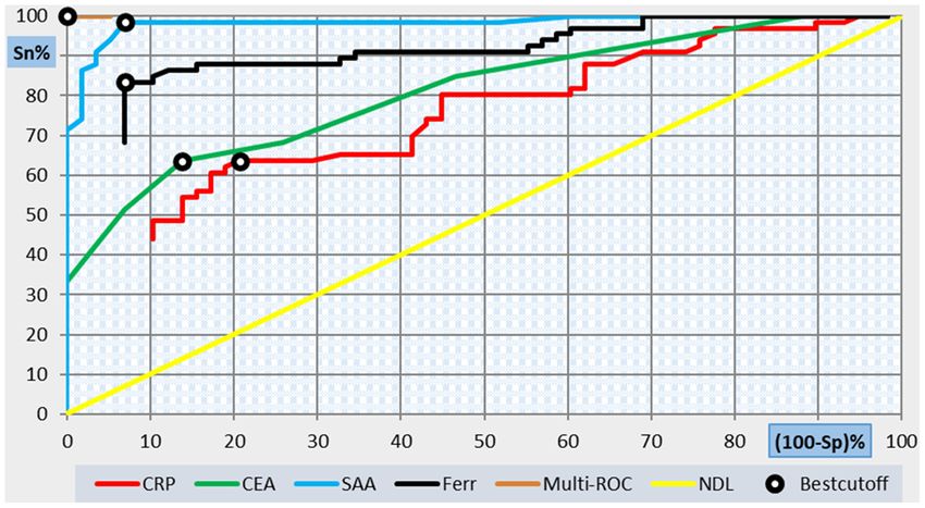
Table VI. Cutoff and performance characteristics of SAA, CEA, ferritin and CRP in predicting severity in COVID-19 patients.
Sensitivity, Specificity, Positive Negative Area under
Parameter Cutoff % % predictive value predictive value Efficiency the curve
SAA, mg/l 16 98.5 93.1 94.2 98.2 96 0.928
CEA, ng/ml 7 63.6 86.2 84 67.6 74.2 0.78
Ferritin, ng/ml 397 83.3 93.1 93.2 83.1 87.9 0.86
CRP, mg/l 54 63.6 79.3 77.8 65.7 71 0.616
SAA + ferritin 100 100 100 100 100 1.000
SAA, serum amyloid A; CEA, carcinoembryonic antigen; CRP, C-reactive protein.
Figure 1. Individual and multi‑ROC curves showing the performance of all studied parameters in predicting severity in COVID‑19 patients. SP, specificity;
Sn, Sensitivity; CRP, C‑reactive protein; CEA, carcinoembryonic antigen; SAA, serum amyloid A; Ferr, ferritin; ROC, receiver operating characteristic; NDL:
Non‑Diagnostic Line.
women (31). In the present study, several social, cultural and Fundamental differences in the immune response between
behavioral differences between the sexes may have contributed males and females are likely to be the driving factor behind
to the high overall male to female ratio (79 to 21%, respec‑ the significant sex bias observed in severe COVID‑19 cases.
tively) seen in this cohort, including habits more common in Sex differences in innate and adaptive immunity have been
men, such as smoking (32,33) and sex‑based differences in reported, which may account for the reduced risk of severe
hygiene. Additionally, based on anecdotal evidence, men are disease in females. A robust antiviral innate IFN response
likely to leave their houses and enter crowded areas in Egypt. and enhanced adaptive immunity in females, higher numbers
Furthermore, differences in health‑seeking behaviors, unequal of CD4 + T cells, robust CD8+ T cells and increased B cell
access to healthcare facilities and testing between sexes may production of immunoglobulin compared to males, may lead
have skewed the data further towards a male bias (34,35). to more effective viral control in females, and a relatively
However, a consistent feature of the COVID‑19 pandemic lower risk of developing severe disease (37,38). The X chro‑
is the male bias towards severe disease (36). This agrees with mosome encodes several immune‑related genes, which can
the results of the present study, as 85% (56 out of 66) of the be variably expressed on both alleles, increasing the diversity
severe cases were men compared with 15% of women. Male of the immune response (39). Hormonal differences also play
sex is associated with nearly a 3x risk of requiring intensive a role; estradiol augments the immune response in contrast
care unit admission and a higher probability of death compared to testosterone, which was found to suppress the immune
to women (31). system (40).10 ABDELHAKAM et al: SAA, FER AND CEA AS BIOMARKERS OF SEVERITY IN COVID-19
The median WBC, PNL and PLT counts were higher in reported elevated serum CEA levels in COVID‑19 patients
the severe group than in the mild group, whereas the lympho‑ with significantly increased CEA levels compared with
cyte count was lower. However, the differences between both healthy controls, and a potential association between CEA
groups regarding these parameters were not statistically and CRP levels was reported (53,54). However, its role in
significant. Of the 124 COVID‑19 positive cases, a high preva‑ predicting clinical outcomes or CT involvement in patients
lence of increased inflammatory marker levels was identified. with COVID‑19 is still under investigation. The present study
These results showed that in all COVID‑19 cases, there was an validated the previous results that CEA levels were related to
overall increased level of inflammatory parameters, including COVID‑19 severity. Moreover, the association between CEA
SAA, CEA, CRP and ferritin, in addition to pro‑coagulation levels and CT scores were also validated, which are in agree‑
markers. D‑Dimer levels were similar to the results of previous ment with the findings of Chen et al (41).
studies (29,41,42), and they were significantly higher at CEA is a biomarker of adenocarcinoma in respiratory or
admission in the severe patient group than in the mild group, digestive system cancers, in addition to non‑neoplastic lung
whereas total WBC, lymphocyte and PLT counts were within diseases (55,56). Increased CEA expression was detected in
the normal range, consistent with the results of Li et al (42) and type II pneumocytes in allergic bronchopulmonary aspergil‑
Wang et al (43). losis (57) and atypical epithelial proliferation in idiopathic
Huang et al (44) showed that patients with severe pulmonary fibrosis (13,58). Bronchiolar and type II alveolar
COVID‑19 had higher levels of IL‑1β, IFN‑ γ, IP‑10 and epithelial cells are the main targets of SARS‑CoV‑2 in
monocyte chemoattractant protein compared with mild cases, the lungs. SARS‑CoV‑2 infection‑induces massive type II
causing Th1 cell activation and stimulating the production of alveolar epithelial cell death and aberrant regeneration of
SAA, CRP, procalcitonin and PLT. These inflammatory factors type II pneumocytes along with the production of CEA, such
may be useful as indicators reflecting the body's response to as that observed in the uncontrolled proliferation in lung
infection (45). adenocarcinoma. Moreover, atypical epithelial and fibroblast
According to Li et al (42) and Wang et al (43) as the disease proliferation may also worsen the obstruction of bronchioles
progressed from mild to severe, SAA and CRP levels increased, and lung consolidation, causing refractory hypoxemia along
while lymphocyte counts gradually decreased. However, WBC with worse CT scores (59,60).
and PLT were all within normal ranges, suggesting that SAA Consequently, it is possible that serum CEA levels may
and CRP are closely related to disease classification, while correlate with the severity and prognosis of COVID‑19. Based
WBC and PLT are of little significance. on the relationship between CEA and type II pneumocyte
SAA showed a highly significant positive correlation with hyperplasia and lung fibrosis, medications such as nintedanib,
age, disease duration, neutrophil count and all inflammatory which target atypical epithelial and fibrotic proliferation, may
indices, including CRP, ferritin, CEA and D‑Dimer in the be a potential therapeutic option to decrease mortality in
present study. Similarly, CEA showed a significant positive patients with COVID‑19 (41).
correlation with age, disease duration and all inflammatory An elevated D‑Dimer level represents microangiopathy and
indices. Additionally, both SAA and CEA levels showed a a hypercoagulable state in COVID‑19 patients (61). COVID‑19
highly significant positive correlation with the CT‑SS. These is implicated in aggressive pro‑inflammatory responses
results agreed with the results from various studies showing causing endothelial cell dysfunction and excessive thrombin
significantly higher SAA levels in PCR positive COVID‑19 formation (62), which can be responsible for oxygen desatura‑
cases, and significant associations between SAA levels tion and respiratory distress seen in severe cases (63,64). In
and the number of COVID‑19 cases, severity and mortality the present study, D‑Dimer levels were significantly higher
rate (29,41,42). in patients with severe COVID‑19, and there was a highly
SAA is an acute phase protein produced by the liver and significant positive correlation with SAA and CEA in addition
is induced by cytokines, including IL‑1β, IL‑6 and TNF‑α. to CRP and ferritin levels. These results are also in agreement
SAA promotes an inflammatory response by activating with a retrospective study of 183 patients with COVID‑19
chemokines and inducing chemotaxis, even at very low performed by Tang et al (65) which showed a significant
concentrations (46,47). In current clinical practices, SAA is increase in D‑Dimer levels and fibrin degradation products,
frequently used as an indicator for monitoring of inflammation and this was indicative of a poor prognosis.
and estimation of prognosis (48,49). SAA showed promising Similarly, CRP is also an acute phase protein, the levels
results when used to monitor the effectiveness of antibiotics of which rise rapidly, and the rate of increase is positively
(cefotiam and Augmentin) in early onset neonatal sepsis correlated with the severity of infection (7,29). A higher CRP
according to a study by Liu et al (49). level is linked to an increased risk of severe COVID‑19 and
Previous studies have shown that patients with severe ARDS may contribute to pneumonia, ARDS and the rapid multiple
have significantly increased levels of SAA, suggesting that organ damage (7,66). According to a study by Liu et al (66),
SAA could be used as a biomarker to monitor the progression elevated CRP and decreased albumin levels were important
of respiratory diseases (50,51). As stated by Cheng et al (52), in factors affecting the prognosis of COVID‑19.
a study involving 89 COVID‑19 patients, dynamic changes in Ferritin is another crucial mediator of the immune
SAA could be used to predict prognosis. response that showed a statistically significant increase in
In the present study, serum CEA levels were significantly severe COVID‑19 cases, and showed a highly significant
higher in patients with severe COVID‑19 than in those with positive correlation with SAA levels in addition to CEA, CRP
a mild infection. Moreover, higher serum CEA levels were and D‑Dimer levels in the present study. Increased ferritin
associated with a higher CT‑SS. A limited number of studies levels may be implicated in the cytokine storm due to itsBIOMEDICAL REPORTS 16: 13, 2022 11
direct immunosuppressive and pro‑inflammatory effects (25). Authors' contributions
Efstathiou et al (67) indicated that viral infections increased
ferritin levels. This finding may be attributed to the fact that DAA and MAEMT designed the study, contributed to data
the inflammatory mediators induce an increase in ferritin collection and interpretation, and wrote the manuscript.
levels, in addition to denaturation and necrosis of cells to NMBE assisted with sample collection, data interpretation
break down cell membranes, causing a leakage of ferritin from and editing of the manuscript. FΜB and MAEM contributed
damaged cells (68). to sample collection, data interpretation and drafting of the
The results of the present study showed that the levels of manuscript. FΜB and MAEMT confirm the authenticity of
SAA, CEA, CRP and ferritin increased as the disease severity all the raw data. All authors have read and approved the final
increased. CT imaging is an important clinical diagnostic manuscript.
tool for evaluating COVID‑19 infections. According to the
correlation analysis in this study, SAA, CEA, CRP and ferritin Ethics approval and consent to participate
levels at admission were highly correlated with the CT‑SS,
suggesting a possible role in predicting disease progression. This study was conducted in accordance with the principles
ROC curve analysis was used to comprehensively and outlined in the Declaration of Helsinki of the World Medical
accurately compare and evaluate the diagnostic performance Association. The study was approved by the Institutional
of SAA, CEA, CRP and ferritin levels on admission and to Ethics Committee of Ain Shams University. Informed consent
explore their clinical and prognostic utility. The results of was obtained from all enrolled participants after receiving an
the current study showed that the AUC was highest for SAA, explanation of the study's aim and procedures.
followed by ferritin, CEA and CRP. According to the multivar‑
iate regression analysis and ROC curve results, the combined Patient consent for publication
use of SAA and ferritin was more sensitive than SAA or
ferritin alone as predictors of severity, as their combination Not applicable.
had the highest predictive value for disease severity, with an
AUC of 1.000. Competing interests
Accordingly, the combined detection of SAA and ferritin
may have guiding significance for assessing the severity, disease The authors declare that they have no competing interests.
progression and prognosis of COVID‑19 cases. This may aid
effective intervention measures to be implemented in timely References
manner and reduce the rates of severe illness and mortality, as
we continue to face upcoming waves of COVID‑19.
1. Gorbalenya AE, Baker SC, Baric RS, De Groot RJ, Drosten C,
The present study has some limitations. The elevations Gulyaeva AA, Haagmans BL, Lauber C, Leontovich AM, et al;
in SAA and CEA may have been due to various other condi‑ Coronaviridae Study Group of the International Committee
tions and comorbidities, which themselves may be associated on Taxonomy of Viruses: The species Severe acute respiratory
syndrome‑related coronavirus: Classifying 2019‑nCoV and
with a higher COVID‑19 risk. Additionally, this study was a naming it SARS‑CoV‑2. Nat Microbiol 5: 536‑544, 2020.
single‑center study with a relatively small cohort, which may 2. Zhou F, Yu T, Du R, Fan G, Liu Y, Liu Z, Xiang J, Wang Y,
have limited the power of the statistical analyses. Song B, Gu X, et al: Clinical course and risk factors for mortality
of adult inpatients with COVID‑19 in Wuhan, China: A retro‑
In conclusion, the combined detection of SAA and ferritin spective cohort study. Lancet 395: 1054-1062, 2020.
may have guiding significance for the severity of COVID‑19 3. Calina D, Docea AO, Petrakis D, Egorov AM, Ishmukhametov AA,
and may be correlated with CT‑SS in patients with COVID‑19. Gabibov AG, Shtilman MI, Kostoff R, Carvalho F, Vinceti M, et al:
Towards effective COVID‑19 vaccines: Updates, perspectives and
Serum CEA levels were correlated with the severity of the CT challenges (Review). Int J Mol Med 46: 3‑16, 2020.
scores and the prognosis of COVID‑19. Therefore, assessment 4. Khan M, Khan H, Khan S and Nawaz M: Epidemiological and
and monitoring of these laboratory markers at the earliest clinical characteristics of coronavirus disease (COVID‑19)
cases at a screening clinic during the early outbreak period: A
stage of the disease may have a significant impact on halting single‑centre study. J Med Microbiol 69: 1114‑1123.2020.
disease progression and decreasing mortality. Further prospec‑ 5. Lu R, Zhao X, Li J, Niu P, Yang B, Wu H, Wang W, Song H,
tive and multicenter studies with validation cohorts should be Huang B, Zhu N, et al: Genomic characterisation and epide‑
miology of 2019 novel coronavirus: Implications for virus origins
performed in the future. and receptor binding. Lancet 395: 565‑574, 2020.
6. Ye Q, Wang B and Mao J: The pathogenesis and treatment of the
Acknowledgements ‘Cytokine Storm’ in COVID‑19. J Infect 80: 607-613, 2020.
7. Tan C, Huang Y, Shi F, Tan K, Ma Q, Chen Y, Jiang X and
Li X: C‑reactive protein correlates with computed tomographic
Not applicable. findings and predicts severe COVID‑19 early. J Med Virol 92:
856‑862, 2020.
8. Wilson PG, Thompson JC, Webb NR, de Beer FC, King VL
Funding and Tannock LR: Serum amyloid A, but not C‑reactive protein,
stimulates vascular proteoglycan synthesis in a pro‑atherogenic
No funding was received. manner. Am J Pathol 173: 1902‑1910, 2008.
9. Vietri L, Fui A, Bergantini L, d'Alessandro M, Cameli P,
Sestini P, Rottoli P and Bargagli E: Serum amyloid A: A potential
Availability of data and materials biomarker of lung disorders. Respir Investig 58: 21‑27, 2020.
10. Goldenberg DM, Neville AM, Carter AC, Go VL, Holyoke ED,
Isselbacher KJ, Schein PS and Schwartz M: CEA (carcinoem‑
The datasets used and/or analyzed during the present study are bryonic antigen): Its role as a marker in the management of
available from the corresponding author on reasonable request. cancer. J Cancer Res Clin Oncol 101: 239‑242, 1981.12 ABDELHAKAM et al: SAA, FER AND CEA AS BIOMARKERS OF SEVERITY IN COVID-19
11. Hao C, Zhang G and Zhang L: Serum CEA levels in 49 different 32. Giovino GA, Mirza SA, Samet JM, Gupta PC, Jarvis MJ,
types of cancer and noncancer diseases. Prog Mol Biol Transl Bhala N, Peto R, Zatonski W, Hsia J, Morton J, et al: Tobacco
Sci 162: 213‑227, 2019. use in 3 billion individuals from 16 countries: An analysis of
12. Liu Q, Wang RS, Qu GQ, Wang YY, Liu P, Zhu YZ, Fei G, Ren L, nationally representative cross‑sectional household surveys.
Zhou YW and Liu L: Gross examination report of a COVID‑19 Lancet 380: 668‑679, 2012.
death autopsy. Fa Yi Xue Za Zhi 36: 21‑23, 2020 (In English, 33. Loffredo CA, Radwan GN, Eltahlawy EM, El‑Setouhy M,
Chinese). Magder L and Hussein MH: Estimates of the prevalence of
13. Fahim A, Crooks MG, Wilmot R, Campbell AP, Morice AH tobacco smoking in Egypt. Open J Epidemiol 5: 129‑135, 2015.
and Hart SP: Serum carcinoembryonic antigen correlates with 34. Spagnolo PA, Manson JAE and Joffe H: Sex and gender
severity of idiopathic pulmonary fibrosis. Respirology 17: differences in health: What the COVID‑19 pandemic can teach
1247‑1252, 2012. us. Ann Intern Med 173: 385‑386, 2020.
14. WHO: World Health Organization. Coronavirus Disease 35. Regitz‑Zagrosek V: Sex and gender differences in health. Science
(COVID‑19) Outbreak. World Health Organization, Geneva, 2020. and society series on sex and science. EMBO Rep 13: 596‑603,
15. Teama MA, Abdelhakam DA, Elmohamadi MA and Badr FM: 2012.
Vitamin D deficiency as a predictor of severity in patients with 36. Singh S, Chowdhry M, Chatterjee A and Khan A: Gender‑based
COVID‑19 infection. Sci Prog 104: 368504211036854, 2021. disparities in COVID‑19: Clinical characteristics and propensity
16. World Medical Association: World Medical Association matched analysis of outcomes. MedRxiv: May 12, 2020 (Epub
Declaration of Helsinki: Ethical principles for medical research ahead of print).
involving human subjects. JAMA 310: 2191‑2194, 2013. 37. Abdullah M, Chai PS, Chong MY, Tohit ERM, Ramasamy R,
17. Prokop M, van Everdingen W, van Rees Vellinga T, Quarles van Pei CP and Vidyadaran S: Gender effect on in vitro lymphocyte
Ufford H, Stöger L, Beenen L, Geurts B, Gietema H, Krdzalic J, subset levels of healthy individuals. Cell Immunol 272: 214‑219,
Schaefer‑Prokop C, et al; COVID‑19 Standardized Reporting 2012.
Working Group of the Dutch Radiological Society: CO‑RADS: 38. Hewagama A, Patel D, Yarlagadda S, Strickland FM and
A categorical CT assessment scheme for patients suspected of Richardson BC: Stronger inflammatory/cytotoxic T‑cell response
having COVID‑19‑definition and evaluation. Radiology 296: in women identified by microarray analysis. Genes Immun 10:
E97‑E104, 2020. 509‑516, 2009.
18. Francone M, Iafrate F, Masci GM, Coco S, Cilia F, Manganaro L, 39. Gal‑Oz ST, Maier B, Yoshida H, Seddu K, Elbaz N, Czysz C,
Panebianco V, Andreoli C, Colaiacomo MC, Zingaropoli MA, et al: Zuk O, Stranger BE, Ner‑Gaon H and Shay T: ImmGen report:
Chest CT score in COVID‑19 patients: Correlation with disease Sexual dimorphism in the immune system transcriptome. Nat
severity and short‑term prognosis. Eur Radiol 30: 6808‑6817, 2020. Commun 10: 4295, 2019.
19. Zhu N, Zhang D, Wang W, Li X, Yang B, Song J, Zhao X, 40. Klein SL and Flanagan KL: Sex differences in immune responses.
Huang B, Shi W, et al: A novel coronavirus from patients with Nat Rev Immunol 16: 626‑638, 2016.
pneumonia in China. N Engl J Med 382: 727-733, 2020. 41. Chen Q, Kong H, Qi X, Ding W, Ji N, Wu C, Huang C, Wu W,
20. Adhikari SP, Meng S, Wu YJ, Mao YP, Ye RX, Wang QZ, Sun C, Huang M, Wu W, et al: Carcinoembryonic antigen: A potential
Sylvia S, Rozelle S, Raat H, et al: Epidemiology, causes, clinical biomarker to evaluate the severity and prognosis of COVID‑19.
manifestation and diagnosis, prevention and control of coro‑ Front Med (Lausanne) 7: 579543, 2020.
navirus disease (COVID‑19) during the early outbreak period: A 42. Li H, Xiang X, Ren H, Xu L, Zhao L, Chen X, Long H, Wang Q
scoping review. Infect Dis Poverty 9: 29. 2020. and Wu Q: Serum amyloid A is a biomarker of severe coronavirus
21. Xu XW, Wu XX, Jiang XG, Xu KJ, Ying LJ, Ma CL, Li SB, disease and poor prognosis. J Infect 80: 646‑655, 2020.
43. Wang D, Hu B, Hu C, Zhu F, Liu X, Zhang J, Wang B, Xiang H,
Wang HY, Zhang S, Gao HN, et al: Clinical findings in a group of Cheng Z, Xiong Y, et al: Clinical characteristics of 138 hospi‑
patients infected with the 2019 novel coronavirus (SARS‑Cov2) talized patients with 2019 novel coronavirus‑infected pneumonia
outside of Wuhan, China: Retrospective case series. BMJ: Feb 19, in Wuhan, China. JAMA 323: 1061‑1069, 2020.
2020 (Epub ahead of print). 44. Huang C, Wang Y, Li X, Ren L, Zhao J, Hu Y, Zhang L, Fan G,
22. Guan W, Ni Z, Hu Y, Liang W, Ou C, He J, Liu L, Shan H, Lei C, Xu J, Gu X, et al: Clinical features of patients infected with 2019
Hui DS, et al: Clinical characteristics of coronavirus disease novel coronavirus in Wuhan, China. Lancet 395: 497‑506, 2020.
2019 in China. N Engl J Med 382: 1708‑1720, 2020. 45. Behrens K and Alexander WS: Cytokine control of megakaryo‑
23. Wu J, Liu J, Li S, Peng Z, Xiao Z, Wang X, Yan R and Luo J: poiesis. Growth Factors 36: 89‑103, 2018.
Detection and analysis of nucleic acid in various biological 46. Connolly M, Rooney PR, Mcgarry T, Maratha AX, Mccormick J,
samples of COVID‑19 patients. Travel Med Infect Dis 37: Miggin SM, Veale DJ and Fearon U: Acute serum amyloid A is
101673, 2020. an endogenous TLR2 ligand that mediates inflammatory and
24. Lin C, Ding Y, Xie B, Sun Z, Li X, Chen Z and Niu M: angiogenic mechanisms. Ann Rheum Dis 75: 1392‑1398, 2016.
Asymptomatic novel coronavirus pneumonia patient outside 47. Sack GH: Serum amyloid A ‑ A review. Mol Med 24: 46, 2018.
Wuhan: The value of CT images in the course of the disease. 48. Wakai M, Hayashi R, Tanaka S, Naito T, Kumada J, Nomura M,
Clin Imaging 63: 7‑9, 2020. Takigawa H, Oka S, Ueno Y, Ito M, et al: Serum amyloid A is a
25. Vargas‑Vargas M and Cortés‑Rojo C: Ferritin levels and better predictive biomarker of mucosal healing than C‑reactive
COVID‑19. Rev Panam Salud Publica 44: e72, 2020. protein in ulcerative colitis in clinical remission. BMC
26. Yang Z, Shi J, He Z, Lu Y, Xu Q, Ye C, Chen S, Tang B, Yin K, Gastroenterol 20: 85, 2020.
Lu Y, et al: Predictors for imaging progression on chest CT 49. Liu C, Zhang Y, Shang Y, Fang C, He Q and Xie L: Clinical
from coronavirus disease 2019 (covid‑19) patients. Aging values of common biomarkers for efficacy monitoring of anti‑
(Albany NY) 12: 6037‑6048.2020. biotics in early onset neonatal sepsis. Transl Pediatr 9: 669‑676,
27. Zheng F, Tang W, Li H, Huang YX, Xie YL and Zhou ZG: 2020.
Clinical characteristics of 161 cases of corona virus disease 50. Yip TT, Chan JW, Cho WC, Yip TT, Wang Z, Kwan TL, Law SC,
2019 (COVID‑19) in Changsha. Eur Rev Med Pharmacol Sci 24: Tsang D, Chan JK, Lee KC, et al: Protein chip array profiling
3404‑3410, 2020. analysis in patients with severe acute respiratory syndrome
28. Ishiguro T, Kagiyama N, Uozumi R, Odashima K, Takaku Y, identified serum amyloid A protein as a biomarker potentially
Kurashima K, Morita S and Takayanagi N: Clinical charac‑ useful in monitoring the extent of pneumonia. Clin Chem 51:
teristics of influenza‑associated pneumonia of adults: Clinical 47‑55, 2005.
features and factors contributing to severity and mortality. Yale J 51. Zinellu A, Paliogiannis P, Carru C and Mangoni AA: Serum
Biol Med 90: 165‑181, 2017. amyloid A concentrations, COVID‑19 severity and mortality:
29. Liu SL, Wang SY, Sun YF, Jia QY, Yang CL, Cai PJ, Li JY, An updated systematic review and meta‑analysis. Int J Infect
Wang L and Chen Y: Expressions of SAA, CRP, and FERR Dis 105: 668‑674, 2021.
in different severities of COVID‑19. Eur Rev Med Pharmacol 52. Cheng L, Yang JZ, Bai WH, Li ZY, Sun LF, Yan JJ, Zhou CL and
Sci 24: 11386‑11394, 2020. Tang BP: Prognostic value of serum amyloid A in patients with
30. CAPMAS, Central Agency for Public Mobilization and Statistics: COVID‑19. Infection 48: 715-722, 2020.
2021. Available from: https://www.capmas.gov.eg/. 53. Wei X, Su J, Yang K, Wei J, Wan H, Cao X, Tan W and Wang H:
31. Peckham H, De Gruijter NM, Raine C, Radziszewska A, Elevations of serum cancer biomarkers correlate with severity of
Ciurtin C, Wedderburn LR, Rosser EC, Webb K and Deakin CT: COVID‑19. J Med Virol 92: 2036‑2041, 2020.
Male sex identified by global COVID‑19 meta‑analysis as a 54. Yang C, Wang J, Liu J, Huang S and Xiong B: Elevated carci‑
risk factor for death and ITU admission. Nat Commun 9: 11: noembryonic antigen in patients with COVID‑19 pneumonia.
6317.2020. J Cancer Res Clin Oncol 146: 3385‑3388, 2020.BIOMEDICAL REPORTS 16: 13, 2022 13
55. He G, Jiang Z, Xue S, Sun X and Wang W: Expression of 63. Gattinoni L, Coppola S, Cressoni M, Busana M, Rossi S and
LDH and CEA in serum in the process of targeted therapy of Chiumello D: COVID‑19 does not lead to a ‘typical’ acute
lung adenocarcinoma and the association between them and respiratory distress syndrome. Am J Respir Crit Care Med 201:
prognosis. Oncol Lett 17: 4550‑4556, 2019. 1299‑1300, 2020.
56. Mohr AM, Gould JJ, Kubik JL, Talmon GA, Casey CA, Thomas P, 64. Oudkerk M, Buller HR, Kuijpers D, van Es N, Oudkerk SF,
Tuma DJ and McVicker BL: Enhanced colorectal cancer metastases McLoud T, Gommers D, Dissel JV, Cate HT and van Beek EJ:
in the alcohol‑injured liver. Clin Exp Metastasis 34: 171‑184, 2017. Diagnosis, prevention, and treatment of thromboembolic compli‑
57. Noguchi T, Yamamoto K, Moriyama G, Saito Y, Kyoyama H, cations in COVID‑19: Report of the national institute for public
Mikami S, Ono R, Kobayashi T, Yamana K and Uematsu K: health of the Netherlands. Radiology 297: E216‑E222, 2020.
Evaluation of serum levels of carcinoembryonic antigen in allergic 65. Tang N, Li D, Wang X and Sun Z: Abnormal coagulation
bronchopulmonary aspergillosis. J Nippon Med School 80: parameters are associated with poor prognosis in patients with
404‑409, 2013. novel coronavirus pneumonia. J Thromb Haemost 18: 844‑847,
58. Abbona GC, Papotti M, Gugliotta P, Pecchio F and Rapellino M: 2020.
Immunohistochemical detection of carcinoembryonic antigen 66. Liu W, Tao ZW, Wang L, Yuan ML, Liu K, Zhou L, Wei S,
(CEA) in non‑neoplastic lung disease. Int J Biol Markers 8: Deng Y, Liu J, Liu HG, et al: Analysis of factors associated with
240‑243, 1993. disease outcomes in hospitalized patients with 2019 novel coro‑
59. Yao XH, Li TY, He ZC, Ping YF, Liu HW, Yu SC, Mou HM, navirus disease. Chin Med J (Engl) 133: 1032‑1038, 2020.
Wang LH, Zhang HR, et al: A pathological report of three 67. Efstathiou SP, Pefanis AV, Tsiakou AG, Skeva II, Tsioulos DI,
COVID‑19 cases by minimally invasive autopsies. Zhonghua Achimastos AD and Mountokalakis TD: Fever of unknown
Bing Li Xue Za Zhi: 49: 411‑417, 2020 (In Chinese). origin: Discrimination between infectious and non‑infectious
60. Fox SE, Akmatbekov A, Harbert JL, Li G, Brown JQ and causes. Eur J Intern Med 21: 137‑143, 2010.
Heide RS: Pulmonary and cardiac pathology in African 68. Kell DB and Pretorius E: Serum ferritin is an important inflam‑
American patients with COVID‑19: An autopsy series from New matory disease marker, as it is mainly a leakage product from
Orleans. Lancet Respir Med 8: 681-686, 2020. damaged cells. Metallomics 6: 748‑773, 2014.
61. Zhang L, Yan X, Fan Q, Liu H, Liu X, Liu Z and Zhang Z:
D‑Dimer levels on admission to predict in‑hospital mortality in This work is licensed under a Creative Commons
patients with Covid‑19. J Thromb Haemost 18: 1324‑1329, 2020.
Attribution-NonCommercial-NoDerivatives 4.0
62. Levi M and Van der Poll T: Coagulation and sepsis. Thromb
Res 149: 38‑44, 2017. International (CC BY-NC-ND 4.0) License.You can also read