Role of nitric oxide in orthodontic tooth movement (Review) - Spandidos ...
←
→
Page content transcription
If your browser does not render page correctly, please read the page content below
INTERNATIONAL JOURNAL OF MOLECULAR MEDICINE 48: 168, 2021

           Role of nitric oxide in orthodontic tooth movement (Review)
                   TONG YAN1,2*, YONGJIAN XIE3*, HONGWEN HE2, WENGUO FAN2 and FANG HUANG1

       1
           Department of Pediatric Dentistry, Hospital of Stomatology, Sun Yat‑sen University, Guangzhou, Guangdong 510055;
           2
             Guangdong Provincial Key Laboratory of Stomatology, Guanghua School of Stomatology, Sun Yat‑sen University,
                   Guangzhou, Guangdong 510080; 3Department of Orthodontic Dentistry, Hospital of Stomatology,
                                  Sun Yat‑sen University, Guangzhou, Guangdong 510055, P.R. China

                                           Received March 22, 2021; Accepted June 8, 2021

                                                      DOI: 10.3892/ijmm.2021.5001

Abstract. Nitric oxide (NO) is an ubiquitous signaling                   tooth movement. Orthodontic tooth movement is a process
molecule that mediates numerous cellular processes associ‑               in which the periodontal tissue and alveolar bone are recon‑
ated with cardiovascular, nervous and immune systems. NO                 structed due to the effect of orthodontic forces. Accumulating
also plays an essential role in bone homeostasis regulation.             evidence has indicated that NO and its downstream signaling
The present review article summarized the effects of NO on               molecule, cyclic guanosine monophosphate (cGMP), mediate
bone metabolism during orthodontic tooth movement in order               the mechanical signals during orthodontic‑related bone
to provide insight into the regulatory role of NO in orthodontic         remodeling, and exert complex effects on osteogenesis and
                                                                         osteoclastogenesis. NO has a regulatory effect on the cellular
                                                                         activities and functional states of osteoclasts, osteocytes and
                                                                         periodontal ligament fibroblasts involved in orthodontic tooth
Correspondence to: Professor Fang Huang, Department of                   movement. Variations of NO synthase (NOS) expression
Pediatric Dentistry, Hospital of Stomatology, Sun Yat‑sen University,    levels and NO production in periodontal tissues or gingival
56 Lingyuan Xi Road, Guangzhou, Guangdong 510055, P.R. China             crevicular fluid (GCF) have been found on the tension and
E‑mail: hfang@mail.sysu.edu.cn                                           compression sides during tooth movement in both orthodontic
Dr Wenguo Fan, Guangdong Provincial Key Laboratory of
                                                                         animal models and patients. Furthermore, NO precursor and
Stomatology, Guanghua School of Stomatology, Sun Yat‑sen                 NOS inhibitor administration increased and reduced the tooth
University, 74 Zhongshan Road 2, Guangzhou, Guangdong 510080,            movement in animal models, respectively. Further research
P.R. China                                                               is required in order to further elucidate the underlying
E‑mail: fanweng@mail.sysu.edu.cn                                         mechanisms and the clinical application prospect of NO in
                                                                         orthodontic tooth movement.
*
    Contributed equally

Abbreviations: NO, nitric oxide; cGMP, cyclic guanosine                  Contents
monophosphate; NOS, nitric oxide synthase; L‑arg, L‑arginine;
nNOS, neuronal NOS; eNOS, endothelial NOS; iNOS, inducible
                                                                         1.   Introduction
NOS; sGC, soluble guanylyl cyclase; PKG, cGMP‑dependent protein
kinases; PDE, phosphodiesterase; PDL, periodontal ligament; CGRP,
                                                                         2.   Orthodontic tooth movement overview
calcitonin gene‑related peptide; M‑CSF, monocyte/macrophage              3.   Effects of NO on orthodontic tooth movement
colony‑stimulating factor; RANKL, receptor activator of nuclear          4.   Conclusions and future perspectives
factor‑κB ligand; OPG, osteoprotegerin; IL, interleukin; TNF, tumor
necrosis factor; PGE2, prostaglandin E2; cAMP, cyclic adenosine
monophosphate; MMPs, matrix metalloproteinases; Runx2,                   1. Introduction
transcription factor runt‑related transcription factor 2; BMP, bone
morphogenetic protein; TGF, transforming growth factor; MAPK,            Nitric oxide (NO) is a water‑soluble, gaseous, short‑lived free
mitogen‑activated protein kinase; HIF, hypoxia‑inducible factor;         radical molecule that plays multifaceted roles in a broad range
VEGF, vascular endothelial growth factor; FSS, fluid shear stress;       of physiological and pathological processes in mammals (1‑3).
ECM, extracellular matrix; Cx, connexin; FAK, focal adhesion
                                                                         NO is produced by NO synthase (NOS) as a consequence of
kinase; ODQ, 1H‑(1,2,4)oxadiazolo‑(4,3‑a)quinoxalin‑1‑one; ERK,
extracellular signal‑regulated kinase; PI3K, phosphoinositide
                                                                         the process of L‑arginine (L‑arg) conversion into L‑citrulline
3‑kinase; Akt, protein kinase B; PFF, pulsed fluid flow; GCF, gingival   with the participation of oxygen and nicotinamide adenine
crevicular fluid; L‑NAME, N(G)‑nitro‑L‑arginine methyl ester             dinucleotide phosphate. Three isoforms of NOS have been
                                                                         identified: Neuronal NOS (nNOS) and endothelial NOS
Key words: NO, cGMP, orthodontic tooth movement, bone                    (eNOS) are constitutively expressed calcium‑dependent
remodeling, osteoblast, osteoclast                                       enzymes, characterized by the rapid production of a small
                                                                         amount of NO; inducible NOS (iNOS) is a calcium‑independent
                                                                         enzyme that is upregulated at the transcriptional level during
2 YAN et al: NITRIC OXIDE IN ORTHODONTIC TOOTH MOVEMENT
inflammation, causing a relatively slow yet increased‑output pressure side, the reduction of blood flow and the distortion
NO production (2,3). The most common target of NO is of nerve endings in PDL may cause hypoxia and the release
soluble guanylate cyclase (sGC), which generates the second of vasoactive neurotransmitters, including substance P, calci‑
messenger cyclic guanosine monophosphate (cGMP) from tonin gene‑related peptide (CGRP), and vasoactive intestinal
guanosine‑5'‑triphosphate within the cell (4,5). cGMP mainly polypeptide. As a result, vasodilatation and the aggregation of
acts on protein kinase G (PKG) and can be degraded by phos‑ circulating leukocytes, monocytes, macrophages, lymphocytes
phodiesterase (PDE), such as PDE5, 6 and 9 (2,6). The effect and mast cells has been observed (22‑26). Growth factors,
of NO on bone mass regulation and bone metabolism has been chemokines and other cytokines also contribute to these
well investigated and reviewed elsewhere; however, studies processes (23,27,28).
on the involvement of NO in orthodontic tooth movement are Osteoclasts are multinucleated cells, that initially differ‑
limited (7‑9). entiate from multipotential hematopoietic precursors in the
Tooth movement induced by orthodontic force is achieved monocyte/macrophage lineage, upon macrophage‑colony
through bone remodeling, as a result of the sequential stimulating factor (M‑CSF) and receptor activator of nuclear
transduction of molecular signals and changes in cellular factor‑κ Β ligand (RANKL) stimulation, which are secreted
behaviors (10,11). It is of utmost significance to determine primarily by cells of the osteoblast lineage (29‑35). M‑CSF
the underlying mechanism of orthodontic tooth movement, in promotes the proliferation, adhesion and migration of osteo‑
order to reduce possible side‑effects and shorten the duration clast precursor cells (36‑38). RANKL promotes the fusion,
of therapy. NO is extensively involved in orthodontic‑related differentiation and bone resorptive function of osteoclasts
biological events, such as aseptic inflammation, mechanical through the activation of RANK on the surface of osteoclast
signal transduction and bone remodeling. Furthermore, the precursors (33,39,40). OPG, a decoy receptor for RANKL,
regulatory effect of NO on bone remodeling has been demon‑ suppresses osteoclastogenesis through the blockage of the
strated to be cGMP‑related (12,13). In the present review, the RANK/RANKL signaling pathway (41,42).
regulatory effects of NO on the functional states of related The aseptic inflammatory response caused by orth‑
cells and tissues during orthodontic tooth movement, as well odontic forces is indispensable for tooth movement (11,43).
as the possible mechanisms involved are discussed, with the Interleukin (IL)‑1β, IL‑6, tumor necrosis factor (TNF)‑ α
aim of providing helpful insight towards the application of and prostaglandin E2 (PGE2) can induce the release of
effective therapeutic interventions in orthodontics. RANKL and MCS‑F to stimulate osteoclast precursor
differentiation (41,44‑47). In addition to the enhancement
2. Orthodontic tooth movement overview of osteoclastogenic factor expression, TNF‑α also activates
osteoclast precursors directly through it binding to TNF
Orthodontic tooth movement relies upon periodontal ligament receptor (32,48,49). PGE2 enhances the bone‑resorbing
(PDL) and alveolar bone remodeling. The PDL is a dense activity of osteoclasts through the increase of intracellular
connective tissue that plugs the tooth to the adjacent alveolar cyclic adenosine monophosphate (cAMP) levels or the partial
bone (14,15). It contains collagen fiber bundle, blood vessel, mediation of TNF‑α (50). Mature osteoclasts occupy small
nerves, interstitial fluids and multiple cell types, including cavities termed Howship's lacunae, in which hydrogen ions
fibroblasts, osteoclasts, osteoblasts and macrophages (10,14). and proteolytic enzymes are released, including cathepsin K
The alveolar bone consists of bone cells (osteoclasts, osteo‑ and matrix metalloproteinases (MMPs), in order to degrade
blasts and osteocytes) and the mineralized matrix (14,16). The the bone matrix (39,51,52). When the magnitude of the force
force applied to the tooth triggers cell‑signaling cascades in decreases, osteoclasts become inactive and detach from the
the PDL and the alveolar bone, leading to tissue remodeling bone (53).
and tooth movement (11,17).
Orthodontic tooth movement can be organized into three Tension side: Osteoblasts and bone formation. Bone depo‑
phases: i) The initial phase; ii) lag phase; and iii) post‑lag sition induced by osteoblasts presents is the predominant
phase (18). In the initial phase, tooth movement occurs due event on the tension side (20,54). Derived from bone marrow
to the deformation of PDL and tooth displacement within the mesenchymal stem cells, osteoblasts secrete an organic matrix
alveolar socket 24 to 48 h after the application of force to the known as the osteoid, which is then incorporated further into
teeth. The lag phase follows the initial phase, during which the mature bone (55). During bone formation, some osteo‑
little or no tooth movement is observed due to PDL hyaliniza‑ blasts transform into bone lining cells on the bone surface,
tion in the compression region. This phase lasts 20‑30 days. or osteocytes embedded in the bone matrix. Osteocytes are
Following the removal of necrotic tissue by macrophages, connected and communicate through cytoplasmic processes
tooth movement resumes in the post‑lag phase (19,20). This in tiny canals, called canaliculi (56,57).
phase usually occurs 40 days after the initial application of Transcription factor runt‑related transcription factor 2
force. (Runx2), also known as ore‑binding factor subunit alpha‑1
(Cbfa1) and the Wnt/β ‑catenin pathway provide the initial
Cellular and molecular mechanisms of orthodontic tooth and essential stimulus for osteoblast differentiation (34,58).
movement Bone morphogenetic protein (BMP), as a member of the
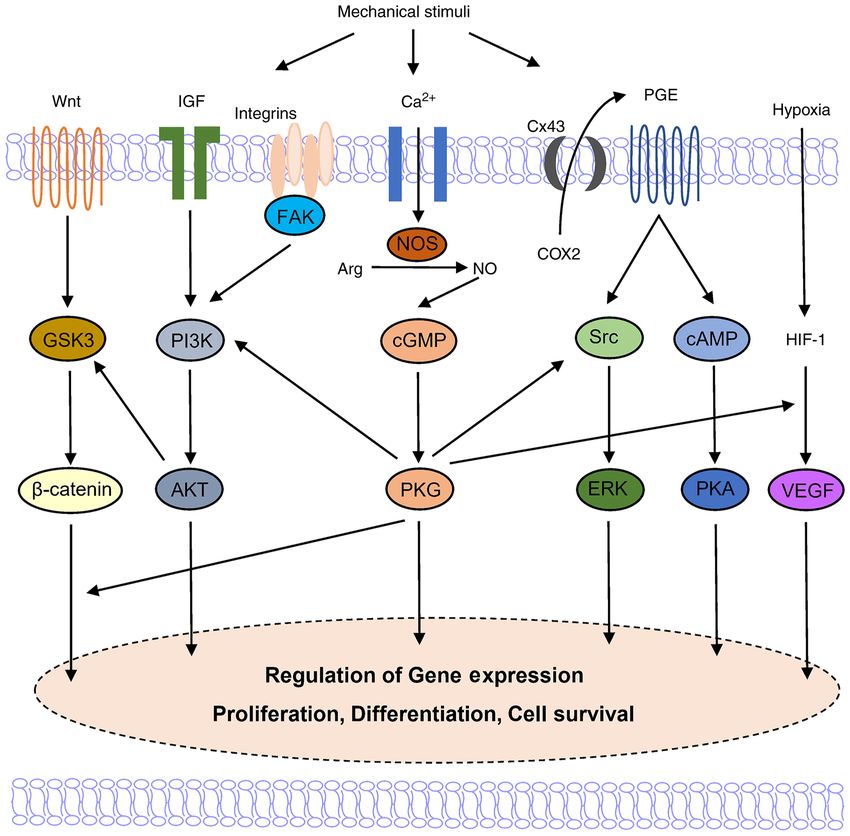
Pressure side: Osteoclasts and bone resorption. The transforming growth factor β (TGF‑β) superfamily, induces
pressure‑tension theory describes orthodontic tooth move‑ the differentiation of osteoprogenitor cells and promotes
ment as an outcome of bone resorption in the compression osteoblast function through the stimulation of Runx2 expres‑
region and bone formation in the tension region (21). On the sion via the small mother against decapentaplegic or p38INTERNATIONAL JOURNAL OF MOLECULAR MEDICINE 48: 168, 2021 3
mitogen‑activated protein kinase (MAPK) pathways (59‑62). controlling bone remodeling by coordinating the activity of
In addition, TGF‑β also suppresses bone resorption activity osteoblasts and osteoclasts.
through the upregulation of the tissue inhibitor of metallopro‑ Fibroblasts are involved in mechanosensation and
teinases expression (43,63). IL‑10 induces an overall reduction mechanotransduction in connective tissues. The application
in RANK signaling, through the facilitation of OPG expres‑ of mechanical stretching activates integrin and causes confor‑
sion and the reduction of RANKL production (43,64,65). mational changes in focal adhesion kinase (FAK), inducing a
Regional hypoxia caused by orthodontic force induces signaling cascade that modulates cytoskeletal dynamics and
hypoxia‑inducible factor (HIF)‑1 expression and upregulates gene transcription in fibroblasts (98,99).
the transcription of vascular endothelial growth factor (VEGF)
in PDL fibroblasts and osteoblasts. VEGF is associated with 3. Effects of NO on orthodontic tooth movement
osteogenic differentiation and matrix mineralization under
the regulation of BMP, corroborating the concept that angio‑ Expression of NO in bone tissue. Three NOS isoforms in
genesis and osteogenesis are combined (66,67). Furthermore, total are expressed in osteoblasts, osteoclasts and osteo‑
HIF‑1 and VEGF also stimulate osteoclast differentiation via cytes (100‑102). iNOS and eNOS are expressed in human
the upregulation of RANKL, contributing to the combination PDL stem cells (103,104). Previous studies revealed the
of bone resorption and bone formation (68‑70). presence of sGC and cGMP in mouse bone marrow macro‑
Some molecules that regulate the response of PDL fibro‑ phages (105), osteoclasts (105‑107), and osteocytes (108).
blasts to the orthodontic forces have been identified in previous Davidovitch et al (109,110) performed immunohistochem‑
studies, such as CC chemokine receptor 5 (CCR5) and CCR5 istry (IHC) on alveolar bone sections obtained from cats
ligands axis (71), relaxin (Rln) and Rln family peptides (Rxfps) and revealed that cGMP expression was increased in the
axis (72), and secretory leucocyte peptidase inhibitor (73). The PDL fibroblast cells stained intensely for; however, most
expression levels of these molecules were upregulated in the cGMP expression was not detected through IHC staining in
PDL, due to compression and tension force; however, their osteoblasts. However, cGMP expression increased due to the
downstream effects were different. Another consequence was subjection of the alveolar bone to mechanical force (111,112).
the upregulation of the osteoclastogenesis‑relating factors, The application of electric currents to the bone, also led to the
including RANKL, MCSF and MMPs, on the compression upregulation of cGMP in osteoblast and PDL fibroblast cells,
side, and osteoclast activity inhibiting factors, including accompanied by bone deposition near the cathode (113‑115).
Runx2, IL‑6, and IL‑12, that may induce osteoblast differen‑ Since a piezoelectric current can be generated by mechanical
tiation on the tension side. stress, the above findings suggest that NO/cGMP is an impor‑
tant signaling pathway, which mediates bone cell response to
Mechanotransduction: Osteocytes and fibroblasts. Osteocytes mechanical force (116).
are critical for the transduction of mechanical stimuli into
biochemical signals (74‑76). When a force is exerted on the Role of NO in cells associated with orthodontic tooth move-
tooth, the squeeze of the interstitial fluid causes fluid shear ment. Mounting evidence indicates that NO regulates multiple
stress (FSS) in the extracellular matrix (ECM) (77). The fluid cellular behaviors related to orthodontic movement (Fig. 1 and
flow hypothesis describes the response of osteocytes to FSS Table I).
as an essential mechanism during orthodontic treatment. FSS
stimulates an increase in the intracellular calcium concentra‑ Osteoclasts. A number of studies have demonstrated that NO
tion and the release of intercellular molecules in osteocytes exerts biphasic effects on osteoclast formation and function.
through the activation of integrin, a transmembrane protein In several cases, NO promotes osteoclastogenesis and bone
that connects ECM macromolecules to the internal cytoskel‑ resorption. NO mediates pre‑osteoclasts fusion through
eton (78‑80). The FSS‑related up‑regulation of NO, PGE2, the upregulation of actin cytoskeleton remodeling (117).
TFG‑β, and insulin‑like growth factor alters the osteocyte Histopathological studies have demonstrated that osteoclasts,
metabolic state and osteoblast/osteoclast functions (81,82). Howship's lacunae and new capillaries were increased in rats
Gap junctions formed by connexin (Cx) also participate in that received an injection of the NO precursor L‑arg during
the osteocyte‑osteoblast communication (83,84). For example, tooth movement (118‑120).
Cx is involved in the release of PGE2, which enhances Runx2 iNOS is an important regulator of osteoclast differ‑
DNA binding activity through the simultaneous activation of entiation under bacterial infection‑induced inflammatory
the cAMP/cAMP‑dependent protein kinase and MAPK path‑ conditions (121‑123). iNOS was previously found to mediate
ways and the subsequent stimulation of RANKL expression in alveolar bone loss and periapical infectious bone resorption
osteoblasts (85‑88). following the oral administration of Porphyromonas gingi-
The inhibitory effect of osteocytes on osteoblastic activity valis (124) or lipopolysaccharide (122). In another study,
can be induced by the secretion of sclerostin, which antago‑ histochemical analysis revealed that the osteoclast number in
nizes BMP effect and blocks canonical Wnt signaling (89‑91). iNOS(‑/‑) mice in comparison to wild‑type mice was consid‑
Osteocytes regulate osteoclastic differentiation via the alter‑ erably decreased (123). Tooth eruptions are similar to tooth
nation of major osteoclast regulators, namely RANKL and movement in terms of monocyte recruitment and osteoclast
M‑CSF (92‑94). Moreover, osteocyte apoptosis induction is differentiation. Evidence indicates that increased levels of
an important event in the recruitment and differentiation of iNOS are associated with a greater number of osteoclasts in
osteoclasts (95‑97). These findings confirm that osteocytes mice with accelerated tooth eruption, indicating that iNOS
play a key role in the response to biomechanical stimuli and may be a bone resorption modulator candidate (125).4 YAN et al: NITRIC OXIDE IN ORTHODONTIC TOOTH MOVEMENT
Figure 1. A schematic representation of bone remodeling and NO regulation during orthodontic tooth movement at the compression and tension sides. After
orthodontic force is applied to the teeth, bone remodeling in the compression region mainly manifests as osteoclastogenesis and bone resorption, while that
in the tension region presents as osteogenesis and bone formation. The regulation of related factors on bone remodeling is indicated by black arrows. NO
regulation of osteoclasts and osteoblasts differentiation is indicated by red arrows. HIF‑1, hypoxia‑inducible factor‑1; VEGF, vascular endothelial growth
factor; RANKL, receptor activator of nuclear factor‑κ B ligand; RANK, receptor activator of nuclear‑κ B; M‑CSF, macrophage‑colony stimulating factor;
IL‑1β/6, interleukin‑1β/6; TNF‑α, tumor necrosis factor‑α; PGE2, prostaglandin E2; MMPs, matrix metalloproteinases; BMP, bone morphogenetic protein;
OPG, osteoprotegerin; TGF‑β, transforming growth factor‑β; IGF, insulin‑like growth factor; TIMP, tissue inhibitor of metalloproteinases.
As previously demonstrated, M1‑like macrophage polar‑ resorption in vivo. In an iNOS(‑/‑) mouse model of apical peri‑
ization and an enhanced M1/M2 macrophage ratio increase odontitis, enhanced osteoclast differentiation and increased
the number of osteoclasts in rats or mice, accompanied by bone resorption were observed in comparison with the control
an increase in M1 macrophage marker expression (TNF‑ α group, accompanied by increased IL‑1β, TNF‑ α, RANK,
and iNOS) on the compression side, during tooth move‑ RANKL and monocyte chemoattractant protein‑1 (MCP‑1)
ment (126,127). TNF‑α stimulates the survival of differentiated levels (138,139). These results suggest that NO deficiency
osteoclasts through the induction of iNOS‑dependent NO is associated with an imbalance in the host inflammatory
generation (128). In the rheumatism inflammatory environ‑ response, resulting in severe bone loss.
ment, the TNF‑α promoting effect on alveolar bone resorption Moreover, iNOS exerts an inhibitory effect on osteoclast
is partly mediated through the activation of iNOS and the differentiation through other pathways. Zheng et al (140)
resulting production of NO (129). demonstrated that iNOS was a RANKL‑induced autocrine
It has been observed that the promoting effect of iNOS negative feedback inhibitor of RANKL‑mediated osteoclasto‑
on osteoclasts is mediated through the NO/cGMP pathway. genesis. RANKL triggered iNOS expression and NO release,
Kaneko et al (105) revealed that 8‑nitro‑cGMP, a NO‑dependent and subsequently inhibited RANKL‑induced osteoclast
derivative of cGMP in mammals, increased RANKL mRNA formation in a cGMP‑independent manner.
expression, and enhanced osteoclast differentiation. The The inconsistent effects of NO on osteoclastogenesis may
reduction in cGMP levels due to the inhibition of NOS caused be attributed to the differences in NO synthesis quantity, cell
RANKL‑induced osteoclast differentiation suppression. types and development states. NO action is also affected by
By contrast, evidence has revealed an inhibitory effect the cytokines in the microenvironment. Multiple factors influ‑
of NO on osteoclasts at low concentrations. NO has been ence the downstream signaling of NO, and further studies are
reported to increase osteoclast and osteoclast precursor cell required to elucidate the specific mechanism of NO regulation.
apoptosis (101,130‑132). A novel NO donor, nitrosyl‑cobin‑
amide (NO‑Cbi), has been found to reduce the RANKL/OPG Osteoblasts. NO is also involved in the bidirectional regulation
gene expression ratio or directly inhibit osteoclast differentia‑ of osteoblasts. Decreased NO concentrations promote osteo‑
tion in vitro and in vivo (133). Nicorandil, an agent that can blast proliferation, differentiation and survival (133,141‑143).
increase NO production in osteoclasts, was previously shown Mineralized nodule formations and mRNA expression levels
to suppress osteoclast differentiation via activating sGC (134). of osteoblastic genes, such alkaline phosphatase, osteocalcin
NO causes osteoclast detachment and downregulates osteo‑ and collagen‑1 genes, have been shown to be enhanced by NO
clast bone‑resorbing activity via the NO/cGMP/PKG pathway donors and 8‑Br‑cGMP, an analog of cGMP (141‑143). This
in vitro (101,107,135‑137). Of note, the selective inhibition effect was blocked by 1H‑(1,2,4)oxadiazolo‑(4,3‑a)quinox‑
of iNOS was previously found to markedly promote bone alin‑1‑one (ODQ), a competitive blocker that prevents sGCTable I. Role of NO signaling in the regulation of cells related to orthodontic movement.
Cell type Agent (concentration) Regulation Downstream pathways (Refs.)
Murine osteoclasts 8‑Nitro‑cGMP (30 µM) Promoted osteoclast formation Enhances the mRNA expression of RANK via PKG (105)
RAW264.7 murine NOC‑12 (15 µM), NOC‑18 (5 µM) Promoted osteoclast formation Regulated actin cytoskeleton remodeling and (117)
osteoclasts NOC‑12 (>25 µM), NOC‑18 (>10 µM) Decreased osteoclast survival pre‑osteoclast fusion
RAW264.7 murine AG (2‑500 µM) Promoted osteoclast formation RANKL/IFN‑β‑induced iNOS/NO as a negative (140)
osteoclasts feedback signal during osteoclastogenesis
UMR‑106 and SNAP (0‑1000 µM) Promoted osteoclastic activity Augmented the TNF‑α‑stimulated MMP‑1 mRNA (122)
MC3T3‑E1
Murine osteoclasts NOC‑18 (10‑500 nM) Increased osteoclast survival Mediated the TNF‑α‑induced osteoclast survival by (128)
NOC‑18 (1‑50 µM) reducing the activity of caspase 3
Rat osteoclasts YC‑1 (100 nM) Decreased osteoclast survival Activated caspase‑3/caspase‑8 activity and inhibited (130)
Inhibited osteoclast activity Src activity
Murine osteoclasts SNAP (300 µM) Decreased osteoclast survival Mediated apoptosis of osteoclast progenitors induced (131)
by TNF‑α and IFN‑γ
Murine osteoclasts L‑NMMA (0.1‑10 mM) Increased osteoclast survival Mediated cell apoptosis of osteoclast progenitors (132)
induced by IL‑12 and IL‑18
Murine osteoclasts NO‑Cbi (3‑30 µM) Inhibited osteoclast formation Reduced the RANKL/OPG gene expression ratio (133)
Murine osteoclasts Nicorandil (1‑100 µM) Inhibited osteoclast formation Via cGMP (134)
Human osteoclasts SNAP (20 mM) Inhibited osteoclast activity Downmodulated acid secretion and inhibited (135‑137)
SNP (100 µM) integrin attachments via cGMP/PKG I/VASP/
IP3R1/IRAG
FLG 29.1 human SIN‑1 (50‑200 µM) Decreased cell proliferation ‑ (101)
preosteoclast cell line
Avian osteoclasts SNP (unknown) Inhibited osteoclast activity Reduced osteoclast membrane HCI transport (107)
activity via PKG
Murine osteoblasts NO‑Cbi (3‑30 µM) Promoted osteoblast proliferation and Stimulated ERK/Akt and Wnt/β‑catenin signaling (133)
differentiation via cGMP/PKG
INTERNATIONAL JOURNAL OF MOLECULAR MEDICINE 48: 168, 2021
Rat osteoblasts NOC‑18 (10 µM) Promoted osteoblast differentiation Via cGMP (141)
Murine osteoblasts SNP (0.01 µM ‑1 mM) Promoted osteoblast differentiation Via cGMP (142)
DEA‑NO (0.1‑100 nM)
Rat osteoblasts 8‑Br‑cGMP (10‑100 µM) Promoted osteoblast differentiation ‑ (143)
Murine osteoblasts Sildenafil and vardenafil (10 nM‑1 mM) Promoted osteoblast differentiation Increased the expression of VEGF and VEGFR2 (144)
Rat osteoblasts NOC‑18 (10‑50 µM), SNP (100 µM) Promoted osteoblast survival and Via cGMP (145)
differentiation, decreased osteoblast survival
MC3T3‑E1 osteoblasts DEA‑NO (100 µM) Promoted osteoblast differentiation Regulated MMP‑13 expression via cGMP/PKG/Runx2 (146)
MC3T3‑E1 osteoblasts SNP (1.5‑3 mM) Decreased osteoblast survival Increased expression levels of p62, ATG7, Beclin‑1 (155)
and LC3‑II via AMPK
56 YAN et al: NITRIC OXIDE IN ORTHODONTIC TOOTH MOVEMENT
activation and lowers cGMP/PKG activity. It has been recently
methyl‑20‑furyl)‑1‑benzyl‑indazole; L‑NMMA, NG‑methyl‑L‑arginine; NO‑Cbi, nitrosyl‑cobinamide; SNP, sodium nitroprusside; VASP, vasodilator‑stimulated phosphoprotein; IP3RI, inositol
DETA‑NONOate/DEA‑NO/NOC‑18, 2,2'‑(hydroxynitrosoydrazino)bis‑ethanamine; AG, aminoguanidine; SNAP, S‑nitroso‑N‑acetyl‑penicillamine; IFN‑β, interferon β; YC‑1, 3‑(50‑hydroxy‑
1,4,5‑trisphosphate receptor I; IRAG, IP3RI‑associated protein; SIN‑1, 3‑morpholinosydnonimine; ATG7, autophagy related 7; LC3‑II, light chain 3‑II; DEA‑NO, diethylamine NONOate; HO‑1, heme
(157,158)
(Refs.) stated that PDE5 inhibitors, which can significantly increase
(159)
(103)
(162)
intracellular cGMP levels, induce osteoblast differentiation
and enhance bone regeneration in osteopenic mice via the
cGMP/VEGF pathway (144). These findings further support
the involvement of NO/cGMP/PKG pathway in the regulation
Akt/ERK and phosphorylating BAD via PKG Iα and
of osteoblast activity (133,143,145).
Increased iNOS expression and NO levels have been
Increased Bax and cytochrome c, and reduced
Mediated the effects of estradiol by activating
observed during osteoblast differentiation in vitro. iNOS has
been reported to mediate the regulation of Runx2 transloca‑
caspase‑3 via JNK, ERK and p38 MAPK
tion and downstream events (146). In eNOS knockout mice,
Downstream pathways
osteoblast growth has been shown to be inhibited (147).
Evidence suggests that eNOS activation promotes cell survival
and enhances osteoblastic gene expression in osteoblasts via
pathway cascades involving Src/extracellular signal‑regulated
kinase (ERK), phosphoinositide 3‑kinase (PI3K)/protein
Via HO‑1/ERK/NF‑κB
kinase B (Akt) and Wnt/β‑catenin (148,149).
NO mediates the action of several local and systemic factors,
Via JNK MAPK
including mechanical stimulation, hormones and other
signaling molecules in osteoblasts (13,150). It has also been
revealed that 1,25‑dihydroxyvitamin D(3) regulates bone
PKG II
mass via the upregulation of iNOS expression and NO produc‑
tion (151). CGRP has been found to promote mandibular
bone fracture healing in vivo and stimulate the eNOS activity
through the increase of intracellular calcium concentrations in
osteoblasts in vitro (152,153). Furthermore, it has been observed
No influence on proliferation and survival,
that 17 β‑estradiol, a major endogenous estrogen, may promote
eNOS expression and osteoblast differentiation through Akt
phosphorylation in a dose‑dependent manner (154). It has been
promoted osteogenic and reduced
previously demonstrated that the bone‑protective effects of
Promoted cell differentiation
Increased osteocyte survival
Decreased cell proliferation
estrogen rely upon the NO/cGMP pathway (147,150). High
Regulation
adipogenic differentiation
concentrations of NO negatively impact osteoblast prolifera‑
Decreased cell survival
tion and survival (145). NO simultaneously induces cell death
and autophagy in osteoblasts (155).
Osteocytes and PDL fibroblasts. The effect of NO on osteo‑
cytes is similar to that of osteoblasts. Parathyroid hormone
and 17β ‑estradiol levels increase the expression of cGMP
oxygenase‑1; Bax, BCL2‑associated X protein; JNK, c‑Jun N‑terminal kinase.
expression in osteocytes (108). Cinaciguat, an activator of sGC
that has been declared as a potential drug target for osteo‑
osteocyte‑like cells
fibroblasts
porosis, was previously found to reverse osteocyte apoptosis
and enhance bone formation in mice subjected to ovariec‑
tomy (156). NO/cGMP/PKG signaling mediated 17β‑estradiol
Agent (concentration)
anti‑apoptotic effect on osteocytes through either the activation
DETA‑NONOate (3 µM)
of the pro‑survival kinases, ERK and Akt, mediated by type II
PKG, or direct phosphorylation of protein related to cell death
SNP (0.5‑1.0 mM)
by type I (PKG) (157,158). However, inflammation‑induced
SNP (1‑4 mM)
iNOS activation and elevated concentrations of NO can lead to
SNP (75 µM)
osteocyte apoptosis (132).
NO/cGMP/PKG signaling has been shown to regulate
human PDL fibroblast proliferation and differentiation, with the
involvement of MAPK and nuclear factor κ‑light‑chain‑enhancer
of activated B cells pathways (103,159‑161). However, the
effect of NO on cell proliferation in PDL has not yet been fully
Human PDL stem cells
clarified. A previous study revealed that NO did not influence
Table I. Continued.
Human PDL cells
PDL stem cell proliferation (103). In other studies, it has been
MLO‑Y4 murine
revealed that exogenous NO inhibits proliferation and induces
Human PDL
apoptosis of PDL fibroblasts (159,162). This discrepancy could
Cell type
be attributed to differences in the cellular differentiation levels
and varying applied agent concentrations.INTERNATIONAL JOURNAL OF MOLECULAR MEDICINE 48: 168, 2021 7
The PDL and the alveolar bone are developed from the of nitrite and denitrosylation of some proteins), in case a
dental follicle during tooth development. The literature was specific NOS isoform is absent (195‑197). The aforemen‑
reviewed and it was observed that studies of NO regulation tioned ultrasound results can only prove the role of nNOS
impact upon the dental follicle during tooth development has or iNOS in ultrasound‑induced promotion on osteoblasts;
not been reported yet, to the best of our knowledge. It was however, those results do not contradict the involvement of
surmised that the exploration of the underlying mechanism of eNOS.
NO on the development of the PDL and alveolar bone may These findings suggest that NO plays a complex role in
provide novel insights into the role of NO in the tissue remod‑ mechanotransduction under stress in the periodontal tissue,
eling observed during orthodontic tooth movement. and further research on this topic is required.
NO signaling in bone mechanotransduction NO is a factor
that mediates early cellular response to applied mechanical Effects of NO in orthodontic tooth movement. Many studies
forces in the PDL and bone (Fig. 2) (13,163,164). NO synthesis have focused on the differential expression of NOS isoforms
in osteoclasts (165), osteoblast (116,151,163,165‑167), osteo‑ between areas of compression and tension during orthodontic
cytes (164,168), PDL fibroblasts (169), fibroblasts (170), and tooth movement. Experiments in rats revealed that the changes
dental pulp cells (171,172) increased following the applica‑ in NOS activity in the PDL could be detected as soon as 1 h
tion of mechanical loading, pulsed fluid flow (PFF), electrical after teeth were subjected to orthodontic force (198). The
stimulation, or pulsed electromagnetic field stimulation. increased expression of iNOS on the pressure side and eNOS on
The mechanical loading‑induced activation of the the tension side was observed 24 h after initiating mechanical
Wnt/β ‑catenin pathway is an important signaling event in loading, while increased nNOS expression mainly occurred
osteoblasts, osteocytes, and PDL fibroblasts, and is mediated after 3 h (199). An increase of iNOS‑positive osteocytes in
by a NO‑dependent mechanism involving the FAK, Src/ERK the compression area was detected 6 h after force application,
and PI3K/Akt signaling pathways (169,173,174). PFF increases while eNOS‑positive osteocytes in the tension area increased
NO synthesis in osteoblasts, resulting in PKG II‑dependent after 24 h (200). As indicated above, it is generally accepted
activation of Src and PI3K‑dependent phosphorylation of Akt. that iNOS dominates bone resorption at the compression site
The nuclear translocation of β‑catenin is induced and the gene while eNOS mediates the osteogenic effect in the tension
expression of c‑fos is upregulated, initiating a proliferative area (200,201).
response in mechanically stimulated osteoblasts (175‑177). The availability of studies related to the changes in NO
When the osteoblast and osteocyte cytoskeleton system of levels in human periodontal tissues before and after orth‑
disrupted, PFF‑induced NO production is affected (178). odontic treatment is limited. Analysis of gingival tissue
PFF induces the release of multiple soluble factors collected from orthodontic patients revealed that eNOS and
that promote osteogenesis and inhibits bone resorption. iNOS levels increased dramatically 2 weeks after the appli‑
This process is partially dependent on the generation of ance placement (202). A variety of biomarkers in gingival
NO (74,179‑181). PFF‑induced NO inhibited osteocyte apop‑ crevicular fluid (GCF) are often analyzed, in order to facilitate
tosis through the downregulation of B‑cell lymphoma‑2 (Bcl‑2) the improvement of clinical treatment. In various studies, many
and caspase‑3 (182). NO also modulates mechanically induced of which recent, it has been mentioned that NO expression
VEGF expression, contributing to angiogenesis during bone levels in GCF is related to orthodontic treatment (203‑206).
remodeling (183,184). Ford et al (203) revealed that NO concentration in GCF on the
The main NOS isoform that produces NO in osteoblasts compression side of the central incisor increased significantly
and osteocytes under the mechanical force action has not 1 h after the application of fixed orthodontic appliances. In
yet been elucidated. The activation of eNOS is associated patients who received rapid maxillary expansion therapy, the
with the phosphorylation or dephosphorylation at several NO levels in GCF were elevated on day 1 and 10 and were still
functional sites on eNOS, which may be induced by FSS, elevated after 3 months of retention (204,205). However, no
estrogens, VEGF and insulin (185‑187). Several studies significant difference was detected in NO levels in GCF on the
have revealed that FSS‑induced NO production is attrib‑ tension side, during the above treatment. These results further
uted to the calcium‑dependent eNOS activation in bone support the different regulatory effects of NO on the tension
cells (13,185,188,189). It has been revealed that the occlusal and pressure side, which are related to the presence of different
force led to iNOS and eNOS increased expression in hypo‑ NOS isoforms on different sides.
functional and normal PDL fibroblasts (100,104). The role of NO in orthodontic treatment has also been
Additionally, it has been suggested that eNOS may be confirmed in animal experiments. Tooth movement was mark‑
not indispensable for mechanically‑induced NO synthesis in edly promoted in rats that received L‑arg injection, whereas a
cultured osteoblasts or eNOS (‑/‑) mice (190,191). It has also significant reduction of tooth movement was observed in the
been mentioned that ultrasound‑induced bone formation may L‑NAME (eNOS inhibitor) group. Histological results also
be mediated through nNOS and iNOS upregulation in osteo‑ revealed a greater number of osteoclasts in the group with
blasts (82,163,192,193). Furthermore, osteopontin has been greater tooth movement (119,120,207). Notably, decreased
shown to suppress the osteoblast response to ultrasound by force‑induced root resorption was noted in this group in
inhibiting the expression of nNOS and iNOS through FAK comparison with the control group, although the number of
downregulation (194). This inconsistency may be explained osteoclasts increased in the L‑arg injection group (119).
in view of the possibility of an alternative way of NO Influences of NO and oral microbiota on the orthodontic
production induction by other NOS isoforms and through a tooth movement are also notable. In addition to being
non‑enzymatic NO production manner (including reduction synthesized by the body, NO can be produced by oral8 YAN et al: NITRIC OXIDE IN ORTHODONTIC TOOTH MOVEMENT
Figure 2. A schematic representation of signaling pathways activated by mechanical stimuli and mechanically‑induced NO regulation in osteoblastic cells.
Mechanical loads induce signal transduction through the activation of several signaling pathways, resulting in the increased expression of pro‑osteogenic factors,
thus providing an environment which contributes to osteoblast proliferation and differentiation. NO/cGMP/PKG pathway is widely involved in the regulation
of the above signaling pathways, indicating its important role in the mechanical transduction process. GSK3, glycogen synthase kinase 3; IGF, insulin‑like
growth factor; PI3K, phosphoinositide 3‑kinase; Akt, protein kinase B; NOS, nitric oxide synthase; Arg, arginine; NO, nitric oxide; cGMP, cyclic guanosine
monophosphate; PKG, protein kinase G; Cx43, connexin 43; COX‑2, cyclooxygenase‑2; PGE2, prostaglandin E2; ERK, extracellular signal‑regulated kinase;
cAMP, cyclic adenosine monophosphate; PKA, protein kinase A; HIF‑1, hypoxia‑inducible factor 1; VEGF, vascular endothelial growth factor.
bacteria under hypoxic conditions through the transfor‑ fibroblasts, and has been shown to promote the proliferation,
mation of saliva nitrate into nitrite (208‑210). It has been differentiation, or inhibition of survival and function of cells.
observed that NO production is upregulated during the As an inflammatory factor and a key second messenger in
deposition of dental plaque (211). In diseases related to mechanical transduction, NO is differentially expressed on
plaque accumulation, including periodontitis, an increase in the tension and compression side during tooth movement,
NO levels in both blood and saliva was reported (212‑214). suggesting its complex involvement in bone remodeling. The
Additionally, apart from the oral bacteria‑originating NO facilitation of NO precursor and the inhibition of NOS inhibitor
production, this has also been ascribed to the inflammatory in orthodontic tooth movement have also been confirmed in
response of the body. It has been previously demonstrated animal experiments. Additional studies are required, in order
that an enhanced osteoclast formation and accelerated orth‑ to evaluate the role and impact of NO on tooth movement
odontic tooth movement may be observed in patients with in clinical practice. As NO exerts complex effects on both
periodontitis (215). It is reasonable to speculate that NO osteoblastic and osteoclastic activities, the spatiotemporal
may be involved in this process, but more direct evidence is generation of NO may determine its specific biological effect
necessary in order to confirm this (203‑207). on bone remodeling. The precise and controlled delivery of
NO to periodontal tissue via NO‑releasing polymeric nano‑
4. Conclusions and future perspectives materials may be a promising approach for the acceleration of
orthodontic tooth movement.
NO is widely involved in the biomechanical response of the
periodontium to orthodontic forces. NO exerts dose‑dependent Acknowledgements
and biphasic effects on the functional status and cell fate deter‑
mination of osteoblasts, osteoclasts, osteocytes, and PDL Not applicable.INTERNATIONAL JOURNAL OF MOLECULAR MEDICINE 48: 168, 2021 9
Funding 15. Hassell TM: Tissues and cells of the periodontium. Periodontol 3:
9‑38, 1993.
16. Bartold PM and McCulloch CA: Information generation and
The present study was funded by the National Natural processing systems that regulate periodontal structure and func‑
Science Foundation of China (nos. 81870737 and 81771098), tion. Periodontol 63: 7‑13, 2013.
17. Antoun JS, Mei L, Gibbs K and Farella M: Effect of orthodontic
the Natural Science Foundation of Guangdong Province treatment on the periodontal tissues. Periodontol 74: 140‑157,
(no. 2021A1515011779) and Guangdong Financial Fund for 2017.
High‑Caliber Hospital Construction (no. 174‑2018‑XMZC‑0 18. Burstone C: The biomechanics of tooth movement. In: Kraus
BS, Riedel BA (eds.): Vistas in orthodontics. Lea, Febiger,
001‑03‑0125/D‑02). Philadelphia 197‑213, 1962.
19. Dhenain T, Côté F and Coman T: Serotonin and orthodontic
Availability of data and materials tooth movement. Biochimie 161: 73‑79, 2019.
20. Asiry MA: Biological aspects of orthodontic tooth movement: A
review of literature. Saudi J Biol Sci 25: 1027‑1032, 2018.
Not applicable. 21. Martin Schwarz A: Tissue changes incident to orthodontic tooth
movement. Int J Orthodontia Oral Surg Radiography 18: 331‑352,
1932.
Authors' contributions 22. Norevall LI, Forsgren S and Matsson L: Expression of neuro‑
peptides (CGRP, substance P) during and after orthodontic tooth
TY, YX and FH conceived the review. TY performed literature movement in the rat. Eur J Orthod 17: 311‑325, 1995.
23. Middleton J, Patterson AM, Gardner L, Schmutz C and
search and manuscript writing. YX contributed to the manu‑ Ashton BA: Leukocyte extravasation: Chemokine transport
script writing and the preparation of figures and tables. HH, and presentation by the endothelium. Blood 100: 3853‑3860,
WF and FH revised the manuscript. TY and FH confirm the 2002.
24. Lee SK, Pi SH, Kim SH, Min KS, Lee HJ, Chang HS, Kang KH,
authenticity of all the raw data. All authors read and approved Kim HR, Shin HI, Lee SK and Kim EC: Substance P regulates
the final manuscript. macrophage inflammatory protein 3alpha/chemokine C‑C ligand
20 (CCL20) with heme oxygenase‑1 in human periodontal liga‑
ment cells. Clin Exp Immunol 150: 567‑575, 2007.
Ethics approval and consent to participate 25. Yamaguchi M, Kojima T, Kanekawa M, Aihara N, Nogimura A
and Kasai K: Neuropeptides stimulate production of inter‑
Not applicable. leukin‑1 beta, interleukin‑6, and tumor necrosis factor‑alpha in
human dental pulp cells. Inflamm Res 53: 199‑204, 2004.
26. Kvinnsland I and Kvinnsland S: Changes in CGRP‑immunoreactive
Patient consent for publication nerve fibres during experimental tooth movement in rats. Eur J
Orthod 12: 320‑329, 1990.
27. Ren Y, Hazemeijer H, de Haan B, Qu N and de Vos P: Cytokine
Not applicable. profiles in crevicular fluid during orthodontic tooth movement of
short and long durations. J Periodontol 78: 453‑458, 2007.
Competing interests 28. Kapoor P, Kharbanda OP, Monga N, Miglani R and Kapila S:
Effect of orthodontic forces on cytokine and receptor levels in
gingival crevicular fluid: A systematic review. Prog Orthod 15:
The authors declare that they have no competing interests. 65, 2014.
29. Teitelbaum SL and Ross FP: Genetic regulation of osteoclast
References development and function. Nat Rev Genet 4: 638‑649, 2003.
30. Xie R, Kuijpers‑Jagtman AM and Maltha JC: Osteoclast differ‑
entiation during experimental tooth movement by a short‑term
1. Loscalzo J: Nitric oxide and vascular disease. N Engl J Med 333: force application: An immunohistochemical study in rats. Acta
251‑253, 1995. Odontol Scand 66: 314‑320, 2008.
2. Förstermann U and Sessa WC: Nitric oxide synthases: Regulation 31. Wada T, Nakashima T, Hiroshi N and Penninger JM: RANKL-
and function. Eur Heart J 33: 829‑837, 837a‑837d, 2012. RANK signaling in osteoclastogenesis and bone disease. Trends
3. Snyder SH: Nitric oxide. No endothelial NO. Nature 377: 196‑197, Mol Med 12: 17‑25, 2006.
1995. 32. Azuma Y, Kaji K, Katogi R, Takeshita S and Kudo A: Tumor
4. Moncada S and Higgs A: The L‑arginine‑nitric oxide pathway. necrosis factor‑alpha induces differentiation of and bone resorp‑
N Engl J Med 329: 2002‑2012, 1993. tion by osteoclasts. J Biol Chem 275: 4858‑4864, 2000.
5. Sawa T, Ihara H, Ida T, Fujii S, Nishida M and Akaike T: 33. Udagawa N, Takahashi N, Jimi E, Matsuzaki K, Tsurukai T,
Formation, signaling functions, and metabolisms of nitrated Itoh K, Nakagawa N, Yasuda H, Goto M, Tsuda E, et al:
cyclic nucleotide. Nitric Oxide 34: 10‑18, 2013. Osteoblasts/stromal cells stimulate osteoclast activation through
6. Francis SH, Busch JL, Corbin JD and Sibley D: cGMP‑dependent expression of osteoclast differentiation factor/RANKL but not
protein kinases and cGMP phosphodiesterases in nitric oxide macrophage colony‑stimulating factor: Receptor activator of
and cGMP action. Pharmacol Rev 62: 525‑563, 2010. NF‑kappa B ligand. Bone 25: 517‑523, 1999.
7. van't Hof RJ and Ralston SH: Nitric oxide and bone. Immunology 103: 34. Katagiri T and Takahashi N: Regulatory mechanisms of osteo‑
255‑261, 2001.
8. Wimalawansa SJ: Nitric oxide and bone. Ann NY Acad Sci 1192: blast and osteoclast differentiation. Oral Dis 8: 147‑159, 2002.
391‑403, 2010. 35. Thirunavukkarasu K, Halladay DL, Miles RR, Yang X,
9. Evans DM and Ralston SH: Nitric oxide and bone. J Bone Miner Galvin RJ, Chandrasekhar S, Martin TJ and Onyia JE: The
Res 11: 300‑305, 1996. osteoblast‑specific transcription factor Cbfa1 contributes to the
10. Li Y, Jacox LA, Little SH and Ko CC: Orthodontic tooth move‑ expression of osteoprotegerin, a potent inhibitor of osteoclast
ment: The biology and clinical implications. Kaohsiung J Med differentiation and function. J Biol Chem 275: 25163‑25172,
Sci 34: 207‑214, 2018. 2000.
11. Krishnan V and Davidovitch Z: On a path to unfolding the 36. Suda T, Takahashi N and Martin TJ: Modulation of osteoclast
biological mechanisms of orthodontic tooth movement. J Dent differentiation. Endocr Rev 13: 66‑80, 1992.
Res 88: 597‑608, 2009. 37. Takahashi N, Udagawa N, Akatsu T, Tanaka H, Shionome M and
12. Kalyanaraman H, Schall N and Pilz RB: Nitric oxide and cyclic Suda T: Role of colony‑stimulating factors in osteoclast develop‑
GMP functions in bone. Nitric Oxide 76: 62‑70, 2018. ment. J Bone Miner Res 6: 977‑985, 1991.
13. Klein‑Nulend J, van Oers RF, Bakker AD and Bacabac RG: Nitric 38. Liggett W Jr, Shevde N, Anklesaria P, Sohoni S, Greenberger J
oxide signaling in mechanical adaptation of bone. Osteoporos and Glowacki J: Effects of macrophage colony stimulating factor
Int 25: 1427‑1437, 2014. and granulocyte‑macrophage colony stimulating factor on osteo‑
14. Nanci A and Bosshardt DD: Structure of periodontal tissues in clastic differentiation of hematopoietic progenitor cells. Stem
health and disease. Periodontol 40: 11‑28, 2006. Cells 11: 398‑411, 1993.10 YAN et al: NITRIC OXIDE IN ORTHODONTIC TOOTH MOVEMENT
39. Boyle WJ, Simonet WS and Lacey DL: Osteoclast differentiation 61. Wu M, Chen G and Li YP: TGF‑β and BMP signaling in osteo‑
and activation. Nature 423: 337‑342, 2003. blast, skeletal development, and bone formation, homeostasis and
40. Tanaka S, Nakamura K, Takahasi N and Suda T: Role of RANKL disease. Bone Res 4: 16009, 2016.
in physiological and pathological bone resorption and therapeu‑ 62. Lee KS, Hong SH and Bae SC: Both the Smad and p38 MAPK
tics targeting the RANKL‑RANK signaling system. Immunol pathways play a crucial role in Runx2 expression following
Rev 208: 30‑49, 2005. induction by transforming growth factor‑beta and bone morpho‑
41. Aubin JE and Bonnelye E: Osteoprotegerin and its ligand: A new genetic protein. Oncogene 21: 7156‑7163, 2002.
paradigm for regulation of osteoclastogenesis and bone resorp‑ 63. Leivonen SK, Lazaridis K, Decock J, Chantry A, Edwards DR
tion. Medscape Womens Health 11: 905‑913, 2000. and Kähäri VM: TGF‑β‑elicited induction of tissue inhibitor of
42. Udagawa N, Takahashi N, Yasuda H, Mizuno A, Itoh K, Ueno Y, metalloproteinases (TIMP)‑3 expression in fibroblasts involves
Shinki T, Gillespie MT, Martin TJ, Higashio K and Suda T: complex interplay between Smad3, p38α, and ERK1/2. PLoS
Osteoprotegerin produced by osteoblasts is an important regu‑ One 8: e57474, 2013.
lator in osteoclast development and function. Endocrinology 141: 64. Park‑Min KH, Ji JD, Antoniv T, Reid AC, Silver RB,
3478‑3484, 2000. Humphrey MB, Nakamura M and Ivashkiv LB: IL‑10 suppresses
43. Garlet TP, Coelho U, Silva JS and Garlet GP: Cytokine expres‑ calcium‑mediated costimulation of receptor activator NF‑kappa
sion pattern in compression and tension sides of the periodontal B signaling during human osteoclast differentiation by inhibiting
ligament during orthodontic tooth movement in humans. Eur J TREM‑2 expression. J Immuno 183: 2444‑2455, 2009.
Oral Sci 115: 355‑362, 2007. 65. Zhang L, Ding Y, Rao GZ and Miao D: Effects of IL‑10 and
44. Liu XH, Kirschenbaum A, Yao S and Levine AC: Cross‑talk glucose on expression of OPG and RANKL in human periodontal
between the interleukin‑6 and prostaglandin E(2) signaling ligament fibroblasts. Braz J Med Biol Res 49: e4324, 2016.
systems results in enhancement of osteoclastogenesis 66. Kusumbe AP, Ramasamy SK and Adams RH: Coupling of
through effects on the osteoprotegerin/receptor activator of angiogenesis and osteogenesis by a specific vessel subtype in
nuclear factor‑{kappa B} (RANK) ligand/RANK system. bone. Nature 507: 323‑328, 2014.
Endocrinology 146: 1991‑1998, 2005. 67. Sivaraj KK and Adams RH: Blood vessel formation and function
45. Zhang YH, Heulsmann A, Tondravi MM, Mukherjee A and in bone. Development 143: 2706‑2715, 2016.
Abu‑Amer Y: Tumor necrosis factor‑alpha (TNF) stimulates 68. Park HJ, Baek KH, Lee HL, Kwon A, Hwang HR, Qadir AS,
RANKL‑induced osteoclastogenesis via coupling of TNF type Woo KM, Ryoo HM and Baek JH: Hypoxia inducible factor‑1α
1 receptor and RANK signaling pathways. J Biol Chem 276: directly induces the expression of receptor activator of nuclear
563‑568, 2001. factor‑ κ B ligand in periodontal ligament fibroblasts. Mol
46. Tani‑Ishii N, Tsunoda A, Teranaka T and Umemoto T: Autocrine Cells 31: 573‑578, 2011.
regulation of osteoclast formation and bone resorption by IL‑1 69. Dandajena TC, Ihnat MA, Disch B, Thorpe J and Currier GF:
alpha and TNF alpha. J Dent Res 78: 1617‑1623, 1999. Hypoxia triggers a HIF‑mediated differentiation of peripheral
47. Miyaura C, Inada M, Matsumoto C, Ohshiba T, Uozumi N, blood mononuclear cells into osteoclasts. Orthod Craniofac
Shimizu T and Ito A: An essential role of cytosolic phospho‑ Res 15: 1‑9, 2012.
lipase A2alpha in prostaglandin E2‑mediated bone resorption 70. Knowles HJ and Athanasou NA: Hypoxia‑inducible factor is
associated with inflammation. J Exp Med 197: 1303‑1310, 2003. expressed in giant cell tumour of bone and mediates paracrine
48. Kitaura H, Yoshimatsu M, Fujimura Y, Eguchi T, Kohara H, effects of hypoxia on monocyte‑osteoclast differentiation via
Yamaguchi A and Yoshida N: An anti‑c‑Fms antibody inhibits induction of VEGF. J Pathol 215: 56‑66, 2008.
71. Lee SY, Yoo HI and Kim SH: CCR5‑CCL Axis in PDL during
orthodontic tooth movement. J Dent Res 87: 396‑400, 2008. orthodontic biophysical force application. J Dent Res 94:
49. Abu‑Amer Y, Erdmann J, Alexopoulou L, Kollias G, Ross FP 1715‑1723, 2015.
and Teitelbaum SL: Tumor necrosis factor receptors types 1 and 72. Yang SY, Kim JW, Lee SY, Kang JH, Ulziisaikhan U, Yoo HI,
2 differentially regulate osteoclastogenesis. J Biol Chem 275: Moon YH, Moon JS, Ko HM, Kim MS and Kim SH: Upregulation
27307‑27310, 2000. of relaxin receptors in the PDL by biophysical force. Clin Oral
50. Yamasaki K: The role of cyclic AMP, calcium, and prostaglan‑ Investig 19: 657‑665, 2015.
dins in the induction of osteoclastic bone resorption associated 73. Lee SY, Moon JS, Yang DW, Yoo HI, Jung JY, Kim OS, Kim MS,
with experimental tooth movement. J Dent Res 62: 877‑881, 1983. Koh JT, Chung HJ and Kim SH: SLPI in periodontal Ligament
51. Domon S, Shimokawa H, Matsumoto Y, Yamaguchi S and is not sleepy during biophysical force‑induced tooth movement.
Soma K: In situ hybridization for matrix metalloproteinase‑1 and J Clin Periodontol 48: 528‑540, 2021.
cathepsin K in rat root‑resorbing tissue induced by tooth move‑ 74. Dallas SL, Prideaux M and Bonewald LF: The osteocyte: An
ment. Arch Oral Biol 44: 907‑915, 1999. endocrine cell and more. Endocr Rev 34: 658‑690, 2013.
52. Apajalahti S, Sorsa T, Railavo S and Ingman T: The in vivo 75. Tresguerres FGF, Torres J, López‑Quiles J, Hernández G,
levels of matrix metalloproteinase‑1 and ‑8 in gingival crevicular Vega JA and Tresguerres IF: The osteocyte: A multifunctional
fluid during initial orthodontic tooth movement. J Dent Res 82: cell within the bone. Ann Anat 227: 151422, 2020.
1018‑1022, 2003. 76. Tatsumi S, Ishii K, Amizuka N, Li M, Kobayashi T, Kohno K,
53. Bonafe‑Oliveira L, Faltin RM and Arana‑Chavez VE: Ito M, Takeshita S and Ikeda K: Targeted ablation of osteocytes
Ultrastructural and histochemical examination of alveolar bone induces osteoporosis with defective mechanotransduction. Cell
at the pressure areas of rat molars submitted to continuous orth‑ Metab 5: 464‑475, 2007.
odontic force. Eur J Oral Sci 111: 410‑416, 2003. 77. Goulet GC, Cooper DM, Coombe D and Zernicke RF: Influence
54. Krishnan V and Davidovitch Z: Cellular, molecular, and of cortical canal architecture on lacunocanalicular pore pressure
tissue‑level reactions to orthodontic force. Am J Orthod and fluid flow. Comput Methods Biomech Biomed Engin 11:
Dentofacial Orthop 129: 469.e1‑432, 2006. 379‑387, 2008.
55. Ducy P, Schinke T and Karsenty G: The osteoblast: A sophis‑ 78. Wang Y, McNamara LM, Schaffler MB and Weinbaum S: A model
ticated fibroblast under central surveillance. Science 289: for the role of integrins in flow induced mechanotransduction in
1501‑1504, 2000. osteocytes. Proc Natl Acad Sci USA 104: 15941‑15946, 2007.
56. Franz‑Odendaal TA, Hall BK and Witten PE: Buried alive: How 79. Phillips JA, Almeida EA, Hill EL, Aguirre JI, Rivera MF,
osteoblasts become osteocytes. Dev Dyn 235: 176‑190, 2006. Nachbandi I, Wronski TJ, van der Meulen MC and Globus RK:
57. Capulli M, Paone R and Rucci N: Osteoblast and osteocyte: Role for beta1 integrins in cortical osteocytes during acute
Games without frontiers. Arch Biochem Biophys 561: 3‑12, musculoskeletal disuse. Matrix Biol 27: 609‑618, 2008.
2014. 80. Liedert A, Kaspar D, Blakytny R, Claes L and Ignatius A: Signal
58. Kundu M, Javed A, Jeon JP, Horner A, Shum L, Eckhaus M, transduction pathways involved in mechanotransduction in bone
Muenke M, Lian JB, Yang Y, Nuckolls GH, et al: Cbfbeta inter‑ cells. Biochem Biophys Res Commun 349: 1‑5, 2006.
acts with Runx2 and has a critical role in bone development. Nat 81. Heino TJ, Hentunen TA and Väänänen HK: Conditioned medium
Genet 32: 639‑644, 2002. from osteocytes stimulates the proliferation of bone marrow
59. Canalis E, Economides AN and Gazzerro E: Bone morphoge‑ mesenchymal stem cells and their differentiation into osteoblasts.
netic proteins, their antagonists, and the skeleton. Endocr Rev 24: Exp Cell Res 294: 458‑468, 2004.
218‑235, 2003. 82. Li L, Yang Z, Zhang H, Chen W, Chen M and Zhu Z:
60. Chen G, Deng C and Li YP: TGF‑ β and BMP signaling in Low‑intensity pulsed ultrasound regulates proliferation and
osteoblast differentiation and bone formation. Int J Biol Sci 8: differentiation of osteoblasts through osteocytes. Biochem
272‑288, 2012. Biophys Res Commun 418: 296‑300, 2012.INTERNATIONAL JOURNAL OF MOLECULAR MEDICINE 48: 168, 2021 11
83. Yellowley CE, Li Z, Zhou Z, Jacobs CR and Donahue HJ: 105. Kaneko K, Miyamoto Y, Tsukuura R, Sasa K, Akaike T,
Functional gap junctions between osteocytic and osteoblastic Fujii S, Yoshimura K, Nagayama K, Hoshino M, Inoue S, et al:
cells. J Bone Miner Res 15: 209‑217, 2000. 8‑Nitro‑cGMP is a promoter of osteoclast differentiation
84. Taylor AF, Saunders MM, Shingle DL, Cimbala JM, Zhou Z induced by RANKL. Nitric Oxide 72: 46‑51, 2018.
and Donahue HJ: Mechanically stimulated osteocytes regu‑ 106. Korkmaz Y, Baumann MA, Schröder H, Behrends S, Addicks K,
late osteoblastic activity via gap junctions. Am J Physiol Cell Raab WH and Bloch W: Localization of the NO‑cGMP
Physiol 292: C545‑C552, 2007. signaling pathway molecules, NOS III‑phosphorylation sites,
85. Cherian PP, Cheng B, Gu S, Sprague E, Bonewald LF and ERK1/2, and Akt/PKB in osteoclasts. J Periodontol 75:
Jiang JX: Effects of mechanical strain on the function of Gap 1119‑1125, 2004.
junctions in osteocytes are mediated through the prostaglandin 107. Dong SS, Williams JP, Jordan SE, Cornwell T and Blair HC:
EP2 receptor. J Biol Chem 278: 43146‑43156, 2003. Nitric oxide regulation of cGMP production in osteoclasts.
86. Cheng B, Kato Y, Zhao S, Luo J, Sprague E, Bonewald LF and J Cell Biochem 73: 478‑487, 1999.
Jiang JX: PGE(2) is essential for gap junction‑mediated intercellular 108. Nagayama K, Miyamoto Y, Kaneko K, Yoshimura K, Sasa K,
communication between osteocyte‑like MLO‑Y4 cells in response Akaike T, Fujii S, Izumida E, Uyama R, Chikazu D, et al:
to mechanical strain. Endocrinology 142: 3464‑3473, 2001. Production of 8‑nitro‑cGMP in osteocytic cells and its upregu‑
87. Kanno T, Takahashi T, Tsujisawa T, Ariyoshi W and lation by parathyroid hormone and prostaglandin E2. In Vitro
Nishihara T: Mechanical stress‑mediated Runx2 activation is Cell Dev Biol Anim 55: 45‑51, 2019.
dependent on Ras/ERK1/2 MAPK signaling in osteoblasts. 109. Davidovitch Z, Montgomery PC and Shanfeld JL: Cellular local‑
J Cell Biochem 101: 1266‑1277, 2007. ization and concentration of bone cyclic nucleotides in response
88. Franceschi RT and Xiao G: Regulation of the osteoblast‑specific to acute PTE administration. Calcif Tissue Res 24: 81‑91, 1977.
transcription factor, Runx2: Responsiveness to multiple signal 110. Davidovitch Z, Montgomery PC and Shanfeld JL: Guanosine
transduction pathways. J Cell Biochem 88: 446‑454, 2003. 3',5'‑monophosphate in bone: Microscopic visualization by an
89. Sapir‑Koren R and Livshits G: Osteocyte control of bone remod‑ immuno‑histochemical technique. Calcif Tissue Res 24: 73‑79,
eling: Is sclerostin a key molecular coordinator of the balanced 1977.
bone resorption‑formation cycles? Osteoporos Int 25: 2685‑2700, 111. Davidovitch Z, Montgomery PC, Yost RW and Shanfeld JL:
2014. Immuno‑histochemical localization of cyclic nucleotides in
90. ten Dijke P, Krause C, de Gorter DJ, Löwik CW and van mineralized tissues: Mechanically‑stressed osteoblasts in vivo.
Bezooijen RL: Osteocyte‑derived sclerostin inhibits bone Anat Rec 192: 363‑373, 1978.
formation: Its role in bone morphogenetic protein and Wnt 112. Graziani E, Zelent ME and Pelliccioni GA: Biochemical aspects
signaling. J Bone Joint Surg Am 90 (Suppl 1): S31‑S35, 2008. of orthodontic movement: Recent findings and trends. Mondo
91. Galli C, Passeri G and Macaluso GM: Osteocytes and WNT: Ortod 16: 77‑84, 1991 (In Italian).
The mechanical control of bone formation. J Dent Res 89: 113. Davidovitch Z, Finkelson MD, Steigman S, Shanfeld JL,
331‑343, 2010. Montgomery PC and Korostoff E: Electric currents, bone
92. Kitaura H, Marahleh A, Ohori F, Noguchi T, Shen WR, Qi J, remodeling, and orthodontic tooth movement. II. Increase in
Nara Y, Pramusita A, Kinjo R and Mizoguchi I: Osteocyte‑related rate of tooth movement and periodontal cyclic nucleotide levels
cytokines regulate osteoclast formation and bone resorption. Int by combined force and electric current. Am J Orthod 77: 33‑47,
J Mol Sci 21: 5169, 2020. 1980.
93. Plotkin LI, Gortazar AR, Davis HM, Condon KW, Gabilondo H, 114. Davidovitch Z, Finkelson MD, Steigman S, Shanfeld JL,
Maycas M, Allen MR and Bellido T: Inhibition of osteocyte Montgomery PC and Korostoff E: Electric currents, bone
apoptosis prevents the increase in osteocytic receptor activator remodeling, and orthodontic tooth movement. I. The effect
of nuclear factor κ B ligand (RANKL) but does not stop bone of electric currents on periodontal cyclic nucleotides. Am J
resorption or the loss of bone induced by unloading. J Biol Orthod 77: 14‑32, 1980.
Chem 290: 18934‑18942, 2015. 115. Davidovitch Z, Steigman S, Finkelson MD, Yost RW,
94. Lin C, Jiang X, Dai Z, Guo X, Weng T, Wang J, Li Y, Feng G, Mont gom e r y P C, Sh a n feld J L a nd Ko r o st of f E:
Gao X and He L: Sclerostin mediates bone response to Immunohistochemical evidence that electric currents increase
mechanical unloading through antagonizing Wnt/beta‑catenin periosteal cell cyclic nucleotide levels in feline alveolar bone
signaling. J Bone Miner Res 24: 1651‑1661, 2009. in vivo. Arch Oral Biol 25: 321‑327, 1980.
95. Cheung WY, Simmons CA and You L: Osteocyte apoptosis regu‑ 116. Karanth HS and Shetty KS: Orthodontic tooth movement and
lates osteoclast precursor adhesion via osteocytic IL‑6 secretion bioelectricity. Indian J Dent Res 12: 212‑221, 2001.
and endothelial ICAM‑1 expression. Bone 50: 104‑110, 2012. 117. Nilforoushan D, Gramoun A, Glogauer M and Manolson MF:
96. Al‑Dujaili SA, Lau E, Al‑Dujaili H, Tsang K, Guenther A and Nitric oxide enhances osteoclastogenesis possibly by mediating
You L: Apoptotic osteocytes regulate osteoclast precursor cell fusion. Nitric Oxide 21: 27‑36, 2009.
recruitment and differentiation in vitro. J Cell Biochem 112: 118. Erez A, Nagamani SC, Shchelochkov OA, Premkumar MH,
2412‑2423, 2011. Campeau PM, Chen Y, Garg HK, Li L, Mian A, Bertin TK, et al:
97. Jilka RL, Noble B and Weinstein RS: Osteocyte apoptosis. Requirement of argininosuccinate lyase for systemic nitric oxide
Bone 54: 264‑271, 2013. production. Nat Med 17: 1619‑1626, 2011.
98. Wang JH, Thampatty BP, Lin JS and Im HJ: Mechanoregulation 119. Shirazi M, Nilforoushan D, Alghasi H and Dehpour AR: The
of gene expression in fibroblasts. Gene 391: 1‑15, 2007. role of nitric oxide in orthodontic tooth movement in rats. Angle
99. Wang HB, Dembo M, Hanks SK and Wang Y: Focal adhesion Orthod 72: 211‑215, 2002.
kinase is involved in mechanosensing during fibroblast migra‑ 120. Akin E, Gurton AU and Olmez H: Effects of nitric oxide in
tion. Proc Natl Acad Sci USA 98: 11295‑11300, 2001. orthodontic tooth movement in rats. Am J Orthod Dentofacial
100. Warita H, Watarai H and Soma K: Nitric oxide synthase expres‑ Orthop 126: 608‑614, 2004.
sion is increased by occlusal force in rat periodontal ligament. 121. Armour KE, Van'T Hof RJ, Grabowski PS, Reid DM and
Orthod Craniofac Res 7: 122‑126, 2004. Ralston SH: Evidence for a pathogenic role of nitric oxide in
101. Brandi ML, Hukkanen M, Umeda T, Moradi‑Bidhendi N, inflammation‑induced osteoporosis. J Bone Miner Res 14:
Bianchi S, Gross SS, Polak JM and MacIntyre I: Bidirectional 2137‑2142, 1999.
regulation of osteoclast function by nitric oxide synthase 122. Lin SK, Kok SH, Kuo MY, Lee MS, Wang CC, Lan WH,
isoforms. Proc Natl Acad Sci USA 92: 2954‑2958, 1995. Hsiao M, Goldring SR and Hong CY: Nitric oxide promotes
102. Helfrich MH, Evans DE, Grabowski PS, Pollock JS, Ohshima H infectious bone resorption by enhancing cytokine‑stimulated
and Ralston SH: Expression of nitric oxide synthase isoforms in interstitial collagenase synthesis in osteoblasts. J Bone Miner
bone and bone cell cultures. J Bone Miner Res 12: 1108‑1115, Res 18: 39‑46, 2003.
1997. 123. Cuzzocrea S, Mazzon E, Dugo L, Genovese T, Di Paola R,
103. Yang S, Guo L, Su Y, Wen J, Du J, Li X, Liu Y, Feng J, Xie Y, Ruggeri Z, Vegeto E, Caputi AP, Van De Loo FA, Puzzolo D
Bai Y, et al: Nitric oxide balances osteoblast and adipocyte and Maggi A: Inducible nitric oxide synthase mediates bone
lineage differentiation via the JNK/MAPK signaling pathway loss in ovariectomized mice. Endocrinology 144: 1098‑1107,
in periodontal ligament stem cells. Stem Cell Res Ther 9: 118, 2003.
2018. 124. Gyurko R, Shoji H, Battaglino RA, Boustany G, Gibson FC III,
104. Watarai H, Warita H and Soma K: Effect of nitric oxide on the Genco CA, Stashenko P and Van Dyke TE: Inducible nitric oxide
recovery of the hypofunctional periodontal ligament. J Dent synthase mediates bone development and P. gingivalis‑induced
Res 83: 338‑342, 2004. alveolar bone loss. Bone 36: 472‑479, 2005.You can also read