Role of Cardiovascular Magnetic Resonance in Native Valvular Regurgitation: A Comprehensive Review of Protocols, Grading of Severity, and ...
←
→
Page content transcription
If your browser does not render page correctly, please read the page content below
REVIEW
                                                                                                                                           published: 07 July 2022
                                                                                                                                  doi: 10.3389/fcvm.2022.881141

                                            Role of Cardiovascular Magnetic
                                            Resonance in Native Valvular
                                            Regurgitation: A Comprehensive
                                            Review of Protocols, Grading of
                                            Severity, and Prediction of Valve
                                            Surgery
                                            Emmanuelle Vermes 1* , Laura Iacuzio 2 , Franck Levy 2 , Yohann Bohbot 1,3 , Cédric Renard 4 ,
                                            Bernhard Gerber 5 , Sylvestre Maréchaux 6 and Christophe Tribouilloy 1,3*
                           Edited by:
                                            1
                      Daniel A. Morris,       Department of Cardiology, Amiens University Hospital, Amiens, France, 2 Department of Cardiology, Center
    Charité-Universitätsmedizin Berlin,     Cardio-Thoracique de Monaco, Monaco, Monaco, 3 UR UPJV 7517, Jules Verne University of Picardie, Amiens, France,
                                            4
                             Germany          Department of Radiology, Amiens University Hospital, Amiens, France, 5 Division of Cardiology, Department
                                            of Cardiovascular Diseases, Cliniques Universitaires St. Luc, Pôle de Recherche Cardiovasculaire (CARD), Institut
                       Reviewed by:         de Recherche Expérimentale et Clinique (IREC), Université catholique de Louvain, Brussels, Belgium, 6 Department
                        Fabrizio Ricci,     of Cardiology, Heart Valve Center, Lille Catholic University Hospital, Lille, France
 University of Studies G. d’Annunzio
              Chieti and Pescara, Italy
                 Claudia Romagnoni,         Valvular regurgitation is common in developed countries with an increasing prevalence
      IRCCS Ca’ Granda Foundation
   Maggiore Policlinico Hospital, Italy
                                            due to the aging of the population and more accurate diagnostic imaging methods.
                        Saul Myerson,       Echocardiography is the gold standard method for the assessment of the severity of
University of Oxford, United Kingdom        valvular heart regurgitation. Nonetheless, cardiovascular magnetic resonance (CMR) has
                    *Correspondence:        emerged as an additional tool for assessing mainly the severity of aortic and mitral valve
                    Emmanuelle Vermes
     emmanuellevermes@hotmail.com           regurgitation in the setting of indeterminate findings by echocardiography. Moreover,
                   Christophe Tribouilloy   CMR is a valuable imaging modality to assess ventricular volume and flow, which are
  tribouilloy.christophe@chu-amiens.fr
                                            useful in the calculation of regurgitant volume and regurgitant fraction of mitral valve
                   Specialty section:       regurgitation, aortic valve regurgitation, tricuspid valve regurgitation, and pulmonary
         This article was submitted to      valve regurgitation. Notwithstanding this, reference values and optimal thresholds to
              Cardiovascular Imaging,
               a section of the journal
                                            determine the severity and prognosis of valvular heart regurgitation have been studied
 Frontiers in Cardiovascular Medicine       lesser by CMR than by echocardiography. Hence, further larger studies are warranted to
         Received: 22 February 2022         validate the potential prognostic relevance of the severity of valvular heart regurgitation
           Accepted: 07 June 2022
                                            determined by CMR. The present review describes, analyzes, and discusses the use of
            Published: 07 July 2022
                                            CMR to determine the severity of valvular heart regurgitation in clinical practice.
                            Citation:
         Vermes E, Iacuzio L, Levy F,       Keywords: valvular regurgitation, cardiovascular magnetic resonance, echocardiography, regurgitant volume,
      Bohbot Y, Renard C, Gerber B,         regurgitant fraction
Maréchaux S and Tribouilloy C (2022)
    Role of Cardiovascular Magnetic
        Resonance in Native Valvular
    Regurgitation: A Comprehensive
                                            INTRODUCTION
        Review of Protocols, Grading
  of Severity, and Prediction of Valve
                                            The prevalence of valvular regurgitation is increasing worldwide, especially in high-income
                             Surgery.       countries (1, 2). In the setting of either atrioventricular or ventricular–arterial valve regurgitation,
 Front. Cardiovasc. Med. 9:881141.          a large regurgitant volume is accommodated by chamber dilation of the ventricles to preserve
    doi: 10.3389/fcvm.2022.881141           compliance and forward cardiac output. However, after some time, ventricular remodeling and

Frontiers in Cardiovascular Medicine | www.frontiersin.org                       1                                            July 2022 | Volume 9 | Article 881141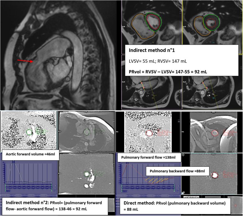
Vermes et al.                                                                                Cardiovascular Magnetic Resonance in Valvular Regurgitation

afterload mismatch eventually progress to a stage in which                  the ventricles, and we will propose a CMR-specific cut-off to help
wall stress can no longer be maintained and preload reserve is              in the decision-making process for valve replacement/repair.
overwhelmed, leading to heart failure. Thus, patients undergo
progression from a compensated stage to subclinical ventricular
dysfunction prior to decompensation and irreversible myocardial             MITRAL REGURGITATION
damage. These hemodynamic and pathological responses take
place insidiously so that patients often remain asymptomatic for            Moderate or severe mitral regurgitation (MR) is frequent in
a long duration despite the occurrence of adverse ventricular               the general population and represents an important cause of
remodeling. This makes the optimal timing of intervention in the            morbidity and mortality worldwide (9). In the United States, its
setting of most valvular regurgitations difficult.                          prevalence in patients ≥ 75 years has risen to > 4% (10) and
    Current guidelines are mostly symptom-based and                         almost to 8% in the United Kingdom (11).
recommend intervention for severe valvular regurgitation                       Cardiac imaging is critical for evaluating the cause of
in symptomatic patients or the presence of certain high-risk                MR (primary or secondary), assessing its severity and the
events (3, 4). The two main objectives of clinicians are, therefore,        consequences for the LV, and defining the best surgical timing.
to accurately diagnose severe valvular regurgitation and identify
high-risk features of early adverse ventricular remodeling in               Cardiovascular Magnetic Resonance for
asymptomatic patients.                                                      Assessing the Cause of Mitral
    Echocardiography is the most practical diagnostic method
for the assessment of valvular pathology and the current first-
                                                                            Regurgitation
line imaging modality for this purpose due to the excellent                 2D Cine Imaging for Valve Morphology
visualization of valve anatomy it affords, its availability, and            Assessing the cause of MR requires visualization of the anterior
its ease of use. This cardiovascular imaging technique allows               and posterior mitral valve (MV) leaflets, the mitral annulus,
the user to analyze valve morphology and motion and the                     the chordae, the anterolateral and posterolateromedial papillary
valvular annulus, quantify valvular regurgitation using different           muscle, and left ventricular (LV) wall motion abnormalities.
methods, and assess ventricle size and function (5). The                    Although echocardiography (transthoracic and especially 3D
EACVI/ASE guidelines propose a multiparametric approach                     transesophageal) is an excellent imaging of the MV which CMR
for the evaluation of valvular regurgitation severity with the              cannot match, CMR can help in identifying MV morphology
quantitative estimation of effective regurgitant orifice and                and MR mechanisms. With a high signal-to-noise ratio and
regurgitant volume, the preferred technique when available (6).             an excellent blood–myocardium contrast-to-noise ratio, cardiac
However, echocardiography assessment of valvular regurgitation              balanced steady-state free precession (bSSFP) imaging is the
suffers from several pitfalls, including a poor acoustic window for         method of choice for cine (motion) images. Retrospective cardiac
certain patients and specific issues concerning the vena contracta          gating, corresponding to the continuous acquisition of both ECG
and flow convergence methods, especially in the presence of                 and image signals collected over several consecutive heartbeats
eccentric jets, non-circular regurgitant orifices, constrained PISA,        and used after image reconstruction, should be used to allow
variable jet intensity during the cardiac cycle, and multiple               optimized temporal and spatial resolution. In the setting of
regurgitant jets (7, 8). The evaluation of regurgitation of the right       atrial fibrillation, however, prospective gating may result in better
heart and particularly the pulmonary valve is even more difficult           image quality. According to recent SCMR recommendations and
by echocardiography.                                                        a consensus on the use of CMR in MR (12, 13), the morphology
    Over the last 20–25 years, cardiovascular magnetic resonance            and motion of the MV apparatus can be studied using
(CMR) has emerged not only as the gold standard method for                      - A stack of contiguous thin (slice thickness between 5 and
assessing left and right ventricular volume, mass, and function                   7 mm) bSSFP cines in the short axis covering the MV.
but also as a robust and accurate tool for evaluating volumetric                - A stack of contiguous cines through-plane on the MV
quantification and accurate flow using a number of methods                        and perpendicular to the mitral commissure to cover all
independent of jet morphology. However, recent guidelines                         of the mitral scallop; from A1–P1 to A2–P2 and A3–
still consider CMR as a “second tool” when echocardiographic                      P3 (Figure 1).
parameters are inconsistent due to the absence of large                         - Standard long-axis cine images, including a 2-chamber
prospective studies defining the clinical impact of valvular                      view (vertical), a 3-chamber view [left ventricular outflow
regurgitation quantification by CMR and due to its availability,                  tract (LVOT)-1 view], and a 4-chamber view.
time, and cost. It is therefore essential to define, in light of
the most recent available data, the crucial role of this modality,             Leaflet motion abnormalities can be described as classically
which can provide, in an “all-in-one technique,” a unique                   defined by echocardiography (prolapse, flail) (Figure 1).
approach to study both valve lesions and their consequences                 However, a flail leaflet in CMR can be masked by the
on the heart chambers and hemodynamics. In this review, we                  regurgitant flow. Maximum mitral leaflet thickness is measured
discuss the emerging potential of CMR for the diagnosis and                 in diastole. Standard long-axis cine images are also informative
prognosis of regurgitant lesions. We will detail for each type of           for visualizing and sizing the mitral annulus (14). With
valvular regurgitation (mitral, aortic, tricuspid, and pulmonary)           CMR reference ranges, mitral annular dilatation, one of the
its ability to assess regurgitation severity, the consequences for          mechanisms in secondary MR, can be identified. The mitral

Frontiers in Cardiovascular Medicine | www.frontiersin.org              2                                           July 2022 | Volume 9 | Article 881141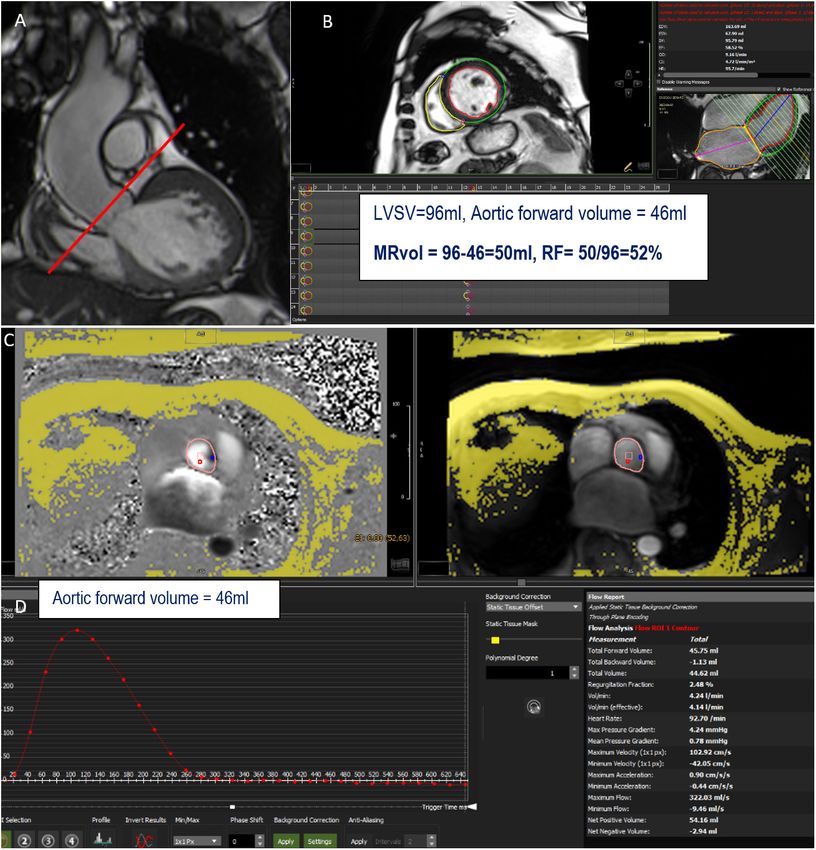
Vermes et al.                                                                                               Cardiovascular Magnetic Resonance in Valvular Regurgitation

annular diameter is measured from the anterior commissure to                            with a slightly darker-than-myocardium signal in bSSFP
the posterior commissure in the diastole (Figure 1). In primary                         sequences cannot be properly identified by CMR (16). Thus,
MR (mitral valve prolapse especially), CMR can detect mitral                            echocardiography (transthoracic and/or transesophageal) is
annulus disjunction, a frequent component of myxomatous                                 still the gold standard for the assessment of MV morphology
mitral valve disease (15) using standard long-axis cine images.                         and MR mechanisms.
   The LVOT-1 view can be used for determining the MV leaflet
length, MV tenting, and tenting height, major determinants
                                                                                        2D Cine Imaging for Regurgitant Jet Visualization
of ischemic MR. However, there are no data comparing CMR
measurement of the mitral annulus or MV length to surgical                              On cine images, regurgitant jets can be visualized as a signal loss
findings, and echocardiography remains the gold standard in the                         (dark/low signal) created by the turbulence of flow in the left
perioperative assessment of primary MR.                                                 atrium. The jet can be difficult to be clearly identified on fixed 2D
                                                                                        images, especially if the direction of the jet is not linear. Moreover,
Technical Considerations                                                                the size and length of the jet can be reduced or increased by
With a slice thickness between 5 and 7 mm, imaging the                                  just changing parameters or sequences. Rapid spoiled gradient-
length and thickness of the mitral chordae is less accurate                             recalled echo sequences with longer repetition and echo times or
than echocardiography. In addition, since CMR images                                    gradient echo (GRE) or hybrid GRE echo-planar sequences have
are reconstructed over several heartbeats, thin and very                                been proposed for higher sensitivity (13, 17). The identification
mobile structures such as vegetation, cordal rupture, or                                of such a flow void provides gross information about the location
fibroelastoma are often poorly visualized when their motion                             and direction of the jet, which can help in determining the
is variable in the cardiac cycle. Moreover, due to lower spatial                        etiology of the MR (for example, a central or eccentric jet,
resolution than echocardiography (especially transesophageal                            suggesting secondary or MV prolapse, respectively) (Figure 1).
echocardiography), annular or leaflet calcifications seen                               Compared to gradient-echo imaging, SSFP imaging results in

  FIGURE 1 | Anterior and posterior mitral valves and their scallops (A1, A2 and A3 and P1, P2, P3) visualized in diastole using balanced steady-state free precession
  (bSSFP) cine images in the plane of the mitral valve (A). Cine 4 chamber view in a patient with posterior and anterior mitral valve prolapse (red arrows) (B). Example
  of mitral annular measurement from the anterior commissure to the posterior commissure (red line) in the 2-chamber view (C). A central mitral regurgitation (flow void
  in the left atrium, red arrow) secondary to mitral annular dilatation visualized in the 4-chamber view (D).

Frontiers in Cardiovascular Medicine | www.frontiersin.org                          3                                               July 2022 | Volume 9 | Article 881141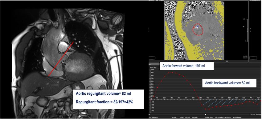
Vermes et al.                                                                                              Cardiovascular Magnetic Resonance in Valvular Regurgitation

TABLE 1 | Various CMR methods used in clinical practice to quantify MR, with their advantages and limitations.

                                                                Indirect                                                                   Direct

CMR method              1: Recommended                                         2                                 Not recommended

CMR sequences           SSFP short-axis images for LVSV                        Volumetric                        Long-axis cine SSFP images and phase-contrast
required                                                                       SSFP short-axis images            velocity mapping placed on the atrial side of the MV,
                                                                               for LVSV and RVSV                 perpendicular to the direction of the jet
                        Phase-contrast velocity mapping at the sinotubular
                        junction for AFF
                        In aortic stenosis: Phase-contrast velocity mapping
                        of the main pulmonary artery for PFF
Formula                 MRvol (ml) = LVSV—AFF                                  MRvol = LVSV—RVSV                 Direct quantification of MRvol
                        RF (%) = MRvol/LVSV
                        In aortic stenosis:
                        MRvol = LVSV—PFF
                        In aortic regurgitation:
                        MRvol = LVSV- (AFF + ABF)
Advantages              Reproducible                                           Fast and simple                   Valid for patients with multiple regurgitations or
                        Independent of jet morphology                                                            intracardiac shunts
                        Valid for patients with multiple regurgitation
Limitations             Careful basal slice selection at the LV base for       Inaccurate for patients with      High velocity jets can cause spin dephasing and
                        systolic volume in mitral prolapse                     multiple regurgitations and       displacement artifacts
                        Careful perpendicular slice selection                  intracardiac shunts               Difficult to select the plane position for patients with
                        Phase-offset errors                                    Careful basal slice selection     eccentric jets with mobile valves
                        Ensure correct maximal velocity encoding               of LV and RV                      Inaccurate for patients with high heart rate variability

CMR, cardiac magnetic resonance; SSFP, steady-state free precession; LVSV, left ventricular stroke volume; RVSV, right ventricular stroke volume; AFF, aortic forward
flow; ABF, aortic backward flow; RF, regurgitant fraction; PFF, pulmonary forward flow; MRvol, mitral regurgitant volume.

significantly reduced signal voids in regurgitant jets in general                      or the aortic valve but not at the mid-ascending aorta due to
and can easily underestimate the degree of regurgitation. These                        the risk of overestimation (18). This sequence generates two
sequences are much less sensitive for identifying regurgitation                        types of images: a magnitude image showing the anatomy of
than echocardiography and do not represent a reliable method                           the aortic valve and a phase map encoding the velocities within
for evaluating MR severity.                                                            each voxel. A dedicated post-processing analyzes the blood flow
                                                                                       through the aortic plane generating a flow curve, which allows
Cardiovascular Magnetic Resonance for                                                  the calculation of the aortic forward flow (AFF) (Figures 2, 3).
Quantifying Mitral Regurgitation                                                       An average of two to three flow measurement acquisitions
There are several CMR methods for evaluating MR severity; all                          is optimal.
are independent of the characteristics of the mitral regurgitant
jet and do not require calculation using a complex equation.                           Advantages
These methods are less accurate in arrhythmic patients or poor                         This method is highly reproducible (19, 20), independent of the
breath-holders. A free-breathing phase-contrast sequence with                          number, shape, or morphology of regurgitant jets (particularly
an increased number of averages (≥3) can be applied in these                           useful for patients with eccentric or multiple jets), and is not
cases. The methods available in clinical practice are summarized                       affected by tricuspid or pulmonary regurgitation. In cases with
in Table 1.                                                                            concomitant aortic regurgitation, aortic diastolic flow (aortic
                                                                                       backward flow: ABF), obtained directly from the diastolic flow
Indirect Method n◦ 1: 2D Cine Imaging and 2D Cine                                      of the aortic phase-contrast image, must be added to the AFF in
Phase-Contrast Velocity Mapping                                                        the equation (Table 1) (17).
The most widely used CMR method to quantitatively assess the                               In cases with aortic stenosis, the through-plane flow imaging
severity of MR is an indirect method combining 2D cine imaging                         slice is placed beyond the turbulent jet. In exceptional cases,
and phase-contrast velocity mapping to quantify the regurgitant                        the pulmonary forward flow (PFF), instead of the AFF due
volume and fraction.                                                                   to non-laminar and high aortic velocity in the ascending
   Mitral regurgitant volume (MRvol) is expressed as the                               aorta, can be used.
difference between the left ventricular stroke volume (LVSV) and
aortic forward flow (AFF), and the regurgitant fraction (RF) as                        Technical Considerations
the MRvol divided by the LVSV, expressed as a percentage.                              Technical considerations are detailed in Table 1. Although CMR
   The LVSV can be obtained from multiple LV short-axis cine                           is the gold standard for volumetric assessment, variability may
bSSFP images, and the AFF from phase-contrast velocity flow in                         arise due to basal slice selection for LVSV measurement which
a trough-plane acquired at the level of the sinotubular junction                       can be challenging in patients with MR due to prolapse with

Frontiers in Cardiovascular Medicine | www.frontiersin.org                         4                                                July 2022 | Volume 9 | Article 881141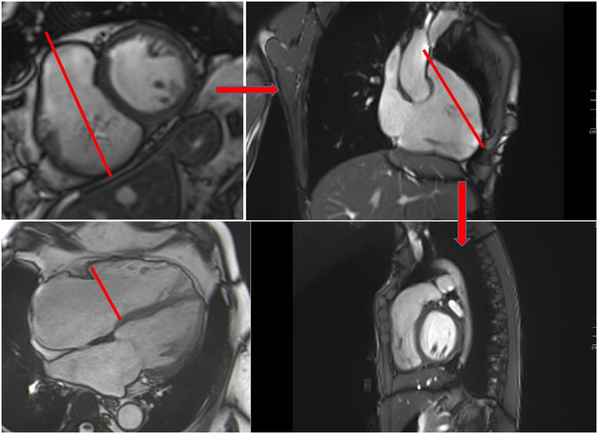
Vermes et al.                                                                                               Cardiovascular Magnetic Resonance in Valvular Regurgitation

  FIGURE 2 | Example of mitral regurgitation (MR) assessment using indirect method n◦ 1. LVOT bSSFP images show the perpendicular line above the aortic valve (red
  line) indicating the slice position for phase-contrast velocity mapping (A). Assessment of left ventricular stroke volume (LVSV) from short-axis cine SSFP images (B).
  Phase and magnitude images with delineation of the aorta (C) generating flow curves (D). This patient has a severe MR with a mitral regurgitant volume (MRvol) of
  50 ml and a regurgitant fraction (RF) of 52%.

Barlow disease, with increased annular excursion due to mitral                          in patients with AR by measuring RF (= ARvol/AFF, Figure 3),
annular disjunction (21). Accordingly, we suggest measuring the                         is very good with an excellent correlation coefficient (0.956 and
LV end systolic volume at the base of the LV rather than at                             0.998 respectively) and small standard errors of the estimate
the leaflets in these patients, resulting in a better assessment of                     (1.19% and 0.34% respectively) (24).
LV function and MR severity, with higher LV ejection fraction
(EF), MRvol, and RF.                                                                    Indirect Method n◦ 2: Volumetric Method: 2D Cine
   The accuracy of the AFF may be compromised by potential                              Imaging
errors:                                                                                 An alternative and simple approach using only bSSFP sequences
                                                                                        in the short axis is the “volumetric method,” expressed as the
     - Mismatch of encoding velocity: the velocity encoding value                       difference between LVSV and right ventricular stroke volume
       (Venc) should be set within 25% of the true maximal                              (RVSV), which represents the quantity of MR in the absence of
       velocity in the aorta to avoid aliasing.                                         other types of valvular regurgitation or intracardiac shunts.
     - Misalignment of imaging plane: to avoid inaccurate aortic
                                                                                        Advantages
       peak velocity, the imaging plane should be orthogonal to
                                                                                        Using a single method is less prone to measurement error than
       the direction of flow.
                                                                                        using two different techniques.
     - Phase offset errors, due to local magnetic field
       inhomogeneities, can be corrected by using a background                          Technical Considerations
       correction.                                                                      Due to the shape of the right ventricle (RV) and extensive
     - During analysis, noise pixels should not be included in the                      trabeculations, RV contouring is more prone to errors in
       contouring of the aorta.                                                         RVSV measurements.

   Validation of AFF by CMR has been established                                        Direct Method
by comparison to left ventricular stroke volume with                                    Direct quantification of MR flow is theoretically feasible with
echocardiography with good correlation (Table 2) (22, 23).                              phase-contrast velocity mapping by directly measuring the
Inter- and intra-observer variability of AFF, indirectly assessed                       regurgitant flow across the MV.

Frontiers in Cardiovascular Medicine | www.frontiersin.org                          5                                               July 2022 | Volume 9 | Article 881141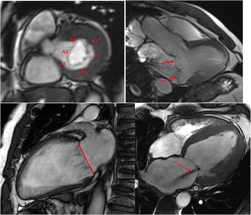
Vermes et al.                                                                                             Cardiovascular Magnetic Resonance in Valvular Regurgitation

                                                                                       severity is also the high velocity of the MR jet (> 5 m/s),
                                                                                       which may result in aliasing and which can be more difficult
                                                                                       to correct in 4D flow imaging and may require multiple
                                                                                       velocity encodings.

                                                                                       Late Gadolinium Enhancement Imaging and T1
                                                                                       Mapping for Additional Information
                                                                                       Late gadolinium enhancement (LGE) imaging, acquired after
                                                                                       gadolinium administration, has the unique ability to provide
                                                                                       tissue characterization of the myocardium by assessing and
                                                                                       quantifying regions of fibrosis and scars across the left atrium
                                                                                       (27), the LV (28), and the papillary muscle (29).
                                                                                           Late gadolinium enhancement sequences [pulse sequences
                                                                                       according to published guidelines (12)] should be performed
                                                                                       at least 10 min after gadolinium injection in the short axis
                                                                                       and the three long-axis planes (13). High-resolution dark
  FIGURE 3 | (A) (left) Slice position for the phase-contrast velocity mapping         blood LGE CMR is required to identify small papillary muscle
  sequence. (B) Flow curve (mL/s) through the aorta at the level of the line on
  panel (A). Aortic forward volume is calculated as the area under the curve
                                                                                       enhancement (30).
  (AUC) of the forward flow on the flow curve (green area); aortic backward                In ischemic MR, the quantification of MR and myocardial
  volume is calculated as the AUC of the backward flow on the flow curve               infarct size with CMR can help for risk stratification in patients
  (orange area). (C) Formula to obtain mitral and aortic regurgitant volume            with advanced ischemic cardiomyopathy (31). In primary MR,
  (MRvol and ARvol) and regurgitant fraction (RF) are shown at the left bottom.
                                                                                       especially MV prolapse, a high prevalence of focal replacement
                                                                                       fibrosis, particularly in the segments adjacent to the posterior
                                                                                       papillary muscle, can be observed (29). However, the LGE
   However, given the significant dynamic motion of the MV                             technique is not able to characterize diffuse fibrosis due
during systole, high velocity, a non-circular orifice, and jet                         to the absence of a normal myocardium as the reference.
angulation, positioning a fixed slice across the MV (without                           A recent parametric mapping technique (T1 mapping) allows for
annular valvular tracking) can be challenging and inappropriate,                       better characterization of global myocardial tissue composition
especially for patients with eccentric MR, multiple jets with                          by direct measurement of T1 relaxation times before and
different directions, and high heart-rate variability. Therefore, we                   after contrast administration (32) and by calculation of the
do not recommend this approach for clinical practice.                                  extracellular volume (ECV) (33).
                                                                                           In MR, little is known about diffuse interstitial fibrosis. One
4D Flow                                                                                recent study suggested that in primary MR, ECV increases with
4D flow velocity-encoded CMR imaging is an emerging                                    MR severity, regardless of MV prolapse or the presence of
technique that involves phase-contrast acquisition with flow                           replacement fibrosis (34). Diffuse interstitial fibrosis in primary
encoding in all three spatial directions and to the dimension                          MR may be more closely related to chronic LV volume overload
of time (3D + time = 4D). It allows the visualization of flow                          than the etiology of MR.
in multiple orientations to follow the spatial motion of the
heart valves and the variable direction of the jets over time.
The use of such a retrospective tracking method in a single
                                                                                       Assessment of MRVol by
volume acquisition covering the entire heart may enable direct                         Echocardiography and Cardiovascular
MR jet volume quantification, overcoming certain limitations                           Magnetic Resonance
of 2D sequences (25). Recent reports have demonstrated its                             Quantification of MR by echocardiography is based on
feasibility in mild to severe MR (26). However, its accuracy                           2D echocardiography, color flow, and Doppler parameters.
has usually been poor in comparison to 2D flow, especially                             Current echocardiographic guidelines consider severe MR as
in primary MR (25). This promising technique, which is                                 MRvol ≥ 60 ml (or effective regurgitant orifice ≥ 40 mm2 )
constantly improving, is not yet commercially available for all                        by the PISA method and RF ≥ 50% (35). Strikingly, the
scanners. Its principal disadvantage is the long scanning time                         echocardiographic cut-off of MRvol ≥ 60 ml and or RF ≥ 50%
(up to 10 min). Furthermore, it requires a post-processing                             were also considered for severe MR in the vast majority of CMR
software. Another limitation for direct measurement of MR                              studies and the current guidelines (35, 36) (Table 3).

TABLE 2 | Validation of forward flow in the aorta by comparison to echocardiography.

First author, year (Ref #)                   Number of patients                                Reference method                              Correlation coefficient

Bogren et al. (22)                             24 normal patients                     Left stroke volume in echocardiograhpy                           0.93
Van Rossum et al. (23)                       17 healthy volunteers                    Left stroke volume in echocardiograhpy                           0.76

Frontiers in Cardiovascular Medicine | www.frontiersin.org                        6                                              July 2022 | Volume 9 | Article 881141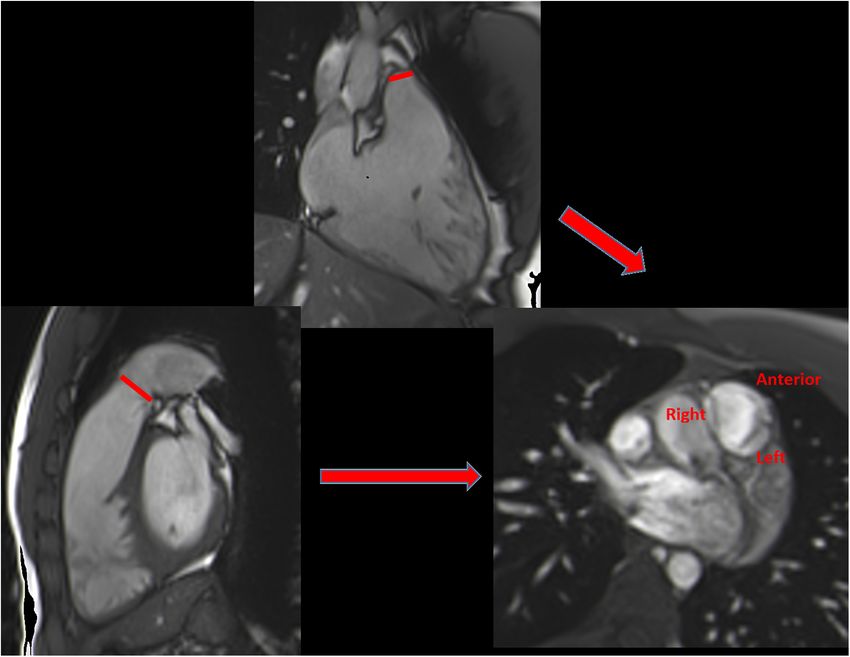
Vermes et al.                                                                                              Cardiovascular Magnetic Resonance in Valvular Regurgitation

TABLE 3 | Cutoff of CMR parameters to define severe MR and AR according to              cohort. Gelfand et al. (44) proposed that RF CMR cutoff value
current American (36) and European recommendations (35).
                                                                                        of 42% (indirect method) for severe MR correlates well with
                                     Severe MR                  Severe AR               Doppler echocardiography. Interestingly, one study used the
                                                                                        multiparametric approach by Doppler echocardiography as the
ASE recommendations (36)            RF ≥ 50%                    RF ≥ 50%                reference standard and compared it to CMR RF. MR severity
                                   MRvol ≥ 60 ml               ARvol ≥ 60 ml
                                                                                        graded by two experts, showed excellent agreement. The authors
ESC recommendations (35)              RF ≥ 50%                   RF ≥ 50%
                                                                                        observed that significant MR (moderate to severe or severe) could
CMR, cardiac magnetic resonance; MR, mitral regurgitation; AR, aortic                   be very accurately identified by CMR using a RF cutoff value of
regurgitation; ASE, American Society of Echocardiography; ESC, European Society         35% (45).
of Cardiology; RF, regurgitant fraction; MRvol, mitral regurgitant volume; ARvol,
aortic regurgitant volume.                                                                  Myerson et al. (46), on initially asymptomatic patients with
                                                                                        moderate or severe MR followed up for up to 8 years, showed that
                                                                                        MRvol and RF by CMR were the most discriminatory parameters
Comparison Between Echocardiographic MRvol and                                          to determine the need for surgery (symptoms or other indications
Cardiovascular Magnetic Resonance MRvol                                                 for surgery), with cut-off thresholds of 55 ml for MRvol and
Most studies have shown only limited agreement in the                                   40% for RF, and an increasing risk with increasing values of the
assessment of MR between CMR and echocardiography (20,                                  parameters. The RF cutoff for severe MR in this study was very
37–39). MRvol values truly differ among volumetric methods                              similar to a recent study by Polte et al. (47) (RF > 41%) and
and the PISA method. There is a consistent tendency to                                  lower than echocardiographic criteria (RF ≥ 50%). As the MRvol
obtain higher MRvol values by PISA relative to CMR (40–                                 is proportional to LV size in primary MR, for two patients with
42), resulting in higher MR severity assessment with a risk                             similar CMR MRvols, the patient with a smaller LV will have
of undergoing surgery without severe MR by CMR (20). The                                more severe MR; calculation of the RF may overcome this issue
discrepancy between these two modalities is more pronounced                             as it normalizes the MRvol to the size of the LV.
in patients with late systolic or multiple jets than central                                Interestingly, integrating ECV may also provide an additional
and holosystolic jet (38). Therefore, using the same thresholds                         benefit for the selection of asymptomatic patients for mitral
to define MR severity and the need for surgery may be                                   correction. Recently, Kikungvan et al. (48), in a prospective
inappropriate. The present available literature indicates that                          observational registry of patients with at least moderate primary
PISA MRvol thresholds and MRvol values by CMR are different                             MR, showed that RF and elevated ECV were independently
and not interchangeable. Interestingly, the MRvol was similar                           associated with events, with a cutoff of 40% for RF and 30% for
using the volumetric method by 3D echocardiography and                                  ECV for mitral surgery.
CMR (43). Recent reports have suggested that 3D-transthoracic                               Another approach to evaluate the accuracy of an imaging
echocardiography is less dependent on geometric assumptions                             modality to quantify MR is to assess LV remodeling after surgery.
and allows better visualization of the vena contracta regurgitant                       Some authors have found a good correlation between MRvol by
orifice. However, characterization of the Doppler jet is still                          CMR and a decrease in LV volume post-MR correction (20, 40).
challenging, with additional limitations of lower spatial and
temporal resolution (36).                                                               Main Message for the Clinician
                                                                                        Based on the most recent guidelines (4), echocardiography
Cardiovascular Magnetic Resonance to Identify                                           remains the first-choice tool to grade MR based on qualitative,
Significant MR and for the Timing of Intervention                                       semiqualitative, quantitative, and structural criteria. Current
There is a paucity of data on specific CMR thresholds to define                         class I surgical recommendations for severe MR are based on
MR severity due to the lack of large trials with a validation                           symptoms or, for asymptomatic patients, on LV dilatation or

TABLE 4 | Indications, preferred methods and evidence level of CMR to determine severity of mitral, aortic, tricuspid and pulmonary regurgitation according to current
American and European guidelines (3, 4, 35, 36).

                            Severe MR                          Severe AR                                  Severe TR                            Severe PR

When CMR is                 Discrepancy between MR             Discrepancy between AR severity            Discrepancy between TR               Discrepancy between PR
indicated                   severity on echo and               on echo and clinical findings              severity on echo and                 severity on echo and
                            clinical findings                  Inconclusive echo on AR severity or        clinical findings                    clinical findings
                            Inconclusive echo on MR            indeterminate AR (3, 4, 35, 36)            Inconclusive echo on TR              Inconclusive echo on PR
                            severity or indeterminate          Patients with bicuspid aortic valve        severity or indeterminate            severity or indeterminate
                            MR (3, 4, 35, 36)                  with unsatisfactory assessment of          TR (3, 4, 35, 36)                    PR (3, 35, 36)
                                                               aorta morphology (36)                      RV assessment (35)                   RV assessment (35)
Preferred CMR               Indirect methods (35, 36)          Direct method (35, 36)                     Indirect methods (35, 36)            Direct method (35, 36)
methods
Class of                    I/B-NR (3)                         I/B-NR (3)                                 I/B-NR (3)                           –
recommendation/
Level of evidence

CMR, cardiac magnetic resonance; MR, mitral regurgitation; AR, aortic regurgitation; TR, tricuspid regurgitation; PR, pulmonary regurgitation; NR, not randomized.

Frontiers in Cardiovascular Medicine | www.frontiersin.org                          7                                                 July 2022 | Volume 9 | Article 881141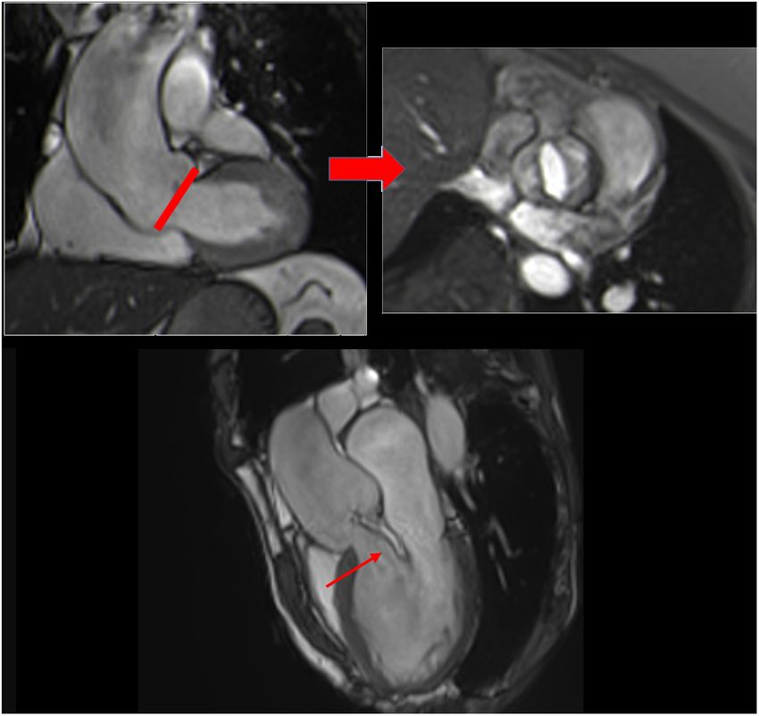
Vermes et al.                                                                                               Cardiovascular Magnetic Resonance in Valvular Regurgitation

TABLE 5 | Relevant studies on validation of CMR parameters in MR and AR.

First author, year (Ref #)       Number of                       Reference method                          Optimal CMR parameters cutoff for concordance with
                                  patients                                                                 echo or for severe regurgitation (sensibility/specificity)

Mitral regurgitation
Polte et al. (47)                    40                 Indication for surgery based on echo                                 RF > 41% (96%/80%)
                                                                  recommendation                                            MRvol > 64 ml (96%/87%)
Le Goffic et al. (45)                34                  TTE based on integrative approach                                   RF > 35% (86%/100%)
Gelfand et al. (44)                  83                            TTE (qualitative)                                              RF > 42% (NA)
Aortic regurgitation
Polte et al. (47)                    38                 Indication for surgery based on echo                                 RF > 30% (87%/67%)
                                                                  recommendation                                            ARvol > 40 ml (87%/73%)
Gabriel et al. (62)                  107                     TTE based on ASE guidelines                                          RF ≥ 30% (NA)

CMR, cardiac magnetic resonance; MR, mitral regurgitation; AR, aortic regurgitation; RF, regurgitant fraction; MRvol, mitral regurgitant volume; ARvol, aortic regurgitant
volume; TTE, transthoracic Echocardiography; ASE, American Society of Echocardiography; NA, not available.

dysfunction (LVESD ≥ 40 mm and/or LVEF ≤ 60%). CMR                                         (left, right, and non-coronary). Contiguous cine images 3 mm
is a “second tool,” indicated when various echocardiographic                               above and below this slice are required (51). During diastole,
parameters are inconsistent (Table 4). Based on the relevant                               three normal leaflets form a three-pointed star “Mercedes-Benz
publications discussed in this review illustrating the accuracy of                         emblem,” but a bicuspid aortic valve can be identified (Figure 4).
CMR to assess LV volume, MR severity, and predict LV reverse                                   Assessing the aortic diameter is crucial for determining
remodeling after correction (Tables 5, 6), this imaging modality                           AR etiology and follow-up. The diameter of the ascending
should be considered not only for patients with severe MR by                               aorta can be measured using non-contrast-enhanced MR
echocardiography to confirm the severity and help guide surgical                           angiography (NCE-MRA) with a respiratory navigator or breath-
decision-making but also for patients for whom the severity of                             hold contrast-enhanced MRA (CE-MRA). Measurements are
MR is unclear by echocardiography. In light of studies discussed                           performed at end-diastole using the internal diameter of the
above, MR is very likely to be severe if the mitral RF is ≥ 40%                            aorta at the level of the sinus, the sinotubular junction, and
of CMR, even if large trials with a validation cohort to define the                        the level of the pulmonary trunk on the ascending aorta (52)
best CMR cutoff are needed.                                                                or at the level of maximum dilatation. Ideally, measurement
                                                                                           should be performed in 3D using a double oblique angulation
                                                                                           perpendicular to the vessel axis. Measurement in standard 2D
AORTIC REGURGITATION                                                                       axial view should be avoided.
Aortic regurgitation (AR) is the second most frequently                                    2D Cine Imaging for Regurgitant Jet Visualization
occurring type of regurgitation after MR (49). As for                                      Aortic regurgitant jets can be visualized on cine images using
MR, echocardiography is the first-line modality for AR                                     long-axis LVOT views (3-chamber and coronal oblique LVOT) as
assessment, and CMR is recommended as a complementary                                      a dark jet due to a signal void from turbulence back flowing into
technique when image quality is suboptimal or in situations                                the LVOT during diastole (Figure 4). The location and direction
of discordance results (3, 4). However, dynamic or multiple                                of the jet can provide additional information about the AR
jets, bicuspid valve or aortic valve calcifications, a non-                                etiology. For example, a central jet can be more highly related to
hemispheric shape, or multivalvular disease are among the                                  aortic dilatation and an eccentric jet more to a cusp abnormality
echocardiographic limitations that can affect the accuracy of                              (prolapse) (Figure 4). However, the appearance of the signal void
AR assessment. Below, we describe how CMR can help in                                      is dependent on the hemodynamics, echo time, and flip angle
defining the etiology, assessing the consequences of AR on                                 (53), and does not directly reflect the aortic regurgitant volume
LV remodeling, grading AR severity, and defining the timing                                (ARvol); therefore, a more reliable assessment is required.
for AR correction.
                                                                                           Cardiovascular Magnetic Resonance for
Cardiovascular Magnetic Resonance for
                                                                                           Quantifying Aortic Regurgitation
Assessing the Cause of Aortic                                                              Various CMR methods used in clinical practice to assess AR
Regurgitation                                                                              severity are summarized in Table 7.
2D Cine Imaging for LV Volume and Aortic Valve and
Root Morphology                                                                            Direct Method: 2D Cine Phase-Contrast Velocity
With a high contrast between the valve leaflets and blood pool,                            Mapping
together with a high signal-to-noise ratio, SSFP imaging can be                            Cardiovascular magnetic resonance has the ability to measure
used to visualize aortic valve anatomy in any plane, irrespective                          flow velocity and direction over time. Unlike MR (with multiple,
of cardiac anatomy (50). A perpendicular plane through the                                 turbulent, irregularly shaped jets), direct flow quantification of
coronal oblique LVOT view allows the visualization of each cusp                            AR is feasible. The most commonly used CMR method to

Frontiers in Cardiovascular Medicine | www.frontiersin.org                             8                                            July 2022 | Volume 9 | Article 881141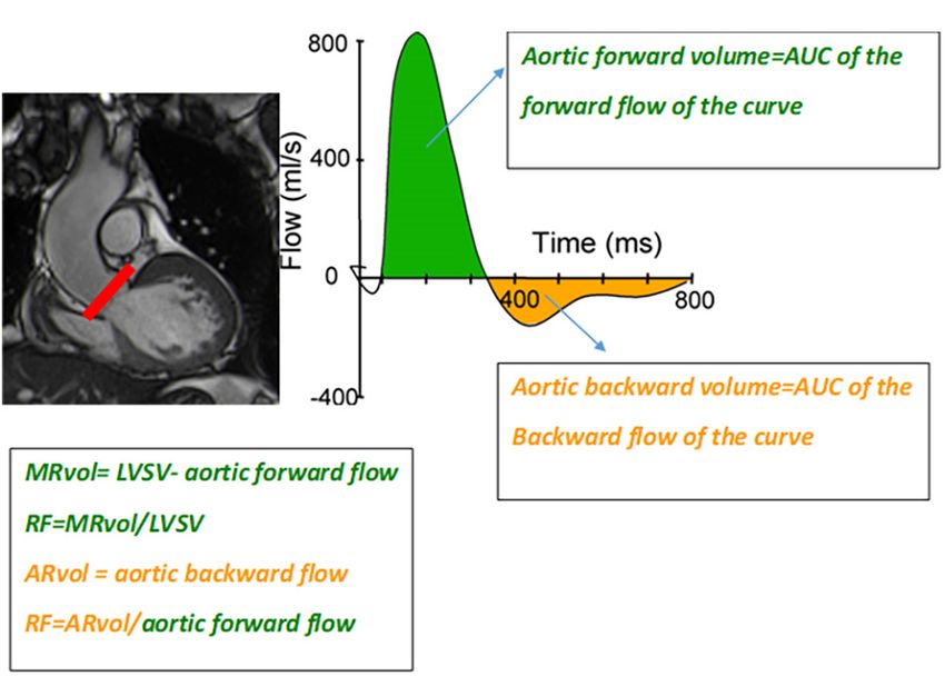
Vermes et al.                                                                                                     Cardiovascular Magnetic Resonance in Valvular Regurgitation

TABLE 6 | Relevant studies assessing prediction of outcome of severe MR and AR assessed by CMR.

First author, year (Ref #)          Number of                 Follow up            Primary outcome                             Optimal CMR parameters cutoff for
                                     patients                                                                                  primary endpoint (sensibility/specificity)

Mitral regurgitation
Cavalcante et al. (31)           578 (ischemic MR)             Median at           All-cause mortality or heart                RF ≥ 35% (NA/84%)
                                                               4.9 years           transplantation at 1 year
Myerson et al. (46)              109 (organic MR)                Mean              Development of indications for              RF > 40% (76%/74%)
                                                            2.5 ± 1.9 years        surgery                                     MR vol > 55 ml (72%/87%)
Aortic regurgitation
Faber et al. (69)                        66                Median 5.1 years        Prediction of valve surgery                 RF > 32% (NA)
Harris et al. (64)                       29                      Mean              Need for valve surgery and heart            RF ≥ 37% (100%/75%)
                                                            4.4 ± 1.5 years        failure                                     ARvol ≥ 50 ml (100%/NA)
Myerson et al. (68)                      113                     Mean              Development of indications for              RF > 33% (85%/92%)
                                                            2.6 ± 2.1 years        surgery

CMR, cardiac magnetic resonance; MR, mitral regurgitation; AR, aortic regurgitation; RF, regurgitant fraction; MRvol, mitral regurgitant volume; ARvol, aortic regurgitant
volume; NA, not available.

quantitatively assess the severity of AR is a direct measurement                           valid in cases of coexisting valvular lesions, and post-processing
using through-plane velocity mapping performed just above the                              is rapid.
aortic valve (2D phase-contrast imaging: 2D-PC) in a plane
perpendicular to the direction of blood flow (21). A dedicated                             Technical Considerations
post-processing analyzes the blood flow through the aortic plane                           An incorrect velocity encoding setting (below the peak velocity in
generating a flow curve, which allows calculation of the AFF,                              the vessel) results in aliasing and an underestimated peak velocity
aortic regurgitant volume (ARvol = area under the backward                                 (56). We recommend starting with an encoding velocity slightly
flow curve during the diastolic phase of the cardiac cycle), and                           below the peak velocity of AR found in echocardiography and
RF (ARvol/AFF) (54) (Figures 3, 5).                                                        repeating the flow measurement with a higher encoding velocity
                                                                                           until it corrects the aliased data set. In the presence of associated,
Advantages                                                                                 aortic stenosis, the peak systolic velocity can be underestimated
This method is highly reproducible and accurate, especially for                            due to lower temporal resolution relative to echocardiography,
laminar regurgitant flow, with an imaging plane perpendicular                              leading to an underestimation of the AFF.
to the aorta (55). It requires only a single breath-hold, is
                                                                                           Additional Parameter
                                                                                           According to recent CMR guidelines (12), velocity-encoded
                                                                                           imaging in a through-plane perpendicular to the descending
                                                                                           aorta is recommended to explore for diastolic flow reversal,
                                                                                           similar to echocardiography (35). In, Bolen et al. (57) found
                                                                                           that HFR (≥ 10 ml/s) is highly sensitive (100%) and specific
                                                                                           (93%) in predicting severe AR. More recently, a study by
                                                                                           Kammerlander et al. showed an association between HFR and
                                                                                           increased cardiovascular events (58).

                                                                                           Indirect Method n◦ 1: 2D Cine Phase-Contrast
                                                                                           Velocity Mapping
                                                                                           ARvol can be measured indirectly as the difference between
                                                                                           the AFF and PFF in the absence of other regurgitation or
                                                                                           intracardiac shunts.

                                                                                           Advantages
                                                                                           This method requires only 2 single breath-holds and allows for
                                                                                           rapid post-processing. If there are concerns about the direct
                                                                                           method, this method can serve for internal validation.
  FIGURE 4 | Coronal oblique bSSFP sequence through the LVOT (A); red line
  indicates image plane for direct visualization of the aortic cusps (B) showing a         Technical Considerations
  true bicuspid aortic valve (without raphe). Eccentric aortic jet (flow void in the
  left ventricle, red arrow) visualized in the coronal LVOT view (C).
                                                                                           This method is not valid in situations of pulmonary regurgitation
                                                                                           or intracardiac shunts.

Frontiers in Cardiovascular Medicine | www.frontiersin.org                             9                                                 July 2022 | Volume 9 | Article 881141
Vermes et al. Cardiovascular Magnetic Resonance in Valvular Regurgitation
TABLE 7 | Various CMR methods used in clinical practice to quantify AR, with their advantages and limitations.
CMR method Direct recommended Indirect
1 2
Phase contrast mapping Volumetric
CMR sequences required Phase-contrast velocity mapping above the aortic Phase-contrast velocity mapping of SSFP short-axis images for
valve the proximal aorta and the main LVSV and RVSV
Phase-contrast velocity mapping in the descending pulmonary artery for AFF and PFF
aorta for HFR
Formula Direct quantification ARvol (ml) = area under the ARvol = AFF—PFF ARvol = LVSV—RVSV
diastolic flow curve
RF (%) = ARvol/AFF
Advantages Reproducible, particularly for laminar jets Rapid Rapid and simple
Valid for multiple regurgitation
Fast post-processing
Limitations Less accurate for no laminar jets and in cases of Inaccurate for multiple Inaccurate for multiple
associated aortic stenosis regurgitations regurgitations
Careful placement 5 mm above the aortic valve Careful perpendicular slice selection Careful basal slice selection for
perpendicular to the jet is necessary Phase-offset errors LV and RV
Phase-offset errors Ensure correct maximal velocity
Ensure correct maximal velocity encoding encoding
CMR, cardiac magnetic resonance; SSFP, steady state free precession; LVSV, left ventricular stroke volume; RVSV, right ventricular stroke volume; AFF, aortic forward
flow; RF, regurgitant fraction; PFF, pulmonary forward flow; ARvol, aortic regurgitant volume; HFR, holodiastolic flow reversal.
FIGURE 5 | Example of the direct quantification of aortic regurgitation by phase-contrast imaging. Perpendicular line (red) above the aortic valve on LVOT view
indicating the slice position for phase-contrast velocity mapping (A) generating phase images (B) and flow curves images (C). The aortic regurgitant volume (ARvol)
is represented by the area under the diastolic flow curve (blue hatch lines).
Indirect Method n◦ 2: Volumetric Method: 2D Cine Technical considerations are described in the MR
Imaging chapter.
Similar to MR, the volumetric method can be used to quantify
ARvol using only bSSFP sequences in the short axis. ARvol is
4D Flow, Late Gadolinium Enhancement Imaging, and
expressed as the difference between LVSV and RVSV in the T1 Mapping
absence of other types of valvular regurgitation (54). 4D flow with its ability for measuring eccentric, non-laminar flow
in any orientation in space could be in the near future appropriate
Advantages for AR quantification (59).
This method is simple and does not require specific acquisitions T1 mapping, with its potential to assess cellular and
because bSSFP cine sequences are performed during every extracellular compartments, could be of interest in AR as a
CMR examination. prognostic marker for clinical outcomes (60).
Frontiers in Cardiovascular Medicine | www.frontiersin.org 10 July 2022 | Volume 9 | Article 881141Vermes et al. Cardiovascular Magnetic Resonance in Valvular Regurgitation
surgery (68). These CMR thresholds are much lower than
established echocardiographic guideline criteria (Rvol ≥ 60 ml
and RF ≥ 50%) (35, 36) and could explain the frequent mismatch
between moderate to severe AR by 2D echocardiography and
mild to moderate AR by CMR (65).
In the absence of a reference method, it is difficult to determine
which imaging modality can better predict the need for aortic
correction, particularly for asymptomatic patients. However,
several studies have reported a better prediction with CMR (64,
65, 69).
Main Message for the Clinician
Echocardiography is the first-choice tool to grade AR based
on qualitative, semiqualitative, and quantitative criteria (4).
Current class I surgical recommendations for severe AR are
based on symptoms or for asymptomatic patients with LV
FIGURE 6 | Cardiovascular magnetic resonance planes used for evaluation of dilatation or dysfunction (LVESD ≥ 50 mm or > 25 ml/m2
the right ventricle. Short axis view (A), 3-chamber view (B) and RVOT (C). or LVEF ≤ 50%). CMR is a “second tool,” indicated when
Example of tricuspid annular measurement in the 4-chamber view various echocardiographic parameters are inconsistent (Table 4).
(red line) (D). Although there is a lack of large, prospective, comparative studies,
the publications discussed above show that CMR could improve
the diagnosis and surgical timing of patients with AR relative
Comparison of Cardiovascular Magnetic to echocardiography (Tables 5, 6). CMR should be considered
Resonance and Echocardiography on for patients with severe AR by echocardiography to confirm the
severity and guide surgical decision-making. From a practical
ARvol and Aortic Regurgitation Severity
point of view and in light of studies discussed above, AR is very
Modest Correlation With Echocardiography,
likely to be significant if aortic RF ≥ 30%, even if large trials to
Particularly for Eccentric Jets define specific CMR thresholds are needed.
Unlike MR, there is a paucity of large and prospective studies
comparing these two modalities. In all studies, the direct CMR
method was used and thresholds to define severe AR were the
TRICUSPID REGURGITATION
same as those used for echocardiography: Rvol ≥ 60 ml and
RF ≥ 50% according to recommendations (35, 36, 61) (Table 3). Unlike MR and AR, less data are available about the prevalence
In most studies, the correlation between these two modalities was of moderate to severe tricuspid regurgitation (TR), which
modest (62–65), particularly in eccentric jets (66). is probably underestimated. In a recent study performed
2D PISA is limited to the alignment of the flow and by in the United States (Olmsted County, Minnesota), the age
the need for computation and geometric assumptions. 3D and sex-related prevalence of TR (diagnosed by Doppler
echocardiography, not restricted by any imaging plane, could echocardiography) was 0.55%, higher among women, and
overcome these limitations and provide a better correlation with increased with age (4.4% among women aged ≥ 75 years and
CMR, especially in eccentric (67). 3.1% among men) (70). Moderate to severe TR is associated
with adverse outcomes, independent of LVEF or pulmonary
Cardiovascular Magnetic Resonance for Grading artery pressure (71), probably due to the development of right-
Severe Aortic Regurgitation and for the Timing of heart failure.
Surgery Causes of TR can classically be divided into two types:
As previously noted, ARvol and/or RF measured by CMR are
categorized based on established echocardiographic guidelines - Primary TR due to a primary lesion of the tricuspid valve
(35), defined as: caused by congenital or acquired disease.
- Secondary (or functional) TR, with a structurally normal
- Mild to moderate AR: 30 mL ≤ ARvol < 45 ml, valve, more commonly associated with left-sided heart
- Moderate to severe AR: 45 mL ≤ ARvol < 60 ml, disease, pulmonary hypertension, RV dysfunction,
- Severe AR: ARvol ≥ 60 ml; RF ≥ 50% dilatation, or without a detectable cause and thus called
isolated (or idiopathic) TR, but often associated with
As for MR, there is a paucity of data on specific CMR atrial fibrillation.
thresholds for ARvol and RF, with a lack of large trials
with a validation cohort. ARvol > 40 ml and RF ≥ 30% Whether isolated TR or not, understanding TR, its
have been proposed to define severe AR with CMR that consequences, and accurately assessing its severity is crucial,
correlates best with echocardiography (47, 62) or for identifying particularly since the emergence of transcatheter interventions,
patients who developed symptoms or needed aortic correction which have moved the tricuspid valve from the shadows into
Frontiers in Cardiovascular Medicine | www.frontiersin.org 11 July 2022 | Volume 9 | Article 881141Vermes et al. Cardiovascular Magnetic Resonance in Valvular Regurgitation
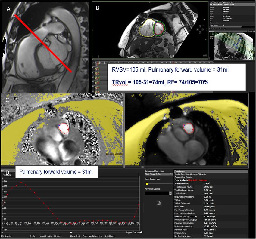
FIGURE 7 | Example of TR assessment using indirect method n◦ 1. RVOT bSSFP images showing slice position in the pulmonary artery (red line) for phase-contrast
velocity mapping to obtain pulmonary forward volume (A). Assessment of right ventricular stroke volume (RVSV) from short-axis cine SSFP images (B). Phase and
magnitude images with delineation of the pulmonary artery (C) allowing flow curves (D). This patient has a severe TR with a tricuspid regurgitant volume (TRvol) of
74 ml and a regurgitant fraction (RF) of 70%.
light. Echocardiography is the preferred and recommended Cardiovascular Magnetic Resonance for
initial imaging technique to assess TR based on an integrative Assessing Tricuspid Regurgitation
approach, including qualitative and quantitative criteria. The
2D Cine Imaging for RV Volume and Tricuspid Valve
use of conventional quantitative parameters (vena contracta
Morphology
and PISA) is based on geometrical assumptions (circular jet
Due to the complex interplay between TR and the RV (TR can
and flat orifice) extrapolated from the MV (72). However, due
cause RV dilatation and vice versa), assessing TR requires the
to the complex non-planar geometry of the tricuspid annulus,
accurate assessment of RV structure and function. CMR is the
these assumptions may not be valid for the tricuspid valve
method of reference to assess RV volume, function, and mass,
(73). Although there are few studies that have assessed TR by
without geometric assumptions (74).
CMR, this modality has the unique advantage of non-invasively
RV structure can be studied using
measuring the flow through vessels and to be the reference
standard modality for RV volume and function assessment, - a stack of contiguous bSSFP short-axis images, the same as
important parameters to take into account in TR. In particular, those used for LV, with careful placement of the basal slice
CMR allows a more precise evaluation of RV volumes and on the myocardial side of the RV (75). In cases of complex
EF than the echo. congenital disease, dedicated axial orientation on the RV
Frontiers in Cardiovascular Medicine | www.frontiersin.org 12 July 2022 | Volume 9 | Article 881141Vermes et al. Cardiovascular Magnetic Resonance in Valvular Regurgitation
(slice thickness between 5 and 6 mm without interval gaps) This is the most widely used CMR method and is valid in
is recommended (36). the absence of intra-ventricular shunts (79). The advantages and
- Long-axis cine images, including three specialized views for technical considerations are similar to those for MR and have
the RV: RV out-flow tract (RVOT) and RV vertical long axis already been discussed in the MR chapter.
(3-chamber) (Figure 6).
Indirect Method n◦ 2: Volumetric Method: 2D Cine
The RV regional wall-motion abnormalities can be Imaging
determined visually. Recently, feature tracking, similar to As for MR and AR, the volumetric method can be used to
speckle tracking in echocardiography, has emerged to more quantify the TRvol, expressed as the difference between RVSV
accurately detect changes in wall motion and earlier ventricular and LVSV in the absence of other forms of valvular regurgitation.
dysfunction, which may be an independent predictor of mortality The advantages and technical considerations are similar to
in severe TR (76). those for left-sided valvular regurgitation and have already been
Tricuspid anatomy can be assessed by short- and long-axis discussed in the MR and AR chapter.
views, as already described, allowing simultaneous visualization These two indirect methods can be performed in addition
of the three leaflets (septal, anterior, and posterior) in the short- to echocardiography to increase the confidence in the
axis view. Leaflets can be described as prolapsed, restricted, assessment of TR severity.
thickened, or with tenting (77). Tenting height and area may
be measured and considered abnormal if they are > 7 mm Direct Method: 2D Cine Phase-Contrast Velocity
(3 mm/m2 ) and 1.1 cm2 (> 0.5 cm2 /m2 ), respectively (14). Mapping
However, tricuspid leaflets are thin, as well as the chordae 2D PC imaging also allows the direct measurement of TR
and papillary muscles, and can be difficult to visualize. regurgitant flow through the tricuspid valve. As for MR, this
Therefore, echocardiography remains the cornerstone for TR technique is limited by significant motion and the saddle-
anatomy assessment. shape of the tricuspid annulus and is not commonly used in
The tricuspid annular diameter, a relevant parameter included clinical practice.
in the guidelines for tricuspid intervention, can be measured in
the 4-chamber view in early diastole, with the upper limits of 4D Flow
the normal value recently reported to be 43 mm (22 mm/m2 ) Data concerning 4D flow in TR are very scarce. One recent study
(78) (Figure 6). It is important to note that for patients with have suggested high concordance of 4D flow with standard 2D
pacemakers or defibrillator leads, generated metallic artifacts PC (indirect method) (80).
decrease the accuracy of CMR for the assessment of RV
volume structure and tricuspid anatomy, as well as for patients
Late Gadolinium Enhancement Imaging and T1
with arrhythmias. Mapping
Unlike LV, tissue characterization (LGE and T1 mapping)
2D Cine Imaging for Regurgitant Jet Visualization of the RV is more complex due to the thin RV wall
The qualitative assessment of TR can be performed using bSSPF (thickness 1–3 mm), which creates partial volume effects.
imaging (2 specialized long-axis and 4-chamber views). TR is Transmural RV enhancement can be visualized, but non-
visualized as a dark jet created by flow turbulence generating transmural lesions are very challenging to visualize. LGE
spin dephasing back flowing into the right atrium. As for patterns may help to understand the underlying mechanism
MR and AR, this is a gross assessment and is not reliable of TR, as it allows to detect ischemic patterns associated
on its own. In the presence of a turbulent jet, regurgitant with pulmonary hypertension, or fibrosis associated with
flow appears as a low signal that can mask the visualization cardiomyopathies such as arrhythmogenic right ventricular
of the leaflets. cardiomyopathy. T1 assessment in TR has not yet been
reported.
Cardiovascular Magnetic Resonance for
Comparison of Cardiovascular Magnetic
Quantifying Tricuspid Regurgitation
Quantitative assessment of TR by CMR can be performed using
Resonance and Echocardiography on
two indirect and one direct method. TRvol and Tricuspid Regurgitation
Severity
Indirect Method n◦ 1: 2D Cine Imaging and 2D Cine Calculation of TRvol by echocardiography is more challenging
Phase-Contrast Velocity Mapping than for MR and AR, and the current guidelines place CMR-
Similar to MR, the regurgitant volume of TR (TRvol) can derived TRvol as an alternative (4) (Table 4). Unlike left-sided
be measured indirectly as the difference between RVSV regurgitant valvular lesions, quantitative assessment of TR by
and PFF (or AFF in the absence of AR) using 2D cine CMR is largely unexplored, and studies comparing these two
(for RVSV) and retrospective acquisition with velocity- imaging modalities are scarce (80, 81).
encoded phase-contrast sequence in a through–plane on the The largest and more recent study to compare
pulmonary valve (or the aortic valve), 5 mm above the valve echocardiography (integrative approach) and CMR (2D PC
(12) (Figure 7). indirect method) has used the same thresholds defined in
Frontiers in Cardiovascular Medicine | www.frontiersin.org 13 July 2022 | Volume 9 | Article 881141Vermes et al. Cardiovascular Magnetic Resonance in Valvular Regurgitation
the ASE guidelines (36): TRvol mild: < 30 ml, moderate: assessment. Since CMR can provide unrestricted image planes
30–44 ml, and severe: ≥ 45 ml. Modest but significant for PV anatomy and RV function, it plays a crucial role in
correlations were found between quantitative echocardiographic PR assessment, especially for patients with repaired tetralogy
measurements (vena contracta, effective regurgitant orifice of Fallot (rTOF).
area, and PISA-derived TRvol) and CMR (from 0.3 to
0.49) (81). Cardiovascular Magnetic Resonance for
Cardiovascular Magnetic Resonance for Grading Assessing the Cause of Pulmonary
Severe Tricuspid Regurgitation and for the Timing of Regurgitation
Surgery 2D Cine Imaging for Right Ventricle Volume and
As for MR and AR, using echocardiographic thresholds is Pulmonary Valve Morphology
inaccurate and extrapolating data from MR is too simplistic The assessment of RV structure and function is essential for
given the differences in anatomy, regurgitant orifice and shape, determining the prognosis and planning surgical/percutaneous
and hemodynamics between the two valves. A recent study by intervention for patients with rTOF (84).
Zhan et al. proposed a different approach by defining optimal Contrary to echocardiography, CMR provides good
CMR thresholds for TRvol and RF for a low, intermediate, visualization of RVOT anatomy using a stack of SSFP cine
and high risk of mortality (81). The study by Zhan et al. images in the short and long axes (RVOT and 3-chamber). The
has the merit of providing CMR thresholds for mortality risk, anatomy of the pulmonary valve, with three semi lunar leaflets
independent of the strong confounding effect of RV dilatation (anterior, left and right), can be visualized in through-plane
and dysfunction. Due to the lack of randomized studies using images on the valve (Figure 8).
these CMR thresholds, the benefit of TR correction (surgery
or percutaneous device) is still unknown. Current guidelines, 2D Cine Imaging for Regurgitant Jet Visualization
mostly based on expert opinion, recommend intervention PR can be seen as a dark jet in cases of flow turbulence in diastole,
for patients with severe TR and concomitant left-sided valve extending into the RVOT. However, in cases of laminar flow with
surgery (class I) (4). In the setting of severe isolated TR, a wide jet, there is almost no turbulence, making the jet nearly
surgery is recommended for symptomatic patients without invisible. As for other forms of valvular regurgitation, it is a gross
RV dysfunction (class I) or asymptomatic patients with RV assessment and is not alone reliable.
dilatation or RV dysfunction (class IIa), without defined
thresholds. However, CMR could contribute to defining these Cardiovascular Magnetic Resonance for
thresholds. Indeed, in a cohort of 76 patients undergoing
tricuspid surgery, Park et al. found that CMR-based RVEF was
Quantifying Pulmonary Regurgitation
an independent predictor for cardiac mortality and major post- Direct Method: Phase-Contrast Velocity Mapping
operative cardiac events, with a cutoff of 46% (82). A recent Similar to AR, direct measurement of pulmonary backward
observational prospective study by Rodriguez et al., on 43 flow is feasible using 2D-PC with retrospective acquisition in a
patients with severe echocardiography-based TR undergoing through-plane acquisition on the pulmonary valve, 5 mm to 1 cm
tricuspid annuloplasty suggested that RVEDV indexed by CMR is above the level of the valve (12) (Figure 9).
the best independent predictor of overall mortality at follow-up, This is the most widely used method and the current
with a cut-off of 104 ml/m2 , associated with higher cardiovascular reference standard technique (85). In patients with rTOF
mortality (83). or dilated pulmonary artery, pulmonary flow can be non-
Aside from further pending CMR studies on specific laminar and turbulent, leading to a possible underestimation
thresholds, those defined by Zhan et al. (81) could be used in of pulmonary regurgitant volume (PRvol). In this case, the
clinical practice and added to the established relevant value of volumetric method or 2D-PC acquisition perpendicular to the
CMR for RV volume and function. right and left pulmonary arteries can be performed to confirm
the findings.
PULMONARY REGURGITATION Indirect Method n◦ 1: Volumetric Method: 2D Cine
Imaging
Trace to mild pulmonary regurgitation (PR) is common in the As for other forms of valvular regurgitation, the volumetric
general population, with little clinical significance. By contrast, method can be used to quantify PRvol by comparing
significant PR is uncommon and usually related to either RVSV and LVSV on bSSFP short-axis cine images
congenital heart disease (primary PR with abnormal leaflets) (Figure 9), in the absence of other regurgitant disease or
or pulmonary hypertension [secondary PR, normal pulmonary intra-cardiac shunt.
valve (PV) leaflets]. Echocardiography is the initial imaging
modality to assess PR. However, the position of the valve behind Indirect Method n◦ 2: 2D Phase-Contrast Velocity
the sternum, which makes visualizing the PV and RVOT difficult Mapping
and thus the derived measurement for PR quantification, does PRvol can be measured indirectly as the difference between PFF
not position echocardiography as the preferred method for PR and AFF in the absence of tricuspid regurgitation (Figure 9).
Frontiers in Cardiovascular Medicine | www.frontiersin.org 14 July 2022 | Volume 9 | Article 881141You can also read