Review Article Acupuncture for Parkinson's Disease: Efficacy Evaluation and Mechanisms in the Dopaminergic Neural Circuit - Hindawi ...
←
→
Page content transcription
If your browser does not render page correctly, please read the page content below
Hindawi
Neural Plasticity
Volume 2021, Article ID 9926445, 23 pages
https://doi.org/10.1155/2021/9926445

Review Article
Acupuncture for Parkinson’s Disease: Efficacy Evaluation and
Mechanisms in the Dopaminergic Neural Circuit

 Yadan Zhao,1 Zichen Zhang,1 Siru Qin ,1 Wen Fan,2 Wei Li,1 Jingyi Liu,1 Songtao Wang ,1
 Zhifang Xu ,1,3 and Meidan Zhao 1,3
 1
 Research Center of Experimental Acupuncture Science, Tianjin University of Traditional Chinese Medicine, Tianjin 301617, China
 2
 Suzuka University of Medical Science, Suzuka 5100293, Japan
 3
 School of Acupuncture & Moxibustion and Tuina, Tianjin University of Traditional Chinese Medicine, Tianjin 301617, China

 Correspondence should be addressed to Zhifang Xu; xuzhifangmsn@hotmail.com and Meidan Zhao; zhaomeidan2012@163.com

 Received 25 March 2021; Revised 10 May 2021; Accepted 27 May 2021; Published 16 June 2021

 Academic Editor: Feng Zhang

 Copyright © 2021 Yadan Zhao et al. This is an open access article distributed under the Creative Commons Attribution License,
 which permits unrestricted use, distribution, and reproduction in any medium, provided the original work is properly cited.

 Parkinson’s disease (PD) is a chronic and progressive neurodegenerative disease caused by degeneration of dopaminergic neurons
 in the substantia nigra. Existing pharmaceutical treatments offer alleviation of symptoms but cannot delay disease progression and
 are often associated with significant side effects. Clinical studies have demonstrated that acupuncture may be beneficial for PD
 treatment, particularly in terms of ameliorating PD symptoms when combined with anti-PD medication, reducing the required
 dose of medication and associated side effects. During early stages of PD, acupuncture may even be used to replace medication.
 It has also been found that acupuncture can protect dopaminergic neurons from degeneration via antioxidative stress, anti-
 inflammatory, and antiapoptotic pathways as well as modulating the neurotransmitter balance in the basal ganglia circuit. Here,
 we review current studies and reflect on the potential of acupuncture as a novel and effective treatment strategy for PD. We
 found that particularly during the early stages, acupuncture may reduce neurodegeneration of dopaminergic neurons and
 regulate the balance of the dopaminergic circuit, thus delaying the progression of the disease. The benefits of acupuncture will
 need to be further verified through basic and clinical studies.

1. Introduction demands on patients and their families, incurring over
 US$14 billion annually [4].
Parkinson’s disease (PD) is a chronic and progressive neuro- Pathologically, PD is characterized by the loss and degen-
degenerative disease typically affecting middle-aged and eration of dopaminergic neurons in the substantia nigra
elderly people. The key clinical manifestation is the dysfunc- (SN), α-synuclein (α-syn) deposition, and Lewy body forma-
tion of voluntary motor regulation, including bradykinesia, tion [5]. Current treatment options for PD include medica-
resting tremor, myotonia, and postural instability [1]. In tion, surgery, and exercise therapy, although medication is
addition to motor dysfunction, nonmotor symptoms may the first choice for PD, with the most commonly prescribed
arise during progression of PD, including cognitive dysfunc- drugs being dopaminergic and anticholinergic agents [2].
tion, emotional disorders such as apathy, depression, anxiety, These have been shown to control the initial symptoms of
and hallucinations, and autonomic nervous dysfunction, patients rapidly and effectively [6], but after a so-called “hon-
such as constipation, hyposmia, and sleep disorders [2]. PD eymoon period” of treatment, the effectiveness often
has a prevalence of 0.3% and is thus the second most com- decreases and medication may no longer be beneficial for
mon neurodegenerative disease after Alzheimer’s disease patients with advanced PD. The fundamental reason for this
[3]. Tragically, the physical status of PD patients declines phenomenon is that existing anti-PD drugs offer only symp-
even when treated, often deteriorating to severe disability tomatic treatment but cannot delay disease progression.
and death. Treatment costs impose enormous financial Moreover, anti-PD drugs can also lead to considerable side
2 Neural Plasticity

effects which can aggravate PD symptoms, and some of teria. Any disagreement was resolved by discussion between
which still lack effective treatment [7]. Many patients with the authors.
PD have therefore turned to complementary and alternative
therapies which may be used in the early stages of PD or prior 3. Clinical Efficacy of Acupuncture in
to the use of levodopa (L-DOPA) to relieve symptoms and Parkinson’s Disease Patients
delay PD progression, with the potential expectation that
these therapies may also reverse or delay neuronal degenera- Acupuncture is commonly and globally used as both an adju-
tion [8]. vant therapy and monotherapy for PD treatment [8]. Com-
 Acupuncture is an economical therapy in traditional Chi- prehensive evidence for the efficacy and safety of
nese medicine originating from China where it has been acupuncture in PD has been provided by a total of twelve sys-
developed for more than 4,000 years. It can promote health tematic reviews (SRs) or meta-analyses (MAs) assessing this
or treat diseases via various different techniques such as man- topic, involving 179 randomized controlled trials (RCTs)
ual acupuncture (MA), electroacupuncture (EA), moxibus- with a total of 11,717 participants [12]. These SRs/MAs have
tion, and acupressure on specific anatomical locations (i.e., compared the effects of acupuncture combined with medica-
acupoints) [9]. Numerous clinical trials have shown that acu- tion versus medication alone, demonstrating better efficacy at
puncture exhibits significant therapeutic benefits for PD symptom alleviation with the combination of acupuncture
patients, reducing both motor and nonmotor symptoms. with medication compared to medication alone [13]. The
Moreover, acupuncture may help to reduce the dose and fre- Unified Parkinson’s Disease Rating Scale (UPDRS) provides
quency of anti-PD drugs as well as alleviate their side effects a comprehensive, efficient, and flexible means of monitoring
[10]. The therapeutic mechanisms of acupuncture have also PD-related disabilities and impairments as well as treatment
been shown to involve a reduction of mitochondrial dysfunc- evaluation, assessing behavior and mood, activities of daily
tion, oxidative stress, protein aggregation, impaired autoph- living, motor functions, and therapeutic complications [14].
agy, and neuroinflammation [11]. In the present review, we Acupuncture combined with dopaminergic drugs was supe-
aim to address the clinical efficacy and potential mechanisms rior to medication only for improving the UPDRS score
of acupuncture in PD treatment, providing novel insights for [15]. Interestingly, when comparing acupuncture only with
the clinical application of acupuncture for the reversal the medication only, there is no significant difference in the
degeneration of dopaminergic neurons and delay of PD scores. Moreover, evidence-based medicine (EBM) studies
progression. have analyzed 13 parallel RCTs and found no difference in
 the efficacy of acupuncture compared with medication for
 PD treatment [10], suggesting that acupuncture may achieve
2. Methods an efficacy close to that of medication. The Webster Scale is
2.1. Search Strategy. We searched the PubMed database for applied to assess the clinical severity of PD, rating levels of
studies published between January 2000 and December akinesia, rigidity, upper limb swing, gait, tremor, facial
2020 containing the keywords “Acupuncture” and “Parkin- appearance, speech, and self-care [16]. Compared with med-
son’s disease”. Only studies in English language were ication only, acupuncture and medication combined have
included. This search identified relevant 183 articles. been shown to result in a greater Webster score reduction,
 indicating an alleviation of clinical severity [12], and com-
 pared with medication only, acupuncture alone similarly
2.2. Study Selection. Following the search engine selection of exhibited a positive effect for decreasing the Webster score
183 articles, a manual search was performed by screening the [10], suggesting that acupuncture alone may be able to effec-
reference list for articles that met our inclusion criteria based tively reduce the severity of PD. EBM reviews reveal that even
on the titles and abstracts. We excluded 43 articles due to with an optimized oral dose of L-DOPA, there is a risk for a
absence of an abstract, unavailability of the full text, or irrel- worsening of movement disorders, and a variety of anti-PD
evance to the subject, leaving 140 articles, including 43 basic drugs may also induce or exacerbate nonmotor symptoms
research articles, 38 clinical research articles, and 59 review [17]. Hence, treatment with L-DOPA and other anti-PD
articles or meta-analyses. The full texts of the 43 basic drugs should be delayed as much as possible in patients with
research and 38 clinical research articles were obtained and early-stage PD. If acupuncture achieves a similar therapeutic
assessed carefully. Out of the 38 clinical studies, 17 were effect to currently available medication, it may represent a
excluded as they described single case reports, editorials, new therapeutic option for PD, especially for early stages.
and uncompleted or uncontrolled trials, resulting in 21 clin- There is no evidence for any serious adverse reactions associ-
ical articles meeting our inclusion criteria. All 43 basic ated with acupuncture, suggesting that it is a relatively safe
research articles met our inclusion criteria. Thus, a total of choice for PD patients. Therefore, the current evidence indi-
64 articles were included in our review. A flow chart of this cates that acupuncture may enhance the therapeutic efficacy
search process is illustrated in Figure 1. of drugs and reduce the required dose of medication, thus
 also alleviating adverse effects.
2.3. Data Extraction. Two authors independently assessed Besides UPDRS and Webster scales, there are more tar-
the titles and abstracts of the retrieved articles and evaluated geted evaluation criteria corresponding to different PD
the full-text articles. The data from finally included articles symptoms [18]. The characteristics of the 15 clinical studies
were validated and extracted according to the predefined cri- included in our analysis are summarized in Table 1. Using
Neural Plasticity 3

 PubMed database
 Identification
 (n = 183)
 Excluded:
 Absence of abstract or full text;
 irrelevant articles (n = 43)
 Full-text articles assessed
 Screening
 (n = 140)
 Excluded:
 Review articles or meta-analyses (n = 59)
 Case reports, editorials, insufficient
 statistical data (n = 17)
 Full-text articles assessed
 Eligibility
 (n = 64)

 Basic researches (n = 43)
 Included
 Clinical trials (n = 21)

 Figure 1: Flow chart of the review processes.

wearable sensors, Lei et al. [19] found that EA had overall there are various types of acupuncture methods, such as
beneficial impact on different temporal and spatial parame- MA, EA, and bee venom acupuncture (BVA). A combination
ters of gait (gait speed and stride length) as well as dynamic of body and scalp acupoints is typically used in PD patients,
posture control (increase of midswing angular velocity and with scalp acupoints located in the occipital, frontal, and
decrease of double support). Kong et al. [20] evaluated parietal lobes as well as in motor and chorea-tremor con-
changes in the General Fatigue score of the Multidimensional trolled areas [33]. It has been shown that scalp acupoints can-
Fatigue Inventory after acupuncture treatment and found not only directly activate and adjust functional areas of the
that 5-week acupuncture treatment effectively alleviated corresponding cerebral cortex but also convey certain thera-
moderate and severe fatigue of PD patients. These results peutic advantages for movement disorders [34]: EA at scalp
are similar to reports of an RCT by Kluger et al. assessing acupoints has been found to reduce the loss of dopaminergic
the relief of PD with fatigue by acupuncture [21]. Using func- neurons in the SN and delay the degeneration of dopaminer-
tional magnetic resonance imaging (fMRI), Yu et al. [22] gic neurons, thereby alleviating clinical symptoms and post-
found enhanced connectivity in four junctions via fMRI after poning the progression of PD [35]. Therefore, scalp
acupuncture. There was a significant correlation between acupoints are an indispensable part of PD acupuncture treat-
changes in functional connectivity and the King’s Parkin- ment. In clinical PD acupuncture trials, the acupoints LR3
son’s Disease Pain Scale, indicating that acupuncture relieves (Taichong), GB34 (Yanglingquan), and ST36 (Zusanli) were
pain in PD patients via modulation of brain regions related to the primary body acupoints used, followed by LI4 (Hegu),
both sensory-discriminative and emotional aspects. In an LI11 (Quchi), and SI3 (Houxi) [2]. Previous reports showed
unblinded trial including 20 PD patients, no adverse reac- that GB34 significantly improves PD symptoms, in particular
tions were reported with an improvement of individual motor dysfunction, and attenuates dopaminergic neuronal
symptoms including tremor, walking, writing, bradykinesia, loss in animal models of PD [36]. Other acupoints such as
pain, sleep, depression, and anxiety in 85% of patients [23]. ST36 [37] and LR3 [38] displayed the same effects. Several
An RCT by Cristian et al. [24] showed a trend toward an preclinical randomized studies have demonstrated that acu-
improvement in the Parkinson’s Disease Questionnaire, daily puncture stimulation at GB34, GV20, and LR3 modulates
living activities, and UPDRS motor score following acupunc- PD-associated brain regions and results in an amelioration
ture, suggesting that acupuncture has a potential effect for of locomotor function closely associated with a reduction of
alleviation of both movement and nonmotor symptoms neuronal apoptosis in the striatum (ST) and SN [39].
including pain, depression, and autonomic symptoms. Using Thereby, these three acupoints have been recommended as
the Parkinson Disease Sleep Scale, Aroxa et al. [25] found the basic setting of acupuncture in clinical treatment of PD
that acupuncture was beneficial for the alleviation of sleep according to the World Health Organization standards [40].
disorders in PD patients. Furthermore, acupuncture may be In the clinical application of acupuncture, acupoints are
beneficial for improving oral cavity function as observed by also selected according to different PD symptoms and com-
increased mean tongue pressure and decreased average saliva plications [26, 27]. For instance, acupoints LR3, KI6 (Zhao-
swallowing reflex latency in PD patients after acupuncture hai), and KI7 (Fuliu) located in the lower leg are expected
[26]. to relieve pain; acupoints HT3 (Shaohai), LI7 (Wenliu), and
 Taken together, the clinical studies above have concluded SI3 are stimulated in severe tremor; moxibustion at acupoints
that acupuncture exhibits a comprehensive beneficial effect SP21 (Dabao) and LR14 (Qimen) or acupuncture at acu-
on symptoms associated with dyskinesia, psychology, behav- points DU16 (Fengfu), DU14 (Dazhui), and Extra (Baliao)
ior, emotion, and cognition of PD patients. Additionally, is used for obvious neck stiffness; acupoints DU4 (Mingmen)
4
 Table 1: The clinical efficacy of acupuncture in the patients with Parkinson’s disease.

 Clinical trial Clinical Acupuncture
Study Intervention Comparison Acupoints Outcomes
 design condition parameters
 GV20, GV14; bilateral Asymmetric biphasic
 Primary outcome: STHW↑,
 foot motor, sensory square wave pulse, 100 μS
Lei et al. Sham EA STFW↑, DTFW↑, and DTHW↑
 RCT PD patients EA (n = 10) area, balance area, LI4, pulse width, “deqi”, 100 Hz
(2016) [19] (n = 5) Secondary outcome:
 ST36, GB34, BL40, or 4 Hz, three times a
 UPRDS scores↓
 SP6, KI3, LR3 week, 3 weeks
 5-30 inches, no flicking or
 PD patients Sham rotation of needles after Primary outcome: MFI-GF↓
Kong et al. CV6; bilateral PC6,
 RCT with moderately Acupuncture (n = 20) acupuncture insertion, 20 minutes, twice Secondary outcome: MFI-total
(2017) [20] ST36, LI4, SP6, KI3
 severe fatigue (n = 20) a week, at least 3 days apart, score↓, UPDRS-motor↓
 5 weeks, 10 sessions
 0.5-1 cm, twisted three Primary outcome: MFIS (total,
 PD patients Sham GV20, GV24;
Kluger et al. times to the right, 30 physical, cognitive, psychosocial)↓
 RCT with moderate EA (n = 47) acupuncture bilateral LI10, HT7,
(2016) [21] minutes, every two Secondary outcomes: PDQ-39↓,
 or severe fatigue (n = 47) ST36, SP6
 weeks, 6weeks HADS (depression)↓, ESS↓, AES↓
 Primary outcome: KPPS↓,
 5-10 mm, “deqi” for 20 s, UPDRS (total)↓, enhanced
 Acupuncture
Yu et al. Comparison PD patients Medication GV20; bilateral 30 minutes, 1-3 sessions connectivity at the four
 +medication
(2019) [22] trials with pain (n = 7) 77.18, GB34 per week, separated by areas, a significant correlation
 (n = 9)
 at least 1 day, 8 weeks between functional
 connectivity changes and KPPS
 Intensity knob: 0; electric
 Idiopathic Bilateral LI4, GB34, exciter switch: 1; wave
Shulman et al. Controlled Sleep and rest category: SIP
 PD patients EA (n = 20) Self-control ST36, K3, KI7, SP6, form: intermittent;
(2002) [23] trial (sleep and rest)↓
 (stages I-III) SI3, TB5 twice a week, 10/16
 treatments
 GV20; bilateral K3,
Cristian et al. PD patients Sham acupuncture K10, BL60, L3, ST41, 4 Hz, 20 minutes, 5
 RCT EA (n = 7) NS
(2005) [24] (stages II-III) group (n = 7) ST36, GB34, bafeng sessions, 2 weeks
 points, MH6, LI4
 Idiopathic Acupuncture+ Bilateral LR3, SP6, LI4,
Aroxa et al. Medication 30 minutes, once a Sleep disorders
 RCT PD patients medication TE5, HT7, PC6, LI11,
(2016) [25] (n = 11) week, 8 weeks evaluation: PDSS↑
 (stages I-III) (n = 11) GB20
 Tongue function: mean
 Bilateral ST36, SP6, Inserted perpendicularly,
Fukuda et al. Controlled PD patients Acupuncture tongue pressure↑, mean
 Self-control LR3, LI4, LI11, GB20, 10-20 mm, 10-15 minutes,
(2016) [26] trial (stages I-III) (n = 13) swallowing reflex
 BL18, BL23 only once
 latency↓, saliva swallow↓
 Short-term: UPDRS (total
 MA, 5-30 mm,
 scores and subscores I, II,
 Acupuncture+ DU20; bilateral GB20, 15 minutes,
Chen et al. Comparison Medication III, and IV)↓, BDI-II
 PD patients medication LI11, LI10, LI4, GB31, twice a week, 18
(2015) [2] clinical trial (n = 20) score↓, WHO-QOL score↓
 (n = 20) ST32, GB34, GB38 (short-term)/36
 Long-term: UPDRS (total
 (long-term) weeks
 scores and subscores I, II,
 Neural Plasticity
Table 1: Continued.

 Clinical trial Clinical Acupuncture
Study Intervention Comparison Acupoints Outcomes
 design condition parameters
 III, and IV)↓, BDI-II
 score↓
 Neural Plasticity

 The Webster’s cumulative
 MA at Extra6, GB13, and
 scores↓, correlation
 Group 1: DU20; GB20, then electric
 analysis of ABP indices
 bilateral GB20, stimulator with continuous
 and the cumulative scores
 Controlled PD patients Acupuncture Blank control LI4, SP6, LR3; group 2: wave for 15 minutes;
Wang et al. (2002) [27] in Webster’s scale↓, the
 trial (stages I-III) (n = 29) (n = 14) Extra6; bilateral LI11, the rest acupoints: uniform
 latent period of V
 SJ5, GB34, ST36, reinforcing-reducing
 wave and the intermittent
 ST40, Extra21 method, 40 minutes; once
 periods of III-V peak and
 every other day, 3 months
 I-V peak↓
 UPDRS (total, 1, 2, 3,
 and4) score↓, BDI-II
 scores↓, neural responses
 (thalamus, cingulate
 Right GB34, LR3 (EA) EA: 15 minutes, 120 Hz,
 gyrus, anterior cingulate,
Yeo et al. Controlled Idiopathic EA+MA Bilateral LR3, LI11, twice a week, 8 weeks; MA:
 Self-control lingual gyrus,
(2018) [28] trial PD patients (n = 10) ST36, GB20, SP6, LI4, 3 to 15 mm, 15 minutes;
 parahippocampal gyrus,
 GB34 (MA) twice a week, 8 weeks
 lateral globus pallidus,
 mammillary body, middle
 temporal gyrus, cuneus,
 and fusiform gyrus)↑
 Primary outcome:
 BVA: injected diluted bee
 Sham treatment UPDRS (part II+III)
 venom 0.1 ml; EA: 1-1.5
 Idiopathic (n = 29)/ scores↓
Cho et al. Acupuncture+ Bilateral GB20, LI11, cm, rotated at 2 Hz for 10
 RCT PD patients conventional Secondary outcomes:
(2017) [29] BVA (n = 29) GB34, ST36, LR3 seconds to achieve “deqi”,
 (stages I-IV) treatment UPDRS (part II and III)
 15 minutes; twice a week,
 (n = 15) scores↓, gait speed↓, gait
 12 weeks
 number↑, PIGD↓
 UPDRS (I and II) scores↓,
 Acupuncture+ Bilateral ST42, SP3, 7-10 minutes, 8-14 Hz,
Eng et al. Controlled Idiopathic UPDRS (III) scores↑,
 Yin Tui Na Self-control LI11, LI15, LI20, ST7, once a week, 6 months,
(2006) [30] trial PD patients PDQ-39 total score↓,
 (n = 25) ST36 24 sessions
 BDI↓
 Primary outcome:
 BVA: injected diluted bee
 UPDRS (part II+III)
 venom 0.1 ml; EA: 1-1.5
 Acupuncture scores↓
Doo et al. Controlled Idiopathic Bilateral GB20, LI4, cm, rotated at 2 Hz for 10
 and BVA+conventional Self-control Secondary outcomes:
(2015) [31] trial PD patients GB34, ST36, LR3 seconds to achieve “deqi”,
 treatment (n = 11) UPDRS (part II and III)
 15 minutes; twice per week,
 scores↓, gait speed ↓,
 12 weeks
 PDQL score↑
 From TE14 to TE2, Tap: with a plum-blossom
 Total effective rate↑, the
Ren (2008) Controlled Acupuncture+ Madopar from PC2 to PC7 (areas needle, until skin turns red;
 PD patients minimum dose of
[32] trial Madopar (n = 50) (n = 30) being tapped) MA: uniform reinforcing
 Madopar needed↓
 Bilateral TE4, LI5, PC7, and reducing method,
 5
6

 Table 1: Continued.

 Clinical trial Clinical Acupuncture
Study Intervention Comparison Acupoints Outcomes
 design condition parameters
 SI6, LI11, LU5, PC3, 30 minutes; once a day,
 HT3, TE14, LI15, SI9, 10 sessions a course, 3-5
 LR4, KI3, ST41, SP9, days between courses,
 GB34, BL40, GB30, 2 courses
 BL36 (MA)
RCT: randomized controlled trial; PD: Parkinson’s disease; EA: electroacupuncture; GV20: Baihui; GV14: Dazhui; LI4: Hegu; ST36: Zusanli; GB34: Yanglingquan; BL40: Weizhong; SP6: Sanyinjiao; KI3: Taixi; LR3:
Taichong; STHW: single-task habitual walking; STFW: single-task fast walking; DTFW: dual-task fast walking; DTHW: dual-task habitual walking; UPDRS: Unified Parkinson’s Disease Rating Scale; PC6: Neiguan;
CV6: Qihai; MFI-GF: General Fatigue score of the Multidimensional Fatigue Inventory; GV24: Shenting; LI10: Shousanli; HT7: Shenmen; MFIS: Modified Fatigue Impact Scale; PDQ-39: 39-item Parkinson’s Disease
Questionnaire; HADS: Hospital Anxiety and Depression Scale; ESS: Epworth Sleepiness Scale; AES: Apathy Evaluation Scale; 77.18: Shenguan; KPPS: King’s Parkinson’s Disease Pain Scale; KI7: Fuliu; SI3: Houxi;
TB5: Waiguan; SIP: Sickness Impact Profile; KI10: Yingu; BL60: Kunlun; NS: no significance; ST41: Jiexi; MH6: Yinwei; TE5: Waiguan; LI11: Quchi; GB20: Fengchi; PDSS: Parkinson’s Disease Sleep Scale; BL18:
Ganshu; BL23: Shenshu; DU20: Baihui; GB31: Fengshi; ST32: Futu; GB38: Juegu; MA: manual acupuncture; BDI-II: Beck Depression Inventory-Version 2; WHO-QOL: WHO quality of life; Extra6: Sishencong; SJ5:
Waiguan; ST40: Fenglong; Extra21: Huatuojiaji; GB13: Benshen; ABP: auditory evoked brain stem potential; TE14: Jianliao; TE2: Yemen; PC2: Tianquan; PC7: Daling; TE4: Yangchi; LI5: Yangxi; SI6: Yanglao; LU5:
Chize; PC3: Quze; HT3: Shaohai; LI15: Jianyu; SI9: Jianzhen; LR4: Zhongfeng; SP9: Yinlingquan; GB30: Huantiao; BL36: Chengfu; BVA: bee venom acupuncture; PIGD: postural instability and gait disturbance;
PDQL: Parkinson’s Disease Quality of Life Questionnaire; ST42: Chongyang; SP3: Taibai; LI20: Yingxiang; ST7: Xiaguan.
 Neural Plasticity
Neural Plasticity 7

 Table 2: The neuroprotective mechanisms of acupuncture in the treatment of Parkinson’s disease.

Study PD model Intervention Acupoints Acupuncture parameters Behavioral measurements Biochemical measurements
 Grm5↓, Itpr1↓, Itpr2↓,
 Pde1c↓, Prkcb1↓, Plcb4↓,
 Atp2b3↓, Slc8a3↑, Htr5a↑,
 Adcy1↑, Drd5↓, Npy1r↓,
Hong et al. MPTP Turned at a rate of two
 MA GB34 — Gpr156↓, Cxcl10↓, Rxfp2↑,
(2010) [49] mice spins per second for 15 s
 Agtrl1↑, Inhbb↓, Cxcl5↓,
 IL21↓, Cox6a1↓, Uqcrq↓,
 Ndufc1↓, Atp5e↓, Mapt↓,
 Ngfr↓, Nefh↓
 TH levels in the ST and the
 SNpc↑, DAT↑, Ctla2a↓,
Choi et al. MPTP GB34, Turned at a rate of two EG383229↓, Ppbp↓,
 MA —
(2011) [50] mice LR3 spins per second for 15 s Ube2l6↓, EG665033↑,
 ENSMUSG00000055323↑,
 Obox6↑, Pbp2↑, Tmem150↑
 TH in the ST, SNpc, and
Yeo et al. MPTP GB34, Turned at a rate of two thalamus↑, Dusp4↑, Mafg↑,
 MA —
(2013) [51] mice LR3 spins per second for 15 s Pgm5↑, Ndph↑, Dnase1l2↑,
 Ucp2↓, Sp2↓, Serinc2↓
 TH in the ST and SNpc↑,
 Cdh1↓, Slc6a13↓, Tph2↓,
Yeo et al. MPTP GB34, Turned at a rate of two Mpzl2↓, Serping1↓, Itih2↓,
 MA —
(2015) [39] mice LR3 spins per second for 15 s Slc6a20a↓, Slc6a4↓, Ucma↓,
 Rdh9↓, 4921530L21Rik↑,
 Gm13931↑
 TH-positive cells in the SN↑,
 CyPA in the SN↑, MBP↑,
 Ube2N↑, LMP6↑, LMP2↑,
Jeon et al. MPTP 2/100 Hz, 1 mA for 20 Pole test: time on the lipocalin/cytosolic fatty acid-
 EA GB34
(2008) [52] mice minutes pole↓ binding protein↑, eIF5A↑,
 CLP↑, ATPase, H1-
 transporting V1 subunit F↑,
 NSF↑
 Open-field test: total
Wang et al. MPTP 0/100 Hz, 1-1.25-1.5 mA movement time↓,
 EA ST36, SP6 —
(2013) [53] mice for 30 minutes movement velocity↓, total
 movement distance↓
 The survival rate of TH-
 Rotational behavior test:
Kim et al. 6-OHDA Left in acupoint for 20 positive neurons↑, the
 MA ST36 the average net number of
(2005) [54] rats minutes density of TH-positive
 turns/min↓
 fibers↑
 TH-positive neurons (RA)↑,
 DA in the SN↓, the intensity
 Left in acupoint for 15 of radionuclide or
Yang et al. MPTP minutes/2 and 15 Hz, Pole test: descending radiopharmaceutical uptake
 RA/EA PC7
(2011) [55] mice alternatively, 1 mA for time↓ (RA)↑, the peak time of
 15 minutes [123I]IBZM uptake (RA)↓,
 HVA concentration in the
 SN (EA)↓
Yeo and Lim MPTP GB34, Turned at a rate of two TH in the ST and SNpc↑, α-
 MA —
(2019) [56] mice LR3 spins per second for 15 s syn in the SN↓, SGK1↑
 TH in the ST and SNpc↑, α-
Yeo et al. MPTP GB34, Turned at a rate of two syn in the ST and SN↓, p-α-
 MA —
(2020) [57] mice LR3 spins per second for 15 s syn 32↓, p-α-syn 16↓, p-α-
 syn 32/total α-syn values↑
 α-syn in the SNpc↓,
 Turned at a rate of two autophagosome
Tian et al. MPTP Rotarod test: the ORP
 MA GB34 spins per second for 15 s, accumulation↓, LC3-II↓,
(2016) [58] mice scores↑
 retained for 10 minutes LAMP1↓, the number and
 function of DA neurons↑,
8 Neural Plasticity

 Table 2: Continued.

Study PD model Intervention Acupoints Acupuncture parameters Behavioral measurements Biochemical measurements
 density of TH-positive
 neurons in the SNpc and
 ST↑, DA concentration↑,
 synaptophysin protein↑
 TH-immunoreactive
 ST36, Placed 2-3 cm above neurons↑, α-syn-positive
Wang et al. Rotenone
 Moxibustion CV4, each acupoint, retained Behavioral scoring [60]↓ aggregates↓, p-mTOR and p-
(2018) [59] rats
 GV16 for 10 minutes p70S6K in the SNpc↓, LC3-II
 in the SNpc↑
 Choroid plexus cells
 TH in the SNpc↑, Bax↓,
Song et al. MPTP Acupoint were injected into ST36, Pole test: exercise
 ST36 cytochrome C↓, BCL-2↑,
(2020) [37] mice injection a depth of 3 mm, capacity↑
 COX-2↓, iNOS↓
 maintained for 1 week
 TH-positive neurons in the
 Rotarod test: latency SN↑, TH-positive fibers↑, 40
 MPTP Turned at a rate of two
 MA GB34 time↑; cylinder test: the of 76 differentially expressed
 mice spins, per second for 15 s
 number of wall touches↑ genes are involved in the p53
Park et al. signaling network
(2015) [61]
 MPTP
 +P53 Turned at a rate of two TH-positive cells and fibers
 MA GB34 —
 knockout spins, per second for 15 s slightly↑
 mice
 TH-positive neurons in the
Doo et al. MPTP SN↑, DA fibers in the ST↑, p-
 BVA GB34 — —
(2010) [62] mice c-Jun-positive cells in the
 SN↓
 TH-positive neurons on the
 lesion side↑, BDNF mRNA
Liang et al. GV14, 2/100 Hz, 1-2-3 mA for expression on the lesioned
 MFB rats EA —
(2002) [63] GV20 30 minutes side of the VTA (100 Hz)↑,
 BDNF mRNA in the SNpc
 (100 Hz)↑
 BDNF mRNA expression in
Wang et al. Rotenone GV16, 100 Hz, 1-1.25-1.5 mA
 EA — the SN↑, GDNF mRNA
(2013) [64] rats LR3 for 30 minutes
 expression in the SN↑
 GDNF mRNA-positive cells
 Rotational behavior test: in the SNpr (100 Hz)↑,
 day 14 the average net GDNF mRNA expression in
Liang et al. GV14, 2/100 Hz, 1-2-3 mA for number of turns/min↓, the GP of the unlesioned side
 MFB rats EA
(2003) [48] GV20 30 minutes day 28 the average net (2 Hz)↑, GDNF mRNA level
 number of turns/min in both the lesioned and
 (100 Hz)↓ unlesioned side of GP (100
 Hz)↑
 Dopaminergic fibers and
 neurons in the ST and SN↑,
 TH-positive neurons in the
 ST and SN↑, TH in the ST
 Cylinder test: the number and SN↑, Bax↓, Bcl-2↑,
 of wall touches↑; rotarod GFAP↓, Iba-1↓, NF-κB↓,
 test: latency time↑; TNF-α↓, the Chao1 index↑,
 akinesia test: latency the number of observed
Jang et al. MPTP GB34, Turned at a rate of two time↓; open-field test: the OTUs↑, the Shannon index↑,
 MA
(2020) [65] mice ST36 spins per second for 30 s total distance↑, central Rikenellaceae↑,
 distance↑, the ratio of Vallitaleaceae↑, Alistipes↑,
 central/total distance↑, Vallitalea↑,
 the number of crossings in Lachnoclostridium↑,
 the central zone↑ Pseudoclostridium↑,
 Bacteroides xylanolyticus↑,
 Vallitalea pronyensis↑,
 Clostridium aerotolerans↑,
 Pseudoclostridium
Neural Plasticity 9

 Table 2: Continued.

Study PD model Intervention Acupoints Acupuncture parameters Behavioral measurements Biochemical measurements
 thermosuccinogenes↑,
 Roseburia faecis↑,
 holdemania↓,
 frisingicoccus↓,
 aestuariispira↓,
 sporobacter↓,
 rhodospirillum↓,
 Bifidobacterium↓,
 turicibacter↓,
 marvinbryantia↓,
 desulfovibrio↓,
 phascolarctobacterium↓,
 erysipelatoclostridium↓,
 butyricimonas↑,
 gracilibacter↑, phocea↑,
 desulfitobacterium↑,
 oscillibacter↑, acutalibacter↑,
 flintibacter↑,
 photosynthesis↑, glutathione
 metabolism↑, tetracycline
 biosynthesis↑,
 photosynthetic proteins↑,
 methane metabolism↓, drug
 metabolism↓
 TH-immunoreactive
 neurons in the ipsilateral SN
 Acu1:
 (Acu1)↑, TH-
 GB34, Turned at a rate of two Rotational behavior test:
Park et al. 6-OHDA immunoreactive terminals in
 MA LR3; spins per second for 15 s, the average net number of
(2003) [66] rats the dorsolateral ST ipsilateral
 Acu2: LI4, retained for 60 s turns/min (Acu1)↓
 to the lesion (Acu1)↑, TrkB-
 LI11
 positive cells in the SN
 (Acu1)↑
 DA content in the ST↑, TH-
 positive neurons in the SN↑,
 Rotarod test: day 9 latency
 degeneration in the ST↓, DA
Lin et al. MPTP GB34, 0/50 Hz, 1 mA for 20 time↑; rotational behavior
 EA depletion in the ipsilateral
(2017) [67] rats LR3 minutes test: the average net
 side↓, mature BDNF, Bcl-2
 number of turns/min↓
 and TH levels in the
 ipsilateral side↑, pAkt↑
 TH in the midbrain↑, BDNF+
 Rotarod test: latency
 GV20, 2 Hz, 1.5 mA for 10 cells↑, pAkt↑, pERK↑,
 MFB rats EA time↑; tail suspension test:
 GV29 minutes functional TrkB FL↑, the
Zhao et al. the immobility time↓
 ratio of TrkB FL/TrkB T1↑
(2019) [68]
 Rotarod test: latency
 EA+K252a GV20, 2 Hz, 1.5 mA for 10 TH in the midbrain↓, BDNF+
 MFB rats time↓; tail suspension test:
 injection GV29 minutes cells↓, pAkt↓, pERK1/2↓
 the immobility time↑
 MPTP Turned at a rate of two Rotarod test: latency
 MA GB34 pAkt↑
 mice spins per second for 15 s time↑, the ORP scores↑
Kim et al. pAkt↓, TH-positive
(2011) [69] MPTP MA Turned at a rate of two Rotarod test: latency dopaminergic neurons in
 GB34
 mice +LY294002 spins per second for 15 s time↓, the ORP scores↓ SN↓, TH-positive
 dopaminergic fibers in ST↓
 Rotarod test: latency TH-positive dopaminergic
 MA time↑; cylinder test: the neurons in the SNpc↑, TH
Hwang et al. MPTP Turned at a rate of two
 +Chunggan GB34 number of wall touches↑; protein in the SNpc and ST↑,
(2019) [70] mice spins per second for 30s
 formula pole test: descending pIκBα↓, pAkt↑, pGSK3β↑,
 times↓ pERK↑, pCREB↑, BDNF↑
 TH-positive cells in the SN
 Acu1: Pole test: day 5
 (GB34)↑, TH
Jeon et al. MPTP GB34; Turned at a rate of two descending times
 MA immunoreactivity in the ST
(2017) [71] mice Acu2: spins per second for 30 s (GB34)↓, day 12
 (GB34)↑, BrdU-positive cells
 HT7 descending times↓
 in the SVZ (GB34)↑,
10 Neural Plasticity

 Table 2: Continued.

Study PD model Intervention Acupoints Acupuncture parameters Behavioral measurements Biochemical measurements
 BrdU/DCX-double stained
 cells in the SVZ↓,
 BrdU/GFAP-double stained
 cells in the SVZ (GB34)↑,
 GFAP-positive cells in the
 ST↑
 GB34,
 Rotational behavior test:
Yu et al. 6-OHDA LR3, Left in acupoints for 20 SOD↑, GSH-Px↑, GSH↑,
 MA the average net number of
(2010) [38] rats ST36, minutes MDA↓
 turns/min↓
 SP10
 TH immunoreactivity↑, day
 3 DA, DOPAC, and HVA in
 the ST↑, H2O2 content in the
Wang et al. MPTP 0/100 Hz, 1-1.25-1.5 mA
 EA ST36, SP6 — ST↓, day 3 GSH content↑,
(2011) [47] mice for 30 minutes
 days 3 and 7 GSH-Px↓, days
 7 and 14 SOD activity↑, day 7
 MDA content in the ST↓
 TH-positive neurons in the
 SN (GB34)↑, caspase-3 in the
 Acu1:
Lee et al. MPTP Turned at a rate of two Pole test: descending ST (GB34)↓, DJ-1 in the ST
 MA GB34;
(2018) [72] mice spins per second for 30 s times (GB34)↓ (GB34)↑, SOD activity
 Acu2: SI3
 (GB34)↑, CAT activity
 (GB34)↑
 TH-immunoreactive in the
 ST↑, TH-positive neurons in
Kang et al. MPTP GB34, Turned at a rate of two
 MA — the SN↑, MAC-1↓, COX-2↓,
(2007) [73] mice LR3 spins per second for 15 s
 iNOS↓, DA, DOPAC, and
 HVA in the ST↑
 TH-positive neurons in the
 CV12, Rotational behavior test: SN↑, TNF-α↓, IL-1β↓, Fe
Li et al. (2019) 6-OHDA Left in acupoints for 15
 MA ST25, the average net number of levels↓, ratio of Fpn1/DMT1
[74] rats minutes
 CV4 turns/min↓ mRNA↓, α-syn in the
 duodenum↓
Yang et al. MPTP
 MA GB34 Turned at 2 Hz for 15 s — NS
(2017) [75] mice
 Pitx3- Three-paw dyskinesia
 deficient MA+L- Turned at a rate of two test, abnormal three-paw Pmch↑, Tac2 mRNA↑, Lcn2
 GB34
 ak/ak DOPA spins per second for 15 s movements↓; AIM mRNA↑
 mice scores↓
Kim et al. Pitx3-
 MA+L- Three-paw dyskinesia
(2019) [76] deficient Turned at a rate of two
 DOPA+TC- GB34 test: abnormal three-paw —
 ak/ak spins per second for 15 s
 MCH7c movements↑
 mice
 6-OHDA MA+L- Turned at a rate of two
 GB34 AIM scores↓ Pmch↑
 mice DOPA spins per second for 15 s
 Neuron density in CA3 and
 Continuous blue laser dentate gyrus↑, AChE in the
Wattanathorn
 beam, wavelength of 405 Morris water maze: escape hippocampus↓, MAO-B
and 6-OHDA Laser
 HT7 nm, output power 100 latency (days 7 and 14)↓, activity in the
Sutalangka rats acupuncture
 mW, a spot diameter of retention time (day 14)↑ hippocampus↓, GSH-Px
(2014) [77]
 500 μm for 10 minutes activity in the right SN↑,
 MDA in the right SN↓
 TH-positive neurons in the
 SN↑, dopaminergic fibers in
 Rotarod test: latency the ST↑, Pmch in the
Park et al. MPTP Turned at a rate of two
 MA GB34 time↑; cylinder test: the hypothalamus↑, double-
(2017) [78] mice spins per second for 15 s
 number of wall touches↑ labeled c-Fos+/MCH+ cells↑,
 MCH concentration in the
 SN↑, the expression levels of
Neural Plasticity 11

 Table 2: Continued.

Study PD model Intervention Acupoints Acupuncture parameters Behavioral measurements Biochemical measurements
 TH and synaptophysin in the
 SN↑, pAkt↑, and pCREB↑
 Rotarod test: latency TH-positive neurons in the
 MPTP MA+TC- Turned at a rate of two
 GB34 time↓; cylinder test: the SN↓, dopaminergic fibers in
 mice MCH7c spins per second for 15 s
 number of wall touches↓ the ST↓
 Pitx3−/+ Turned at a rate of two Rotarod test: latency
 MA GB34 —
 mice spins per second for 15 s time↑
 Pitx3−/+ MA+TC- Turned at a rate of two Rotarod test: latency
 GB34 —
 mice MCH7c spins per second for 15 s time↓
 pGSK-3β and pAMPK in the
 Rotarod test: latency
 A53T Tg Turned at a rate of two SN↑, BDNF expression in the
 MA GB34 time↑; cylinder test: the
 mice spins per second for 15 s SN↑, synuclein expression in
 number of wall touches↑
 the SN↓
MPTP: 1-methyl-4-phenyl-1,2,3,6-tetrahydropyridine; MA: manual acupuncture; GB34: Yanglingquan; Grm5: glutamate receptor metabotropic 5; Itpr1:
inositol 1,4,5-triphosphate receptor 1; Itpr2: inositol 1,4,5-triphosphate receptor 2; Pde1c: phospho-diesterase 1C; Prkcb1: protein kinase beta 1; Plcb4:
phospholipase C beta 4; Atp2b3: ATPase calcium-transporting plasma membrane 3; Slc8a3: solute carrier family 8, sodium/calcium exchange member 3;
Htr5a: 5-hydroxytryptamine (serotonin) receptor 5A; Adcy1: adenylate cyclase 1; Drd5: dopamine receptor 5; Npy1r: neuropeptide Y receptor Y1; Gpr156:
G protein-coupled receptor 156; Cxcl10: chemokine (C–X–C motif) ligand 10; Rxfp2: relaxin/insulin-like family peptide receptor 2; Agtrl1: angiotensin
receptor-like 1; Inhbb: inhibin beta-B; Cxcl5: chemokine (C–X–C motif) ligand 5; IL21: interleukin 21; Cox6a1: cytochrome c oxidase, subunit VI a,
polypeptide 1; Uqcrq: ubiquinol-cytochrome c reductase, complex III subunit VII; Ndufc1: NADH dehydrogenase (ubiquinone) 1, subcomplex unknown, 1;
Atp5e: ATP synthase, epsilon subunit; Mapt: microtubule-associated protein tau; Ngfr: nerve growth factor receptor (TNFR superfamily, member 16); Nefh:
neurofilament, heavy polypeptide; LR3: Taichong; TH: tyrosine hydroxylase; ST: striatum; SNpc: substantia nigra pars compacta; DAT: dopamine
transporter; Ctla2a: cytotoxic T lymphocyte-associated protein 2 alpha; EG383229: predicted gene EG383229; Ppbp: proplatelet basic protein; Ube2l6:
ubiquitin-conjugating enzyme E2L 6; EG665033: predicted gene EG665033; ENSMUSG00000055323: predicted gene ENSMUSG00000055323; Obox6:
oocyte-specific homeobox 6; Pbp2: phosphatidylethanolamine-binding protein 2; Tmem150: transmembrane protein 150; Dusp4: dual specificity
phosphatase 4; Mafg: v-maf musculoaponeurotic fibrosarcoma oncogene family, protein G; Pgm5: phosphoglucomutase 5; Ndph: Norrie disease homolog;
Dnase1l2: deoxyribonuclease 1-like 2; Ucp2: uncoupling protein 2; Sp2: Sp2 transcription factor; Serinc2: serine incorporator 2; Cdh1: cadherin 1; Slc6a13:
solute carrier family 6 (neurotransmitter transporter, GABA), member 13; Tph2: tryptophan hydroxylase 2; Mpzl2: myelin protein zero-like 2; Serping1: the
serine (or cysteine) peptidase inhibitor, clade G, member 1; Itih2: interalpha trypsin inhibitor, heavy chain 2; Slc6a20a: SLC6 (neurotransmitter transporter),
member 20A; Slc6a4: SLC6 (neurotransmitter transporter, serotonin), member 4; Ucma: upper zone of growth plate and cartilage matrix associated; Rdh9:
retinol dehydrogenase 9; 4921530L21Rik: RIKEN cDNA 4921530L21 gene; Gm13931: predicted gene 13931; EA: electroacupuncture; CyPA: cyclophilin A;
MBP: myelin basic protein; Ube2N: ubiquitin-conjugating enzyme E2N; LMP6: proteasome subunit C7-I; LMP2: 20S proteasome subunit; eIF5A:
eukaryotic translation initiation factor 5A; CLP: coactosin-like protein; NSF: N-ethylmaleimidesensitive fusion protein; ST36: Zusanli; SP6: Sanyinjiao; 6-
OHDA: 6-hydroxydopamine; LI4: Hegu; RA: retained acupuncture; PC7: Daling; DA: dopamine; [123I] IBZM: [123I] iodobenzamide; HVA: homovanillic
acid; α-syn: α-synuclein; SGK1: serum/glucocorticoid-regulated kinase 1; p-α-syn: phosphorylated α-synuclein; ORP: overall rod performance; LC3-II:
microtubule-associated protein 1 light chain 3 II; LAMP1: lysosome-associated membrane protein 1; CV4: Guanyuan; GV16: Fengfu; p-mTOR:
phosphorylated mammalian target of rapamycin; p-p70S6k: phosphorylated ribosomal protein S6 kinase; BVA: bee venom acupuncture; Bax: Bcl-2-
associated X protein; MDA: malondialdehyde; GSH: glutathione; TNF-α: tumor necrosis factor-α; IL-1β: interleukin-1β; Bcl-2: B cell lymphoma-2; GFAP:
glial fibrillary acidic protein; Iba-1: ionized calcium-binding adaptor molecule-1; NF-κB: nuclear factor kappa-B; OTUs: operational taxonomic units; COX-
2: cyclooxygenase; iNOS: inducible NO synthase; p-c-Jun: phosphorylated c-Jun kinase; MFB: medial forebrain bundle; GV14: Dazhui; GV20: Baihui;
BDNF: brain-derived neurotrophic factor; VTA: ventral tegmental area; GDNF: glial cell-derived neurotrophic factor; SNpr: substantia nigra pars reticulata;
GP: globus pallidus; LI11: Quchi; TrkB: tyrosine kinase receptor B; GV29: Yintang; PKB/Akt: protein kinase B; p-Akt: phosphorylated Akt; pERK:
phosphorylated extracellular-regulated protein kinase; TrkB FL: full-length TrkB; TrkB T1: splicing truncated isoforms TrkB; pIκBα: phosphorylated
inhibitor kappa B alpha; pGSK3β: phosphorylated glycogen synthase kinase 3β; pCREB: phosphorylated cAMP-response element-binding protein; HT7:
Shenmen; BrdU: 5-bromo-2 ′ -deoxyuridine; SVZ: subventricular zone; DCX: doublecortin; SP10: Xuehai; SOD: superoxide dismutase; GSH-Px: glutathione
peroxidase; DOPAC: dihydroxyphenylacetic acid; SI3: Houxi; CAT: catalase; MAC-1: macrophage antigen complex-1; CV12: Zhongwan; ST25: Tianshu;
Fpn1: ferroportin1; DMT1: recombinant divalent metal transporter 1; NS: no significance; L-DOPA: levodopa; AIM: abnormal involuntary movement; Pmch:
pro-melanin-concentrating hormone; Tac2: tachykinin 2; Lcn2: lipocalin 2; CA3: the CA3 region of the hippocampus; AChE: acetyl cholinesterase; MAO-B:
monoamine oxidase-B; MCH: melanin-concentrating hormone; pAMPK: phosphorylated cAMP-response phosphorylated adenosine 5-monophosphate-
activated protein kinase.

and UB23 (Shenshu) are stimulated for low back pain; BL15 effects [41]. However, there are several limitations in current
(Xinshu), LR3, and SP6 (Sanyinjiao) points for depression acupuncture studies. First, outcome measures of most studies
and anxiety; acupoints CV23 (Lianquan) and ST4 (Dicang) are subjective and vulnerable to potential biases, potentially
for dysphagia; acupoints SP15 (Daheng), ST25 (Tianshu), reducing the credibility of the efficacy of acupuncture. Sec-
CV6 (Qihai), and SJ6 (Zhigou) for constipation; acupoints ond, only few articles have defined acupoints according to
CV11 (Jianli) and P6 (Neiguan) for fullness in the chest different pathological stages or symptoms. Appropriate acu-
and epigastrium; and LR3, KI7, and BL18 (Ganshu), BL15, points should be added according to different symptoms of
GB34, and UB13 (Feishu), UB20 (Pishu), and CV6 for hot patients. Third, the acupuncture protocol and stimulating
flashes and paroxysmal sweating. parameters differ between trials. In the future, research on
 Taken together, a host of clinical studies have shown that acupuncture treatment for PD should be carried out with
acupuncture is beneficial for PD patients, especially for higher methodological quality, optimized acupoint selection,
reducing the required dose of medication and related side frequency, duration, and study of long-term effects.
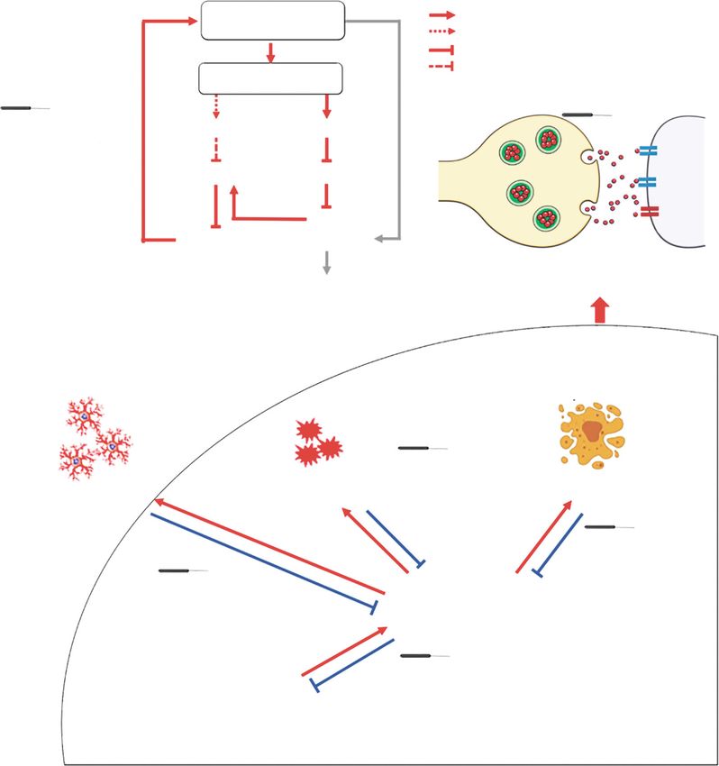
12 Neural Plasticity

4. Mechanism of Acupuncture Protecting from inhibits increased levels of p-α-syn 32 and p-α-syn 16 at ser-
 Dopaminergic Neurodegeneration ine 129 in the nigral dopaminergic neurons, providing evi-
 dence that acupuncture may reduce neurotoxicity via
Extensive studies have shown that genetic predisposition [42], inhibiting the level of α-syn and p-α-syn, thereby protecting
environmental factors [43], and a variety of intracellular and dopaminergic neurons [57].
extracellular pathogenic factors contribute to the development Acupuncture has also been demonstrated to promote the
of PD, which is characterized by selective loss of dopaminergic degradation of α-syn by restoring autophagy. Autophagy is
neurons in the SN, depletion of dopamine (DA) in the ST, and essential for the removal of toxic α-syn aggregates in order to
the formation of Lewy bodies that are mainly composed of α- maintain intracellular homeostasis [81]. Mammalian target of
syn [44]. The reduction of striatal DA levels disrupts the neu- rapamycin (mTOR) is a negative regulator of cellular autoph-
rotransmitter balance in the basal ganglia and thalamus, agy and it has been shown that 1-methyl-4-phenyl-1,2,3,6-tet-
resulting in motor symptoms such as paralysis agitans, brady- rahydropyridine (MPTP) upregulates microtubule-associated
kinesia, and rigidity [45]. Loss of DA and changes in neuro- protein 1 light chain 3 II (LC3-II) in a PD model, while down-
transmitters such as serotonin (5-HT), norepinephrine (NE), regulation lysosomal-associated membrane protein 1 (LAMP1,
γ-aminobutyric acid (GABA), and glutamate (Glu) in differ- lysosomal structural protein) indicates that an impairment of
ent brain regions and the peripheral nervous system lead to lysosomes and the interruption of autophagosome-lysosome
the occurrence of nonmotor symptoms, such as autonomic fusion may lead to an accumulation of autophagosomes in
nervous dysfunction, neuropsychiatric disorders, sleep disor- the substantia nigra pars compacta (SNpc) of MPTP mice
ders, and gastrointestinal symptoms [46]. [58]. Tian et al. observed that after acupuncture treatment for
 After reviewing 31 basic studies on acupuncture treat- 4 days, LC3-II was reduced by approximately 40% and LAMP1
ment of PD models (Table 2), we found that MA and EA by approximately 20%, and more than 50% of α-syn in the
were most commonly utilized, although BVA, moxibustion, SNpc was cleared, suggesting that acupuncture at GB34
acupoint injection, and laser acupuncture were also reported. restores lysosomal structures and reduces the accumulation of
It is worth mentioning that EA at high frequencies shows bet- autophagosomes, enhancing the clearance of autophagosomes
ter efficacy than low frequencies in some animal models of and degradation of α-syn. This group further found that acu-
PD [47, 48]. The most commonly selected acupoint was puncture did not change levels of upstream proteins of lyso-
GB34 followed by LR3. Thirty studies showed that acupunc- somal autophagy system (LAS), p-mTOR, p-p70s6k, and
ture improves motor function in PD models as assessed by ULK1, which indicated that acupuncture activates the indepen-
the rotarod test, the cylinder test, and the pole test. Basic dent mTOR pathway to enhance the autophagic clearance of α-
studies suggest that acupuncture may achieve its effectiveness syn. After activation of mTOR, p70S6K and 4E-binding protein
for PD treatment by preventing the DA neurons from α-syn 1 (4E-BP1) are activated to form p-p70S6K and p-4E-BP1,
aggregation, apoptosis, oxidative stress, and modulating neu- which then inhibit autophagy. Wang et al. [59] found that
roinflammation and the basal ganglia circuit around DA moxibustion at ST36, CV4 (Guanyuan), and GV16 (Fengfu)
neurons, which we have outlined in detail below. decreased p-mTOR, p-p70s6k and α-syn levels while increasing
 LC3-II levels, suggesting moxibustion exerts neuroprotective
4.1. α-Synuclein Aggregation. During the pathogenesis of PD, effects by promoting clearance of α-syn and enhancing autoph-
soluble α-syn monomers are thought to progressively aggre- agy via the mTOR pathway. As rapamycin (mTOR antagonist)
gate to large insoluble α-syn fibrils, called Lewy bodies [79]. treatment results in considerable side effects in PD patients
Overproduction and aggregation of α-syn furthermore such as dyslipidemia, antiproliferative toxicity, and renal dys-
induces mitochondrial dysfunction, oxidative stress, and function [82], acupuncture may represent as the alternative
neuroinflammation, leading to damage of dopaminergic neu- strategy to target mTOR.
rons in the SN and ST.
 It is suggested that acupuncture may inhibit increased 4.2. Apoptosis. It is established that nigral dopaminergic neu-
levels of α-syn for its neuroprotective effects. Serum/gluco- rons in PD patients undergo apoptosis and the formation of
corticoid-regulated kinase 1 (SGK1) is a serine threonine- apoptotic bodies [83]. Acupuncture may possess antiapopto-
specific protein kinase which may regulate α-syn. Yeo and sis properties via blocking apoptosis pathways of dopaminer-
Lim [56] found that acupuncture at GB34 and LR3 upregu- gic neurons. BVA has been found to decrease caspase-3
lated SGK1 and inhibited an α-syn increase. In SGK1 siRNA activity and downregulate caspase-3 and Bax gene expres-
knockdown SH-SY5Y cells, the authors observed a downreg- sion, suggesting that it may inhibit the mitochondrion-
ulation of SGK1 in dopaminergic neurons along with an dependent apoptotic pathway of dopaminergic neurons
increase in α-syn expression, suggesting that acupuncture [84]. MA and acupoint injection exhibited similar effects:
may inhibit the increase of α-syn expression by downregula- MA or acupoint injection at ST36 has been shown to down-
tion of SGK1. Other research shows that overexpression of regulate Bax, cytochrome C, and upregulate Bcl-2 [65]. Park
SGK1 exerts neuroprotective functions by reducing the pro- et al. [61] found that p53 may be involved in the neuropro-
duction of reactive oxygen species (ROS) and mitochondrial tective effect of acupuncture: 40 of 76 differentially expressed
dysfunction [80]. It has been reported that in the process of genes following acupuncture were involved in the p53 signal-
α-syn aggregation, phosphorylation at serine 129 increases ing network, and conditionally knocking down p53 pathway
the neurotoxicity of α-syn. A previous study has shown that genes in midbrain dopaminergic neurons attenuated the neu-
acupuncture suppressed α-syn levels in the SN and ST and roprotective effect of acupuncture. The c-Jun N-terminal
Neural Plasticity 13

kinase (JNK) signaling may mediate apoptosis through phos- process of protein synthesis in DA neurons, destroying the
phorylation of c-Jun [85]. Doo et al. [62] observed that BVA structure of the cellular membrane and ultimately resulting
at GB34 can rescue dopaminergic neurons from apoptosis by in the death of DA neurons. The neuroprotective effects of
inhibiting c-Jun activation in MPTP mouse, while another acupuncture treatment may be mediated through the regula-
study suggested that MA at GB34 does not change p-c-Jun tion of antioxidant systems, as a study reported that MA at
levels [69]. Therefore, the effects of different acupuncture GB34, LR3, ST36, and SP10 increases levels of superoxide
stimulation methods on JNK need to be further clarified. dismutase (SOD), glutathione (GSH), and glutathione perox-
 Several reports have established neurotrophic factors idase (GSH-Px), and decreases levels of malondialdehyde
(NTFs) as a major player in the neuroprotective effects of acu- (MDA), along with improved rotarod behavior [38], and
puncture in PD treatment. Liang et al. [63] found that long- similar results were also observed in EA and BVA treatment.
term high-frequency EA at GV14 and GV20 effectively pre- For example, high-frequency EA at ST36 and SP6 increases
vented the degeneration of ventral dopaminergic neurons and levels of GSH and SOD and decreases striatal H2O2 and
upregulated the levels of brain-derived neurotrophic factor MDA levels [47]. BVA at GB34 increases GSH and
(BDNF) in the ventral subregions of the midbrain, which paraoxonase-1 activities and decreases MDA levels [84]. In
induced the regeneration of injured dopaminergic neurons. addition to increased SOD and catalase (CAT) activities,
Subsequently, the authors also reported that EA at 2 Hz Lee et al. also observed upregulated DJ-1, which exists widely
increased glial cell-derived neurotrophic factor (GDNF) in peripheral tissues, neurons, and glial cells, playing an
mRNA in both sides of the globus pallidus, and 100 Hz EA essential role in antioxidation via regulating the activity of
increased GDNF mRNA in both sides of the globus pallidus SOD and CAT [72]. It is therefore speculated that the eleva-
and unlesioned side of SN pars reticulata, speculating that EA tion of DJ-1 caused by acupuncture at GB34 may exert anti-
could regulate the retrograde transport of GDNF from gan- oxidant effects by enhancing the activity of striatal SOD and
glion to SN and restore the balance of different nuclei in the CAT. These results indicate that acupuncture protects DA
basal ganglia circuit which contributes to the behavioral neurons from oxidative stress by restoring the balance
improvement of medial forebrain bundle- (MFB-) lesioned rats between oxides and antioxidants.
[48]. Another study confirmed that EA upregulated BDNF and
GDNF mRNA in the SN of PD models [64]. Tyrosine kinase 4.4. Neuroinflammation. Microglia, the primary immune
receptor B (TrkB) is a high-affinity BDNF receptor whose acti- cells of the central nervous system, play a vital role in the
vation results in the maintenance of neuronal differentiation neuroinflammation in PD [89]. Injury signaling in degener-
and survival [86]. Acupuncture at GB34 and LR3 increases ated DA neurons can shift microglia to a pro-inflammatory
the TrkB expression in the damaged SN of 6-hydroxydopa- “M1” phenotype, resulting in the release of ROS and cyto-
mine- (6-OHDA-) induced PD rats [66]. TrkB consists of dif- kines such as tumor necrosis factor-α (TNF-α), interleukin-
ferent subtypes, including full-length (TrkB FL) and truncated 1β (IL-1β), interleukin-6, and interleukin-12, which exacer-
(TrkB T1 or T2) TrkB. TrkB T1 is regarded as a dominant neg- bate oxidative stress and inflammation, ultimately leading
ative form of TrkB, which may suppress the neurotrophic to DA neuron apoptosis [90]. Inducible nitric oxide synthase
activity of the BDNF/TrkB signaling pathway [87]. Hence, bal- (iNOS) expressed in glial cells causes nitric oxide production,
ancing TrkB FL and TrkB T1 is essential for neuroprotection which in turn activates microglia in conjunction with various
[88]. It has been reported that the TrkB inhibitor (K252A) proinflammatory “M1” phenotype cytokines. Acupuncture
eliminates the neuroprotective effect of EA, and another study at GB34 and LR3 has been shown to attenuate the expression
revealed that EA may reverse the imbalance between TrkB FL of macrophage antigen complex-1, a marker of microglial
and TrkB T1 to upregulate p-Akt, p-ERK1/2, and BDNF activation, and mitigate increases in cyclooxygenase-2
[68]. 50 Hz EA at GB34 and LR3 has been shown to increase (COX-2) and iNOS expression in an MPTP-induced PD
BDNF and downstream p-Akt levels [67], improving rotational models [73]. Similarly, striatal DA levels were shown to
behavior in a rat model of unilateral MPP injury, and another increase from 46% to 78% within 7 days, suggesting that acu-
study revealed that inhibition of the PI3K/Akt signaling path- puncture exerts neuroprotective effects by attenuating
way blocked the protective effect of acupuncture on DA neu- MPTP-induced glial activation and neuroinflammation.
rons [69]. Acupuncture at GB34 combined with KD5040 Injecting choroid plexus cells at ST36 likewise decreased
downregulated pIκBα and upregulated pAkt, pGSK3β, pERK, iNOS and COX-2 expression and enhanced exercise capacity
pCREB, and BDNF, which significantly improve motor func- of MPTP-induced mice [37], while BVA at GB34 has been
tion [70]. Therefore, the results presented above suggest that suggested to protect dopaminergic neurons by downregulat-
BDNF/TrkB and their downstream signaling pathways may ing inflammatory factors such as TNF-α and IL-1β [84].
mediate the neuroprotective effects of acupuncture in PD treat- Acupuncture can also inhibit neuroinflammation by reg-
ment. Besides, acupuncture has been found to increase the ulating the brain-gut axis, thus alleviating movement dys-
number of 5-bromo-2 ′ -deoxyuridine- (BrdU) positive cells function in PD. An altered gut microbiota has been
and to restore neurogenesis in the subventricular zone [71], reported to induce microglial activation and neuroinflamma-
which provides a new pathway for studying the molecular tion, which may promote α-syn overexpression and contrib-
mechanisms of acupuncture treatment for PD. ute to motor dysfunction in PD models [91]. Jang et al. [65]
 observed that acupuncture changed the relative abundance
4.3. Oxidative Stress. Aggregation of free radicals causes lipid of Butyricimonas, Holdemania, Frisingicoccus, Gracilibacter,
peroxidation, which leads to excessive oxidation during the Phocea, and Aestuariispira, which showed significant
You can also read