Reprogramming cholesterol metabolism in macrophages and its role in host defense against cholesterol-dependent cytolysins
←
→
Page content transcription
If your browser does not render page correctly, please read the page content below
www.nature.com/cmi

                  REVIEW ARTICLE                         OPEN

                  Reprogramming cholesterol metabolism in macrophages and
                  its role in host defense against cholesterol-dependent
                  cytolysins
                                                                     1,2 ✉
                  Min-Sub Lee1 and Steven J. Bensinger

                  © The Author(s) 2022

                     Cholesterol is a critical lipid for all mammalian cells, ensuring proper membrane integrity, fluidity, and biochemical function.
                     Accumulating evidence indicates that macrophages rapidly and profoundly reprogram their cholesterol metabolism in response to
                     activation signals to support host defense processes. However, our understanding of the molecular details underlying how and why
                     cholesterol homeostasis is specifically reshaped during immune responses remains less well understood. This review discusses our
                     current knowledge of cellular cholesterol homeostatic machinery and introduces emerging concepts regarding how plasma
                     membrane cholesterol is partitioned into distinct pools. We then discuss how proinflammatory signals can markedly reshape the
                     cholesterol metabolism of macrophages, with a focus on the differences between MyD88-dependent pattern recognition receptors
1234567890();,:

                     and the interferon signaling pathway. We also discuss recent work investigating the capacity of these proinflammatory signals to
                     selectively reshape plasma membrane cholesterol homeostasis. We examine how these changes in plasma membrane cholesterol
                     metabolism influence sensitivity to a set of microbial pore-forming toxins known as cholesterol-dependent cytolysins that
                     specifically target cholesterol for their effector functions. We also discuss whether lipid metabolic reprogramming can be leveraged
                     for therapy to mitigate tissue damage mediated by cholesterol-dependent cytolysins in necrotizing fasciitis and other related
                     infections. We expect that advancing our understanding of the crosstalk between metabolism and innate immunity will help
                     explain how inflammation underlies metabolic diseases and highlight pathways that could be targeted to normalize metabolic
                     homeostasis in disease states.

                     Keywords: Cholesterol; Macrophages; Innate Immunity; Metabolism

                     Cellular & Molecular Immunology (2022) 19:327–336; https://doi.org/10.1038/s41423-021-00827-0

                  INTRODUCTION                                                                             Macrophages rapidly reprogram their metabolic state to
                  Macrophages are key players in the innate immune system and                           facilitate inflammation, resolution, and effector functions [4, 5].
                  are tasked with responding to a diverse array of pathogens.                           The metabolic reprogramming of macrophages targets nearly all
                  Macrophages rely on recognizing pathogen-associated molecular                         aspects of core metabolic pathways, including glycolysis, oxidative
                  patterns (PAMPs) through the engagement of pattern-recognition                        metabolism, the redox state, and nucleotide, protein, and lipid
                  receptors (PRRs) and other similar receptors [1, 2]. Rapid                            composition [4–6]. Without these coordinated changes in cellular
                  recognition of foreign elements results in the production of                          metabolism, macrophages have deficits in their immune functions
                  proinflammatory cytokines and chemokines, phagocytosis, oxida-                         and contribute to disease pathogenesis in many contexts. This
                  tive bursts to clear microbial pathogens, the activation of                           review focuses on one interesting and less understood aspect of
                  neighboring immune cells, and the recruitment of other immune                         metabolic reprogramming: the abrupt and profound shift in
                  cells to the site of infection [3]. Macrophages are also critical for                 cellular cholesterol homeostasis induced by PRR signals and other
                  the resolution of inflammation, the restoration of tissue home-                        proinflammatory stimuli. We provide a brief introduction to
                  ostasis through their ability to remove cellular debris and                           cellular cholesterol metabolism and the molecular mechanisms
                  apoptotic cells, and the coordination of wound repair processes                       underlying the rapid alterations in cholesterol homeostasis as they
                  [3]. The ability of macrophages to detect these distinct and                          are currently understood. We then discuss the potential reasons
                  disparate signals and subsequently integrate this information into                    why inflammatory signals drive the intracellular redistribution of
                  physiological responses makes them ideal cells to mechanistically                     cholesterol in macrophages. We specifically highlight one inter-
                  study how immune responses are regulated at the molecular level.                      esting aspect of gram-positive bacterial infections that rely on

                  1
                   Department of Molecular and Medical Pharmacology, University of California, Los Angeles, CA 90095, USA. 2Department of Microbiology, Immunology and Molecular Genetics,
                  University of California, Los Angeles, CA 90095, USA. ✉email: sbensinger@mednet.ucla.edu

                  Received: 23 August 2021 Accepted: 7 December 2021
                  Published online: 11 January 2022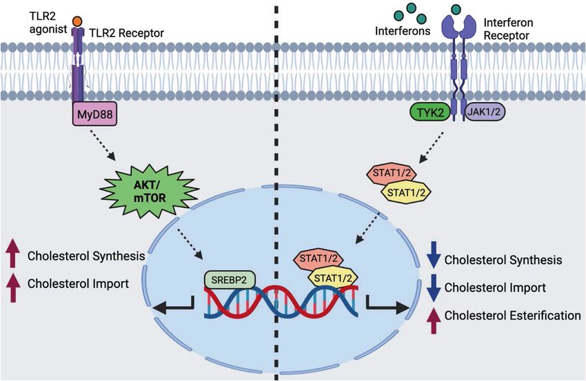
M.-S. Lee and S.J. Bensinger
328
      membrane cholesterol to induce cytotoxicity and the resultant              activated by receptor-mediated kinase signaling pathways. For
      tissue damage. Finally, we discuss how manipulating cholesterol            example, TCR, BCR, and select PRR signaling can increase
      metabolism might be an attractive therapeutic approach to                  cholesterol biosynthetic flux in both innate and adaptive immune
      bolster host defense and spare tissues from pathogen-mediated              cells, usually through the AKT/mTOR signaling pathway [16–19].
      damage.                                                                       The expression levels of cholesterol biosynthetic enzymes are
                                                                                 also directly regulated by the pool of intracellular sterols. The rate-
                                                                                 limiting enzyme in cholesterol biosynthesis is the ER-residential
      REGULATION OF CELLULAR CHOLESTEROL HOMEOSTASIS                             protein, 3-hydroxy-3-methyl glutaryl coenzyme A reductase
      Among the thousands of lipids found in the mammalian lipidome,             (HMGCR). Increased amounts of oxysterols in the ER, such as 25-
      cholesterol is the most abundant lipid species found in cells,             hydroxycholesterol (25HC), can induce the proteolytic degradation
      accounting for up to 30% of total lipids [7]. Cholesterol is               of HMGCR, resulting in decreased cholesterol biosynthesis [10, 20].
      exceedingly hydrophobic and composed of four planar rings, and             In addition, cholesterol, 25HC, and other oxysterols potently retain
      as such, it must reside within lipid bilayers of membranes or be           the SCAP-SREBP2 complex in the ER, preventing SCAP-SREBP2
      stored in its esterified form within lipid droplets. Cholesterol is an      translocation to the Golgi body and the subsequent processing
      essential lipid for all mammalian cells, playing indispensable roles       and nuclear import of SREBP2. Of particular importance in
      in establishing cell membrane biochemical and biophysical                  inflammation and macrophage biology, CH25H, the enzyme
      properties, including the organization of lipid microdomains,              responsible for generating 25HC, is considered a canonical
      receptor distribution, bilayer fluidity, and ultimately membrane            interferon (IFN)-regulated gene [21–23]. The IFN-mediated upre-
      integrity [7–9]. An interesting and important feature of cholesterol       gulation of CH25H and resulting production of 25HC appear to
      metabolism is that cholesterol can be synthesized within cells or          underlie many of the changes in cholesterol homeostasis
      directly imported by the internalization of lipoproteins [10]. Thus,       observed in macrophages during inflammatory responses to
      requisite cholesterol levels can be met through the combined               viruses and some microbes [24–26]. The production of 25HC has
      actions of synthesis and import pathways.                                  also been shown to activate liver X receptors (LXRs) to promote
         Cholesterol cannot be degraded by mammalian cells, and                  the resolution of inflammation and effectively link the regulation
      excess cholesterol is either stored in intracellular depots as lipid       of the SREBP2 and LXR pathways downstream of IFN receptor
      droplets or exported from the cell via the cholesterol efflux               signaling [10, 21, 27, 28].
      pathway [10]. Excess accumulation of cellular cholesterol can
      result in severe cellular dysfunction and activation of the                Cholesterol import
      inflammasome, leading to IL-1β-mediated inflammation [11].                   Despite the intrinsic ability to synthesize cholesterol, immune cells
      Given the essential but potentially toxic nature of cholesterol,           may preferentially utilize cholesterol import via receptor-mediated
      the biosynthetic, import, export, and esterification mechanisms             endocytosis to meet their cholesterol requirements. The mechan-
      must be tightly and coordinately regulated to ensure that                  istic details of cholesterol import have recently been reviewed
      sufficient but not excessive cholesterol is available to cells.             elsewhere [10]. In brief, the import of cholesterol canonically
      Disrupted regulation of cholesterol metabolism is linked with              occurs through the import of low-density lipoproteins (LDL) from
      many types of severe congenital human diseases, such as Tangier            the extracellular space via the low-density lipoprotein receptor
      disease, familial hypercholesterolemia, Niemann-Pick type C                (LDLR). LDLR is a ubiquitously expressed cell surface glycoprotein
      disease, and Schnyder corneal disease [10]. Importantly, the               and is a direct transcriptional target of SREBP2; therefore, its
      balance of these pathways is rapidly altered in the context of             function is directly correlated with SREBP2 activity [10]. Surface
      inflammation, infections, autoimmunity, cancer, allergy, and                LDLR captures circulating LDL, which becomes incorporated into
      wound repair [12].                                                         clathrin-coated vesicles that enter the endocytic pathway.
                                                                                 Following endocytosis, LDL-carried cholesteryl esters are hydro-
      Cholesterol biosynthesis                                                   lyzed by lysosomal acid lipases (LAL) in lysosomes, liberating free
      The details of the cholesterol biosynthetic pathway are exceed-            cholesterol. Cholesterol is then exported from the lysosomal
      ingly complex but well defined [10]. Cholesterol is synthesized             lumen via the concerted actions of Niemann-Pick Type C1 (NPC1)
      through the enzymatic activity of over 20+ distinct metabolic              and NPC2 [10, 29]. These free cholesterols are delivered to other
      steps in the mevalonate pathway and the downstream cholesterol             subcellular compartments, such as the ER and plasma membrane
      synthesis pathway. The enzymes involved in cholesterol biosynth-           (PM), for subsequent equilibration with the cellular cholesterol
      esis are largely found in the endoplasmic reticulum (ER)                   pool and distribution to other compartments of the cell as
      membrane. These enzymes are subject to multiple layers of                  required [10].
      transcriptional and posttranslational regulation to ensure tight
      control over synthetic capacity and import to avoid deleterious            Cholesterol export
      accumulation [10].                                                         Unlike other lipid metabolites, such as long-chain fatty acids,
         The expression of cholesterol biosynthetic enzymes is tran-             phospholipids, and ceramides, mammalian cells do not have a
      scriptionally regulated by the transcription factor, sterol regulatory     built-in catabolic pathway for cholesterol. Instead, excess cholesterol
      element-binding protein 2 (SREBP2). SREBP2 has complex regula-             in cells is either exported out of the cell or stored as cholesterol esters
      tion, and we direct readers to excellent reviews that discuss the          in lipid droplets. Cholesterol efflux is transcriptionally controlled by
      biology of this transcription factor [13, 14]. In brief, SREBP2 resides    the lipid-activated nuclear receptors, liver X receptors (LXR α and β)
      in the ER in association with two other proteins: SREBP-cleavage-          [30]. Upon excess cholesterol accumulation in macrophages, LXRs
      activating protein (SCAP) and insulin-induced genes (INSIGs)               transactivate the genes encoding the transporters Abca1 and Abcg1
      [10, 15]. This protein complex is sensitive to ER sterol levels and        [10]. The exact mechanism of ABCA1’s ability to move cholesterol out
      controls SREBP2 function. Under sterol-replete conditions, SREBP2          of the cell remains under investigation, but it is generally thought
      is held in the ER, effectively decreasing cholesterol synthesis.           that ABCA1 flips phospholipids and cholesterol through the
      When cholesterol levels in the ER drop below the cell threshold,           membrane, which then allows apolipoproteins (e.g., Apo-AI) to
      SREBP2 translocates to the Golgi, where it is sequentially cleaved         accept the newly accumulated cholesterol that now sits on the outer
      by site 1 and site 2 proteases (S1P and S2P, respectively) [10, 15].       leaflet [31]. In contrast, ABCG1 is expressed intracellularly in
      Cleaved SREBP2 then moves to the nucleus to transactivate the              endosomes, where it facilitates the movement of cholesterol from
      genes encoding enzymes in the cholesterol synthesis program. In            the ER to the inner leaflet via endosomal vesicles [32]. LXR activity,
      immune cells, the SREBP2 transcriptional program can also be               and correspondingly, ABCA1 and ABCG1 expression, can be altered

                                                                                                     Cellular & Molecular Immunology (2022) 19:327 – 336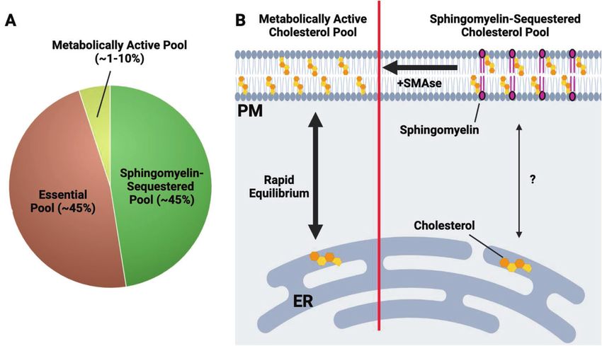
M.-S. Lee and S.J. Bensinger
                                                                                                                                                       329

Fig. 1 Opposing transcriptional programs ensure cholesterol homeostasis in macrophages. Cholesterol homeostasis is transcriptionally
regulated by liver X receptors (LXRα and β) and sterol regulatory element binding transcription factor 2 (SREBP2) signaling. Excess cholesterol
accumulation in cells activates LXRs, leading to increased cholesterol efflux and reduced cholesterol import. Activating the SREBP2
transcriptional pathway increases de novo cholesterol biosynthesis and increases cholesterol import

by a wide array of inflammatory signals in macrophages. LXRs and              Based on these studies, it is thought that the plasma membrane
SREBP2 help establish macrophage cholesterol homeostasis (Fig. 1).        contains at least three distinct pools of cholesterol [36] (Fig. 2). One
Importantly, we now know that proinflammatory signals control both         pool is termed the accessible or metabolically active pool. This
the LXR and SREBP transcriptional axes, supporting the idea that          fraction of PM cholesterol is very small and appears to be highly labile,
rapidly modulating cholesterol synthesis and efflux in innate immune       meaning that the pool size can be rapidly altered in response to
cells facilitates their effector function [17, 21, 27].                   changes in total cellular cholesterol concentrations. This metabolically
                                                                          active pool is thought to be in equilibrium with the cholesterol pool in
Cholesterol esterification                                                 the ER and thereby influences the cholesterol sensing apparatus in
As mentioned previously, cholesterol esterification is an important        the ER (e.g., SCAP/INSIG-SREBP). As such, the accessible cholesterol
method for storing excess cholesterol in lipid droplets. Two major        pool in the PM plays a critical role in linking PM cholesterol levels with
isozymes regulate cholesterol esterification: acyl coenzyme A:             the cholesterol pool in the ER and helps to set overall cellular
cholesterol acyltransferase 1 (ACAT1) and ACAT2. ACAT1 is highly          cholesterol homeostasis. The exact distribution of the metabolically
expressed throughout the body, whereas ACAT2 has a more restricted        active pool of cholesterol in the membrane remains unknown, and
expression pattern [10]. ACAT enzymes utilize free cholesterol as an      further work is needed to determine if this pool is spatially restricted
allosteric activator and a substrate, subsequently conjugating fatty      across the PM.
acyl-CoA into the hydrophobic end of the cholesterol molecule                The importance of the metabolically active pool of cholesterol
[10, 33]. This conjugation results in the production of cholesteryl       in pathogen lifecycle and host defense processes requires further
esters, which become stored inside lipid droplets. Interestingly,         investigation. Nevertheless, we suspect that this pool will be a
neither ACAT1 nor ACAT2 contains regulatory elements specific for          critical target for microbes and viruses that exploit cholesterol
the transcription factors SREBP2 and LXR. However, the Soat1 and          metabolism as part of their pathogenesis. Indeed, it is worth
Soat2 genes that encode the cholesterol esterification enzymes ACAT        noting that mechanistic work on this distinct cholesterol pool in
1 and 2, respectively, are upregulated by cytokines such as interferons   the PM of cells relies on specific domains of microbial proteins
and tumor necrosis factor, indicating a role for esterification in         that directly bind cholesterol (e.g., domain 4- of cholesterol-
controlling inflammation and host defense [21, 34, 35].                    dependent cytolysins) [40]. Thus, it seems reasonable to infer that
                                                                          controlling specific cholesterol pools in the PM with inflammatory
                                                                          cytokines or other host defense machinery will be a component of
NEW CONCEPTS IN PLASMA MEMBRANE CHOLESTEROL                               innate immune responses to microbes and viruses. This concept is
HOMEOSTASIS                                                               further explored below.
Cholesterol is essential for plasma membrane function. In the                A second, larger pool of cholesterol in the PM is tightly
absence of sufficient plasma membrane cholesterol, cells cannot            associated with sphingomyelin (SM) and other phospholipid (PL)
maintain lipid bilayer integrity and have perturbed microdomain           species [36]. This pool is known as the SM-sequestered pool, and
assembly, thereby influencing signaling and endocytosis, among             the tight association of cholesterol with sphingomyelin forms the
other functions. Although cholesterol in the plasma membrane              biophysical basis for the assembly of lipid microdomains or lipid
represents the largest cholesterol pool within the cell, cholesterol      rafts within the plasma membrane [41]. Similar to work using
is unevenly distributed throughout the plasma membrane.                   CDCs to define the metabolically active cholesterol pool, this
Accumulating evidence indicates that several distinct pools of            distinct pool of cholesterol can be identified using the fungal-
cholesterol can be found within the plasma membrane, each                 derived protein ostreolysin A (OlyA) [42]. The SM-sequestered pool
serving unique roles in PM function and cellular physiology [36].         size does not rapidly change in response to acute cellular
Recent elegant biochemical studies that leveraged microbial               cholesterol deprivation or overload. Thus, it is thought that the
products that selectively bind to cholesterol in membranes have           SM-sequestered pool does not directly equilibrate with the ER
shed light on this exciting and important concept [37–39].                cholesterol pool or the metabolically active pool. However, brief

Cellular & Molecular Immunology (2022) 19:327 – 336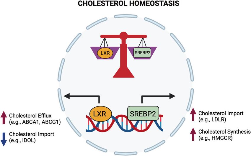
M.-S. Lee and S.J. Bensinger
330

      Fig. 2 An overview of the three-cholesterol pool model in the plasma membrane. A The plasma membrane (PM) contains at least three
      distinct pools of cholesterol. The metabolically active or accessible cholesterol pool makes up ~1–10% of total PM cholesterol. Sphingomyelin-
      sequestered cholesterol and the essential pool are each estimated to be ~45% of total PM cholesterol. B Depiction of the metabolically active
      and sphingomyelin-sequestered cholesterol pools. On the left side of the diagram, the metabolically active cholesterol pool rapidly
      equilibrates with the ER cholesterol pool, thereby providing a metabolic conduit for ER-resident cholesterol sensing machinery (e.g., SCAP-
      SREBP, HMGCR) to sense PM cholesterol levels. On the right, the sphingomyelin-sequestered cholesterol pool is composed of cholesterol
      molecules that are tightly associated with sphingomyelin and likely other phospholipid species. In contrast with the metabolically active
      cholesterol pool, the SM-associated cholesterol pool does not rapidly equilibrate with the ER cholesterol pool. Treatment of cells with bacterial
      sphingomyelinase (SMase) liberates SM-associated cholesterol, allowing it to enter the metabolically active pool. The essential pool (not
      shown) is less well understood and thought to play a fundamental role in the bilayer integrity of the plasma membrane

      treatment of cells with bacterial sphingomyelinase (SMase), which          the coordinated upregulation of both cholesterol biosynthesis and
      hydrolyzes sphingomyelin into ceramides and phosphorylcholine              lipoprotein import to meet the anabolic lipid requirements
      [43], decreases OlyA binding and rapidly increases the size of the         associated with growth and proliferation. In other contexts,
      accessible cholesterol pool. This finding indicates that newly              phagocytes reprogram their cholesterol metabolism in a signal-
      liberated cholesterol from the sphingomyelin-associated pool               specific manner, resulting in distinct metabolic outcomes [17].
      flows into other PM-resident pools [36, 43]. This observation may           Lipidomics and isotope tracer analyses have revealed that
      be particularly relevant for microbial infections and the potential        macrophages rapidly upregulate cholesterol synthesis when
      interplay between SMases and other toxins that rely on                     activated through MyD88-dependent PRRs, resulting in the
      cholesterol for their effector functions.                                  general accumulation of total cellular cholesterol [17, 21]. In
         The remaining cholesterol appears to exist in a third pool              contrast, interferon (IFN) signaling and PRRs that generate IFN
      known as the essential pool. This cholesterol pool appears to be           responses (e.g., TLR3-TRIF signaling) downregulate cholesterol
      equivalent in size to the SM-sequestered pool, but it has been             biosynthesis and increase cholesterol storage in the form of
      largely defined through exclusionary characteristics [36]. The exact        cholesterol esters [21, 44]. Thus, it has been proposed that
      nature of the essential pool of cholesterol remains poorly                 macrophages shift their cholesterol metabolism in a context-
      understood. However, we can hypothesize that this pool plays a             specific manner to facilitate specific effector functions and ensure
      fundamental role in bilayer integrity through its ability to foster        the generation of particular forms of inflammation (i.e., IFNs versus
      molecular interactions between different classes of phospholipid           IL-1β) (Fig. 3).
      species (i.e., PC, PE, PI, and PS) and other bilayer-associated               Interestingly, changes in synthesis and total cellular cholesterol
      proteins to establish fundamental biophysical characteristics [36].        levels do not necessarily correlate. For example, interferon
      The essential cholesterol pool requires harsh chemical treatment           signaling (both type I and II) decreases cholesterol biosynthesis
      of cells to remove it from the membrane, and its removal results in        via transcriptional and posttranslational mechanisms. Despite a
      the loss of cellular viability. The regulation of the essential pool       decrease in cholesterol biosynthetic capacity, macrophages
      size and the spatial distribution of this pool within the membrane         maintain or modestly increase their cholesterol levels in response
      have not been clearly defined. There is the idea that microbes and          to IFNs [21, 45]. This observation suggests that interferon signaling
      viruses specifically target distinct pools of cholesterol for their         regulates other cholesterol homeostatic pathways (e.g., increasing
      pathogenesis. However, there is no direct evidence that the                cholesterol import or reducing cholesterol efflux pathways).
      essential pool is specifically targeted or exploited by pathogens to        Further analysis of plasma membrane cholesterol pools suggests
      facilitate their lifecycle.                                                that interferon signaling specifically decreases the metabolically
                                                                                 active pool of cholesterol in the plasma membrane without
                                                                                 disturbing the sphingomyelin-sequestered cholesterol pool or the
      METABOLIC REPROGRAMMING OF CHOLESTEROL                                     essential pool [21]. The molecular events underlying selective
      HOMEOSTASIS IN MACROPHAGES                                                 reprogramming of plasma membrane cholesterol pools remain a
      It is well understood that many types of immune cells reprogram            poorly understood but is an active area of investigation. It is
      their cholesterol metabolism in response to activation and                 known that some aspects of IFN-mediated reprogramming are
      proinflammatory signals. The activation of lymphocytes results in           dependent on the production of oxysterol 25HC [10]. The capacity

                                                                                                    Cellular & Molecular Immunology (2022) 19:327 – 336
M.-S. Lee and S.J. Bensinger
331
Fig. 3 Signal-specific reprogramming of cholesterol metabolism in macrophages by proinflammatory signals. On the left side,
macrophages receive an input stimulus that signals through MyD88-dependent PRRs (e.g., TLR2). MyD88 signaling activates AKT/mTOR, which
increases SREBP2 transcriptional activity, resulting in increased cholesterol biosynthesis and cholesterol import. On the right side: type I and II
IFN signaling reprograms macrophage cholesterol metabolism in a JAK-STAT-dependent manner. IFN receptors drive the downregulation of
cholesterol biosynthesis, reduce cholesterol import, and upregulate cholesterol esterification
of 25HC to specifically decrease the accessible cholesterol pool is One well-characterized example of the interplay between
thought to increase cholesterol esterification, decrease cholesterol cholesterol metabolism and microbial pathogenesis can be found
biosynthesis, and activate the LXR pathway [10, 34, 46]. This in Mycobacteria infections. One hallmark of M. tuberculosis (M. tb)
observation further highlights the exquisite specificity by which infection of cells is the marked accumulation of intracellular lipids
IFNs can remap the cellular cholesterol landscape and indicates resulting in foam cell formation [49]. It has been shown that M. tb
that shifting specific cholesterol pools of the cell is an important has the ability to degrade cholesterol for use in bacterial
component of host defense against viruses and aspects of metabolism and that this cholesterol catabolism may be a
microbial infections. mechanism by which M. tb persists in IFN-activated macrophages
In contrast to IFN signaling, MyD88-dependent PRRs, such as Toll- [50]. Similarly, M. leprae uses the oxidation of cholesterol to
like receptor (TLR)-2, TLR-7, and TLR-9, lead to an overall increase in facilitate energetics and cell wall biosynthesis [51]. Importantly,
cholesterol biosynthesis and total cholesterol in macrophages [21]. interfering with the ability of these bacteria to use cholesterol
This increase in cholesterol is dependent on the upregulation of the decreases intracellular survival in host cells. Thus, cholesterol is a
SREBP transcriptional axis. The exact mechanism linking requisite host metabolite that is used as a nutrient source by
MyD88 signaling with the SREBP transcriptional axis has yet to be Mycobacteria for persistence and pathogenicity.
fully elucidated but is dependent on AKT/mTOR and likely the In other instances, obligate intracellular bacteria use cholesterol
transcription factor, NRF2 [17]. Due to increased cholesterol synthesis, to facilitate entry into host cells. The hydrophobic nature of
MyD88-dependent PRRs expand the accessible cholesterol pool in the cholesterol means that this lipid is primarily embedded in cellular
plasma membrane [21]. Despite this increase in total and membrane membranes. Some intracellular microbes specifically target the
cholesterol levels, MyD88-dependent PRRs do not significantly pool of cholesterol in cholesterol-rich microdomains in the PM for
increase the levels of cholesterol esters. Thus, this reciprocal their entry [48]. Microbe-induced alterations in cholesterol
relationship between MyD88 and IFN signaling in cholesterol metabolism have also been shown to perturb cellular phagolyso-
homeostasis in macrophages supports the concept that rapid and some function and intracellular organelle trafficking [48, 52].
profound regulation of cholesterol homeostasis is an integral aspect Pharmacologic or genetic disruptions of lipid microdomains or
of macrophage’s ability to respond to a broad array of pathogens and intracellular cholesterol trafficking pathways decreases the effi-
inflammatory stimuli. ciency of microbe entry and intracellular pathogen persistence
[48, 53]. While these are just a few examples of how microbes
exploit host cholesterol metabolism for their pathogenesis, they
CHOLESTEROL AND BACTERIAL PATHOGENESIS support the concept that reprogramming cholesterol homeostasis
The conserved use of cholesterol in mammalian cells likely underlies through inflammation is an innate immune mechanism used in
cholesterol targeting by microbes and viruses to facilitate their host defense. These observations also support the idea that
lifecycle and pathogenicity. These strategies have been described in pharmacologically targeting cellular cholesterol metabolism could
several reviews, and we direct the reader to the literature to gain a be an adjunctive therapeutic approach to facilitate the clearance
greater appreciation of the specifics of the different classes or types of of microbes.
pathogens [47, 48]. In this brief review, we restrict our discussion to
some interesting aspects of cholesterol metabolism in bacterial
pathogenesis, followed by a more detailed discussion of cholesterol- CHOLESTEROL-DEPENDENT CYTOLYSINS
dependent cytolysins (CDCs), a family of pore-forming toxins that rely As discussed previously, microbial proteins can target lipids in
on cholesterol for their effector functions. membrane to facilitate pathogenesis. One well-defined group of
Cellular & Molecular Immunology (2022) 19:327 – 336M.-S. Lee and S.J. Bensinger
332
virulence factors that target host cholesterol are cholesterol- In our studies on PRRs, we were surprised to find that TLR3
dependent cytolysins (CDCs). CDCs are pore-forming toxins that activation rendered macrophages very resistant to CDC-mediated
are secreted as soluble monomers and subsequently oligomerize cytotoxicity. These data led us to surmise that type I IFNs mediated
on host membranes to form pores [40]. Approximately thirty this resistance and that TLR3 was an “accidental” mechanism by
distinct gram-positive bacteria have been identified that produce which we discovered this interesting response. Indeed, subse-
CDCs, including several species that mediate severe diseases in quent gain- and loss-of-function studies showed a critical and
humans (e.g., Clostridium perfringens, Streptococcus pyogenes, and essential role for type I IFN signaling in this protective mechanism
Bacillus anthracis) [54, 55]. CDCs contain four domains. Domains [21]. The activation of PRRs that lead to type I interferon
1–3 primarily play structural roles that facilitate oligomerization production [58] (e.g., NOD2 and STING) also induced protection
into a prepore intermediate once cholesterol recognition occurs. against CDCs. Importantly, delivery of either type I IFN or type II
Domain 4 contains a tryptophan-rich region(s) that is involved in (IFN-γ) in trans resulted in protection, expanding the impact of this
cholesterol recognition and membrane binding. Once Domain immunometabolic reprogramming event. We observed that IFN-
4 successfully binds to cholesterol in the membrane, the CDC stimulated macrophages retained functionality, even when
monomers oligomerize into a prepore intermediate, which then challenged with CDCs, as evidenced by their ability to phagocy-
becomes inserted into the membrane [56]. tose microbes or apoptotic cells. We also found that the protective
The cellular consequences of CDCs vary depending on the CDC effect of IFNs could be observed in freshly isolated neutrophils,
dosage, duration, and cell type. CDCs create a pore that is ~250 Å in indicating that a generalized cellular mechanism, at least for
diameter, which is large enough to allow the leakage of biomolecules phagocytes, underlie this response [21]. Whether this effect is also
(amino acids, nucleotides, and small proteins), as well as ions (Ca2+, true for nonimmune cells stimulated with IFNs has not been
K+, Na+, and Cl−) [56]. Pores also allow the influx of water into cells, determined.
which leads to blebbing and apoptosis due to osmotic shock. The The molecular mechanism of IFN-mediated protection against
mechanisms underlying CDC-mediated cell lysis have been char- CDCs also lies in the ability of IFNs to alter cholesterol synthesis in
acterized in erythrocytes because these cells have a minimal capacity macrophages. Isotope labeling studies showed that IFN signaling
to repair membrane damage. Unlike erythrocytes, nucleated cells decreases cholesterol synthesis and that this decrease in
have an intrinsic membrane repair system that is triggered upon Ca2+ cholesterol synthesis was dependent on the upregulation of
influx during pore formation [56]. Repair processes induced by CDC CH25H and the subsequent production of 25HC [21]. The
pore formation include patch repair, clogging, and microvesicle generation of 25HC by macrophages results in inhibition of the
shedding [56, 57]. Whether changes in cholesterol metabolism SREBP2 transcriptional axis and the direct degradation of HMGCR
induced by microbes or host inflammation alter the efficiency of [20, 59, 60]. Consistent with this finding, genetic ablation of CH25H
membrane repair is largely unknown and should be investigated. rendered naïve macrophages highly sensitive to CDCs and
However, it is easy to hypothesize that the dramatic alterations in lipid abrogated the ability of IFNs to protect against CDC-mediated
homeostasis observed in response to proinflammatory signals will pore formation. Ch25h-deficient mice also developed severe
have some impact on host membrane repair systems. erythema and larger ulcerative skin lesions when intradermally
challenged with streptolysin O (SLO), a CDC secreted by S.
pyogenes. Conversely, pharmacologic addition of 25HC provided a
INDUCED RESISTANCE TO CDC TOXICITY VIA METABOLIC marked level of protection against CDC challenge, solidifying the
REPROGRAMMING role of CH25H in this interesting immune-metabolic response.
The specific dependency of CDCs on membrane cholesterol to Consistent with a role for oxysterols in mediating protection to
execute their effector function led to the hypothesis that CDCs, both 25HC and 27HC protect endometrial cells from the
alterations in membrane cholesterol homeostasis could provide CDC pyolysin, which is produced by Trueperella pyogenes [46].
some form of resistance to CDC-mediated cellular damage. Interestingly, this effect was partially dependent on the ability of
Indeed, induced cholesterol efflux through pharmacologic activa- these oxysterols to activate LXRs and reduce accessible choles-
tion of the LXR transcriptional pathway or genetic manipulation of terol, likely through cholesterol efflux.
cholesterol metabolism provides some measure of protection The molecular events mediating the ability of IFNs to protect
against CDC-mediated cellular toxicity [21, 46]. However, it is against CDCs remain incompletely defined, but we have been able
unclear whether physiological signals in the context of inflamma- to gain some understanding of the pathways required for this
tory responses to infections would have similar protective effects. effect. Using fluorescently labeled ALO-D4 (the D4 domain of the
Leveraging previous work that showed that PRR and cytokine CDC Anthrolysin O) [39], we were able to show that IFNs
signaling influence macrophage cholesterol homeostasis, we decreased ALO-D4 binding to the membrane. These data suggest
explored whether activating macrophages might intrinsically that the cholesterol levels in the plasma membrane dropped
induce resistance to CDCs. Working from the hypothesis that below those required for effective CDC binding and oligomeriza-
the recognition of gram-positive bacteria by PRRs would be tion [21]. Reprogramming cholesterol synthesis appears to be
required for such a mechanism, we set out to determine whether important for altering CDC binding to the plasma membrane, but
activating macrophages with TLR2 agonists could influence CDC- how these changes in cholesterol synthesis directly translate into
mediated loss of membrane integrity. However, we found that this protection at the PM remains less clear.
supposition was incorrect and that the opposite appeared to be One possibility is that inhibiting cholesterol biosynthesis
true. We observed that activating macrophages via TLR2 and other globally reduces cholesterol levels in the PMs of cells. However,
MyD88-dependent TLRs resulted in a modest but highly mass spectrometry data showed that cholesterol levels in the PM
reproducible increase in sensitivity to CDC-mediated cellular were largely maintained in IFN-stimulated macrophages, and we
toxicity. Thus, TLR2-mediated recognition of gram-positive bac- observed decreases in ALO-D4 binding as little as two hours after
teria by macrophages does not appear to induce resistance to IFN treatment. Likewise, a brief 20-minute treatment of macro-
CDCs. Rather, this signal primes heightened CDC sensitivity. The phages with sphingomyelinase, which effectively liberated cho-
increased sensitivity to CDCs is dependent on the ability of TLR2 to lesterol associated with sphingomyelin, quickly restored CDC
drive cholesterol biosynthesis, and inhibiting this pathway binding and sensitivity to CDC-mediated toxicity. Thus, we
ameliorates the increase in CDC toxicity. Whether this circuit of concluded that a small and difficult-to-quantify cholesterol pool
TLR2 agonism, heightened cholesterol synthesis and increased in the PM must be rapidly decreased in response to IFN signaling
sensitivity to CDCs is important for microbial pathogenesis to mediate protection [21]. These data also suggest that the
remains to be tested [21]. production of, microbial sphingomyelinases [61] in the context of
Cellular & Molecular Immunology (2022) 19:327 – 336M.-S. Lee and S.J. Bensinger
333
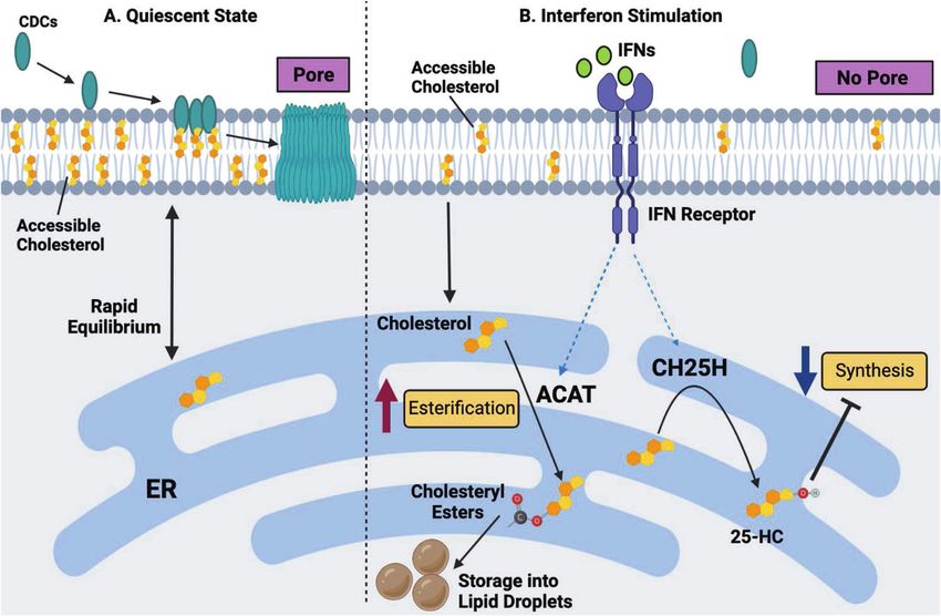
Fig. 4 A working model of interferon-mediated protection of macrophages against CDC-induced cytotoxicity. A In a quiescent state, CDCs
target metabolically active (or accessible) cholesterol in the plasma membrane of macrophages, resulting in pore formation and the
subsequent loss of membrane integrity. B IFN stimulation markedly decreases the size of the accessible cholesterol pool, resulting in reduced
CDC binding and pore formation on the plasma membrane. Alterations in the accessible cholesterol pool in the plasma membrane are driven
by a reduction in cholesterol biosynthesis and heightened cholesterol esterification. The inhibition of cholesterol synthesis and esterification is
dependent, in part, on the upregulation of the interferon-stimulated gene, Ch25h, and the production of oxysterol 25HC. 25HC decreases
synthesis via the degradation of the HMGCR enzyme and the inhibition of the SREBP2 transcriptional pathway. 25HC also facilitates
cholesterol esterification via the ER-residential enzymes ACAT1 and ACAT2.
polymicrobial infections will sensitize host cells to the harmful effects ACAT enzymes increased the sensitivity of macrophages to CDCs,
of CDCs and quickly overcome the protective effects induced by IFNs. even in the absence of CH25H. Thus, we proposed that IFNs
It has also been shown that IFN signaling, downstream of TLR4, results induce a robust but highly selective cholesterol redistribution
in the accumulation of lanosterol, a sterol intermediate of the program that moves cholesterol targeted by CDCs out of the PM,
cholesterol biosynthetic pathway, in the PMs of macrophages [62]. redistributes it to the ER, and subsequently stores esterified
This increase in lanosterol levels alters membrane fluidity, which cholesterol if needed. Inhibiting cholesterol synthesis with 25HC is
potentiates phagocytosis by macrophages and the killing of E. coli necessary to prevent this small but highly labile pool from refilling
[62]. Therefore, it remains possible that the accumulation of lanosterol and resensitizing macrophages to CDC toxins. A working model of
or other sterol intermediates in the PM in response to IFN signaling how IFN-mediated reprogramming of cholesterol homeostasis
contributes to this protective effect, perhaps through the dilution of promotes resistance to CDCs is shown in Fig. 4, however many of
the accessible cholesterol pool. However, this hypothesis needs to be the mechanistic details of this proposed model need to be better
formally tested. defined and tested.
It also remains unclear where the cholesterol targeted by CDCs A recent complementary study showed that IFN-mediated
is moved in response to IFN signaling. One possibility is that production of 25HC increased cellular immunity to L. monocyto-
cholesterol moves into another cholesterol pool within the plasma genes and Shigella flexneri by inhibiting cell–cell spreading [34].
membrane. We measured sphingomyelin-associated cholesterol This study found that 25HC interfered with the ability of these
by imaging macrophages with the mushroom toxin protein, microbes to traverse the plasma membrane of infected cells and
ostreolysin (OlyA), to test this possibility. In contrast to ALO-D4 enter uninfected neighboring cells through double plasma
staining, imaging macrophages with recombinant OlyA protein membrane structures. The molecular mechanism is also under
revealed little difference in staining between the IFN and control investigation but appears to be dependent on rapid internaliza-
groups [21]. Thus, it does not appear that cholesterol from the tion of the accessible cholesterol pool via the activation of ACATs
CDC-targeted pool flows into the sphingomyelin-associated pool and subsequent storage of this cholesterol as esters in a process
in response to IFNs. We were unable to test whether cholesterol that is highly analogous to that seen in macrophages [34].
moves into the essential pool since we cannot define this pool in However, it is important to note that 25HC was not effective at
macrophages. Additional biochemical studies on membrane blocking L. monocytogenes escape from the phagocytic vacuole,
fractions will be required to determine whether the lateral which is a process that requires listeriolysin O (LLO), a CDC
movement of cholesterol occurs in response to IFN and mediates produced by these microbes. Therefore, the mechanism by which
protection of the PM to CDCs. IFN-induced changes in the accessible cholesterol pool alter CDC
An alternative explanation is that the cholesterol required for sensitivity may only be relevant to the plasma membrane.,
CDC recognition is rapidly moved into another subcellular Nevertheless, this study reinforces the growing concept that
location. In support of this concept, we observed that IFNs reprogramming the pool of accessible plasma membrane
upregulated several genes involved in intracellular cholesterol cholesterol with IFNs can protect cells from damage induced by
movement (e.g., Gramd1b, Stard3, and Npc1/2) and storage (Soat microbes.
1/2). Moreover, we found that IFNs induced the accumulation of a Based on these new concepts in membrane cholesterol
small amount of cholesterol esters in macrophages. Inhibiting homeostasis, it will be interesting to revisit whether other
Cellular & Molecular Immunology (2022) 19:327 – 336M.-S. Lee and S.J. Bensinger
334
intracellular microbes that exploit cellular cholesterol for their determine the extent to which activation signals reshape lipid
lifecycle depend on the accessible cholesterol pool. Given that composition in the context of infections. Much of our knowledge
both type I and type II IFNs regulate the size of the metabolically about lipid metabolic reprogramming is predicated on knowledge
active cholesterol pool, we suspect that this small pool of gained using highly reductionist systems. The intrinsic complexity
cholesterol will also be necessary for viruses that rely on of infections will undoubtedly muddy the water of our current
cholesterol for entry. 25HC has been shown to block viral entry working models. We predict that there will be instances in which
[22, 23, 63], so it is possible that 25HC-mediated regulation of the pathogens misdirect macrophages to acquire the wrong lipidome,
metabolically active cholesterol pool in the PM is one of the ostensibly interfering with requisite effector functions to clear
molecular mechanisms underlying the antiviral effect of 25HC. infections. It will be exciting for the field to generate comprehen-
sive pathogen-based immune metabolic studies to guide our
thinking and shape models.
FUTURE DIRECTIONS: NECROTIZING FASCIITIS AND A Another series of questions for the field to address center
SPECULATIVE LINK TO CHOLESTEROL METABOLISM around the issues of durability and plasticity. The approaches we
Necrotizing fasciitis (NF), also known as flesh-eating disease, is a and others have taken reasonably focus on short-lived inflamma-
subset of an aggressive skin and soft tissue infection consisting of tory macrophages. It remains unclear how durable these changes
liquefying necrosis of dermal and subcutaneous tissues [64]. NF is in lipid composition are and whether macrophages can undergo
mediated by select gram-positive microbes that secrete toxins, secondary reshaping of their lipidome in response to newly
including CDCs and hemolytic toxins into infected and surround- received information. For example, we envision that exposure to
ing tissues [65]. The observation that metabolic reprogramming of different cytokines throughout an immune response will continu-
cholesterol metabolism attenuates CDC-mediated cytotoxicity and ally reshape the lipidome to match required effector functions. .
tissue damage in the skin is striking. It is tantalizing to hypothesize Alternatively, it is possible that initial exposure to a specific
that the dysregulation of tissue lipid homeostasis could influence proinflammatory cytokine (i.e., IFN-γ) may render cells refractory to
the extent of tissue damage associated with necrotizing soft tissue any changes in lipid composition, essentially removing any
infection., Development of NF is rare and it remains unclear plasticity in metabolism. Macrophages have variable lifespans,
why individuals develop NF. However, it is worth noting that and resident tissue macrophages are long-lived with some self-
comorbidities associated with the development of NF include renewal capabilities [3]. It will be important to determine whether
metabolic diseases, such as obesity and diabetes [66, 67]. Thus, it inflammation-driven metabolic reprogramming of lipid metabo-
is possible that the dysregulation of lipid metabolism, in particular lism indelibly imprints on long-lived macrophages, their progeny
cholesterol homeostasis, sensitizes individuals to the deleterious in tissues, or myeloid stem cells. If such an observation was found
effects of CDCs and other toxins that drive the pathogenesis of NF. to be true, this could be critical for understanding pathogenic
It will be necessary for the field to test these interesting but circuits that link metabolic disease (e.g., atherosclerosis) and
nascent ideas. Likewise, it will be exciting to determine whether inflammation in apparent feed-forward systems. This type of
targeting lipid metabolism in infected tissues attenuates the indelible programming could also help to explain aspects of
development of NF, particularly in individuals who have pre- innate immune memory or the capacity of innate immune cells to
existing lipid metabolic dysregulation. If proven true, this concept generate preferential immunity to pathogens upon subsequent
will open new avenues for developing adjunctive therapies to exposures. Tackling these exciting and important questions will
attenuate these rare but highly pathogenic skin and soft tissue undoubtedly advance our mechanistic understanding of immu-
infections. nometabolism. We also believe that continued research into the
crosstalk between lipid metabolism and the function of macro-
phages will provide essential insights for developing new
CONCLUSIONS therapeutic approaches to control unwanted inflammation,
It is now clear that reshaping lipid composition is an integral and infections, and metabolic diseases.
essential part of myeloid cell differentiation and function. In the
absence of proper lipid metabolic reprogramming, macrophages
exhibit dysregulated inflammatory responses and altered immune REFERENCES
functions. These observations suggest that environmental or 1. Takeda K, Kaisho T, Akira S. Toll-like receptors. Annu Rev Immunol.
metabolic signals that interfere with the metabolic reprogram- 2003;21:335–76.
ming of lipid composition will result in phagocyte dysfunction. An 2. Ozinsky A, Underhill DM, Fontenot JD, Hajjar AM, Smith KD, Wilson CB, et al. The
additional layer of complexity lies in the observation that repertoire for pattern recognition of pathogens by the innate immune system is
macrophages do not converge on a single lipidome irrespective defined by cooperation between Toll-like receptors. Proc Natl Acad Sci.
of the activating signal. Instead, distinct proinflammatory stimuli 2000;97:13766–71.
drive the acquisition of different lipidomes [17]. These data 3. Wynn TA, Chawla A, Pollard JW. Macrophage biology in development, home-
ostasis and disease. Nature. 2013;496:445–55.
indicate considerable specificity in the lipidome that macro-
4. Hubler MJ, Kennedy AJ. Role of lipids in the metabolism and activation of
phages acquire during different inflammatory responses. More- immune cells. J Nutr Biochem. 2016;34:1–7.
over, these specific changes in lipid composition appear to impart 5. Russell DG, Huang L, VanderVen BC. Immunometabolism at the interface
information to the cell that ultimately regulates distinct effector between macrophages and pathogens. Nat Rev Immunol. 2019;19:291–304.
functions and immunity. We expect that there will be instances in 6. Kominsky DJ, Campbell EL, Colgan SP. Metabolic shifts in immunity and inflam-
which reprogramming of lipid composition will be beneficial for mation. J Immunol. 2010;184:4062–8.
some forms of immunity but harmful to other forms of immunity. 7. Lange Y, Swaisgood MH, Ramos BV, Steck TL. Plasma membranes contain half the
For example, reprogramming of cholesterol metabolism may phospholipid and 90% of the cholesterol and sphingomyelin in cultured human
benefit antiviral immune responses but interfere with antimicro- fibroblasts. J Biol Chem. 1989;264:3786–93.
8. Ikonen E. Cellular cholesterol trafficking and compartmentalization. Nat Rev Mol
bial responses. This additional layer of complexity also suggests
Cell Biol. 2008;9:125–38.
that a context-specific approach to correcting the metabolism of 9. van Meer G, Voelker DR, Feigenson GW. Membrane lipids: where they are and
macrophages and other immune cells will be necessary if one how they behave. Nat Rev Mol Cell Biol. 2008;9:112–24.
hopes to normalize function. 10. Luo J, Yang H, Song BL. Mechanisms and regulation of cholesterol homeostasis.
Of course, there remain many important and unresolved Nat Rev Mol Cell Biol. 2020;21:225–45.
questions about lipid metabolic reprogramming that the field of 11. Guo H, Callaway JB, Ting JP. Inflammasomes: mechanism of action, role in dis-
immunometabolism should address. One crucial issue is to ease, and therapeutics. Nat Med. 2015;21:677–87.
Cellular & Molecular Immunology (2022) 19:327 – 336M.-S. Lee and S.J. Bensinger
335
12. Tall AR, Yvan-Charvet L. Cholesterol, inflammation and innate immunity. Nat Rev 40. Cassidy SK, O’Riordan MX. More than a pore: the cellular response to cholesterol-
Immunol. 2015;15:104–16. dependent cytolysins. Toxins (Basel). 2013;5:618–36.
13. Brown MS, Goldstein JL. The SREBP pathway: regulation of cholesterol metabolism 41. Ouweneel AB, Thomas MJ, Sorci-Thomas MG. The ins and outs of lipid rafts:
by proteolysis of a membrane-bound transcription factor. Cell. 1997;89:331–40. functions in intracellular cholesterol homeostasis, microparticles, and cell mem-
14. Madison BB. Srebp2: A master regulator of sterol and fatty acid synthesis. J Lipid branes: thematic review series: biology of lipid rafts. J Lipid Res. 2020;61:676–86.
Res. 2016;57:333–5. 42. Johnson KA, Radhakrishnan A. Accessibility of cholesterol at cell surfaces. J Lipid
15. Lee SH, Lee J-H, Im S-S. The cellular function of SCAP in metabolic signaling. Exp Res. 2020;61:1307.
Mol Med. 2020;52:724–9. 43. Henry B, Ziobro R, Becker KA, Kolesnick R, Gulbins E. Acid sphingomyelinase.
16. Chatterjee S, Szustakowski JD, Nanguneri NR, Mickanin C, Labow MA, Nohturfft A, Handb Exp Pharmacol. 2013;215:77–88.
et al. Identification of novel genes and pathways regulating SREBP transcriptional 44. Platanias LC. Mechanisms of type-I- and type-II-interferon-mediated signalling.
activity. PLoS ONE. 2009;4:e5197. Nat Rev Immunol. 2005;5:375–86.
17. Hsieh WY, Zhou QD, York AG, Williams KJ, Scumpia PO, Kronenberger EB, et al. 45. York AG, Williams KJ, Argus JP, Zhou QD, Brar G, Vergnes L. et al. Limiting
Toll-like receptors induce signal-specific reprogramming of the macrophage cholesterol biosynthetic flux spontaneously engages type I IFN signaling. Cell.
lipidome. Cell Metab. 2020;32:128–43.e5. 2015;163:1716–29.
18. Kidani Y, Elsaesser H, Hock MB, Vergnes L, Williams KJ, Argus JP, et al. Sterol 46. Ormsby TJR, Owens SE, Horlock AD, Davies D, Griffiths WJ, Wang Y, et al. Oxy-
regulatory element-binding proteins are essential for the metabolic program- sterols protect bovine endometrial cells against pore-forming toxins from
ming of effector T cells and adaptive immunity. Nat Immunol. 2013; pathogenic bacteria. Faseb J. 2021;35:e21889.
14:489–99. 47. Sviridov D, Bukrinsky M. Interaction of pathogens with host cholesterol meta-
19. Covarrubias AJ, Aksoylar HI, Horng T. Control of macrophage metabolism and bolism. Curr Opin Lipidol. 2014;25:333–8.
activation by mTOR and Akt signaling. Semin Immunol. 2015;27:286–96. 48. Samanta D, Mulye M, Clemente TM, Justis AV, Gilk SD. Manipulation of host
20. Lu H, Talbot S, Robertson KA, Watterson S, Forster T, Roy D. et al. Rapid pro- cholesterol by obligate intracellular bacteria. Front Cell Infect Microbiol.
teasomal elimination of 3-hydroxy-3-methylglutaryl-CoA reductase by interferon- 2017;12:727.
γ in primary macrophages requires endogenous 25-hydroxycholesterol synthesis. 49. Russell DG, Cardona PJ, Kim MJ, Allain S, Altare F. Foamy macrophages and the
Steroids. 2015;99:219–29. progression of the human tuberculosis granuloma. Nat Immunol. 2009;10:943–8.
21. Zhou QD, Chi X, Lee MS, Hsieh WY, Mkrtchyan JJ, Feng AC, et al. Interferon- 50. Pandey AK, Sassetti CM. Mycobacterial persistence requires the utilization of host
mediated reprogramming of membrane cholesterol to evade bacterial toxins. cholesterol. Proc Natl Acad Sci USA. 2008;105:4376–80.
Nat Immunol. 2020;21:746–55. 51. Marques MA, Berrêdo-Pinho M, Rosa TL, Pujari V, Lemes RM, Lery LM, et al. The
22. Blanc M, Hsieh WY, Robertson KA, Kropp KA, Forster T, Shui G. et al. The tran- essential role of cholesterol metabolism in the intracellular survival of myco-
scription factor STAT-1 couples macrophage synthesis of 25-hydroxycholesterol bacterium leprae is not coupled to central carbon metabolism and energy pro-
to the interferon antiviral response. Immunity. 2013;38:106–18. duction. J Bacteriol. 2015;197:3698–707.
23. Liu SY, Aliyari R, Chikere K, Li G, Marsden MD, Smith JK. et al. Interferon-inducible 52. Teng O, Ang CKE, Guan XL. Macrophage-bacteria interactions-a lipid-centric
cholesterol-25-hydroxylase broadly inhibits viral entry by production of 25- relationship. Front Immunol. 2017;8:1836.
hydroxycholesterol. Immunity. 2013;38:92–105. 53. Bukrinsky MI, Mukhamedova N, Sviridov D. Lipid rafts and pathogens: the art of
24. Cyster JG, Dang EV, Reboldi A, Yi T. 25-Hydroxycholesterols in innate and deception and exploitation. J Lipid Res. 2020;61:601–10.
adaptive immunity. Nat Rev Immunol. 2014;14:731–43. 54. Ramachandran R, Tweten RK, Johnson AE. Membrane-dependent conformational
25. Zhao J, Chen J, Li M, Chen M, Sun C. Multifaceted functions of CH25H and 25HC changes initiate cholesterol-dependent cytolysin oligomerization and inter-
to modulate the lipid metabolism, immune responses, and broadly antiviral subunit beta-strand alignment. Nat Struct Mol Biol. 2004;11:697–705.
activities. Viruses. 2020;12:727. 55. Heuck AP, Moe PC, Johnson BB. The cholesterol-dependent cytolysin family of
26. Bah SY, Dickinson P, Forster T, Kampmann B, Ghazal P. Immune oxysterols: role in gram-positive bacterial toxins. Subcell Biochem. 2010;51:551–77.
mycobacterial infection and inflammation. J Steroid Biochem Mol Biol. 56. Thapa R, Ray S, Keyel PA. Interaction of macrophages and cholesterol-dependent
2017;169:152–63. cytolysins: the impact on immune response and cellular survival. Toxins (Basel).
27. Spann NJ, Glass CK. Sterols and oxysterols in immune cell function. Nat Immunol. 2020;12:531
2013;14:893–900. 57. Blazek AD, Paleo BJ, Weisleder N. Plasma membrane repair: a central process for
28. Bensinger SJ, Tontonoz P. Integration of metabolism and inflammation by lipid- maintaining cellular homeostasis. Physiol (Bethesda). 2015;30:438–48.
activated nuclear receptors. Nature. 2008;454:470–7. 58. Ioannidis I, Ye F, McNally B, Willette M, Flaño E. Toll-like receptor expression and
29. Frolov A, Zielinski SE, Crowley JR, Dudley-Rucker N, Schaffer JE, Ory DS. NPC1 and induction of type I and type III interferons in primary airway epithelial cells. J
NPC2 regulate cellular cholesterol homeostasis through generation of low den- Virol. 2013;87:3261–70.
sity lipoprotein cholesterol-derived oxysterols. J Biol Chem. 2003;278:25517–25. 59. Radhakrishnan A, Ikeda Y, Kwon HJ, Brown MS, Goldstein JL. Sterol-regulated
30. Phillips MC. Molecular mechanisms of cellular cholesterol efflux. J Biol Chem. transport of SREBPs from endoplasmic reticulum to Golgi: oxysterols block
2014;289:24020–9. transport by binding to Insig. Proc Natl Acad Sci USA. 2007;104:6511–8.
31. He P, Gelissen IC, Ammit AJ. Regulation of ATP binding cassette transporter A1 60. DeBose-Boyd RA. Feedback regulation of cholesterol synthesis: sterol-accelerated
(ABCA1) expression: cholesterol-dependent and - independent signaling path- ubiquitination and degradation of HMG CoA reductase. Cell Res. 2008;18:609–21.
ways with relevance to inflammatory lung disease. Respir Res. 2020;21:250. 61. Huseby M, Shi K, Brown CK, Digre J, Mengistu F, Seo KS, et al. Structure and biological
32. Pandzic E, Gelissen IC, Whan R, Barter PJ, Sviridov D, Gaus K, et al. The ATP activities of beta toxin from Staphylococcus aureus. J Bacteriol. 2007;189:8719–26.
binding cassette transporter, ABCG1, localizes to cortical actin filaments. Sci Rep. 62. Araldi E, Fernández-Fuertes M, Canfrán-Duque A, Tang W, Cline GW, Madrigal-
2017;7:42025. Matute J, et al. Lanosterol modulates TLR4-mediated innate immune responses in
33. Chang TY, Li BL, Chang CC, Urano Y. Acyl-coenzyme A: cholesterol acyl- macrophages. Cell Rep. 2017;19:2743–55.
transferases. Am J Physiol Endocrinol Metab. 2009;297:E1–9. 63. Doms A, Sanabria T, Hansen JN, Altan-Bonnet N, Holm GH. 25-Hydroxycholesterol
34. Abrams ME, Johnson KA, Perelman SS, Zhang L-S, Endapally S, Mar KB. et al. production by the cholesterol-25-hydroxylase interferon-stimulated gene
Oxysterols provide innate immunity to bacterial infection by mobilizing cell restricts mammalian reovirus infection. J Virol. 2018;92:e01047-18
surface accessible cholesterol.Nat Microb. 2020;5:929–42. 64. Olsen RJ, Musser JM. Molecular pathogenesis of necrotizing fasciitis. Annu Rev
35. Lei L, Xiong Y, Chen J, Yang JB, Wang Y, Yang XY, et al. TNF-alpha stimulates the Pathol. 2010;5:1–31.
ACAT1 expression in differentiating monocytes to promote the CE-laden cell 65. Bhattacharjee P, Keyel PA. Cholesterol-dependent cytolysins impair pro-
formation. J Lipid Res. 2009;50:1057–67. inflammatory macrophage responses. Sci Rep. 2018;8:6458.
36. Das A, Brown MS, Anderson DD, Goldstein JL, Radhakrishnan A. Three pools of 66. Huttunen R, Syrjänen J. Obesity and the risk and outcome of infection. Int J Obes
plasma membrane cholesterol and their relation to cholesterol homeostasis. Elife. (Lond). 2013;37:333–40.
2014;3:e02882. 67. Karlsson EA, Beck MA. The burden of obesity on infectious disease. Exp Biol Med
37. Gay A, Rye D, Radhakrishnan A. Switch-like responses of two cholesterol sensors do (Maywood). 2010;235:1412–24.
not require protein oligomerization in membranes. Biophys J. 2015;108:1459–69.
38. Das A, Goldstein JL, Anderson DD, Brown MS, Radhakrishnan A. Use of mutant
125I-perfringolysin O to probe transport and organization of cholesterol in
membranes of animal cells. Proc Natl Acad Sci USA. 2013;110:10580–5. ACKNOWLEDGEMENTS
39. Endapally S, Infante RE, Radhakrishnan A. Monitoring and modulating intracel- MSL was supported by NIH Ruth L. Kirschstein National Research Service Award AI
lular cholesterol trafficking using ALOD4, a cholesterol-binding protein. Methods 007323. SJB was supported by NIH HL 146358 and HL 157710. Figures were created
Mol Biol. 2019;1949:153–63. with Biorender software at Biorender.com.
Cellular & Molecular Immunology (2022) 19:327 – 336You can also read