Reconstruction of the Hip in Multiple Hereditary Exostoses
←
→
Page content transcription
If your browser does not render page correctly, please read the page content below
children
Article
Reconstruction of the Hip in Multiple Hereditary Exostoses
Dong Hoon Lee 1 and Dror Paley 2, *

                                          1   Donghoon Advanced Lengthening Reconstruction Institute, Superstar tower 3-5F 10, Wiryeseoil-ro,
                                              Sujeong-gu, Seongnam-si 11962, Gyeonggi-do, Korea; orthopaedee@naver.com
                                          2   Paley Orthopedic and Spine Institute, Kimmel, 901 45th St, West Palm Beach, FL 33407, USA
                                          *   Correspondence: dpaley@paleyinstitute.org; Tel.: +1-561-844-5255; Fax: +1-561-844-5245

                                          Abstract: The hip joint involvement in multiple hereditary exostoses (MHE) occurs in 30–90%,
                                          causing pain and limitation of motion by femoroacetabular impingement, coxa valga, acetabular
                                          dysplasia, hip joint subluxation, and osteoarthritis. The purpose of this study was to investigate the
                                          clinical and radiographic outcomes of ten hips in seven patients treated by surgical dislocation and
                                          corrective osteotomies between 2004 and 2009. Surgical dislocation and excision of the osteochon-
                                          dromas and varus intertrochanteric osteotomies were performed in all cases when the neck–shaft
                                          angle was >150◦ . Common sites of osteochondromas were medial, posterior, and anterior neck of the
                                          femur. Neck–shaft angle of the femur was improved from a mean of 157◦ to 139◦ , postoperatively.
                                          On an average, the center-edge angle improved from 20◦ to 30◦ postoperatively. We believe that
                                          Ganz’s safe surgical dislocation technique is the preferred treatment of MHE. This safeguards the
                                          circulation of the femoral head and the osteochondromas can be resected under direct vision. It can
                                          be combined with additional corrective osteotomies because the hip affected by MHE is frequently
                                          associated with dysplastic changes which can result in premature osteoarthritis.


                                          Keywords: hip; multiple hereditary exostoses (MHE); femuro-acetabular impingement; coxa valga;
                                   surgical hip dislocation; varus intertrochanteric osteotomy
Citation: Lee, D.H.; Paley, D.
Reconstruction of the Hip in Multiple
Hereditary Exostoses. Children 2021,
8, 490. https://doi.org/10.3390/          1. Introduction
children8060490                                 Multiple hereditary exostoses (MHE) is an autosomal-dominant disorder with a
                                          prevalence of 1:50,000 persons within the general population [1]. The EXT1 and EXT2 genes
Academic Editor: Vito Pavone              located at 8q24 and 11p11–p12, respectively, are known to be associated with MHE [2,3].
                                                A variety of problems can be related to MHE and the majority of these problems are
Received: 6 May 2021
                                          related to the bony protrusions affecting the surrounding joints, muscles, tendons, nerves,
Accepted: 6 June 2021
                                          blood vessels, and skin. Common problems are pain, restricted range of motion, and
Published: 8 June 2021
                                          cosmetic concerns. Pain may come from repeated motion over prominent osteochondroma,
                                          bursa formation, and joint impingement [1,4]. Some osteochondromas tether the growth
Publisher’s Note: MDPI stays neutral
                                          plate. This can lead to asymmetric growth of the growth plate and consequent limb
with regard to jurisdictional claims in
                                          deformity. The common deformities of the lower extremity include short stature, leg length
published maps and institutional affil-
                                          discrepancy, and valgus deformities of the knee, ankle, and proximal femur [4,5].
iations.
                                                MHE incidence at the proximal femur and pelvis has been reported from 30% to
                                          90% and 15% to 62%, respectively [1,6,7]. The hip area involvement can lead to coxa
                                          valga [5,8–11], acetabular dysplasia [5,9], femoroacetabular impingement (FAI) [9,10,12],
                                          hip joint subluxation [9,10,12], and osteoarthritis [13,14]. Major concerns in the hip joint are
Copyright: © 2021 by the authors.
                                          pain and limited range of motion by FAI. Dysplastic hip which can be caused by FAI and
Licensee MDPI, Basel, Switzerland.
                                          by coxa valga is also a big problem [9–11]. Thus, proper treatment in MHE hip is important
This article is an open access article
                                          to eliminate pain and limit motion to prevent premature hip arthritis. There have been only
distributed under the terms and
                                          a few reports about the surgical treatment of one- or two-hip case series of hip exostoses
conditions of the Creative Commons
Attribution (CC BY) license (https://
                                          due to MHE [10–12,15–17]. The purpose of this study was to investigate the clinical and
creativecommons.org/licenses/by/
                                          radiographic outcomes of ten hips in seven patients treated by surgical dislocation and
4.0/).                                    corrective osteotomies.

Children 2021, 8, 490. https://doi.org/10.3390/children8060490                                             https://www.mdpi.com/journal/children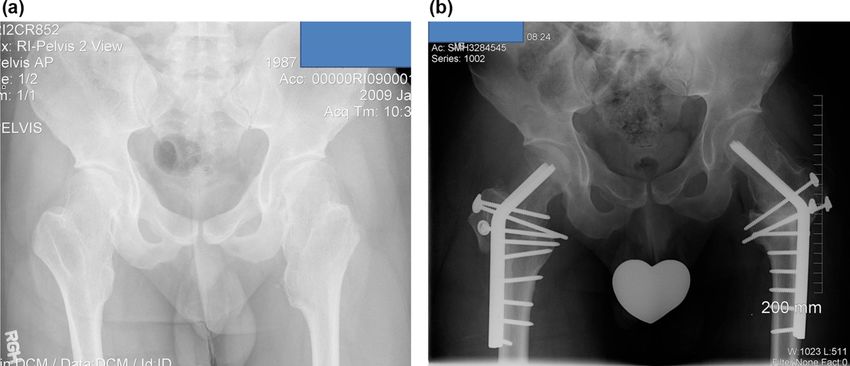
Children 2021, 8, 490                                                                                                              2 of 10

                                   2. Material and Methods
                                       All patients who underwent hip surgery due to MHE were included, and patients who
                                  did not follow-up after surgery or did not have appropriate X-rays or medical records were
                                  excluded from the study. The records of ten hips in seven patients with MHE treated by
                                  surgical dislocation of the hip joint between 2004 and 2009 were retrospectively reviewed
                                  (Table 1). The main reasons for visiting the clinic were hip pain (ten hips) and motion
                                  limitation (nine hips). The hip pain was aggravated with motion especially flexion, internal
                                  rotation, and adduction. One case presented a sciatica-like symptom of the right leg
                                  (Figure 1a–c). The diagnosis was initially made on plain radiographs. One patient was
                                  diagnosed with Langer–Giedion syndrome [18], which had MHE with a unicameral cyst
                                  (Figure 2a,b). Three-dimensional reconstructive computerized tomography and bone
                                  scanning were performed in selected persons for accurate surgical planning if the location
                                  and the shape of the mass was not clearly identified by plain x-ray. The study included four
                                  male and three female patients, and the average age at the time of surgery was 22 (range,
Children 2021, 8, x FOR PEER REVIEW
                                  6–39) years. The right side was involved in one patient, the left side in one patient, and
                                  both sides in five patients. However, two patients underwent surgery on the unilateral side
                                  because the other side was free from any disability among the patients with both lesions.

                                    Figure
                                   Figure   1. 1.
                                                AA    14‐year‐old
                                                  14-year-old         girl complained
                                                                girl complained             of posterior
                                                                                   of posterior  thigh painthigh
                                                                                                              at thepain
                                                                                                                     rightat  the extremity
                                                                                                                           lower  right lowerthatextremit
                                    was    aggravated      with   straight‐leg     raising   (case  no.  2). (a)  Preoperative     anteroposterior
                                   was aggravated with straight-leg raising (case no. 2). (a) Preoperative anteroposterior radiograph                 rad
                                   ofofthe
                                         the  right
                                           right hiphip   shows
                                                      shows  a smalla small
                                                                      bump at bump     at the
                                                                                 the medial     medial
                                                                                             side        side of neck.
                                                                                                  of the femoral   the femoral    neck. (b)
                                                                                                                         (b) Preoperative     Preoperati
                                                                                                                                          lateral
                                    radiograph
                                   radiograph         of the
                                                  of the rightright  hip shows
                                                               hip shows    a large abump
                                                                                      largeatbump     at the side
                                                                                               the posterior   posterior   side ofneck.
                                                                                                                   of the femoral   the femoral
                                                                                                                                         (c) The neck.
                                   sciatica-like
                                    sciatica‐like symptom
                                                      symptomwas completely     resolved resolved
                                                                   was completely         after complete
                                                                                                      afterresection
                                                                                                             completeof the osteochondromas
                                                                                                                          resection            of
                                                                                                                                     of the osteochond
                                   the  posterior  femoral  neck.
                                    the posterior femoral neck.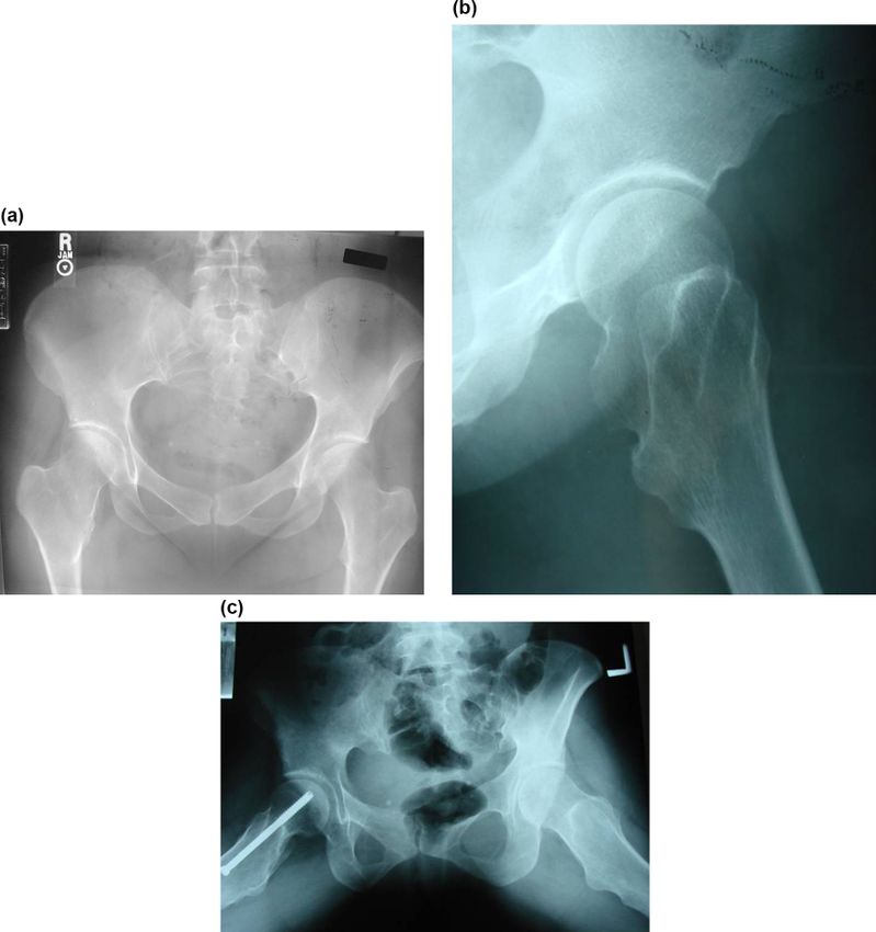
Figure 1. A 14‐year‐old girl complained of posterior thigh pain at the right lower extremi
was aggravated with straight‐leg raising (case no. 2). (a) Preoperative anteroposterior rad
of the right hip shows a small bump at the medial side of the femoral neck. (b) Preoperati
radiograph of the right hip shows a large bump at the posterior side of the femoral neck.
Children 2021, 8, 490 3 of 10
sciatica‐like symptom was completely resolved after complete resection of the osteochond
the posterior femoral neck.
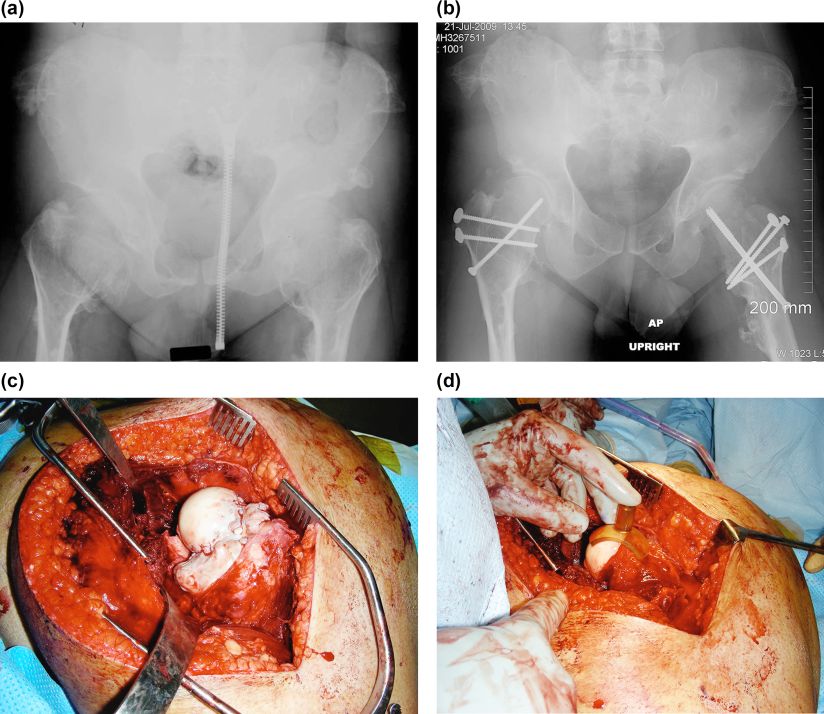
Figure
Figure 2. The
2. The images
images illustrate
illustrate the casethe
of acase of a 14‐year‐old
14-year-old boy with Langer–Giedion
boy with Langer–Giedion syndrome (case syndro
no. 3). (a) Preoperative anteroposterior radiograph of the left hip shows osteochondroma
no. 3). (a) Preoperative anteroposterior radiograph of the left hip shows osteochondromas at the
femoral
femoral head and and
head a completely dislocated
a completely hip. (b) Dislocated
dislocated hip. (b) hip.
Dislocated hip.
Table 1. Demographic data.
Age at Operation Follow-Up Period Site of Tumor Symptoms at
Case Gender Involved Side
(Years) (Months) at Hip Presentation
1 Male R 6 84 A/M/P Hip pain, LOM
Sciatic nerve Sx.,
2 Female R 39 80 M/P
Hip pain
3 Male L 14 97 FH Hip pain, LOM
4 Female L 11 M/P Hip pain, LOM
R: 23 50 A/M/P Hip pain, LOM
5 Male B
L: 26 13 A/M/P Hip pain, LOM
R: 22
6 Male B L: 25 12 A/M/P Hip pain, LOM
R: 26
L: 22 19 A/M/P Hip pain, LOM
7 Female B R: 28 A/M Hip pain, LOM
L: 27 40 A/M/P Hip pain, LOM
A, anterior; M, medial; P, posterior; R, right; L, left; B, both; LOM, limitation of motion; FH, femoral hip.
The radiographic assessment of the proximal femur was performed by evaluating
the location of the mass, the femoral neck–shaft angle, Shenton’s line, and the ratio of
the diameter of the femoral neck to that of the shaft (NSR) as an index of femoral neck
overgrowth and surgical excision amount [1,5]. Moreover, NSR was measured as the
diameter of the femoral neck divided by the diameter of the femoral shaft just distal to the
lesser trochanter (Figure 3a–d). The acetabulum was evaluated by Sharp’s acetabular angle
and the center-edge angle of Wieberg [19,20].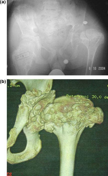
diameter of the femoral neck to that of the shaft (NSR) as an index o
                         growth and surgical excision amount [1,5]. Moreover, NSR was meas
                         of the femoral neck divided by the diameter of the femoral shaft jus
Children 2021, 8, 490    trochanter (Figure 3a,b,c,d). The acetabulum was evaluated  4 ofby
                                                                                         10 Shar

                         and the center‐edge angle of Wieberg [19,20].

                          Figure
                        Figure      3.images
                                3. The  The images
                                               show the caseshow       the caseman
                                                                 of a 23-year-old  of with
                                                                                       a 23‐year‐old         man with
                                                                                            symptomatic bilateral          symptomatic b
                                                                                                                   femoroacetabular
                        impingement pain and limitation of motion (case no. 5). (a) Preoperative anteroposterior radiograph
                          tabular impingement pain and limitation of motion (case no. 5). (a) Preopera
                        shows a large bump at the medial side of the femoral neck. Neck–shaft angle of the femur was
                          radiograph
                        within             shows
                                tolerable range         a large
                                                  (Rt, 140;          bump
                                                             Lt, 150).        at the medial
                                                                        The containment            side
                                                                                          of the hip was of   the femoral
                                                                                                           normal.             neck. Neck
                                                                                                                   (b) Postoperative
                        anteroposterior   within tolerable
                          femur was radiograph            of the hip. range     (Rt, 140;
                                                                         Mediolateral        Lt, 150).
                                                                                       neck–shaft          The containment
                                                                                                    ratio improved                 of the h
                                                                                                                     from 6.7 to 1.6
                        onPostoperative anteroposterior radiograph of the hip. Mediolateral neck–shaf
                            the right hip, 3.6  to  1.7  on the  left hip. A  cortical or cannulated   screw   was inserted  through
                        the femoral neck to prevent femoral neck fracture and the osteotomized greater trochanter was
                          6.7 to 1.6 on the right hip, 3.6 to 1.7 on the left hip. A cortical or cannulated sc
                        fixed with two-three 3.5 mm cortical screws. (c) Intraoperative photograph of the left hip. (d) The
                          through the femoral
                        osteochondroplasty                 neck
                                              of the femoral        tocan
                                                                 neck    prevent    femoral
                                                                           be performed          neckosteotomes
                                                                                          with small      fractureandand    the osteotom
                                                                                                                        a high-speed
                          ter was
                        burr.  Whenfixed      with two‐three
                                       the resection    is satisfactory,3.5   mm template
                                                                          a special cortical(spherometer
                                                                                                screws. (c)gage) Intraoperative
                                                                                                                     can be used to photo
                          (d) The osteochondroplasty of the femoral neck can be performed
                        check  the sphericity  of  the  remaining    femoral  head. The  neck–shaft   ratio improved            with
                                                                                                                      from 3.6 to 1.7 small
                        mediolaterally and 2.7 to 1.3 anteroposteriorly.
                         high‐speed burr. When the resection is satisfactory, a special template (spher
                         usedAllto
                                 tencheck    the sphericity
                                      hips underwent    surgeryof
                                                                to the  remaining
                                                                    excise           femoral
                                                                           osteochondromas     head.
                                                                                           using  safe The  neck–shaft
                                                                                                       surgical hip    r
                        dislocation  [21] by the senior author (DP).  One  female and
                         3.6 to 1.7 mediolaterally and 2.7 to 1.3 anteroposteriorly.  two male patients had staged
                        bilateral surgeries. Varus intertrochanteric osteotomy was performed when the neck–shaft
                        angle was more than 150◦ (Figure 4a,b). After surgery, follow-up was performed at 4-week
                        intervalsAll
                                   untilten  hips
                                         a bony     underwent
                                                union                surgery
                                                       was established,         to excise
                                                                         and thereafter,      osteochondromas
                                                                                         observations were made at usi
                        intervals of 6 months to 1 year. All of the patients were followed with serial radiographs
                         dislocation [21] by the senior author (DP). One female and two male
                        for a mean follow-up of 49 (range, 12–97) months.
                         bilateral surgeries. Varus intertrochanteric osteotomy was perform
                         shaft angle was more than 150° (Figure 4a,b). After surgery, follow‐u
                         4‐week intervals until a bony union was established, and thereafter
                         made at intervals of 6 months to 1 year. All of the patients were follow
                         ographs for a mean follow‐up of 49 (range, 12–97) months.
ldren 2021, 8, x FOR
Children 2021,PEER
8, 490 REVIEW 5 of 10 5o
Figure 4. The case
Figure of acase
4. The 22‐year‐old manman
of a 22-year-old with symptomatic
with symptomatic bilateral femoroacetabular
bilateral femoroacetabular impingement
impingement in MHEin MHE
(case (case no. 6).
no. 6).
(a) A preoperative
(a) A preoperative anteroposterior radiograph of the hip shows a large bump at the medial side of the femoral neck and neck and
anteroposterior radiograph of the hip shows a large bump at the medial side of the femoral
severe coxa valga.
severe coxa(b) Postoperative
valga. anteroposterior
(b) Postoperative anteroposteriorradiograph
radiograph ofofthethe hip.
hip. Valgus
Valgus intertrochanteric
intertrochanteric osteotomies
osteotomies were were
performedperformed
on both on
hipsboth hips using
using bladeblade plates,
plates, and and thetrochanteric
the trochanteric osteotomy
osteotomy sitessites
werewere
fixed fixed
with 3.5 mm 3.5
with cortical
mmscrews.
cortical screws.
2.1. Surgical Procedure
2.1. Surgical Procedure
In most of the cases, surgical dislocation and excision of the osteochondromas were
In most ofwith
performed the the
cases,
use surgical
of surgicaldislocation
dislocation of and
the excision
hip describedof the
by osteochondromas
Ganz et al. [21]. we
performed with the use of
A Kocher–Langenbeck surgical
incision and dislocation of theflip
a greater trochanter hiposteotomy
described byperformed
were Ganz et al. [21]
with the patient in a lateral position on a radiolucent table.
Kocher–Langenbeck incision and a greater trochanter flip osteotomy were perform A trochanteric osteotomy
was performed with a thickness of 1.5 cm along the line of the posterior margin of the
with vastus
the patient in a lateral position on a radiolucent table. A trochanteric osteotomy w
lateralis just anterior to the most posterior insertion of the vastus medius. This
performed
left the with a thickness
posterior 2/3rd of the of piriformis
1.5 cm along tendon the
andline of the
all the posterior
other margin
short external of the vast
rotators,
lateralis just anterior
including to the
the obturator most posterior
externus, insertionthe
intact and preserved of deep
the vastus
branch ofmedius.
the medialThis left t
femoral circumflex artery. The capsule can be exposed through the
posterior 2/3rd of the piriformis tendon and all the other short external rotators, includi interval between the
gluteus minimus and the tendon of piriformis with the trochanteric fragment including
the obturator externus, intact and preserved the deep branch of the medial femoral c
the gluteus minimus flipped anteriorly. The z-shaped capsulotomy was made carefully
cumflex
not artery.
injuring The capsuleThe
the labrum. canhipbe was
exposed through
anteriorly the interval
dislocated, between
which exposes thethe gluteus m
whole
imus femoral
and the tendon
head of piriformis
and acetabulum with
with leg the trochanteric
manipulation. Removal fragment including the glute
of the osteochondromas
was performed with osteotomes and a small high-speed
minimus flipped anteriorly. The z‐shaped capsulotomy was made carefully not burr. Meticulous determination of injuri
the extension
the labrum. The hipof osteochondroplasty was made with
was anteriorly dislocated, whicha spherometer
exposes gage (Wrightfemoral
the whole Medical head a
Technology, Arlington, TN, USA; Figure 3). The acetabulum was then examined to check
acetabulum with leg manipulation. Removal of the osteochondromas was performed w
the status of the cartilage and labrum. The hip was relocated and put through a full range of
osteotomes and
motion to make a sure
smallthathigh‐speed
the impingement burr.
wasMeticulous
eliminated. Adetermination
3.5 mm cortical or of the extension
cannulated
osteochondroplasty
screw was insertedwas alongmade with
the axis a spherometer
of the femoral neck togage (Wright
prevent Medical
incidental fractureTechnology,
of the A
femoral neck (Figure 3). The greater trochanter was reattached
lington, TN, USA; Figure 3). The acetabulum was then examined to check the status of twith three 3.5 mm cortical
screws. The proximal fragment was fixed by the blade plate when varus intertrochanteric
cartilage and labrum. The hip was relocated and put through a full range of motion
osteotomy was undertaken.
make sure that the impingement was eliminated. A 3.5 mm cortical or cannulated scr
2.2. Postoperative
was inserted along Management
the axis of the femoral neck to prevent incidental fracture of the fem
ral neck Patients
(Figurewere 3). allowed
The greaterto touch down weight-bearing
trochanter was reattachedpostoperatively using bilateral
with three 3.5 mm corti
crutches in an abduction brace for 6 weeks.
screws. The proximal fragment was fixed by the blade plate when varus intertrochante
osteotomy wasConsiderations
2.3. Ethical undertaken.
All participants provided informed consent for participation. This article does not
2.2. Postoperative Management
disclose any personally identifiable data of any of the participants in any form. Thus,
Patients were allowed to touch down weight‐bearing postoperatively using bilate
crutches in an abduction brace for 6 weeks.Children 2021, 8, 490 6 of 10
consent for publication is not applicable here. All study-related procedures were conducted
following the rules of the 1975 Declaration of Helsinki.
3. Results
3.1. Clinical Results
The pain was completely resolved with the excision of the mass in all cases. One
patient had a sciatica-like symptom that was suspected to be compression neuropathy.
After surgical decompression with the excision of osteochondromas, the symptoms of the
patient were completely resolved (Figure 2). The range of motion of the hip was improved
as follows: flexion from a mean of 65◦ (range, 10◦ –110◦ ) preoperatively to a mean of 104◦
(range, 90◦ –130◦ ); extension from −3◦ (range, −20◦ –0◦ ) to 0◦ ; adduction from 12◦ (range,
0◦ –30◦ ) to 26◦ (range, 15◦ –40◦ ); abduction from 30◦ (range, 0◦ –60◦ ) to 44◦ (range, 20◦ –60◦ );
internal rotation from 21◦ (range, 0◦ –45◦ ) to 30◦ (range, 15◦ –45◦ ); and external rotation
from 31◦ (range, 10◦ –60◦ ) to 32◦ (range, 10◦ –50◦ ) (Table 2).
Table 2. Changes in range of motion of the hip joint.
Range of Motion (◦ ) Additional Procedure
Case Flexion Extension Adduction Abduction IR ER
Pre Post Pre Post Pre Post Pre Post Pre Post Pre Post
1 10 110 −10 0 5 20 45 45 10 35 60 50 VIO
2 110 110 0 0 20 20 45 45 20 20 40 20 Sciatic nerve decompression
Langer–Giedion syndrome
3 60 90 0 0 0 20 0 20 0 20 20 20
VaIO; DFVO
4 60 130 0 0 0 20 60 60 10 20 40 40 VIO
R 60 100 −20 0 0 15 30 60 0 15 20 40
5
L 60 105 0 0 5 20 20 60 20 20 20 30
R 70 90 0 0 15 30 30 30 20 30 45 45 VIO
6
L 70 100 0 0 20 30 30 30 45 45 10 10 VIO
R 90 100 0 0 20 40 20 45 40 45 20 30 VIO
7 L 60 100 0 0 30 40 20 45 45 45 30 30
R, right; L, left; VIO, varus intertrochanteric osteotomy; VaIO, valgus intertrochanteric osteotomy; DFVO, distal femoral varus osteotomy;
Pre, pre-operation; Post, post-operation.
3.2. Radiographic Results
Osteochondromas were noted around the femoral neck in nine cases. The medial side
of the femoral neck was involved in all patients, posterior in nine of ten hips, and anterior
in eight. Osteochondromas involved the femoral head in the Langer–Giedion syndrome
(Figure 1). NSR in mediolateral width was improved with surgery from a mean of 3.3
(range, 2.1–6.7) to 1.8 (range, 1.0–3.0) and in the anteroposterior width from 2.7 (range,
1.9–4.3) to 1.4 (range, 0.8–2.1). Neck–shaft angle was improved from a mean of 157◦ (range,
135◦ –180◦ ) to 139◦ (range, 130◦ –150◦ ); and center-edge angle from 20◦ (range, 0◦ –30◦ )
to 30◦ (range, 25◦ –40◦ ). Sharp’s acetabular angle was 44◦ on average (range, 40◦ –45◦ )
preoperatively. Shenton’s line was broken in six hips and postoperatively reduced in all
cases (Table 3).
3.3. Combined Surgery and Complications
All the additional correctional osteotomies were performed during the index surgery.
Varus intertrochanteric osteotomy was done in five hips. Valgus intertrochanteric and
distal femoral varus osteotomies were performed in one case (Langer–Giedion syndrome).
However, no pelvic osteotomy was done.
None of the patients has undergone additional reconstructive hip surgery at a mean of
4.1 (range, 1.0–8.1) years postoperatively. Avascular necrosis of the femoral head, fracture
of the femoral neck, fracture of the greater trochanter, and nonunion were not observed.Children 2021, 8, 490 7 of 10
Table 3. Changes in radiographic parameters.
NSA (◦ ) NSR SL CEA (◦ )
Case AP Lateral SAA (◦ )
Preoperative Postoperative Preoperative Postoperative +82- Posterative
Pre Post Pre Post
Broken 45
1 180 135 3.6 2.4 3.3 2.1 Reduced 0 25
(sublux.)
2 145 145 2.1 1.2 1.9 0.8 Intact Intact 45 30 30
Broken
3 135 135 5.0 3.0 4.3 1.9 Reduced 40Children 2021, 8, 490 8 of 10
osteotomy should be considered at index surgery to restore adequate congruency of the
hip joint to prevent this deteriorative course. Six hips (60%) were necessary to undergo
additional corrective osteotomies in this series.
Eight reports have been published regarding surgical treatment of MHE hip [10–12,15–
17,37,38]. Sorel et al. [37] reported 20 symptomatic osteochondromas of the femoral neck.
They observed that the mean range of motion significantly increased in all directions and
postoperatively; the pain associated with the lesion either considerably decreased or was
non-existent. Although some complications were observed in four patients, including
pseudoarthrosis of the trochanteric osteotomy, traumatic separation of the trochanteric
osteotomy, a peritrochanteric femoral fracture, and AVN, they concluded that Ganz’s
surgical dislocation provided a reliable approach to osteochondromas of the femoral neck
and offered noteworthy improvement in the quality of life, including pain and range of
motion of the hip. Shin et al. [17] reported 23 pediatric hip disease cases treated using
Ganz’s surgical hip dislocation technique. Nine hips with MHE were included in this
series, and all nine MHE hips showed FAI due to sessile osteochondromas of the femoral
neck. They were treated by osteochondroplasty via a surgical hip dislocation approach. In
one patient, femoral varization and derotation osteotomy were combined. They observed
that the range of hip flexion improved from a preoperative value of 82.8◦ (range, 60◦ –110◦ )
to a postoperative value of 108.9◦ (range, 90◦ –130◦ ) and that there was no increase in hip
pain or stiffness after the surgical hip dislocation. They observed one case of postoperative
AVN of the femoral head in unstable slipped capital femoral epiphysis. It was highly
likely that the delayed intervention caused AVN rather than the surgical hip dislocation
approach itself, given that surgical intervention was delayed by 1 week for medical reasons,
and the femoral head was found to be avascular intraoperatively. However, no detailed
clinical and radiographic description of MHE cases was found. Among the four reports
of the two-hip case series, Jellicoe et al. [11] gained completely asymptomatic hips with
2 and 3 years of follow-up using Ganz surgical dislocation. However, they observed a
still-subluxed hip at the last follow-up and felt that additional surgery may be needed.
Felix et al. [9] reported an MHE patient with bilateral hip subluxation and acetabular
dysplasia. Their treatment included bilateral femoral varus derotation osteotomies through
posterior approach and bilateral acetabular osteotomies (steel pelvic osteotomy) using an
adductor approach. They observed an asymptomatic patient and obtained a good range
of motion at 2 years of follow-up. Moreover, Woodward et al. [12] reported two cases
of children with MHE in whom painful restriction of hip movement developed due to
intra-acetabular osteochondromata. Excision of the lesions relieved pain and restored joint
movement after 14 and 3 months of follow-up, respectively. The other two case reports by
Bonnomet et al. [16] described the cases of two children with MHE hip. The lesions were
primarily located in the acetabular fossa and caused pain and limitation of range of motion.
The exostoses were removed using hip arthroscopy, which is a less invasive approach.
Although they obtained good results in terms of pain relief and range of motion at 3 years
of follow-up, they did not describe deformities and dysplastic changes of the hip which
is one of the mainstream MHE treatments. These cases constitute a new and interesting
application of hip arthroscopy. In a single-hip case report, Ofiram and Porat [10] reported
a case of intra-articular and extra-articular osteochondromas in the right hip which caused
hip subluxation. They performed arthrotomy using Smith–Peterson approach without
a concomitant osteotomy in the presence of severe acetabular dysplasia. However, they
opened the possibility of second-stage osteotomies. It is believed that the author’s series
on the surgical hip treatment in MHE is one of the largest and the most detailed one.
The limitation of this paper is that the number of cases is not large enough to give
statistical significance, and data on clinical indicators for evaluating pain or quality of
life are not presented. In addition, the results of long-term follow-up are expected to be
necessary in the future.
According to the author’s experience, Ganz’s safe surgical dislocation is the treatment
of choice for the MHE hip. This safeguards the circulation of the femoral head avoidingChildren 2021, 8, 490 9 of 10
AVN of the femoral head. However, good knowledge of the anatomy of the vasculature
around the hip joint is essential. The osteochondromas can be resected under direct vision
and the femoral head templated with a spherical template to ascertain if the femoral head
is spherical. Moreover, it can be combined with a varus osteotomy using a blade plate
for fixation. Furthermore, hips affected by MHE are frequently associated with dysplastic
changes and should be carefully evaluated for the necessity of additional osteotomy to
restore proper coverage and containment.
Author Contributions: Conceptualization, D.P.; data curation, D.P.; formal analysis, D.H.L.; inves-
tigation, D.P.; methodology, D.P.; project administration, D.P.; supervision, D.P.; validation, D.P.;
visualization, D.P.; writing—original draft, D.H.L.; writing, review and editing, D.H.L. All authors
have read and agreed to the published version of the manuscript.
Funding: This research did not receive any specific grant from funding agencies in the public,
commercial, or not-for-profit sectors.
Institutional Review Board Statement: All study-related procedures were conducted following the
rules of the Declaration of Helsinki of 1975.
Informed Consent Statement: All participants provided informed consent for participation. This
article does not disclose any personally identifiable data of any of the participants in any form. Hence,
consent for publication is not applicable here.
Data Availability Statement: The datasets generated during and/or analyzed during the current
study are available from the corresponding author on reasonable request.
Conflicts of Interest: The authors declare no conflict of interest.
References
1. Schmale, G.A.; Conrad, E.U.; Raskind, W.H. The natural history of hereditary multiple exostoses. J. Bone Joint Surg. Am. 1994, 76,
986–992. [CrossRef]
2. Ahn, J.; Lüdecke, H.J.; Lindow, S.; Horton, W.A.; Lee, B.; Wagner, M.J.; Horsthemke, B.; Wells, D.E. Cloning of the putative tumour
suppressor gene for hereditary multiple exostoses (EXT1). Nat. Genet. 1995, 11, 137–143. [CrossRef] [PubMed]
3. Stickens, D.; Clines, G.; Burbee, D.; Ramos, P.; Thomas, S.; Hogue, D.; Hecht, J.T.; Lovett, M.; Evans, G.A. The EXT2 multiple
exostoses gene defines a family of putative tumour suppressor genes. Nat. Genet. 1996, 14, 25–32. [CrossRef] [PubMed]
4. Stieber, J.R.; Dormans, J.P. Manifestations of hereditary multiple exostoses. J. Am. Acad. Orthop. Surg. 2005, 13, 110–120. [CrossRef]
[PubMed]
5. Porter, D.E.; Benson, M.K.; Hosney, G.A. The hip in hereditary multiple exostoses. J. Bone Joint Surg. Br. 2001, 83, 988–995.
[CrossRef]
6. Solomon, L. Hereditary multiple exostosis. Am. J. Hum. Genet. 1964, 16, 351–363. [CrossRef]
7. Shapiro, F.; Simon, S.; Glimcher, M.J. Hereditary multiple exostoses. Anthropometric, roentgenographic, and clinical aspects. J.
Bone Joint Surg. Am. 1979, 61, 815–824. [CrossRef]
8. Weiner, D.S.; Hoyt, W.A., Jr. The development of the upper end of the femur in multiple hereditary exostosis. Clin. Orthop. Relat.
Res. 1978, 137, 187–190. [CrossRef]
9. Felix, N.A.; Mazur, J.M.; Loveless, E.A. Acetabular dysplasia associated with hereditary multiple exostoses. A case report. J. Bone
Joint Surg. Br. 2000, 82, 555–557. [CrossRef]
10. Ofiram, E.; Porat, S. Progressive subluxation of the hip joint in a child with hereditary multiple exostosis. J. Pediatr. Orthop. B
2004, 13, 371–373. [CrossRef]
11. Jellicoe, P.; Son-Hing, J.; Hopyan, S.; Thompson, G.H. Surgical hip dislocation for removal of intraarticular exostoses: Report of
two cases. J. Pediatr. Orthop. 2009, 29, 327–330. [CrossRef]
12. Woodward, M.N.; Daly, K.E.; Dodds, R.D.; Fixsen, J.A. Subluxation of the hip joint in multiple hereditary osteochondromatosis:
Report of two cases. J. Pediatr. Orthop. 1999, 19, 119–121. [CrossRef]
13. Scarborough, M.T.; Moreau, G. Benign cartilage tumors. Orthop. Clin. N. Am. 1996, 27, 583–589. [CrossRef]
14. Moran, M.; Krieg, A.H.; Boyle, R.A.; Stalley, P.D. Bilateral total hip arthroplasty in Severe Hereditary Multiple Exostosis: A report
of two cases. Hip Int. 2009, 19, 279–282. [CrossRef] [PubMed]
15. Gore, D.R. Intra-articular osteochondromas of the hip joint in a child with multiple osteochondromas. Case report. Clin. Orthop.
Relat. Res. 1985, 199, 173–178. [CrossRef]
16. Bonnomet, F.; Clavert, P.; Abidine, F.Z.; Gicquel, P.; Clavert, J.M.; Kempf, J.F. Hip arthroscopy in hereditary multiple exostoses: A
new perspective of treatment. Arthroscopy 2001, 17, E40. [CrossRef] [PubMed]
17. Shin, S.J.; Kwak, H.S.; Cho, T.J.; Park, M.S.; Yoo, W.J.; Chung, C.Y.; Choi, I.H. Application of Ganz surgical hip dislocation
approach in pediatric hip diseases. Clin. Orthop. Surg. 2009, 1, 132–137. [CrossRef] [PubMed]Children 2021, 8, 490 10 of 10
18. Bauermeister, S.; Letts, M. The orthopaedic manifestations of the Langer-Giedion syndrome. Orthop. Rev. 1992, 21, 31–35.
[CrossRef] [PubMed]
19. Fredensborg, N. The CE angle of normal hips. Acta Orthop. Scand. 1976, 47, 403–405. [CrossRef] [PubMed]
20. Cooperman, D.R.; Wallensten, R.; Stulberg, S.D. Acetabular dysplasia in the adult. Clin. Orthop. Relat. Res. 1983, 175, 79–85.
21. Ganz, R.; Gill, T.J.; Gautier, E.; Ganz, K.; Krügel, N.; Berlemann, U. Surgical dislocation of the adult hip a technique with full
access to the femoral head and acetabulum without the risk of avascular necrosis. J. Bone Joint Surg. Br. 2001, 83, 1119–1124.
[CrossRef]
22. Peters, C.L.; Erickson, J.A. Treatment of femoro-acetabular impingement with surgical dislocation and debridement in young
adults. J. Bone Joint Surg. Am. 2006, 88, 1735–1741. [CrossRef] [PubMed]
23. Beaulé, P.E.; Le Duff, M.J.; Zaragoza, E. Quality of life following femoral head-neck osteochondroplasty for femoroacetabular
impingement. J. Bone Joint Surg. Am. 2007, 89, 773–779. [CrossRef] [PubMed]
24. Laude, F.; Sariali, E.; Nogier, A. Femoroacetabular impingement treatment using arthroscopy and anterior approach. Clin. Orthop.
Relat. Res. 2009, 467, 747–752.
25. Ganz, R.; Parvizi, J.; Beck, M.; Leunig, M.; Nötzli, H.; Siebenrock, K.A. Femoroacetabular impingement: A cause for osteoarthritis
of the hip. Clin. Orthop. Relat. Res. 2003, 417, 112–120.
26. Horisberger, M.; Brunner, A.; Herzog, R.F. Arthroscopic treatment of femoroacetabular impingement of the hip: A new technique
to access the joint. Clin. Orthop. Relat. Res. 2010, 468, 182–190. [CrossRef]
27. Clohisy, J.C.; St John, L.C.; Schutz, A.L. Surgical treatment of femoroacetabular impingement: A systematic review of the literature.
Clin. Orthop. Relat. Res. 2010, 468, 555–564.
28. Bedi, A.; Dolan, M.; Hetsroni, I.; Magennis, E.; Lipman, J.; Buly, R.; Kelly, B.T. Surgical treatment of femoroacetabular impingement
improves hip kinematics: A computer-assisted model. Am. J. Spots Med. 2011, 39, 43S–49S. [CrossRef]
29. Philippon, M.J.; Weiss, D.R.; Kuppersmith, D.A.; Briggs, K.K.; Hay, C.J. Arthroscopic labral repair and treatment of femoroacetab-
ular impingement in professional hockey players. Am. J. Sports Med. 2010, 38, 99–104. [CrossRef]
30. Ilizaliturri, V.M.; Nossa-Barrera, J.M.; Rodriguez, E.A.; Galindo, J.C. Arthroscopic treatment of femoroacetabular impingement
secondary to paediatric hip disorders. J. Bone Joint Surg. Br. 2007, 89, 1025–1030. [CrossRef]
31. Domb, B.G.; Stake, C.E.; Botser, I.B.; Jackson, T.J. Surgical dislocation of the hip Versus arthroscopic treatment of femoroacetabular
impingement: A prospective matched-pair study With average 2-year follow-up. Arthroscopy 2013, 29, 1506–1513. [CrossRef]
[PubMed]
32. Nawabi, D.H.; Degen, R.M.; Fields, K.G.; McLawhorn, A.; Ranawat, A.S.; Sink, E.L.; Kelly, B.T. Outcomes After arthroscopic
treatment of femoroacetabular impingement for patients With borderline hip dysplasia. Am. J. Spots Med. 2016, 44, 1017–1023.
[CrossRef] [PubMed]
33. Nwachukwu, B.U.; Chang, B.; Kahlenberg, C.A.; Fields, K.; Nawabi, D.H.; Kelly, B.T.; Ranawat, A.S. ASArthroscopic treatment
of femoroacetabular impingement in adolescents provides clinically significant outcome improvement. Arthroscopy 2017, 33,
1812–1818. [CrossRef] [PubMed]
34. Ahn, Y.S.; Kim, S.; Kim, W.J.; Lim, J.H.; Jung, S.T. Characteristics of hip impingement syndrome in patients with multiple
hereditary exostoses. BMC. Musculoskelet. Disord. 2021, 22, 153. [CrossRef] [PubMed]
35. Pacifici, M. Hereditary Multiple Exostoses: New Insights into Pathogenesis, Clinical Complications, and Potential Treatments.
Curr. Osteoporos Rep. 2017, 15, 142–152. [CrossRef]
36. Malagón, V. Development of hip dysplasia in hereditary multiple exostosis. J. Pediatr. Orthop. 2001, 21, 205–211. [CrossRef]
37. Sorel, J.C.; Schaeffer, M.F.; Homan, A.S.; Scholtes, V.A.B.; Kempen, D.H.R.; Ham, S.J. Surgical hip dislocation according to Ganz
for excision of osteochondromas in patients with multiple hereditary exostoses. J. Bone Joint Surg. Am. 2016, 98–B, 260–265.
[CrossRef]
38. Guindani, N.; Eberhardt, O.; Wirth, T.; Surace, M.F.; Fernandez, F.F. Surgical dislocation for pediatric and adolescent hip deformity:
Clinical and radiographical results at 3 years follow-up. Arch. Orthop. Trauma. Surg. 2017, 137, 471–479. [CrossRef]You can also read