Rapid processing of fearful faces relies on the right amygdala: evidence from individuals undergoing unilateral temporal lobectomy
←
→
Page content transcription
If your browser does not render page correctly, please read the page content below
www.nature.com/scientificreports
OPEN Rapid processing of fearful faces
relies on the right amygdala:
evidence from individuals
undergoing unilateral temporal
lobectomy
David Framorando1, Eleanor Moses1, Lore Legrand2, Margitta Seeck2 & Alan J. Pegna1,2*
Facial expressions of emotions have been shown to modulate early ERP components, in particular the
N170. The underlying anatomical structure producing these early effects are unclear. In this study,
we examined the N170 enhancement for fearful expressions in healthy controls as well as epileptic
patients after unilateral left or right amygdala resection. We observed a greater N170 for fearful faces
in healthy participants as well as in individuals with left amygdala resections. By contrast, the effect
was not observed in patients who had undergone surgery in which the right amygdala had been
removed. This result demonstrates that the amygdala produces an early brain response to fearful
faces. This early response relies specifically on the right amygdala and occurs at around 170 ms. It
is likely that such increases are due to a heightened response of the extrastriate cortex that occurs
through rapid amygdalofugal projections to the visual areas.
Over the last 2 decades, studies investigating the visual processing of facial expressions of emotions have identi-
fied the participation of structures extending beyond the usual visual areas, in particular with the involvement
of the amygdala where fearful faces are c oncerned1–6. On the other hand, some studies have pointed out a crucial
role of the right amygdala in emotion p rocessing2,4,7–9. Furthermore, it has been shown that the amygdala is
activated when faces are presented subliminally to prevent conscious detection. More strikingly, even after the
destruction of the primary visual cortex and the appearance of cortical blindness, amygdala activation remains
present following visual presentations of facial e xpressions9. Although several visual pathways have been sug-
gested to account for such e ffects10, one influential view has postulated that the amygdala might receive informa-
tion regarding facial expressions through a direct, subcortical route, which bypasses the primary visual cortex.
This pathway would allow unconscious processing to occur even when the main thalamo-striate route to the
cortex is damaged or insufficiently a ctivated9,11–14.
Evidence has suggested that this subcortical route engages projections from the retina to the superior collicu-
lus, the pulvinar and the a mygdala15. It is thought that this phylogenetically older pathway may have survived as
an alternative route due to its evolutionary usefulness16–18. Indeed, if rapid avoidance is to occur for threatening
stimuli, a faster, albeit crude, pathway may allow precious milliseconds to be gained for the organism’s response.
Hence, the existence of the subcortical pathway may reside in its rapid responsiveness to emotional or relevant
stimuli. In the healthy brain, it has been hypothesized that this rapid activation of the amygdala may enhance the
visual cortical response to relevant stimuli through retrograde feedback projections to extrastriate regions6. If
the cortical route is damaged, this subcortical pathway would lead to affective blindsight in patients with cortical
blindness, and unconscious processing in subliminal p rocedures11,12,19–21.
One outstanding question regarding this hotly debated subcortical pathway is whether or not it is truly rapid.
To answer this question, an appropriate experimental investigation of this question must involve a method that
uses a high temporal resolution.
A number of event-related potential (ERP) studies have examined the timing of cortical activation in response
to emotional faces and have observed emotion-based modulations of the early components. Both the P 122,23 and
1
School of Psychology, The University of Queensland, Saint Lucia, Brisbane, QLD 4068,
Australia. 2Unit for Presurgical Evaluation of Epilepsy, Neurology Clinic, Geneva University Hospitals, 1205 Geneva,
Switzerland. *email: a.pegna@uq.edu.au
Scientific Reports | (2021) 11:426 | https://doi.org/10.1038/s41598-020-80054-1 1
Vol.:(0123456789)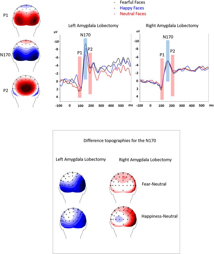
www.nature.com/scientificreports/
Left ATL Right ATL Control
Fearful Faces (− 12.41 µV ± 1.67) (− 7.25 µV ± 1.52) (− 2.63 µV ± 0.93)
Happy Faces (− 10.56 µV ± 1.53) (− 7.07 µV ± 1.40) (− 2.42 µV ± 0.85)
Neutral Faces (− 9.30 µV ± 1.39) (− 7.64 µV ± 1.27) (− 2.18 µV ± 0.77)
Vegetables (− 2.68 µV ± 1.08) (− 2.12 µV ± 0.99) (− 0.21 µV ± 0.61)
Table 1. Mean amplitude values and standard errors of the N170 for fearful, happy, neutral faces and
vegetables for the left and right ATL patients and for the control group.
N170 components21,24–27 are reportedly enhanced by faces expressing fear and by auditory signals of distress28.
Importantly, a more recent r eview29 suggested a more robust effect for the latter component, while P1 and P2
modulations appear to be highly variable and inconsistent. More compellingly, subliminal presentations of
fearful faces also appear to produce an enhanced early fronto-central a ctivity19 or a N170 r esponse21, and lateral
subliminal presentations in dot-probe tasks produce lateralised N170 e ffects27. These observations, coupled with
the observations from fMRI and PET30,31 showing that the amygdala is activated by subliminal faces, suggest that
the early modulation could be driven by the amygdala and would therefore support that its activation occurs
rapidly enough for it to enhance extrastriate activity as suggested by Vuilleumier et al.6.
In an attempt to confirm this, Rothstein et al.5 compared the ERP response to fearful and neutral faces in a
small group of epileptic patients. The findings indicated an enhancement of the P1 for fearful compared to neutral
faces that was correlated with the degree of amygdala atrophy, again suggesting that the early ERP modulation
was dependent on the integrity of the amygdala.
The current investigation therefore aimed to determine the timing of amygdala involvement in the visual
processing of emotional (fearful) facial expressions by examining ERP responses in patients following right or
left anterior temporal lobectomy (ATL) that included the amygdala. We recorded EEG in patients with left and
right ATL and a group of healthy controls who were presented with fearful, happy and neutral faces, as well as a
control category (vegetables; 300 ms) during a 1-back task. The faces were presented at the centre of the screen for
300 ms and were followed by a blank (black) screen that appeared for 1200 ms. ERP components (P1, N170 and
P2) to the three facial expressions and vegetables were compared in the 3 groups. We hypothesised that controls
would show an enhanced N170 due to feedback projections from amygdala, while the absence of an amygdala
in ATL patients would disrupt this effect.
Results
A sensitivity analysis (α err prob = 0.05; total sample sizes: 16 (control group) vs. 8 (patients group); number of
groups: 1(control group) vs. 2 (patients group); number of measurements: 6; correlations among rep measures:
0.8; sphericity: 0.7) showed that the present study is powered to detect effect sizes 15.79, ps < 0.001, η2 > 0.41, whereas happy faces did not significantly differ from
neutral faces (p = 0.374). In addition, we ran focused-cell contrasts between emotions across the three groups.
Faces produced a greater N170 deflection than vegetables (means shown in Table 1) in Left and Right ATL patient
groups, Fs(1, 23) > 4.89, ps < 0.038, η2 > 0.017. In the control group, fearful and happy faces significantly differed
from vegetables, whereas neutral faces fell short of the threshold for significance (p = 0.057).
P2. The 4 × 2 × 3 ANCOVA revealed a main effect of Stimulus Category F(3, 69) = 8.77, p < 0.001, η2 = 0.28 and
a Stimulus Category x Group interaction, F(6, 69) = 20.13, p < 0.001, η2 = 0.64. Focused-cell contrasts revealed that
vegetables (7.73 µV ± 0.67) produced a stronger P2 compared to fearful (2.47 µV ± 0.65), neutral (2.93 µV ± 0.59)
and happy (2.92 µV ± 0.73) faces, Fs(6, 69) > 43.28, ps < 0.001, η2 > 0.65. Fearful faces did not significantly dif-
fer from neutral and happy ones. In addition, we ran focused-cell contrasts between emotions across the three
groups. Faces differed from the vegetables in the Left and Right ATL groups, Fs(1, 23) > 11.53, ps = 0.002, η2 > 0.33,
whereas no significant differences was found for the control group (ps > 0.441).
In order to test specifically our main hypothesis (i.e., effects of lobectomy on the N170 response to fearful
facial expressions), we further carried out two separate repeated-measures ANOVAs on the N170 amplitudes in
the control and patient groups. The two groups were examined separately, comparing neutral, happy and fearful
expressions. As age did not interact with emotion in either group, this factor was not included in the follow-up
Scientific Reports | (2021) 11:426 | https://doi.org/10.1038/s41598-020-80054-1 2
Vol:.(1234567890)www.nature.com/scientificreports/
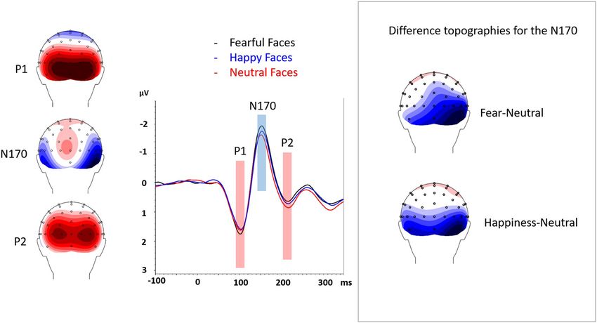
Figure 1. The N170 is shown for fearful faces (black) and neutral faces (red) conditions. Left and right leads are
collapsed. On the right, difference topographies are shown for fearful minus neutral conditions and for happy
minus neutral conditions at the N170.
analysis (two separate ANCOVAs revealed that age was not related to Emotion in the patient and in the control
group, ps > 0.426) (Fig. 1).
Control group. N170. The 3 (Fear, Happy, Neutral) × 2 (Left, Right Hemisphere) ANOVA revealed a main
effect of Hemisphere, F(1,15) = 12.32, p = 0.003, η2 = 0.45, and of Emotion, F(2,30) = 4.41, p = 0.034, η2 = 0.23.
The N170 deflection was greater in the right (-2.79 µV ± 0.44) compared to the left (-2.03 µV ± 0.34) channels. As
illustrated in Fig. 2, focused-cell contrasts revealed that fearful faces (-2.63 µV ± 0.38) produced a stronger N170
deflection compared to neutral (-2.18 µV ± 0.38) and happy (-2.42 µV ± 0.40) faces, Fs(1, 15) > 3.26, ps < 0.045,
η2 > 0.17 (one-tailed), whereas happy faces did not significantly differ from neutral faces (p = 0.103). The Emo-
tion x Hemisphere interaction was not significant, F(2, 30) > 3.04, ps = 0.06, η2 > 0.17.
Patients with amygdala lobectomy. N170. Results revealed a significant Emotion x Amygdala Lobec-
tomy interaction, F(2, 18) = 16.73, p = 0.022, η2 = 0.40. To decompose this interaction, focused-cell contrasts were
run separately for left and right lobectomized patients. As seen in Fig. 2, in patients with left amygdala lobec-
tomy, fearful faces (− 12.41 µV ± 2.58) produced a stronger N170 deflection relative to neutral (− 9.30 µV ± 2.09),
and happy (− 10.56 µV ± 2.33) faces, Fs(1, 9) > 14.62, ps < 0.004, η2 > 0.62, while happy faces did not differ signifi-
cantly from neutral faces (p = 0.199). By contrast, no significant effect emerged in patients with right amygdala
lobectomy (ps > = 0.511). No other effects were significant (ps > 0.062).
Discussion
The results of this study reveal that, in healthy controls, fearful faces produce an enhanced N170 component com-
pared to neutral faces, corroborating a number of previous reports21,24–27,32. More importantly, this enhancement
continued to be observed in a group of patients who had undergone left temporal resection, but not those whose
resection had been on the right, suggesting that the N170 enhancement requires a functional right amygdala.
The idea that early ERP components are enhanced for emotional expressions emerged almost two decades
ago. For example, Batty and Taylor24 reported that fearful faces produced a greater N170 than neutral and happy
faces. Similarly, modulations of the N170 for fearful faces were subsequently reported in tasks where the facial
stimuli were irrelevant25, or when they were presented subliminally21. Similar findings seem to apply to other
emotional expressions such as crying faces26, and even to fearful body expressions32. Other authors suggested an
even earlier modulation, reporting P1 enhancements for fearful faces. For example, Batty & T aylor24 observed a
P1 increase for fearful faces, a finding echoed by o thers23. Moreover, Eimer and H olmes22 also reported effects
in the 100 ms range, but over anterior electrodes, an effect that the authors attributed to enhanced attentional
processes. The reasons for which this early ERP enhancement occurs has remained a point of discussion so
far. One point of view that has been discussed at length is that emotional faces might differ in their low-level
features, which could produce an early modulation of the ERP response. For example, fearful expressions are
generally characterised by widely opened eyes, thereby increasing the brightness in the eye region. This could
Scientific Reports | (2021) 11:426 | https://doi.org/10.1038/s41598-020-80054-1 3
Vol.:(0123456789)www.nature.com/scientificreports/
Figure 2. The N170 for the left and right amygdala lobectomy is shown for fearful faces (black), neutral faces
(red) and happy faces (blue) conditions. Left and right hemispheres are collapsed. On the bottom, we displayed
the difference topographies between the fearful and the neutral conditions and between the happiness and the
neutral conditions for the N1 of patients with left and right amygdala lobectomy.
naturally lead to an increase in the early sensory responses that are sensitive to brightness. With this in mind,
one experiment inverted the faces to disrupt emotion recognition while maintaining the low-level features22. The
effect of this manipulation was to attenuate the ERP differences produced by the emotional expressions. Had the
effects been driven by low-level features rather than emotion, such a manipulation would not have been found.
Another approach consists in ensuring that no systematic difference exists in the brightness of the emotional
stimuli. In doing so, the likelihood of this factor driving the effect is reduced. Along these lines, differential effects
between neutral and fearful faces were still reported when stimuli were controlled in brightness and contrast
using a histogram-based method for equalizing perceptual v ariations25. More recently, a study examined the ERP
response to emotional faces and their scrambled Fourier transformed version, in which low-level visual features
were preserved33. The findings confirmed that emotional expressions modulated both the P1 and N170, however,
the P1 response was modulated by both scrambled and intact versions, while only the N170 was modulated by
Scientific Reports | (2021) 11:426 | https://doi.org/10.1038/s41598-020-80054-1 4
Vol:.(1234567890)www.nature.com/scientificreports/
emotional expression. This suggests that the P1 modulations are due to low-level differences across stimuli, while
the N170 is specifically tied to emotions.
Our current study circumvents any bias due to low-level characteristics since the same stimuli were used for
the comparison across groups of participants (i.e., any difference in luminance between categories is the same
across groups). Hence, the loss of an N170 modulation by the right-lobectomy group alone demonstrates an
emotion-dependent effect of the right amygdala and precludes a low-level explanation for this modulation. This
raises the question about the mechanism underlying the enhancement of the N170 by fearful faces.
Several authors have proposed that emotional/threatening stimuli activate a subcortical pathway that in turn
increase the neuronal activity in visual areas through feedback from the a mygdala6,34. More specifically, emo-
tional/threatening stimuli have been posited to activate a rapid subcortical pathway to the amygdala, allowing
coarse but rapid processing of relevant stimuli18,35. Ample evidence has been provided to corroborate the exist-
ence of this extra-geniculate visual pathway4,18,36, allowing the amygdala to be activated even in the absence of a
primary visual c ortex9. One role of this parallel pathway is thought to be the enhancement of visual processing for
relevant stimuli, such as fearful faces. Addressing this question, Vuilleumier et al.6 compared the BOLD response
in extrastriate visual areas to fearful and neutral faces in patients with temporal lobe epilepsy, and patients who
presented with hippocampal sclerosis which included or excluded the amygdala. The visual extrastriate response
was found to be enhanced for fearful faces compared to neutral expressions in all patients except those with
amygdalar sclerosis. This led to the conclusion that the amygdalae were responsible for the increase in visual
extrastriate activity for emotional faces, suggesting feedback activation from this structure to visual areas. How-
ever, to confirm whether a causal effect is present, the temporal dynamics of neural activation must be examined.
A subsequent study by Rotshtein et al.5 sought to identify the periods during which the amygdala affected the
electrical response of the brain. Their study again examined epileptic patients who presented hippocampal and
amygdalar sclerosis, or hippocampal sclerosis alone, this time using an ERP paradigm with fearful and neutral
faces. Their results revealed that the presence of amygdala sclerosis dampened the P1 enhancement observed
for fearful faces and that this amplitude was correlated with the degree of atrophy. These findings confirmed an
early modulation of the ERP response although the involvement of anterior channels is inconsistent with the
hypothesis of extrastriate activity. On the other hand, our findings identify the N170 as the component of interest,
revealing changes over expected channels and consistent with extrastriate activity (although no source localisa-
tion was performed). Moreover, our current findings were obtained from patients following amygdala resection,
showing that the effect was highly lateralized. Specifically, participants with right amygdala damage showed no
modulation of the N170. By contrast, participants who presented damage to the left, but not the right amygdala
showed that fearful faces produced a stronger N170 compared to happy and neutral faces.
Our current finding is the first combined amygdalectomy/ERP study to reveal an asymmetry of function of
the amygdala for emotion processing. A small number of experiments have previously reported asymmetries
following the presentation of fearful s timuli1,37,38. For instance, one experiment reported that passively watch-
ing fearful faces activated the left amygdala more than watching neutral f aces38. Another experiment, reported
that the left amygdala responded more to fearful faces in a dot-probe t ask37. In this study, a fearful face could
appear simultaneously with a neutral face either in the left or the right visual field. In the control condition, two
neutral faces were presented. Results revealed a heightened left amygdala activation when fearful faces were
present in the left visual field compared to the control condition. The authors therefore proposed that the left
amygdala mediates attentional effects to masked fearful faces. Contradicting these observations however, other
papers have highlighted the role of the right amygdala in emotion p rocessing2–4,7–9,30,31. For example, Cecere
2
et al. reported that consciously perceived happy faces elicited an increased N170 when an unseen fearful face
appeared simultaneously in the blind field of a right hemianopic patient. The authors concluded that implicit
subcortical processing of fearful signals could influence face encoding only when the right hemisphere was
intact. Elsewhere, Morris et al.4 reported that unseen fearful faces and fear-conditioned faces activated the right
but not left amygdala in control participants. These authors concluded to the existence of a subcortical pathway
to the right amygdala that processes behaviorally relevant visual events independently of the extratriate route.
The results of our current study corroborate the latter studies, emphasizing the right amygdala’s essential role
for emotional processing. In line with previous suggestions6, we hypothesize that this may be rendered possible
by rapid subcortical projections to the amygdala that precede the feedforward sweep of information through
the geniculate pathway. Indeed, the amygdala has been posited to project to the primary virtual cortex, and
thus can modulate the processing of visual information in this area34,39. By triggering this fast-track pathway,
fearful faces would activate amygdala, leading to an increase in the extrastriate response. By contrast with right
amygdalectomies, patients with left amygdala removal likely retain the use of the extra-geniculate pathway for
fearful face processing. This in turn could lead to an enhanced extrastriate response and enhanced stimulus
encoding2, thereby giving rise to N170 modulations40.
Taken together, our results demonstrate that the early ERP modulation—in this case the N170—for fearful
faces necessitates the integrity of the amygdala, and more importantly that this N170 enhancement relies on the
right amygdala. It is likely that projections to the right amygdala provide a route for the processing of behaviorally
relevant stimuli such as fearful faces, which lead to a heightened response of the extrastriate cortex via rapid
amygdalofugal projections to the visual areas.
Methods
Participants. The study was approved by The Human Research Ethics Committee at The University of
Queensland. All methods were performed in accordance with the guidelines and regulation at University of
Queensland.
Scientific Reports | (2021) 11:426 | https://doi.org/10.1038/s41598-020-80054-1 5
Vol.:(0123456789)www.nature.com/scientificreports/
Surgery Temporal pole Age of seizure Years since
Patient Gender Age laterality excision onset surgery Seizure Type seizure free
1 M 28 L No 7 14 PC Yes
2 F 42 L No 12 6 PCG Yes
3 F 26 L Yes 1 14 P Yes
4 F 28 L Yes n/a 2 PCG Yes
5 M 49 L Yes 15 14 PC Yes
6 F 47 L Yes 12 8 PCG Yes
7 F 22 R Yes 7 3 PC Yes
8 F 56 R Yes 2 .25 PG No
9 M 35 R Yes 11 10 PG Yes
10 F 46 R Yes 14 17 PC Yes
11 F 31 R Yes 27 1 PC Yes
12 M 42 R No 12 15 PCG Yes
Table 2. Summary of patient group. Age is in years at the time of the experiment. All patients underwent
unilateral removal of the amygdala and hippocampus either on the left (L) or on the right (R). Patients whose
surgery included the anterior temporal pole are indicated as “yes’ or “no” in the 5th column. Type of seizures
are indicated as P = partial simple, PC = partial complex, PCG = partial complex secondarily generalized.
n/a = not available.
Control participants. Sixteen healthy participants (3 males) took part in the study. Participants were recruited
through the University of Queensland research participation program from a first year psychology course, and
were awarded partial course credit for their participation. Mean age was 24.10 years (± 8.34). All had normal or
corrected-to-normal vision and had no self-reported psychiatric or neurological condition.
Amygdalectomy patients. Fourteen patients (8 female) with pharmaco-resistant epileptic seizures who under-
went amygdalohippocampectomy of the left (8 individuals) or right (6 individuals) were initially recruited from
the Unit for Presurgical Evaluation of Epilepsy (Neurology Clinic, Geneva University Hospital). The data of two
patients was discarded for the study due to a high number of EEG artefacts such as for example eyeblinks on
the stimulus. The average age of the resulting group of participants was 37 years; epilepsy onset varied between
1 year of age and 27 years of age. The study was performed between 3 months and 17 years after surgery. Patient
details are provided in Table 2 in method section. They suffered from intractable epilepsy between 6 and 54 years
until surgical alleviation of the disease. Participants had normal or corrected-to-normal vision.
Stimuli. A total of 30 greyscale photographs (236 × 236 pixels) faces expressing fearful, happy or neutral
expressions (10 per category, 5 males) were selected from the K-DEF database41. The faces were cropped at the
hairline to show only facial features. Faces were presented on a black background (RGB: 0, 0, 0). The program
ImageJ (http://rsbweb.nih.gov/ij/index.html) was used to calculate the luminance across categories. All picture
modifications were made with Adobe Photoshop CS3. Faces stimuli were equated for luminance and were pre-
sented in the centre of the monitor (21″ Hewlett Packard, LCD screen) at a refresh rate of 60 Hz, situated at
115 cm from the subject with minimal lighting.
Greyscale photographs of 20 common vegetables (onions, carrots, aubergines, etc.) were added as distracters.
These stimuli were adjusted to the same size and brightness levels as the face stimuli.
Procedure. A one-back procedure was used. The photographs were presented for 300 ms in the centre of
the screen. On 10% of the trials, the stimulus would be presented twice in immediate succession. To ensure that
attention was maintained on the stimuli, participants were instructed to respond to these immediate repetitions
by pressing a key on a standard keyboard. The order of presentation of the stimuli was randomised across par-
ticipants. Instructions were delivered verbally and were repeated in writing on the screen. Participants were told
to maintain their gaze on a central fixation cross. They were informed that photographs of faces and vegetables
would be presented and were asked to press the designated key on the keyboard whenever a picture was pre-
sented twice in succession.
A white fixation cross was presented on a black background for random durations between 500 and 1000 ms.
This was followed by the stimulus that lasted 300 ms. A blank (black) screen then appeared for 1200 ms. At the
end of each block, a self-paced break was observed prior to initiating the following block.
For patient recordings, a camera was attached on the experiment presentation screen to allow for constant
monitoring via the video as well as the EEG recording in case of any difficulty, or the onset of an epileptic seizure,
and the video and EEG were monitored in the experimenter control group during the procedure. The experi-
mental procedure was run using a dedicated software (E-prime v2; www.pstnet.com/eprime).
Three emotional expressions were used (fear, happy and neutral), displayed by 10 different individuals. These
30 photographs were presented randomly for a total of 8 times throughout the experiment, totalling 240 stimuli.
An additional 80 photographs representing vegetables were included, and 10% of the stimuli were randomly
Scientific Reports | (2021) 11:426 | https://doi.org/10.1038/s41598-020-80054-1 6
Vol:.(1234567890)www.nature.com/scientificreports/
presented as an immediate repetition for the one back procedure (= 32 samples). Participants thus viewed 352
stimuli in total, presented in 4 blocks of equivalent duration.
The total duration of the procedure (4 blocks + breaks) was approximately 25 min.
The study was approved by the Ethics Committee of Geneva University Hospitals (epileptic patients) and the
Ethics Committee of the University of Queensland (healthy controls). Participants gave their written informed
consent to participate before beginning the experiment.
ERP recording. Continuous EEG was acquired at 1024 Hz using an AD-Box ActiveTwo amplifier (Amsterdam,
The Netherlands) and 64 equally-spaced scalp electrodes referenced to Cz. Two external electrodes EOG were
placed on the face in order to monitor eye blinks and saccades (one on the outer canthus of the right eye and
one above the right eyebrow). A trigger pulse was sent by the stimulus-delivering PC to the EEG-acquisition
PC upon appearance of the visual stimulus. The timing of the markers was verified during preparation of the
paradigm by comparing stimulus presentation on the screen (using a photodiode) and marker onset on the EEG
signal.
EEG signal analysis was performed using BrainVision Analyzer 2.1 (Brain Products, Gilching Germany).
All trials in which participants responded were excluded from EEG analysis. Epochs were first established from
100 ms before, to 350 after stimulus onset. Bad electrodes were removed and re-interpolated using 3D splines42.
ERPs were then baseline corrected using the 100 ms pre-stimulus period. The signal was filtered offline between
0.1 and 30 Hz, with Cz maintained as the reference. We computed the mean amplitude for the P1, N170, and
P2 components. Mean amplitudes were established by visually determining the electrodes with the maximum
amplitude at the regions of interest (ROIs), as well as the temporal occurrence in the grand means.
ERP Processing. Analyses were performed on the mean amplitudes of the P1, N170 and P2 components.
All 3 components were measured at their maximal occurrence, determined in time windows at respectively:
95–120 ms, 145–170 ms and 205–230 ms for both the patient group and the control group. The sites were based
on electrodes of maximal activity during these intervals and were consistent with those typically used to inves-
tigate these components (PO8/PO7, P7/P8, P9/P10 for the P1; P7/P8, P9/P10 and TP9/TP10 for the N170; and
O1/O2/Oz/POz for P2).
An analysis of covariance was carried out on the P1, N170, and P2 amplitudes using Stimulus Category (Fear,
Happy, Neutral, Vegetable), Electrode Hemisphere (Left vs Right) and Participant Group (Left ATL, Right ATL,
Control) as factors, and age as a covariate. Then two separated ANOVAs were performed for the control and the
patient groups to test the effects of fearful facial expressions on the N170.
For the control group, the analyses consisted of a 3 (Emotion: neutral, fearful, happy faces) × 2 (Hemisphere:
left, right) repeated-measures ANOVA. The analyses of the patient group consisted of a 3 (Emotion: neutral,
fearful, happy faces) × 2 (Hemisphere: left, right) repeated-measures ANOVA with left and right ATL included
as a categorical factor. Violations of sphericity and p-values were corrected according to the epsilon of Green-
house–Geisser or Huynh–Feldt.
Received: 4 August 2020; Accepted: 10 December 2020
References
1. Hardee, J. E., Thompson, J. C. & Puce, A. The left amygdala knows fear: laterality in the amygdala response to fearful eyes. Soc.
Cogn. Affect. Neurosci. 3, 47–54 (2008).
2. Cecere, R., Bertini, C., Maier, M. E. & Làdavas, E. Unseen fearful faces influence face encoding: evidence from ERPs in hemianopic
patients. J. Cog. Neurosci. 26, 2564–2577 (2014).
3. Morris, J. S. & Dolan, R. J. Dissociable amygdala and orbitofrontal responses during reversal fear conditioning. Neuroimage 22,
372–380 (2004).
4. Morris, J. S., Öhman, A. & Dolan, R. J. A subcortical pathway to the right amygdala mediating “unseen” fear. Proc. Natl. Acad. Sci.
96, 1680–1685 (1999).
5. Rotshtein, P. et al. Amygdala damage affects event-related potentials for fearful faces at specific time windows. Hum. Brain Mapp.
31, 1089–1105 (2010).
6. Vuilleumier, P., Richardson, M. P., Armony, J. L., Driver, J. & Dolan, R. J. Distant influences of amygdala lesion on visual cortical
activation during emotional face processing. Nat. Neurosci. 7, 1271–1278 (2004).
7. Baker, K. B. & Kim, J. J. Amygdalar lateralization in fear conditioning: evidence for greater involvement of the right amygdala.
Behav. Neurosci. 118, 15 (2004).
8. Gainotti, G. Emotions, unconscious processes, and the right hemisphere. Neuropsychoanalysis 7, 71–81 (2005).
9. Pegna, A. J., Khateb, A., Lazeyras, F. & Seghier, M. L. Discriminating emotional faces without primary visual cortices involves the
right amygdala. Nat. Neurosci. 8, 24–25 (2005).
10. Pessoa, L. & Adolphs, R. Emotion processing and the amygdala: from a’low road’to’many roads’ of evaluating biological significance.
Nat. Rev. Neurosci. 11, 773–782 (2010).
11. Bertini, C., Pietrelli, M., Braghittoni, D. & Làdavas, E. Pulvinar lesions disrupt fear-related implicit visual processing in hemianopic
patients. Front. Psychol. 9, 2329 (2018).
12. De Gelder, B., Vroomen, J., Pourtois, G. & Weiskrantz, L. Non-conscious recognition of affect in the absence of striate cortex.
NeuroReport 10, 3759–3763 (1999).
13. Morris, J. S., DeGelder, B., Weiskrantz, L. & Dolan, R. J. Differential extrageniculostriate and amygdala responses to presentation
of emotional faces in a cortically blind field. Brain 124, 1241–1252 (2001).
14. Troiani, V., Price, E. T. & Schultz, R. T. Unseen fearful faces promote amygdala guidance of attention. Soc. Cogn. Affect. Neurosci.
9, 133–140 (2014).
15. Tamietto, M. & De Gelder, B. Neural bases of the non-conscious perception of emotional signals. Nat. Rev. Neurosci. 11, 697–709
(2010).
16. LeDoux, J. Emotional networks and motor control: a fearful view. Prog. Brain Res. 107, 437–446 (1996).
Scientific Reports | (2021) 11:426 | https://doi.org/10.1038/s41598-020-80054-1 7
Vol.:(0123456789)www.nature.com/scientificreports/
17. Johnson, M. H. Subcortical face processing. Nat. Rev. Neurosci. 6, 766–774 (2005).
18. Tamietto, M., Pullens, P., de Gelder, B., Weiskrantz, L. & Goebel, R. Subcortical connections to human amygdala and changes
following destruction of the visual cortex. Curr. Biol. 22, 1449–1455 (2012).
19. Kiss, M. & Eimer, M. ERPs reveal subliminal processing of fearful faces. Psychophysiology 45, 318–326 (2008).
20. Pegna, A. J., Darque, A., Berrut, C. & Khateb, A. Early ERP modulation for task-irrelevant subliminal faces. Front. Psychol. 2, 88
(2011).
21. Pegna, A. J., Landis, T. & Khateb, A. Electrophysiological evidence for early non-conscious processing of fearful facial expressions.
Int. J. Psychophysiol. 70, 127–136 (2008).
22. Eimer, M. & Holmes, A. An ERP study on the time course of emotional face processing. NeuroReport 13, 427–431 (2002).
23. Pourtois, G., Dan, E. S., Grandjean, D., Sander, D. & Vuilleumier, P. Enhanced extrastriate visual response to bandpass spatial
frequency filtered fearful faces: time course and topographic evoked-potentials mapping. Hum. Brain Mapp. 26, 65–79 (2005).
24. Batty, M. & Taylor, M. J. Early processing of the six basic facial emotional expressions. Cogn. Brain Res. 17, 613–620 (2003).
25. Blau, V. C., Maurer, U., Tottenham, N. & McCandliss, B. D. The face-specific N170 component is modulated by emotional facial
expression. Behav. Brain Funct. 3, 7 (2007).
26. Hendriks, M. C., van Boxtel, G. J. & Vingerhoets, A. J. An event-related potential study on the early processing of crying faces.
NeuroReport 18, 631–634 (2007).
27. Carlson, J. M. & Reinke, K. S. Spatial attention-related modulation of the N170 by backward masked fearful faces. Brain Cogn. 73,
20–27 (2010).
28. Andrzejewski, J. A. & Carlson, J. M. Electrocortical responses associated with attention bias to fearful facial expressions and audi-
tory distress signals. Int. J. Psychophysiol. 151, 94–102 (2020).
29. Schindler, S. & Bublatzky, F. Attention and emotion: an integrative review of emotional face processing as a function of attention.
Cortex 130, 362–386 (2020).
30. Morris, J. S., Öhman, A. & Dolan, R. J. Conscious and unconscious emotional learning in the human amygdala. Nature 393,
467–470 (1998).
31. Whalen, P. J. et al. The emotional counting Stroop paradigm: a functional magnetic resonance imaging probe of the anterior
cingulate affective division. Biol. Psychiat. 44, 1219–1228 (1998).
32. Stekelenburg, J. J. & de Gelder, B. The neural correlates of perceiving human bodies: an ERP study on the body-inversion effect.
NeuroReport 15, 777–780 (2004).
33. Schindler, S., Bruchmann, M., Gathmann, B., Moeck, R., & Straube, T. Effects of low-level visual information and perceptual load
on P1 and N170 responses to emotional faces. PsyArxiv (2019).
34. Iwai, E., & Yukie, M. (1987) Amygdalofugal and amygdalopetal connections with modality‐specific visual cortical areas in macaques
(Macaca fuscata, M mulatta, and M fascicularis). J. Comp. Neurol. 261, 362-387.
35. LeDoux, J. Rethinking the emotional brain. Neuron 73, 653–676 (2012).
36. Rafal, R. D. et al. Connectivity between the superior colliculus and the amygdala in humans and macaque monkeys: virtual dis-
section with probabilistic DTI tractography. J. Neurophysiol. 114, 1947–1962 (2015).
37. Carlson, J. M., Reinke, K. S. & Habib, R. A left amygdala mediated network for rapid orienting to masked fearful faces. Neuropsy-
chologia 47, 1386–1389 (2009).
38. Thomas, K. M. et al. Amygdala response to facial expressions in children and adults. Biol. Psychiat. 49, 309–316 (2001).
39. Amaral, D. G., Behniea, H. & Kelly, J. L. Topographic organization of projections from the amygdala to the visual cortex in the
macaque monkey. Neuroscience 118, 1099–1120 (2003).
40. Bentin, S., Allison, T., Puce, A., Perez, E. & McCarthy, G. Electrophysiological studies of face perception in humans. J. Cogn.
Neurosci. 8, 551–565 (1996).
41. Lundqvist, D., Flykt, A. & Ohman, A. Karolinska Directed Emotional Faces (Department of Neurosciences, Karolinska Hospital,
Stockholm, 1998).
42. Perrin, F., Pernier, J., Bertnard, O., Giard, M. H. & Echallier, J. F. Mapping of scalp potentials by surface spline interpolation.
Electroencephalogr. Clin. Neurophysiol. 66, 75–81 (1987).
Acknowledgements
This investigation was supported by the Swiss National Science Foundation Grants Nos. P2GEP1_188266 and
#320030-144187.
Author contributions
A.P. provided the idea for the study, developed the experimental procedure, and wrote the paper. D.F. contributed
to developing the procedure, analyzed the data, and wrote the paper. E.M. and L.L. performed the recordings and
contributed to the analysis for the paper. M.S. participated in the development of the experimental procedure,
provided medical expertise and contributed to writing the paper.
Competing interests
The authors declare no competing interests.
Additional information
Correspondence and requests for materials should be addressed to A.J.P.
Reprints and permissions information is available at www.nature.com/reprints.
Publisher’s note Springer Nature remains neutral with regard to jurisdictional claims in published maps and
institutional affiliations.
Scientific Reports | (2021) 11:426 | https://doi.org/10.1038/s41598-020-80054-1 8
Vol:.(1234567890)www.nature.com/scientificreports/
Open Access This article is licensed under a Creative Commons Attribution 4.0 International
License, which permits use, sharing, adaptation, distribution and reproduction in any medium or
format, as long as you give appropriate credit to the original author(s) and the source, provide a link to the
Creative Commons licence, and indicate if changes were made. The images or other third party material in this
article are included in the article’s Creative Commons licence, unless indicated otherwise in a credit line to the
material. If material is not included in the article’s Creative Commons licence and your intended use is not
permitted by statutory regulation or exceeds the permitted use, you will need to obtain permission directly from
the copyright holder. To view a copy of this licence, visit http://creativecommons.org/licenses/by/4.0/.
© The Author(s) 2021
Scientific Reports | (2021) 11:426 | https://doi.org/10.1038/s41598-020-80054-1 9
Vol.:(0123456789)You can also read