Antiproliferative therapy with sirolimus and propranolol for congenital vascular anomalies in newborns (Case reports)
←
→
Page content transcription
If your browser does not render page correctly, please read the page content below
EXPERIMENTAL AND THERAPEUTIC MEDICINE 22: 1097, 2021

      Antiproliferative therapy with sirolimus and propranolol for
       congenital vascular anomalies in newborns (Case reports)
                    CĂTĂLIN CÎRSTOVEANU1,2, ANA MIHAELA BIZUBAC1,2, CRISTINA MUSTEA1,
                   ȘTEFAN MANOLACHE1, ALEXANDRA ISTRATE‑BÂRZAN1, DOINIȚA SFRIJAN2,3,
                  VERONICA MARCU4, DAN‑ALEXANDRU IOZSA5,6 and RADU‑IULIAN SPĂTARU5,6

         1
          Neonatal Intensive Care Unit, ‘Marie S. Curie’ Emergency Clinical Hospital for Children, 077120 Bucharest;
             2
               Department of Pediatrics, ‘Carol Davila’ University of Medicine and Pharmacy, 050474 Bucharest;
                     3
                       Oncology Department, 4Radiology Department and 5Pediatric Surgery Department,
                        ‘Marie S. Curie’ Emergency Clinical Hospital for Children, 077120 Bucharest;
                         6
                           Department of Pediatric Surgery and Orthopedics, ‘Carol Davila’ University
                                   of Medicine and Pharmacy, 050474 Bucharest, Romania

                                            Received May 31, 2021; Accepted June 30, 2021

                                                      DOI: 10.3892/etm.2021.10531

Abstract. We present a series of four newborns diagnosed                showed a marked reduction in the size of the mass, improve‑
with complicated congenital vascular anomalies, with                    ment in overall appearance or even calcification in the liver
different localization: Congenital lymphatic malformation               vascular tumor. No patient showed life threatening side effects
(CLM) on the left hemithorax extending on the left upper                to the treatment. Hypertriglyceridemia was the only side effect
limb; congenital hepatic hemangioma (CHH) with impor‑                   noted in all patients. This is in accordance with several inter‑
tant complications in the first 7 weeks of life; Kaposiform             national studies, which try to demonstrate the importance of
hemangioendothelioma (KHE) of the left lower limb compli‑               sirolimus in neonatal vascular malformations in monotherapy
cated with Kasabach Merritt phenomenon (KMM) and most                   or combined with different drugs.
probable diffuse capillary malformation with overgrowth
(DCMO). All patients were treated with combined antipro‑                Introduction
liferative therapy with sirolimus and propranolol. The initial
dose of sirolimus was 0.45‑0.5 mg/m 2 with doses adjusted               Vascular anomalies describe two pathological entities:
according to plasmatic levels. Therapeutic intervals of siro‑           Congenital vascular malformations (CVMs) and vascular
limus were considered at plasmatic levels of 7‑12 ng/ml. Our            tumors (1). Depending on the presence of arterial flow,
aim was to use the lowest therapeutic dose in order to avoid            these can be described as fast‑flow [vascular malforma‑
possible side effects. Propranolol was initiated in doses of            tions‑arteriovenous malformation, arteriovenous fistula, and
0.5‑1.0 mg/kg/day and was increased up to 3.0 mg/kg/day                 complex malformations such as Parkes‑Weber syndrome
depending on tolerability. Following two months, every patient          and vascular tumors (hemangiomas or neoplasms such as
                                                                        Kaposiform hemangioendothelioma (KHE), hemangio‑
                                                                        pericytoma, angiosarcoma)] and low‑flow, benign lesions
                                                                        [venous malformations, lymphatic malformations, capillary
Correspondence to: Dr Ana Mihaela Bizubac, Neonatal Intensive           malformations such as diffuse capillary malformation with
Care Unit, ‘Marie S. Curie’ Emergency Clinical Hospital for Children,   overgrowth (DCMO, combined/complex malformations, such
20 Constantin Brâncoveanu Boulevard, 077120 Bucharest, Romania          as Klippel‑Trenaunay syndrome (KTS), Proteus syndrome,
E‑mail: anamihaeladogaru@yahoo.com                                      CLOVES syndrome, Sturge‑Weber syndrome] (2‑4).
                                                                            CVMs result from a failure in angiogenesis during the
Abbreviations:      CLM, congenital lymphatic malformation;             development of the vascular system; they are present at
CHH, congenital hepatic hemangioma; KHE, Kaposiform                     birth and usually grow with the infant (3). If arterial flow
hemangioendothelioma; KMP, Kasabach‑Merritt phenomenon;                 is observed through imaging studies, differential diagnosis
DCMO, diffuse capillary malformation with overgrowth; CVM,
                                                                        includes arterio‑venous malformations, arteriovenous fistula
congenital vascular malformation; KTS, Klippel Trenaunay Syndrome;
                                                                        or a complex vascular malformation (2).
PI3K, phosphatidylinositol 3‑kinase; AKT, protein kinase B (also
PKB); mTOR, mammalian target of rapamycin receptor; HDL,                    Capillary malformations are clinically diagnosed and
high‑density lipoprotein; VEGF, vascular endothelial growth factor      present as port‑wine stains and telangiectasia. Most of the
                                                                        times they are isolated anomalies but can be part of a complex
Key words: congenital vascular anomalies, vascular malformations,       vascular malformation (1,3‑5).
antiproliferative therapy, propranolol, sirolimus                           Venous malformations and complex malformations with
                                                                        a venous component have a variable presentation depending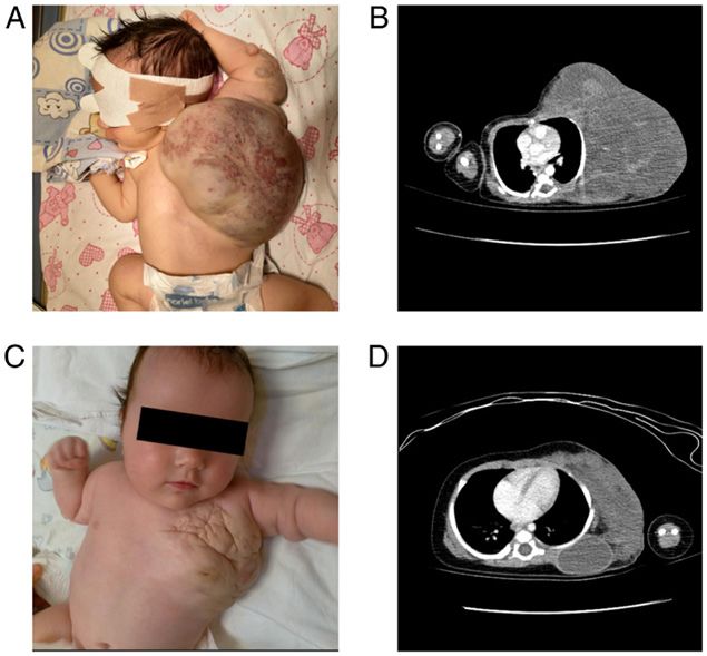
2           CÎRSTOVEANU et al: Sirolimus and Propranolol for newborn Congenital Vascular Anomalies

on their depth and associated complications. Complications            propranolol, a nonselective β‑adrenergic receptor antagonist
associated with venous malformations are venous stasis,               that causes vasoconstriction and used as first line of treat‑
thrombosis and localized intravascular coagulopathy (6).              ment for congenital hemangiomas, inhibits angiogenesis and
Despite being congenital, venous malformations usually                promotes apoptosis (14) by acting on the PI3K/AKT signaling
manifest later in childhood because they grow with the patient.       pathway (27). Studies have shown that protein levels and mRNA
Upon physical examination, they appear as compressible                of both PI3K and AKT were decreased after propranolol
bluish colored, sponge‑like masses (3,6).                             treatment, thus suggesting that a reduction in hemangiomas is
    Lymphatic malformations appear following aberrant                 related to the inhibition of PI3K and AKT (27‑29).
morphogenesis of primordial lymphatic structures and can
be classified as microcystic or macrocystic (7‑9). Lymphatic          Case reports
malformations or complex vascular malformations with a
lymphatic component can lead to disfigurement caused by               We analyzed 4 patients admitted to the Neonatal Intensive Care
tissue hypertrophy and skeletal overgrowth; depending on the          Unit of ‘Marie Curie’ Emergency Clinical Hospital for Children,
organs involved, they can lead to chylous effusions with organ        Bucharest, Romania between August 2019 and September 2020.
compromise. Large lesions can be complicated by fluid loss,           All patients were diagnosed with CVMs and treated with
hypoproteinemia, bleeding, and infection (10).                        combined antiproliferative therapy with propranolol and siro‑
    Vascular tumors develop from the endothelium; they are            limus. We analyzed the medical records and summarized the
fully formed at birth and are divided into congenital heman‑          date available into a case series. Our aim was to monitor the
giomas that can either be rapidly involuting or non‑involuting        patient clinical and radiological responses and possible adverse
and more complex tumors such as KHE or tufted angioma (2).            reactions to the therapy and demonstrate the treatment safety
Complex vascular neoplasms are infiltrative lesions and can           in comparison with other cases reported in the literature. All
cause Kasabach‑Merritt phenomenon (KMP). KMP is a                     patients were started on oral propranolol at 1 mg/kg/day with
consumptive coagulopathy syndrome consisting of platelet              doses adjusted up to 3 mg/kg/day according to the tolerance
trapping with profound thrombocytopenia, enlargement of the           and sirolimus at an initial dose of 0.4 mg/m2 with doses tired
lesion and significant hypofibrinogenemia (10‑12).                    according to plasmatic levels. Blood tests were drawn every
    Currently, there is no established standard of care for the       other week and then monthly to check for possible side effects.
treatment of vascular anomalies. Therapy is guided based on               All of our patients were females, admitted to our unit
symptoms and association of complications. Management                 between 1 and 21 days of life. One patient was diagnosed
is comprised of surgical excision, sclerotherapy or embo‑             prenatally through fetal‑magnetic resonance imaging while
lization (2,3,13). Medical management includes steroids,              3 newborns were diagnosed in our unit. All patients had
vincristine and interferon with potential significant side            distinct congenital vascular anomalies: 1 patient was diagnosed
effects in infants, especially neurotoxicity (10,14). Propranolol     with congenital lymphangioma of the left hemithorax and left
represents the first line of treatment for patients with vascular     upper arm; 1 patient with CHH complicated with liver failure;
tumors such as hepatic hemangiomas (14‑16).                           1 patient with DCMO; and 1 patient with KHE of the left lower
    Ideally, therapies for vascular anomalies would target            limb. Surgical intervention was intended for 2 patients and
specific cellular pathways involved in abnormal cellular prolif‑      later postponed because of the high risk of negative outcomes.
eration and growth.                                                   Treatment was initiated between 4 days of age and 7 weeks.
    In the last few years, multiple cases have been reported          Response to treatment was noted beginning with the first
for the safety of sirolimus as a therapeutic option for various       days of therapy in all patients through changes in the overall
congenital vascular anomalies (9,10,12,17‑23). The phosphati‑         aspect and size of the mass, changes in the overlaying skin,
dylinositol 3‑kinase (PI3K)/protein kinase B (AKT) signaling          and improved mobility. Plasmatic levels of sirolimus were
pathway is pivotal for cellular functions such as growth and          monitored at 7 days of treatment and every other week for the
survival and has been demonstrated to be involved in normal           duration of stay in our unit. After a stable level was achieved,
vascular development and angiogenesis (24,25). Extracellular          plasmatic levels were monitored monthly. Plasmatic levels
signals activating the PI3K/AKT pathway transfer signals to           of sirolimus were difficult to maintain in the recommended
mammalian target of rapamycin receptor (mTOR) which, in               ranges. All patients had initially a higher plasmatic level that
return, increases expression of vascular endothelial growth           later decreased. Hypertriglyceridemia was the only side effect
factors (VEGF)‑A and VEGF‑C. VEGF‑A and VEGF‑C                        observed in all four patients. Duration of treatment varied
are regulators of both angiogenesis and lymphangiogenesis,            between 27 weeks and 20 months. Three patients still undergo
promoting protein synthesis, cellular growth and prolifera‑           the treatment while for one patient the treatment was stopped
tion (9,10,24,25). Over time, inhibitors that target this signaling   after 48 weeks after complete calcification of the liver mass.
pathway have been shown to reduce VEGF secretion and
angiogenesis, thus making them a suitable option for the              Case 1. A female baby was diagnosed prenatally through fetal
treatment of CMVs (24,25). Sirolimus is a direct antagonist           MRI with lymphatic malformation on the left hemythorax
of mTOR that acts by blocking the downstream synthesis of             and left upper arm. The newborn was transferred to our unit
the PI3K/AKT/mTOR pathway resulting in antitumoral and                at 21 h of life, hemodynamically stable, breathing unaided. On
antiangiogenic effects by impairing VEGF production (10,26).          inspection, she presented with a giant lesion on the left hemi‑
mTOR receptors have been shown to be overexpressed in                 thorax (~30/25 cm) extending to the left upper limb; overlaying
vascular tumors, which makes sirolimus suitable as a treatment        skin was purplish and there was a central area of telangiectasis
option for vascular malformations (24). Similar to sirolimus,         (Fig. 1A). There were no differences in color or temperature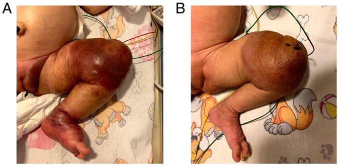
EXPERIMENTAL AND THERAPEUTIC MEDICINE 22: 1097, 2021                                                            3

Figure 1. Case 1. Angio‑CT on admittance (A and B) vs. at 9 weeks of treatment (C and D) showed significant reduction in the size mostly through reduction
of the cystic component. CT, computed tomography.

between the two upper limbs; grasping reflex and spontaneous                   Case 2. A female baby at clinical examination presented with
movements were present.                                                        abdominal distention and a palpable mass in the right hypochon‑
    Chest angio‑computed tomography (CT) at 4 days of                          drium. She was diagnosed at the local maternity department with
life (Fig. 1B) showed a massive [12/8.5 cm (axial), 13.8 cm                    CHH. The diagnosis was based on the angio‑CT that showed
(longitudinal)] soft tissue, cystic lesion with fine septal and                characteristic findings suggestive of CHH: A well‑defined solitary
intralesional hemorrhage. The lesion infiltrated the intersca‑                 lesion of ~66.5/61.5/66 mm in the central hilar portion of the liver
pulo‑costal space without damaging the scapulo‑humeral                         with centripetal enhancement and central sparing (Fig. 2A). Her
articulation. The CT scan revealed only one fine vascular                      evolution was complicated by moderate anemia, thrombocyto‑
branch inside the mass.                                                        penia, progressive hepatic cytolysis, cholestatic and inflammatory
    Surgical removal of the lesion was attempted and later                     syndromes. She was transferred to our clinic at 21 days of life.
postponed following significant hemorrhage after a fine needle                     Upon admittance, the patient was breathing smoothly, had
aspiration was performed in the operating room. Monotherapy                    a stable heart rate and was hemodynamically stable; she was
with propranolol was started on admittance but after 4 weeks of                moderately jaundiced, had a distended abdomen and a palpable
treatment there were no visible effects. On the 20th day of life, we           mass of about 7/6 cm was found on clinical examination.
began combination therapy with sirolimus (0.4 mg/m2 per dose,                      Abdominal ultrasound (US) on admittance showed a
twice daily) and propranolol. After the first week of combined                 central hilar lesion occupying the Vth and VIth liver segments
therapy, the overall size of the mass shrunk, the overlying skin               and partially the IVth, VIIth and VIIIth segments; the tumor
changed to a natural color and the arm movement improved.                      had well‑defined margins, was heterogenous and had a central
    Plasmatic levels of sirolimus were monitored and                           inhomogeneous necrotic zone surrounded by a peripheral,
varied between 2.7‑29 µg/l with doses adjusted between                         well vascularized area (Fig. 2C). Echocardiographic findings
0.32 and 0.66 mg/m2.                                                           were normal.
    The only side effects noted were a slight tendency to prolonged                Upon arrival, she was started on oral propranolol with an
dyslipidemia with hypertriglyceridemia and decreased levels of                 initial dose of 1 mg/kg/day which was increased slowly until a
high‑density lipoprotein (HDL). Massive reduction of the lesion                maintenance dose of 3 mg/kg/day was reached. Ursodeoxycholic
size was noted at 3 months of age (Fig. 1C). Control angio‑CT                  acid was initiated for the cholestatic syndrome at a dose of
performed at that time (11 weeks of combined therapy) showed                   10 mg/kg/day. In evolution, the patient exhibited an associated
a reduction in mass size predominantly through reduction of the                feeding intolerance with frequent postprandial regurgitation
cystic component (maximum axial size, 10/5.7 cm) (Fig. 1D). The                and vomiting because of the mass effect of the lesion.
patient was discharged at home at 3½ months of age, continuing                     Abdominal US studies showed no visible changes after
the combined antiproliferative therapy. Currently, she is still                4 weeks of treatment. Liver function tests remained elevated
undergoing treatment going on 20 months of combined therapy                    over the normal reference ranges and she presented with
and is on clinical, pharmacological and radiological follow‑up.                persistent hypoalbuminemia, for which albumin infusion was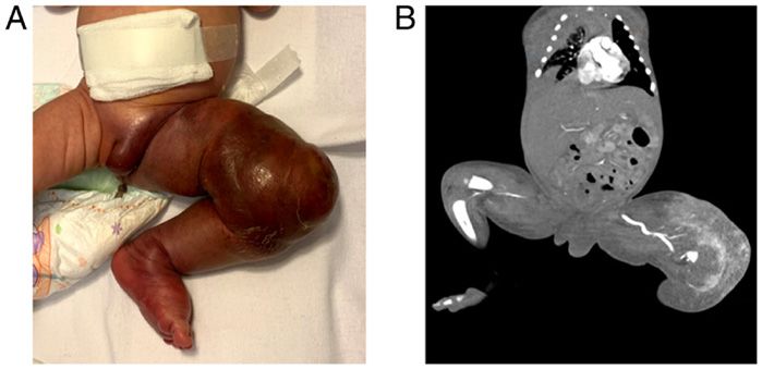
4 CÎRSTOVEANU et al: Sirolimus and Propranolol for newborn Congenital Vascular Anomalies
Figure 2. Case 2. (A and B) Abdominal angio‑CT scan showing a solitary hepatic mass with centripetal enhancement and central sparing vs. after 5 weeks
of combined antiproliferative therapy which showed a reduction in size with increased intralesional calcifications. (C and D) Abdominal ultrasound (US) on
admittance showing a giant liver lesion with a central necrotic zone surrounded by a peripheral well vascularized area vs. US at 2 weeks of combined therapy
which showed increased central necrotic area and intralesional calcifications. CT, computed tomography.
needed. Cholestasis persisted over the course of the treatment Case 3. A female newborn presented with a giant mass encom‑
with propranolol. passing the left thigh, knee and calf. In evolution, the mass
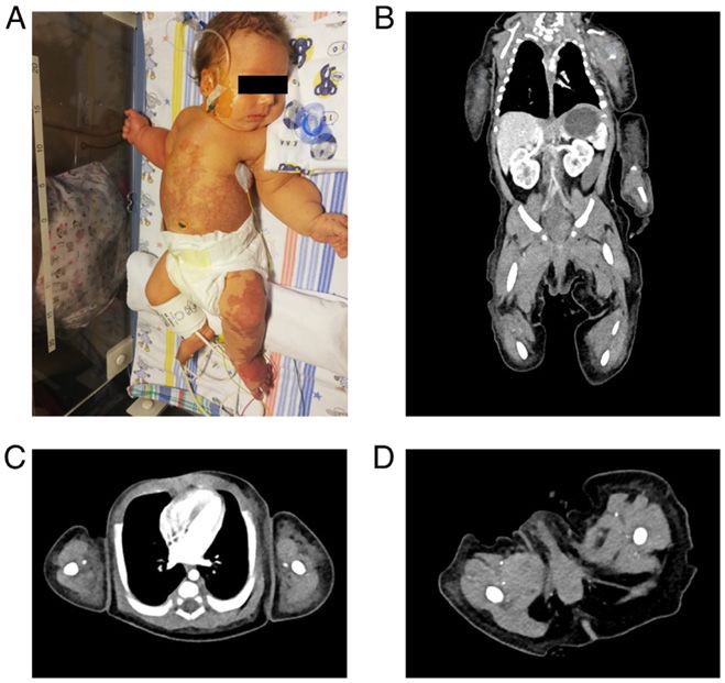
Giving the large size and central localization of the lesion, extended upwards to the inguinal region and downwards to
after a multi‑disciplinary hearing composed of the neona‑ the distal 1/3 of the calf and foot; the limb became infiltrated
tology, pediatric surgery and radiology teams, a decision was with purplish overlaying skin and tender to touch. She was
made to postpone the surgical treatment, given the high risk transferred to our unit at 4 days of age, breathing unaided and
for a negative outcome. hemodynamically stable. Upon inspection, the left lower limb
At the age of 1 month and 3 weeks, the patient was started was swollen. A giant mass was noted with ill‑defined margins,
on combined antiproliferative therapy with oral sirolimus purplish in color with overlaying petechia and telangiectasis.
(0,4 mg/m2 per dose, twice daily) and propranolol (3 mg/kg/day). The limb was tender to touch and warmer than the right lower
The dose of sirolimus ranged between 0.35 and 0.8 mg/m 2, limb; the left thigh girth upon arrival was ~25 cm compared
adjusted according to plasmatic levels (4.4‑16.73 µg/l). to 11 cm around the right thigh, and active movement of the
Abdominal US after 1 week of combined antiproliferative left limb was limited (Fig. 3A). Peripheral pulses were present.
treatment showed diminished intratumoral vascularization and Laboratory tests showed profound thrombocytopenia (PLT
new necrotic areas inside the tumoral tissue. After 2 weeks 5,000/mm3) and coagulation disturbances requiring adminis‑
of treatment, a reduction in size was noticeable (63/56/57 mm tration of FFP and platelet transfusions. Upon arrival, she was
compared to ~66.5/61.5/66 mm) (Fig. 2D). Side effects noted started on combined antiproliferative therapy with propranolol
were persistent dyslipidemia with hypertriglyceridemia and (1 mg/kg/day) and sirolimus (starting dose 0.4 mg/m2) in asso‑
low HDL‑cholesterol levels. Control angio‑CT scan after ciation with methylprednisolone (2 mg/kg/day).
4 weeks of treatment showed moderate reduction of mass Angio‑CT scan performed on admission showed a massive
size (63/54/51 mm) and multiple intratumoral calcifications hypervascular, relatively well‑defined lesion encompassing the
(Fig. 2B). The patient was discharged at home at 3 months left thigh and knee that receives arterial flow from the inferior
and 1 week of age on oral propranolol (3.5 mg/kg/day) and segment of the left femoral artery and left popliteal artery with
sirolimus (~0.55 mg/m2). At present the patient is well at home. early enhancement of the femoral vein; soft tissue edema of
After 30 weeks of treatment, abdominal US showed that the the left labia and thigh was present (Fig. 3B).
mass shrunk to half of its original size (current size 32/29 mm Response to treatment was noted starting the first days of
vs. ~66.5/61.5/66 mm at birth) and is fully calcified. therapy. Upon inspection, the foot and calf were slowly normal‑
Combined antiproliferative therapy was stopped after 48 ized in regards to color, swelling of the limb was diminished as
weeks of treatment. Currently she is under clinical and radio‑ well as the petechiae and telangiectasis, margins of the lesion
logical follow‑up. became well defined, encompassing 2/3 distal thigh and 1/3EXPERIMENTAL AND THERAPEUTIC MEDICINE 22: 1097, 2021 5
Figure 3. Case 3. (A) Presentation on admittance. (B) Angio‑CT at 4 days of life showing a giant hypervascular lesion in the left lower limb with early enhance‑
ment of the femoral vein. CT, computed tomography.
Figure 4. Case 3. Clinical presentation after (A) 4 days and (B) 2 weeks of combined antiproliferative therapy.
proximal calf (Fig. 4A and B). Platelet count increased gradually forced to reduce the dose of sirolimus to 1 mg/m2. Vincristine
in the first 3 weeks of treatment and normalized after 4 weeks of therapy was discontinued after 8 courses.
combined therapy when the PLT count was elevated >200,000/µl. Throughout the course of the treatment, sirolimus doses ranged
Corticosteroid therapy was tapered over the course of 4 weeks between 0.5 and 2.0 mg/m2 with plasmatic levels of 5.2‑14 µg/l.
and stopped when the platelets reached a normal value. The patient is a social case and was transferred to a chronic
After 11 weeks of combined antiproliferative therapy, care facility still undergoing combined therapy, being on
during which the patient showed good response to therapy, her week 30 of treatment.
status was complicated with swelling of the whole left lower
limb including the left major labia. The diameter of the thigh Case 4. A female newborn presented in the delivery room with
increased from 24 to 31 cm in a week. Mobility of the limb cyanosis predominantly on the inferior limbs, hypertrophy on
was good, overlaying skin color was normal and there was the left side of the body with port‑wine stains on the inferior
local warmth compared to the contralateral limb. Laboratory limbs and trunk. She was transferred to our NICU at 7 days
tests were normal. Doppler‑US of the leg showed no arterial or of life with suspicions of DCMO vs. KTS. Upon admission,
venous obstruction; control angio‑CT showed diffused edema she was breathing unaided and was hemodynamically stable.
of the leg without changes in tumor size. Port‑wine stains were observed on inspection, predominantly
Propranolol and sirolimus doses were doubled and methyl‑ on the left lower limb with extension on the truncal skin, while
prednisolon for 4 weeks was reintroduced with no visible effect. the left side of the body was hypertrophied apparently through
At 13 weeks of combined therapy, the patient was started soft tissue overgrowth (Fig. 5A). There was a 2 cm difference
on vincristine at 0.05 mg/m2/week. After 8 rounds of vincris‑ in girth between the left and right inferior limbs.
tine therapy associated with sirolimus and propranolol, we Laboratory tests found elevated D‑Dimers with normal
observed only mild changes in the tumor aspect. Initially, there fibrinogen and coagulation studies. She was started on siro‑
was a reduction in thigh girth with resolution of labial edema limus (0.5 mg/m 2) upon admission and was administered
but after 5 rounds of vincristine the thigh girth increased combined antiproliferative therapy starting on day of life 10.
to 32 cm. Because of severe hypertriglyceridemia, we were The initial dose of propranolol was 0.5 mg/kg/day and it was6 CÎRSTOVEANU et al: Sirolimus and Propranolol for newborn Congenital Vascular Anomalies
Figure 5. Case 4. (A) Clinical presentation on admittance. (B) Angio‑CT scan coronal section showing soft tissue asymmetry on the upper limbs, lower limbs
and trunk with ill‑delimitated dense areas of fluid/ parafluid consistency. (C and D) Axial sections showing soft tissue asymmetry in both upper and lower
limbs. CT, computed tomography.
slowly increased up to 3 mg/kg/day. Whole exome sequencing DCMO] complicated with intralesional bleeding, liver failure
was performed and came back negative for KTS. or consumptive coagulopathy.
Angio‑CT scan performed at 3 weeks of life showed soft Currently there is no consensus regarding the standard
tissue asymmetry on the limbs, thorax and abdominal wall and therapy for CVMs, as literature dealing with vascular anoma‑
diffusely delimitated dense areas of fluid/parafluid consistency lies consists mainly of case reports. Treatment options are
in the soft tissue, without any signs of associated vascular individualized based on the type of malformation, localiza‑
anomalies (arterial or venous) (Fig. 5B‑D). CT scans inciden‑ tion, associated symptoms and complications.
tally found incomplete transposition of the inferior vena cava Sirolimus formerly known as rapamycin is a mTOR inhib‑
to the left of the aorta in the infrarenal segment (suprarenal itor with antiproliferative, immunosuppressive and antitumoral
segment of the IVC at the right of the aorta). effects acting on the mTOR/PI3K/AKT pathway (9). The
After 7 days of treatment, port‑wine stains started to mTOR/PI3K/AKT pathway is a cell signaling pathway impli‑
become paler and the diameter of both left limbs decreased cated in both angiogenesis and lymphangiogenesis resulting
mildly. The sirolimus dose was tapered according to the plas‑ in cellular growth and vascular proliferation (19,24,25,30,31).
matic levels. The initial dose of 0.4 mg/m 2 was decreased to Sirolimus inhibits aberrant vascular proliferation by blocking
0.3 and then 0.2 mg/m2 based on plasmatic levels that ranged the mTOR/PI3K/AKT pathway and decreasing VEGF produc‑
from 16.8 to 26 µg/l. The only side effect noted was inconsis‑ tion (9,26,31).
tent hypertriglyceridemia. A recent paper published by Lin et al evaluated the effect
The patient was discharged at home at 7 weeks of life. of propranolol on infantile hemangiomas and demonstrated
Currently, she is on week 27 of combined therapy and is on regression of the vascular lesions that was related to inhibition
clinical and pharmacological follow‑up. of the PI3K/AKT pathway (27).
Cases 1 and 2 diagnosed with CLM of the left hemithorax
Discussion (case 1) and CHH (case 2) were both initially started on
propranolol without a visible effect after 4 weeks of mono‑
Congenital vascular anomalies are a heterogenous group therapy.
of pathologies comprised of vascular tumors and CVMs. Propranolol was initiated with starting doses of
Reported incidence of CVMs is 0.3‑1.5% while vascular 0.5‑1 mg/kg/day which was increased gradually up to a dose of
tumors such as hemangiomas appear in 2‑3% of newborns (1). 3 mg/kg/day for a better tolerance. Sirolimus was started on the
We reported a case series of four patients admitted to our lower recommended dose of 0.4‑0.5 mg/m2 and was adjusted
NICU Unit between August 2019 and September 2020, diag‑ according to plasmatic levels. Our protocol for monitoring
nosed with distinct vascular anomalies [CLM, CHH, KHE and plasmatic levels of sirolimus was to draw the first test 7 daysEXPERIMENTAL AND THERAPEUTIC MEDICINE 22: 1097, 2021 7
after the beginning of the treatment and then every other week Response to treatment for case 4 was noted during the
for the duration of the patient admission in our unit. After a first days of therapy by changes in the color of the capillary
steady plasmatic level was reached, tests were drawn monthly. lesions which became paler. Over time, no major changes were
Re c om m end e d pla sm at ic levels of si r ol i mus observed on the overall size of the limbs.
vary between reports ranging from 7.5‑10, 10‑15 and Prior to starting the treatment, all patients had blood tests
12‑20 ng/ml (9,10,19,22,30) while reported doses for drawn to check for baseline levels of complete blood count,
newborns are 0.4‑0.8 mg/m2 (9,10,17‑19,32,33). Our aim was to acute inflammatory markers, liver and renal function tests,
use the lowest therapeutic dose of sirolimus to avoid potential lipid profile and plasmatic electrolytes based on the most
side effects associated with the drug. common adverse effects of sirolimus.
Despite suboptimal plasmatic levels, cases 1, 2 and 3 Adverse effects observed in our patients were concor‑
showed impressive results starting in the first days of therapy. dant with reported cases in the literature (9,10,14,17,19‑22).
On the other hand, case 4 diagnosed with DCMO had higher Sirolimus is a new immunosuppressant drug originally
than recommended plasmatic levels despite receiving lower approved for use in kidney transplant recipients, but no patient
doses of sirolimus (maximum plasmatic level of 26 ng/ml showed signs of infection/sepsis or hematologic changes
at a dose of 0.2 mg/m 2). We could not explain this finding throughout the course of treatment. Long‑term administra‑
compared with the other patients in our report. tion at these dosages seems to be safe. All patients showed
Studies on adult renal transplant patients on immunosup‑ increased triglycerides. Cases 1 and 2 showed persistent
pressive therapy with sirolimus showed variations of plasmatic hypertriglyceridemia that resolved after ~10 weeks of therapy
concentrations of sirolimus among patients receiving the same while cases 3 and 4 showed inconsistent high triglyceride
dose and an intrapatient variability in blood concentration. levels. Studies conducted on adult patients demonstrated
These variations are dependent on varying contents of entero‑ that sirolimus increases liver synthesis of triglycerides
cyte P‑glycoprotein and CYP3A4 between individuals; both by increasing lipase activity in the adipose tissue and/or
enterocyte P‑glycoprotein and CYP3A4 being implicated in decreasing lipoprotein lipase activity (37).
the metabolism of sirolimus (29). Before initiation of treatment, case 2 with CHH had elevated
Case 3 diagnosed with KHE of the left lower limb compli‑ liver enzymes that remained over the normal reference range
cated with KMP required associative therapy with propranolol, over the course of the first 20 weeks of therapy but we could
sirolimus and methylprednisolone for 4 weeks until platelet not distinguish if this was an adverse effect of sirolimus or if
levels reached a normal value. the liver enzymes were elevated because of tissue destruction
Currently there is no consensus regarding first line of treat‑ by the liver mass.
ment for patients diagnosed with KHE with or without KMP. In conclusion, combined therapy with propranolol and
Medical therapy for patients with KHE and KMP includes sirolimus is a safe therapeutic choice for patients with congen‑
corticosteroids, vincristine, propranolol, interferon and in ital vascular anomalies with good outcomes and without life
recent years sirolimus (30). Regimens reported in the literature threatening adverse effects. More studies must be conducted to
as first line therapies for patients with KHE are corticosteroid confirm our findings.
plus vincristine or corticosteroid plus sirolimus (11,12,30,34).
Despite showing good initial response to combined antip‑ Acknowledgements
roliferative therapy with sirolimus and propranolol, the patient
developed lymphedema of the ipsilateral limb after 12 weeks Not applicable.
of therapy. Giving the fast increase in thigh girth (8 cm
in 1 week) we decided to increase the dose of sirolimus to Funding
2 mg/m2 and reintroduced methylprednisolone. We based our
decision on a reported case by Chinello et al, where sirolimus No funding was received.
was administered in doses up to 3 mg/m2 to maintain plasmatic
levels between 7.5‑10 ng/ml (30). Availability of data and materials
Vincristine was introduced during the 13th week of
combined antiproliferative therapy. After 8 rounds of vincris‑ More information concerning the 4 cases can be obtained
tine, the patient showed diminished leg swelling (thigh girth from the corresponding author upon reasonable request.
decreased by 2 cm), subcutaneous tissue became softer, less
indurated and mobility of the limb improved. Authors' contributions
A retrospective multicenter analysis conducted in 2018 by
Ji et al concluded that lymphedema is a frequent sequalae found CC conceived the presented idea and the study design, and
in patients diagnosed with KHE and that associated KMP or provided final approval of the version to be published. AMB
sirolimus treatment has no prediction in its development (35). investigated the aspects of the case series. CM developed the
KHE can predispose the patient secondary lymphedema theory and contributed to the computations of the data. SM
through infiltration of the tumor in the lymphatic vessels or and AIB collected the data of the patients for this study and
to primary lymphedema because of aberrant formation of the verified the analytical methods. DS and VM helped supervise
lymphatic vasculature with insufficient vessels (35,36). Giving the project in view of the data collected. DAI and RIS inves‑
the reported evolution of patients with KHE with long term tigated the aspects and supervised the findings of this work.
follow‑up it is difficult to conclude whether vincristine or All authors have read and approved the final manuscript for
sirolimus may have any effect on lymphedema. publication.8 CÎRSTOVEANU et al: Sirolimus and Propranolol for newborn Congenital Vascular Anomalies
Ethics approval and consent to participate 17. Triana P, Miguel M, Díaz M, Cabrera M and López Gutiérrez JC:
Oral sirolimus: An option in the management of neonates with
life‑threatening upper airway lymphatic malformations. Lymphat
The study was approved by the Ethics Committee of Res Biol 17: 504‑511, 2019.
‘Marie S. Curie’ Emergency Clinical Hospital for Children. 18. Wang Z, Yao W, Sun H, Dong K, Ma Y, Chen L, Zheng S and
Li K: Sirolimus therapy for kaposiform hemangioendothelioma
Patients who participated in this study had complete clinical with long‑term follow‑up. J Dermatol 46: 956‑961, 2019.
data. Written informed consent to participate was signed by 19. Adams DM, Trenor CC III, Hammill AM, Vinks AA, Patel MN,
the patient's guardian/next of kin. Chaudry G, Wentzel MS, Mobberley‑Schuman PS, Campbell LM,
Brookbank C, et al: Efficacy and safety of sirolimus in the treatment
of complicated vascular anomalies. Pediatrics 137: e20153257, 2016.
Patient consent for publication 20. Freixo C, Ferreira V, Martins J, Almeida R, Caldeira D, Rosa M,
Costa J and Ferreira J: Efficacy and safety of sirolimus in the treat‑
ment of vascular anomalies: A systematic review. J Vasc Surg 71:
Written informed consent was obtained from the minors' 318‑327, 2020.
guardian/next of kin for publication of any identifiable images 21. Sandbank S, Molho‑Pessach V, Farkas A, Barzilai A and
or data included in this article. The corresponding author is in Greenberger S: Oral and topical sirolimus for vascular anomalies:
A multicentre study and review. Acta Derm Venereol 99: 990‑996,
possession of these documents. 2019.
22. Honnorat M, Viremouneix L, Ayari S, Guibaud L, Coste K,
Competing interests Claris O and Butin M: Early adjuvant medication with the mTOR
inhibitor sirolimus in a preterm neonate with compressive cystic
lymphatic malformation. Front Pediatr 8: 418, 2020.
The authors declare that they have no competing interests. 23. Cirstoveanu C, Bizubac M, Mustea C, Barascu I, Manolache S,
Nine L, Istrate‑Barzan A, Spataru R and Marcu V: Combined
antiproliferative therapy with rapamycin and propranolol for
References giant congenital lymphangioma. Chest 157: A317, 2020.
24. Karar J and Maity A: PI3K/AKT/mTOR pathway in angiogenesis.
1. Rendón‑Elíasa FG, Hernández‑Sánchez M, Albores‑Figueroa R, Front Mol Neurosci 4: 51, 2011.
Montes‑Tapia FF and Gómez‑Danés LH: Congenital vascular 25. Laplante M and Sabatini DM: mTOR signaling at a glance. J Cell
malformations update. Med Univer 16: 184‑198, 2014. Sci 122: 3589‑3594, 2009.
2. Behr GG and Johnson C: Vascular anomalies: Hemangiomas 26. Greenberger S, Yuan S, Walsh LA, Boscolo E, Kang KT,
and beyond‑part I, Fast‑flow lesions. AJR Am J Roentgenol 200: Matthews B, Mulliken JB and Bischoff J: Rapamycin suppresses
414‑422, 2013. self‑renewal and vasculogenic potential of stem cells isolated from
3. Behr GG and Johnson C: Vascular anomalies: Hemangiomas and infantile hemangioma. J Invest Dermatol 131: 2467‑2476, 2011.
beyond‑part 2, Slow flow lesions. AJR Am J Roentgenol 200: 27. Lin Z, Wang L, Huang G, Wang W and Lin H: Propranolol
423‑436, 2013. inhibits the activity of PI3K, AKT, and HIF‑1α in infantile
4. White CL, Olivieri B, Restrepo R, McKeon B, Karakas SP and hemangiomas. Pediatr Surg Int 34: 1233‑1238, 2018.
Lee EY: Low‑flow vascular malformation pitfalls: From clinical 28. Pan WK, Li P, Guo ZT, Huang Q and Gao Y: Propranolol induces
examination to practical imaging evaluation‑part 1, lymphatic regression of hemangioma cells via the down‑regulation of the
malformation mimickers. AJR Am J Roentgenol 206: 940‑951, 2016. PI3K/Akt/eNOS/VEGF pathway. Pediatr Blood Cancer 62:
5. Cox JA, Bartlett E and Lee EI: Vascular malformations: A 1414‑1420, 2015.
review. Semin Plast Surg 28: 58‑63, 2014. 29. Mahalati K and Kahan BD: Clinical pharmacokinetics of
6. Behravesh S, Yakes W, Gupta N, Naidu S, Chong BW, sirolimus. Clin Pharmacokinet 40: 573‑585, 2001.
Khademhosseini A and Oklu R: Venous malformations: Clinical 30. Chinello M, Di Carlo D, Olivieri F, Balter R, De Bortoli M,
diagnosis and treatment. Cardiovasc Diagn Ther 6: 557‑569, 2016. Vitale V, Zaccaron A, Bonetti E, Parisi A and Cesaro S:
7. Zheng W, Aspelund A and Alitalo K: Lymphangiogenic factors, Successful management of kaposiform hemangioendothelioma
mechanisms and applications. J Clin Invest 124: 878‑887, 2014. with long‑term sirolimus treatment: A case report and review of
8. Colbert SD, Seager L, Haider F, Evans BT, Anand R and Brennan PA: the literature. Mediterr J Hematol Infect Dis 10: e2018043, 2018.
Lymphatic malformations of the head and neck‑current concepts 31. Mukhopadhyay S, Frias MA, Chatterjee A, Yellen P and
in management. Br J Oral Maxilofac Surg 51: 98‑102, 2013. Foster DA: The enigma of rapamycin dosage. Mol Cancer
9. Amodeo I, Colnaghi M, Raffaeli G, Cavallaro G, Ciralli F, Gangi S, Ther 15: 347‑353, 2016.
Leva E, Pignataro L, Borzani I, Pugni L and Mosca F: The use of 32. Alaqeel AM, Alfurayh NA, Alhedyani AA and Alajlan SM:
sirolimus in the treatment of giant cystic lymphangioma: Four case Sirolimus for treatment of kaposiform hemangioendothelioma
reports and update of medical therapy. Medicine (Baltimore) 96: associated with Kasabach‑Merritt phenomenon. JAAD Case
e8871, 2017. Rep 2: 457‑461, 2016.
10. Hammill AM, Wentzel MS, Gupta A, Nelson S, Lucky A, 33. Warren D, Diaz L and Levy M: Diffuse hepatic hemangiomas
Elluru R, Dasgupta R, Azizkhan RG and Adams DM: Sirolimus successfully treated using sirolimus and high‑dose propranolol.
for the treatment of complicated vascular anomalies in children. Pediatr Dermatol 34: e286‑e287, 2017.
Clin Med Insights Blood Disord 57: 1018‑1024, 2011. 34. Drolet BA, Trenor CC III, Brandão LR, Chiu YE, Chun RH,
11. Mahajan P, Margolin J and Iacobas I: Kasabach‑merritt Dasgupta R, Garzon MC, Hammill AM, Johnson CM,
phenomenon: Classic presentation and management options. Tlougan B, et al: Consensus‑derived practice standards plan for
Clin Med Insights Blood Disord 10: 1179545X17699849, 2017. complicated Kaposiform hemangioendothelioma. J Pediatr 163:
12. Ji Y, Chen S, Yang K, Xia C and Li L: Kaposiform 285‑291, 2013.
hemangioendothelioma: Current knowledge and future 35. Ji Y, Chen S, Xia C, Zhou J, Jiang X, Xu X, Yang K, Zhang X,
perspectives. Orphaneet J Rare Dis 15: 39, 2020. Kong F, Lu G and Zhang Y: Chronic lymphedema in patients with
13. Zhang B and Ma L: Updated classification and therapy of vascular kaposiform hemangioendothelioma: Incidence, clinical features,
malformations in pediatric patients. Pediatr Invest 2: 119‑123, 2018. risk factors and management. Orphanet J Rare Dis 15: 313, 2020.
14. Raphael MF, Breur JM, Vlasveld FA, Elbert NJ, Liem YT, 36. Konczyk DJ, Goss JA, Maclellan RA and Greene AK: Association
Kon M, Breugem CC and Pasmans SG: Treatment of infantile between extremity kaposiform hemangioendothelioma and
hemangiomas: Therapeutic options in regard to side effects and lymphedema. Pediatr Dermatol 35: e92‑e93, 2018.
adverse events‑a review of the literature. Expert Opin Drug 37. Morrisett JD, Abdel‑Fattah G and Kahan BD: Sirolimus changes
Saf 15: 199‑214, 2016. lipid concentrations and lipoprotein metabolism in kidney trans‑
15. Droitcourt C, Kerbrat S, Rault C, Botrel MA, Happe A, Garlantezec R, plant recipients. Transplant Proc 35(3 Suppl): 143S‑150S, 2003.
Guillot B, Schleich JM, Oger E and Dupuy A: Safety of oral propran‑
olol for infantile hemangioma. Pediatrics 141: e20173783, 2018.
16. Léauté‑Labrèze C, Hoeger P, Mazereeuw‑Hautier J, Guibaud L, This work is licensed under a Creative Commons
Baselga E, Posiunas G, Phillips RJ, Caceres H, Lopez Gutierrez JC, Attribution-NonCommercial-NoDerivatives 4.0
Ballona R, et al: A randomized, controlled trial of oral propranolol International (CC BY-NC-ND 4.0) License.
in infantile hemangioma. N Engl J Med 372: 735‑746, 2015.You can also read