Radiotherapy Experimentelle Strahlentherapie vorgelegt von
←
→
Page content transcription
If your browser does not render page correctly, please read the page content below
Aus der

  Klinik und Poliklinik für Strahlentherapie und Radioonkologie

                          LMU Klinikum

           Ludwig-Maximilians-Universität München

                 Direktor: Prof. Dr. med. Claus Belka

        Image-guided adaptive
          photon and proton
            radiotherapy

Habilitationsschrift zur Erlangung der Venia Legendi im Fach
              Experimentelle Strahlentherapie

                          vorgelegt von
               Dr. rer. nat. Christopher Kurz
                          München 2021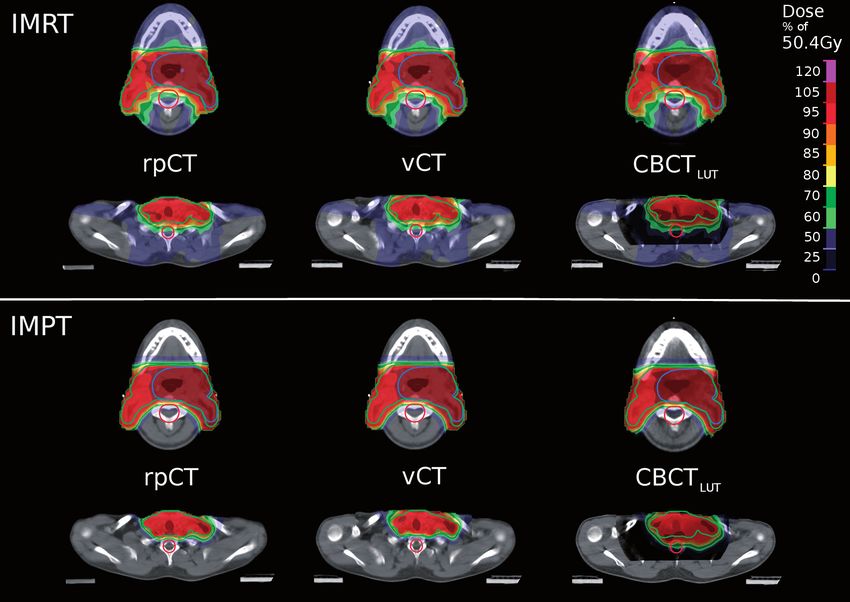
Habilitation Dr. rer. nat. Christopher Kurz

This cumulative habilitation is primarily based on the following peer-reviewed scientific contributions (in
the order of appearance in this thesis):

    1. Kurz C, Dedes G, Resch A, Reiner M, Ganswindt U, Nijhuis R, Thieke C, Belka C, Parodi K, Landry G.
       Comparing cone-beam CT intensity correction methods for dose calculation in adaptive intensity
       modulated photon and proton therapy for head and neck cancer. Acta Oncol. 2015;54(9):1651-7.
       DOI: 10.3109/0284186X.2015.1061206

    2. Kurz C, Kamp F, Park YK, Zöllner C, Rit S, Hansen DC, Podesta M, Sharp GC, Li M, Reiner M, Hofmaier
       J, Neppl S, Thieke C, Nijhuis R, Ganswindt U, Belka C, Winey BA, Parodi K, Landry G. Investigating
       deformable image registration and scatter correction for CBCT-based dose calculation in adaptive
       IMPT. Med Phys. 2016;43(10):5635-46. DOI: 10.1118/1.4962933

    3. Hansen DC, Landry G, Kamp F, Li M, Belka C, Parodi K, Kurz C. ScatterNet: a convolutional neural
       network for cone-beam CT intensity correction. Med Phys. 2018;45(11):4916-26. DOI:
       10.1002/mp.13175

    4. Kurz C, Landry G, Resch A, Dedes G, Kamp F, Ganswindt U, Belka C, Raaymakers BW, Parodi K. A
       Monte-Carlo study to assess the effect of 1.5T magnetic fields on the overall robustness of pencil-
       beam scanning proton radiotherapy plans for prostate cancer. Phys Med Biol. 2017;62(21):8470-
       82. DOI: 10.1088/1361-6560/aa8de9

    5. Maspero M, van den Berg CAT, Landry G, Belka C, Parodi K, Seevinck PR, Raaymakers BW, Kurz C.
       Feasibility of MR-only proton dose calculations for prostate cancer radiotherapy using a
       commercial pseudo-CT generation method. Phys Med Biol. 2017;62(24):9159-76. DOI:
       10.1088/1361-6560/aa9677

    6. Haehnle J, Süss P, Landry G, Teichert K, Hille L, Hofmaier J, Nowak D, Kamp F, Reiner M, Thieke C,
       Ganswindt U, Belka C, Parodi K, Küfer KH, Kurz C. A novel method for interactive multi-objective
       dose-guided patient positioning. Phys Med Biol. 2017;62(1):165-85. DOI: 10.1088/1361-
       6560/62/1/165

                                                    1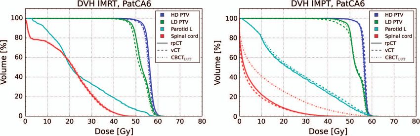
Habilitation Dr. rer. nat. Christopher Kurz

Table of contents
1 Introduction and background .............................................................................................. 3
2 Own scientific contributions ................................................................................................ 8
   2.1 CBCT-guided adaptive radiotherapy ........................................................................................... 8
      2.1.1 Comparing cone-beam CT intensity correction methods for dose calculation in adaptive
      intensity modulated photon and proton therapy for head and neck cancer ....................................... 8
      2.1.2 Investigating deformable image registration and scatter correction for CBCT-based dose
      calculation in adaptive IMPT ................................................................................................................. 9
      2.1.3 ScatterNet: a convolutional neural network for cone-beam CT intensity correction ................ 10
      2.1.4 Further publications related to CBCT-guided adaptive radiotherapy ........................................ 11
   2.2 MRI-guided adaptive proton therapy ....................................................................................... 12
      2.2.1 A Monte-Carlo study to assess the effect of 1.5T magnetic fields on the overall robustness of
      pencil-beam scanning proton radiotherapy plans for prostate cancer .............................................. 12
      2.2.2 Feasibility of MR-only proton dose calculations for prostate cancer radiotherapy using a
      commercial pseudo-CT generation method........................................................................................ 13
      2.2.3 Further publications related to MRI-guided adaptive radiotherapy .......................................... 14
   2.3 Dose-guided patient positioning .............................................................................................. 15
      2.3.1 A novel method for interactive multi-objective dose-guided patient positioning .................... 15
      2.3.2 Further publications related to dose-guided patient positioning .............................................. 16

3 Conclusions and outlook .................................................................................................... 17
4 List of abbreviations ........................................................................................................... 19
5 Literature ........................................................................................................................... 20
6 Acknowledgements ............................................................................................................ 24
7 Declaration ........................................................................................................................ 25
8 Facsimile of relevant scientific contributions ...................................................................... 26

                                                                         2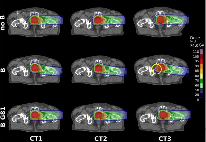
Habilitation Dr. rer. nat. Christopher Kurz

1 Introduction and background
In Germany close to 500,000 new patients are diagnosed with cancer every year [44]. Therapeutic options
comprise surgery, chemo- and targeted therapy, as well as radiotherapy. In total, more than half of all
cancer patients undergo radiotherapy as part of their treatment, with the ultimate goal of sterilizing all
cancer cells by local energy or dose deposition by ionizing radiation. While the dose to the target volume
should be high enough to achieve this aim, the dose to all relevant organs-at-risk (OAR), should be as low
as reasonably possible to prevent severe side effects. In most cases, patients follow the workflow
illustrated in figure 1.

 Figure 1. Overview of the typical radiotherapy workflow: Prior to treatment, a planning CT (mandatory for dose calculation
 during treatment planning) is acquired. Besides, MRI is frequently used for accurate tumor and OAR delineation due to its
 superior soft-tissue contrast. The segmented CT is then used for treatment optimization with a dedicated planning system.
 Several days to weeks later, the patient receives the first irradiation fraction. In-room imaging with the patient in treatment
 position (cone-beam CT (CBCT) or, more recently, MRI) is used for accurate patient alignment. In adaptive radiotherapy, the
 acquired in-room imaging data is not only used for patient set-up, but also for daily adaptation of the treatment to the actual
 patient anatomy. For this, the in-room imaging data have to be rendered suitable for accurate dose calculation (CBCT
 correction or synthetic CT (sCT) generation) and all relevant structures for treatment planning have to be segmented (green).

In modern radiotherapy, image-guidance using various imaging modalities plays an integral role during
treatment planning, as well as during patient treatment itself (in-room imaging). At treatment planning
stage, an X-ray computed tomography (CT) image is generally acquired in order to derive electron density
(photon therapy) or relative stopping power ratio (proton therapy) information, which are required for
accurate dose calculation during treatment plan optimization. More and more, CT imaging is accompanied
by magnetic resonance imaging (MRI) and/or positron emission tomography (PET) scans. While MRI is
typically used to infer more detailed anatomical features for improved tumor and OAR delineation due to
its superior soft-tissue contrast, it is more recently also applied to determine functional properties of the

                                                               3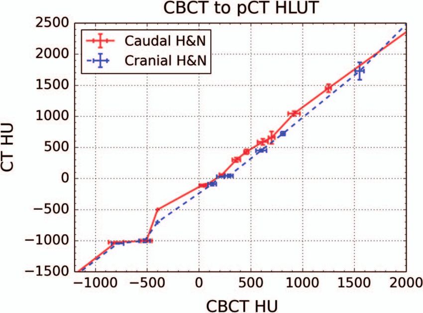
Habilitation Dr. rer. nat. Christopher Kurz

tumor, e.g., by means of diffusion-weighted imaging (DWI) [48]. Also PET imaging aims at determining
functional and metabolic properties of tumor tissues, such as glucose uptake rates or oxygenation levels,
as well as at identifying potential spread to adjacent lymph nodes or even distant metastasis [21].
Information derived from functional imaging, using either MRI or PET, can then be used for treatment plan
personalization, e.g., by defining certain regions in the tumor to receive a higher (boosted) dose level [8].
However, tissue properties derived from functional imaging might not only be considered at treatment
planning stage, but also allow for early response assessment in radiotherapy by monitoring their behavior
as a function of time during the course of fractionated radiotherapy [22].

Besides the previously described imaging taking place outside the treatment room, mostly at treatment
planning stage, in-room imaging [20], aiming at the exact alignment of the patient with respect to the
treatment unit, constitutes an indispensable part of image-guided radiotherapy. In combination with
modern external beam radiotherapy techniques, such as intensity-modulated photon or proton
radiotherapy (IMRT [52], IMPT [9]), in-room imaging theoretically enables tight adjustment of the
delivered dose to the target. However, the full potential of these techniques is currently not exploited in
clinical practice. The reason for this, and generally one of the major challenges in modern radiotherapy, is
the presence of anatomical changes, occurring on time scales from seconds (lung), to minutes (abdomen
and pelvis), days or weeks (head and neck [H&N]) [5, 26]. For the vast majority of patients in today’s
radiotherapy, such anatomical alterations are only considered by introducing safety margins around the
actual tumor volume during treatment planning [50]. This ensures irradiation of the tumor to the
prescribed dose, but increases the irradiated volume, the dose burden to OARs, and eventually limits the
applicable dose and hereby the treatment efficacy. A substantially improved treatment for tumor entities
affected by anatomical changes during fractionated radiotherapy, such as H&N, prostate or pancreatic
cancer, can be realized by online adaptive radiotherapy (ART) [54]. Instead of applying the same irradiation
plan throughout the entire course of treatment (typically several weeks) and assuming the initial planning
anatomy is still valid, the treatment is optimized at each irradiation session on basis of the daily anatomy
in treatment position, as inferred directly from in-room imaging (figure 1). This intrinsically accounts for
anatomical changes and allows for reduced margins and optimal OAR sparing at each fraction, even when
increasing the target dose at the same time.

Despite the anticipated benefits, the fraction of patients currently irradiated in online adaptive scenarios
is still negligible. The main reason is that nearly all patients in photon and proton radiotherapy are treated
using cone-beam CT (CBCT) for in-room imaging. Although CBCT allows for accurate patient alignment, the
image quality is hampered by the detection of scattered photons and not sufficient for accurate dose
calculation, which is indispensable for treatment adaptation. In the last years, various methods have been
proposed for CBCT image correction, aiming at rendering the data suitable for photon and proton therapy
dose calculation [12, 30, 39, 41]. Suggested techniques range from simple look-up-table (LUT)-based
recalibration of the CT numbers, over CT-to-CBCT deformable image registration (DIR) to more
sophisticated projection-based correction algorithms. However, most of them either lack accuracy (LUT-
based), robustness (DIR-based) or speed (projection-based) for online application. Only recently, deep
learning approaches, in particular convolutional neural networks (CNN), which had already shown
impressive results for a variety of medical image processing tasks [3, 11], have been adopted in the context

                                                      4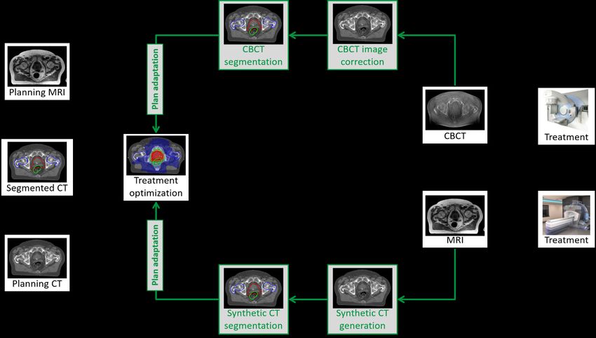
Habilitation Dr. rer. nat. Christopher Kurz

of CBCT correction. Several groups have reported promising results in terms of correction speed and
accuracy utilizing U-shaped CNNs (Unets) [23, 32, 45] or generative adversarial networks (GAN) [13, 17].
These networks enable CBCT intensity correction with accuracy similar to previously published methods,
but with correction times of only few seconds once the network models are trained. In the scope of this
habilitation, CBCT intensity correction, aiming at yielding images suitable for accurate photon and proton
dose calculation in the context of adaptive radiotherapy, was a major focus. Various conventional (LUT-,
DIR-, projection-based) and deep learning-based (Unet, GAN) methods have been investigated in terms of
their dosimetric accuracy and capability for online application. A detailed description of these works will
be given in section 2.1.

Besides the poor image quality, including a comparably low soft-tissue contrast, an important obstacle for
CBCT-guided online ART is the imaging dose, which can amount to a total of 1-2 Gy (therapeutic doses: 50
to 70 Gy) if daily imaging is performed for 30 or more fractions [2]. Thus, CBCTs are often acquired less
frequently, e.g., only once per week, for treatment sites such as the head and neck region, where
anatomical changes are expected to be gradual and occurring over longer time scales.

Due to these limitations of CBCT image-guidance, great efforts have been made over the last decades to
realize the integration of MRI as in-room imaging modality. Substantial technical challenges related to the
electro-magnetic decoupling and, hereby, interference-free side-by-side operation of the MRI and the
treatment machine, had to be overcome. Thus, only during the last few years, integrated MR-linear
accelerators (MR-Linacs) became certified and clinically available at few academic institutions. Ever since,
this technique has aroused considerable interest in the radiotherapy community [28]. The superior soft-
tissue contrast allows for accurate visualization of targets and OARs at no imaging dose, and the vendors
of both certified MR-Linacs (ViewRay MRIdian [36] and Elekta Unity [29]) have implemented basic online
ART workflows for the first time in the history of radiotherapy [1, 6, 43, 53], leading to a paradigm shift in
patient treatment. Besides pre-treatment adaptation, in-room MRI allows for continuous imaging (in 2D)
and tracking of the tumor volume itself without requiring any external surrogate [51]. In combination with
gated beam delivery, i.e., irradiating only when the target is in a pre-defined location, highly accurate
irradiation of tumors affected by intra-fractional motion is feasible. Promising clinical results, among
others for pancreatic cancer, have been reported in initial studies [46]. Worldwide, a large number of
studies on MRI-guided radiotherapy are still being carried in order to assess the promised clinical benefits
from these devices and from online ART workflows.

Due to the substantial personnel, technical and financial outlay of this emerging technique, however, only
three MR-Linacs will be installed in Germany by the end of 2020, including an MRIdian MR-Linac at the
LMU Department of Radiation Oncology that went clinically operational in January 2020. Worldwide the
number of devices is still below one hundred. Additionally, treatments are still limited to entities, such as
the prostate or the pancreas, which allow for hypo-fractionated schemes (
Habilitation Dr. rer. nat. Christopher Kurz

Nevertheless, recently also interest in combining the advantages of MRI-guidance and proton therapy,
currently relying on CBCT as in-room imaging modality, is growing in the radiotherapy community [40]. In
proton therapy, even larger benefits are anticipated from ART due to the higher sensitivity of proton dose
distributions to anatomical changes [24], which might be substantially reduced when using MR-guidance.
Also considering the potentially advantageous proton dose distributions allowing for considerably reduced
integral dose, MR-guided proton therapy has the potential to further boost treatment efficiency in
radiotherapy in the future. A first pre-clinical prototype has just lately been installed in Dresden and
technical feasibility studies are on-going [47]. Similar to photon therapy, a major challenge is to electro-
magnetically decouple the MRI scanner and the beam delivery system. Taking into the consideration that
up-to-date proton therapy facilities utilize pencil-beam scanning dose delivery with magnetic steering of
the beam, it can be acknowledged that substantial technical issues will have to be solved to pave the way
towards clinical implementation. On top, and in contrast to MRI-guided photon therapy, also the impact
of the MRI scanner B-fields on the treatment beam itself has to be carefully modeled and considered
during treatment planning. In the scope of this habilitation, the first study on the feasibility of fully inverse
IMPT optimization in B-fields has been conducted. Moreover, the obtained plans have been investigated
in depth in terms of their robustness against anatomical and positional uncertainties. The corresponding
publication will be described in more detail in section 2.2.

Another main challenge MRI-guided ART with photons, as well as potentially in the future with protons, is
facing, is the conversion of the in-situ in-room MRIs into synthetic CTs (sCT). These are required for
accurate dose calculation and adaptation since the MRI signal cannot be directly converted into the
required electron density or stopping power ratio information [10]. Today, both vendors of clinically
certified MR-Linacs utilize an initial planning CT and CT-to-MRI DIR for sCT generation [6, 53]. But, DIR
accuracy is often limited in regions with pronounced inter-fractional anatomical changes, such as the
abdomen or pelvis, where the largest benefits from ART are actually anticipated. More accurate, fully
automatic and rapid sCT generation for MRI-guided online ART might in the future be achieved by deep
learning. Unets, and also GANs [13], have been shown to yield sCTs suitable for accurate photon dose
calculation [7, 15, 33]. A further advantage of CNNs is that, once trained, no planning CT is required for
sCT generation, thus allowing an MR-only workflow that could reduce dose burden to the patient, as well
as the clinical workload. However, CNN-based sCT algorithms have not yet been clinically applied in the
scope of MRI-guided photon therapy. In proton therapy, even higher CT number accuracy than in photon
therapy will be required for sCT generation, given the sensitivity of proton dose calculations to the
underlying stopping power maps. Nevertheless, several studies on sCT generation for MRI-guided proton
therapy exist and suggest clinically acceptable accuracy for proton dose calculation. The used algorithms
are based either on bulk-assignment, on LUT-based approaches [14, 25, 34, 42] or on the utilization of
deep learning techniques [31, 37, 49]. In the scope of this habilitation, methods for sCT generation using
conventional bulks-assignment techniques, as well as deep CNNs, have been investigated for application
in proton, but also in photon therapy. More details will be provided in section 2.2.

Beyond the previously discussed challenges related to CBCT intensity correction and sCT generation for
accurate dose calculation, image-guided online ART requires accurate target and OAR delineation for
treatment plan optimization. Until today, this remains a manual task requiring a trained physician for tens

                                                       6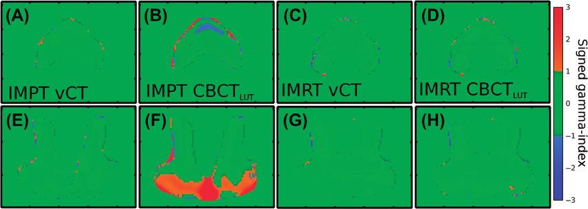
Habilitation Dr. rer. nat. Christopher Kurz

of minutes to hours, which is not acceptable for online ART where the patient has to stay on the treatment
table. In current clinically implemented MRI-guided photon ART workflows, contours are obtained via the
same DIR as the sCT and suffer from the same limited accuracy. Often extensive and time-consuming
manual corrections are required, rendering segmentation one of the bottlenecks in MRI-based ART.
Various deep learning techniques based on CNNs have, however, shown great potential for fast (
Habilitation Dr. rer. nat. Christopher Kurz
2 Own scientific contributions
2.1 CBCT-guided adaptive radiotherapy
2.1.1 Kurz C, Dedes G, Resch A, Reiner M, Ganswindt U, Nijhuis R, Thieke C, Belka C, Parodi K, Landry G.
Comparing cone-beam CT intensity correction methods for dose calculation in adaptive intensity
modulated photon and proton therapy for head and neck cancer. Acta Oncol. 2015;54(9)
In light of the previously described limitations in CBCT image quality, intensity correction strategies to
enable accurate dose calculation for plan adaptation in online ART have been investigated in this study.
More specifically, two different correction methods have been evaluated in terms of their dose calculation
accuracy for photon and proton radiotherapy of the H&N region. The simpler considered approach was to
perform correction via a population-based Hounsfield Unit (HU) rescaling [27]. For this, the HU values on
a reference diagnostic CT and a corresponding CBCT were sampled for air (inside and outside the patient),
fatty tissue, muscle, brain, soft and hard bone using a cohort of 9 H&N cancer patients. With these pairs
of values, a LUT was generated and used for CBCT rescaling. The method was compared to a CT-to-CBCT
DIR-based approach (virtual CT [vCT]), using a Morphon’s algorithm, which had been suggested in a
previous publication [30]. DIR used a metric based on the local image phase, thus focusing on the
alignment of edges in moving (CT) and fixed (CBCT) images, which is deemed superior to an intensity-based
approach when performing cross-modality image registration. To infer the dosimetric accuracy of both
approaches photon and proton treatment plans were recalculated on the obtained corrected CBCTs and
compared to a reference dose calculation using a diagnostic quality replanning CT acquired close in time.
Dose distributions were compared by means of clinically relevant dose-volume-histogram (DVH)
parameters and a gamma-index analysis.
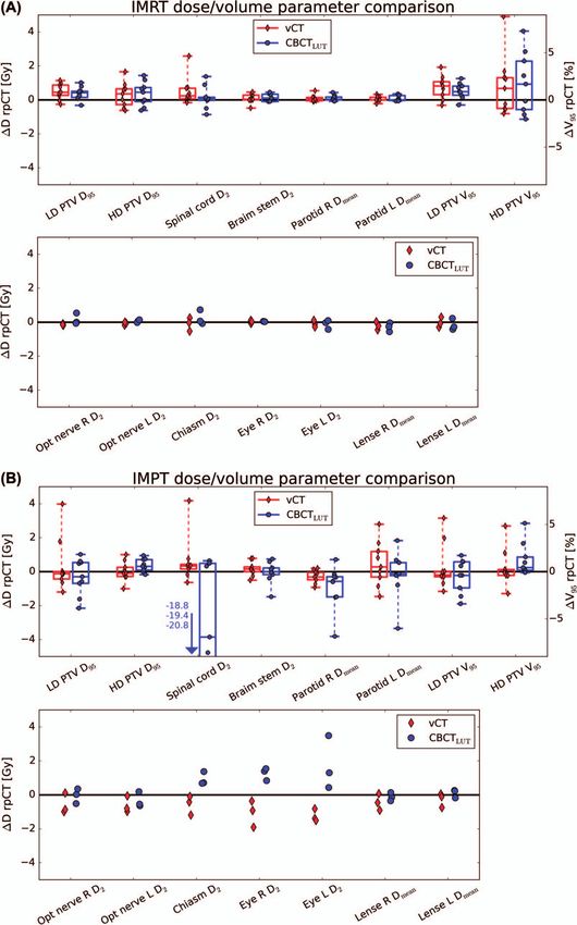
While both approaches, LUT and DIR, were found to yield accurate dosimetric results for photon therapy
of H&N cancer, the vCT clearly outperformed the LUT-based approach for proton therapy, where higher
HU accuracy is required. In particular, the LUT-based approach failed at correcting the CBCT in the area
between the shoulders, which is affected by substantial shadowing artifacts. These could not be overcome
by using a single LUT for CBCT intensity rescaling.
In a follow-up study, the feasibility of using the more accurate vCT and the corresponding warped
structures (target and OARs, using the same CT-to-CBCT deformation field) for automated proton therapy
plan adaptation, mimicking an online ART workflow, was investigated [see section 2.1.4, A1]. For the same
9 H&N patients, a novel treatment plan using the vCT and the corresponding structures was generated
automatically by using the same treatment planning settings as in the initial planning scenario. The new
vCT-based plan was then recalculated on a reference diagnostic replanning CT acquired 30 to 50 days after
the initial planning CT (but within 1 to 3 days of the considered CBCT). It could be shown that the
automatically obtained vCT-based adapted plans yielded clinically preferable plans on this replanning CT
when compared to the initial treatment plan. In particular, hotspots related to patient weight-loss and
tumor shrinkage could be efficiently diminished, thus demonstrating for the first time the feasibility of
automated, DIR-based online proton ART for H&N cancer.
8Habilitation Dr. rer. nat. Christopher Kurz
2.1.2 Kurz C, Kamp F, Park YK, Zöllner C, Rit S, Hansen DC, Podesta M, Sharp GC, Li M, Reiner M, Hofmaier
J, Neppl S, Thieke C, Nijhuis R, Ganswindt U, Belka C, Winey BA, Parodi K, Landry G. Investigating
deformable image registration and scatter correction for CBCT-based dose calculation in adaptive IMPT.
Med Phys. 2016;43(10)
Since the DIR-based vCT approach could be successfully applied for CBCT correction in photon and proton
therapy in the H&N region, this follow-up study aimed at extending it to the pelvic region, considering a
cohort of prostate cancer patients. While the H&N region is mostly affected by gradual anatomical
changes, e.g., due to patient weight-loss, the pelvis is subject to more pronounced and random inter-
fractional changes, e.g., related to variations in bladder and rectum filling. Under these conditions, the DIR
approach was found considerably less accurate than observed in the H&N region. In particular, the
algorithm failed in accurately modeling large volume changes of the rectum or the bladder, which are
typically accompanied by pronounced sliding motion.
Because of this, an alternative CBCT correction technique using the vCT only as a prior to perform
projection-based scatter correction [39, 41] was studied as an alternative. In this approach, a forward
projection of the vCT using the geometry of the gantry-mounted CBCT scanner is first performed. The
contribution of scattered photons, and also of other low-frequency perturbations such as beam hardening
[A2], in the measured CBCT projections is then estimated by subtracting the presumably scatter-free vCT
forward projections from the measured CBCT projections and applying a generous filter (due to the
assumed low spatial frequency). This estimated scatter contribution is then subtracted from the measured
CBCT projections, yielding a set of scatter-corrected projections, which can be reconstructed to obtain an
intensity corrected CBCT (CBCTcor).
vCT and CBCTcor were compared for two cohorts of H&N and prostate cancer patients. For the H&N
cohort, where the vCT had shown to yield dosimetrically accurate results in previous studies, the CBCTcor
was found equivalent. Also for the prostate cohort, vCT and CBCTcor obtained almost similar proton dose
distributions. However, when carefully checking the geometric fidelity of both images, CBCTcor was found
superior for the prostate patients. In particular, as already mentioned above, the vCT yielded incorrectly
shaped internal structures, which became apparent in a comparison with the original (uncorrected) CBCT
image. In particular, the geometry of the OARs, i.e., of the rectum and the bladder, was found inconsistent.
While the impact of these geometric deviations on dose calculation was only minor, they would clearly
hamper treatment evaluation and treatment plan optimization (DVH calculation). When looking at
CBCTcor, remaining artifacts from the incorrect vCT prior were still visible, but the impact was substantially
reduced and accurate delineation of all OARs could be demonstrated.
Given the superior robustness of the CBCTcor approach, it is supposed to be particularly beneficial in
regions suffering from pronounced inter-fractional anatomical changes and, thus, reduced DIR accuracy.
Both approaches, vCT and CBCTcor, were employed in several further studies described in the following
section and listed in section 2.1.4, A1-A6.
9Habilitation Dr. rer. nat. Christopher Kurz
2.1.3 Hansen DC, Landry G, Kamp F, Li M, Belka C, Parodi K, Kurz C. ScatterNet: a convolutional neural
network for cone-beam CT intensity correction. Med Phys. 2018;45(11)
Although the CBCTcor intensity correction approach proved to allow for accurate photon and proton dose
calculation for different tumor sites, one main drawback of the method is the long processing time, being
in the order of 5-7 minutes. Especially when considering its application for online adaptation, with the
patient on the treatment table, this is not acceptable. Also due to the fact that, e.g., in the prostate region,
anatomical changes can progress on similar time-scales. In this contribution, a potential solution has been
investigated: the application of deep CNNs, which have show remarkable performance for many image-
to-image translation tasks in recent years and have extremely short application times once the networks
are trained.
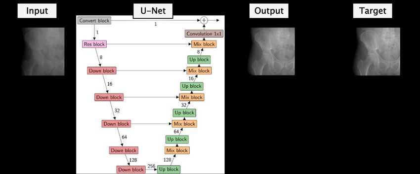
To this aim, a deep Unet was trained and evaluated in terms of dosimetric accuracy for projection-based
intensity correction using clinical CBCT data of 30 prostate cancer patients in this study. The basic idea is
depicted in figure 2: The network is trained to translate a measured projection into a scatter corrected
projection, as obtained from the accurate, but slow CBCTcor reference method. The output of the network
is compared to this reference by means of a loss function (L2 norm in this case) in order to optimize the
free parameters of the network iteratively. The network used a conventional Unet design with an encoding
(down-sampling) and decoding (up-sampling) branch, linked via so-called skip connections which enable a
direct flow of extracted features from the encoding to the decoding branch at each level of the network.
Down- and up-sampling blocks made use of so-called residual blocks, i.e., skip connections of convolutional
layers, to ease training of the network [18]. In total, 7323 projections of 15 patients were used for training
in 2D, using pairs of measured and corrected 2D projections. During training the value of the loss function
was monitored for 7 validation patients in order to determine the optimal stopping point for training.
Eventually, photon and proton dose calculation accuracy was evaluated for the remaining 8 independent
test patients, using CBCTcor as a reference image.
Figure 2. Conceptual design of the Unet investigated for CBCT scatter correction [16]. The network consists of several down-
and up-sampling building blocks and aims at translating measured projections into corrected projections. For training, the
network output is compared to a reference corrected projection by means of an L2 loss function.
10Habilitation Dr. rer. nat. Christopher Kurz
Once the network was trained, which took in the order of 30 to 40 hours, it took only about 0.01 s to
correct a single CBCT projection. For a whole CBCT data set, correction can thus be achieved within few
seconds, rendering this method particularly interesting in the scope of CBCT-guided online ART. In
addition, it was shown that Unet-based CBCT correction is not only fast, but also dosimetrically accurate
for photon and proton therapy, yielding dose distributions in close agreement to CBCTcor. Due to using
the CBCTcor projections for training, the method was also found robust in the presence of pronounced
anatomical changes, e.g., in the bladder.
In a second study on deep learning-based CBCT correction, the versatility of the proposed Unet design for
CBCT intensity correction could be demonstrated. The same network was successfully trained to not only
perform CBCT correction in projection space, but also in image space [A5]. For this, the network was
trained to translate the raw reconstructed CBCT image without applying any corrections into either a vCT
or a CBCTcor. For all three training strategies (projections, vCT, CBCTcor), intensity-corrected CBCT images
were obtained that allowed for accurate photon and proton dose calculation.
As an alternative network design, a cycle-consistent GAN (cycleGAN) was also investigated for performing
CBCT intensity correction in a further study [A6]. The cycleGAN was trained to translate CBCTs into CT
equivalent images. GANs are conceptually different from Unets and consist of a generator and a
discriminator network, which are trained jointly using an adversarial loss function: while the generator
network aims at generating as realistic as possible CT images from input raw CBCT images, the
discriminator aims at distinguishing between fake (generator output) and real CT images. By adding a
dedicated cycle-consistency loss function for conditioning the generator network outwork on the input,
the cycleGAN can be trained using unpaired image data. This is a unique feature for deep CNNs and is
feasible due to the fact that the network output is not compared on a pixel-by-pixel basis to a reference
image. Unpaired training is deemed of particular interest for applications where matching training data is
difficult to obtain, e.g., in the prostate region, where pronounced inter-scan anatomical changes between
diagnostic CT and daily CBCT occur. In the respective study, it could be shown for the first time that
accurate CBCT correction using a cycleGAN and unpaired training is feasible in the scope of adaptive
photon and proton therapy.
2.1.4 Further publications related to CBCT-guided adaptive radiotherapy
A1. Kurz C, Nijhuis R, Reiner M, Ganswindt U, Thieke C, Belka C, Parodi K, Landry G. Feasibility of
automated proton therapy plan adaptation for head and neck tumors using cone beam CT images.
Rad Onc. 2016;11:64
A2. Zöllner C, Rit S, Kurz C, Vilches-Freixas G, Kamp F, Dedes G, Belka C, Parodi K, Landry G.
Decomposing a prior-CT-based cone-beam CT projection correction algorithm into scatter and
beam hardening components. Phys Imag Radiat Oncol (phiRO). 2017;3:49-52
A3. Hofmaier J, Haehnle J, Kurz C, Landry G, Maihoefer C, Schüttrumpf L, Süss P, Teichert K, Söhn M,
Spahr N, Brachmann C, Weiler F, Thieke C, Küfer KH, Belka C, Parodi K, Kamp F. Multi-criterial
patient positioning based on dose recalculation on scatter-corrected CBCT images. Radiother
Oncol. 2017;125(3):464-9
11Habilitation Dr. rer. nat. Christopher Kurz
A4. Niepel K, Kamp F, Kurz C, Hansen DC, Rit S, Neppl S, Hofmaier J, Bondesson D, Thieke C, Dinkel J,
Belka C, Parodi K, Landry G. Feasibility of 4DCBCT-based proton dose calculation: an ex-vivo
porcine lung phantom study. Z Med Phys. 2019;29(3):249-61
A5. Landry G, Hansen DC, Kamp F, Li M, Hoyle B, Weller J, Parodi K, Belka C, Kurz C. Comparing Unet
training with three different datasets to correct CBCT images for prostate radiotherapy dose
calculations. Phys Med Biol. 2019;64(3):035011
A6. Kurz C, Maspero M, Savenije MHF, Landry G, Kamp F, Pinto M, Li M, Parodi K, Belka C, van den
Berg CAT. CBCT correction using a cycle-consistent generative adversarial network and unpaired
training to enable photon and proton dose calculation. Phys Med Biol. 2019; 64(22):225004
2.2 MRI-guided adaptive proton therapy
2.2.1 Kurz C, Landry G, Resch A, Dedes G, Kamp F, Ganswindt U, Belka C, Raaymakers BW, Parodi K. A
Monte-Carlo study to assess the effect of 1.5T magnetic fields on the overall robustness of pencil-beam
scanning proton radiotherapy plans for prostate cancer. Phys Med Biol. 2017;62(21)
Besides CBCT, MRI is playing an increasingly import role for in-room imaging in photon radiotherapy. For
its application in proton therapy, as described above, considerable technical challenges remain to be
solved before clinical introduction. Nevertheless, similar to photon therapy, proton therapy could greatly
benefit from the superior soft-tissue contrast as well as the online real-time imaging capabilities of MRI in
comparison to CBCT and is thus gaining interest. In the study described in this section, one of the crucial
ingredients for performing MRI-guided proton ART, namely the treatment plan optimization under
consideration of the MRI magnetic field, has been addressed.
In contrast to photon therapy, the MRI B-field not only affects the secondary electrons, but also the
primary beam particles. In this proof-of-concept study, inverse pencil-beam scanning IMPT planning in the
presence of a simplified cylindrical 1.5 T magnetic field perpendicular to the proton beam has been
successfully implemented. For the first time also an accurate modeling of the patient geometry in the
underlying Monte-Carlo simulations, based on the given patient CT image, was performed. For treatment
plan optimization, pencil-beams with Bragg peaks in vicinity of the target volume were first selected and
the individual pencil-beam doses calculated under consideration of the B-field using the GEANT4 Monte-
Carlo code. To obtain the final treatment plan, the weights of the individual pencil beams were then
optimized using an in-house developed research treatment plan optimization tool (CERR).
The implemented treatment planning pipeline was also used to infer the robustness of IMPT plans in the
presence of a perpendicular magnetic field. A cohort of 5 prostate cancer patients with 3 repeated CTs
each was included in the study. Different orientations of the patients with respect to the magnetic field
were considered, just as different gantry angles. Robustness against inter-fractional anatomical changes,
as well as robustness against variations in patient set-up (shifts of ±5 mm in anterior-posterior and left-
right direction) were studied for the different scenarios and compared to standard IMPT without magnetic
field. Results showed that MRI-guided proton therapy is feasible yielding similar plan quality and
robustness as found in conventional CT-based proton therapy without magnetic fields. However, to
achieve comparable robustness, adaptation of the treatment geometry (in this case of the gantry angle)
12Habilitation Dr. rer. nat. Christopher Kurz
was required. The adaptation had to be performed in such a way that the (due to the magnetic deflection)
curved proton path within the patient was as close as possible to the proton path in the scenario without
magnetic field. For this, an optimal gantry angle of 81°, instead of the clinically used 90° for the given
patient set-up, was determined (see figure 3). The reduced robustness observed without gantry angle
adaptation (i.e., using an angle of 90°) was related to the fact that the curved proton beam traversed
regions which were subject to more pronounced inter-fractional changes related to rectum filling and body
outline variations. Thus, for other anatomical sites, adaptation of the gantry angles might have to be
performed in a different way, also due to the fact that the curvature of the proton beam will change when
using different initial beam energies.
Figure 3. Proton dose distributions for an exemplary prostate patient optimized on CT1 and recalculated on CT2/3 to infer
the robustness against inter-fractional anatomical changes for different scenarios: Treatment without B-field (top row), as
well as treatment in presence of a 1.5 T B-field perpendicular to the incident beam using gantry angles of 90° (middle row)
and 81° (bottom row). Robustness for the B-field scenario with 81° gantry angle was similar to the conventional scenario
without B-field, while reduced robustness was observed for a gantry angle of 90° in presence of the B-field. A substantial
overshoot on CT 3 is indicated by the yellow circle.
2.2.2 Maspero M, van den Berg CAT, Landry G, Belka C, Parodi K, Seevinck PR, Raaymakers BW, Kurz C.
Feasibility of MR-only proton dose calculations for prostate cancer radiotherapy using a commercial
pseudo-CT generation method. Phys Med Biol. 2017;62(24)
Another crucial input required for MRI-guided adaptive proton therapy is the generation of synthetic CT
images from in-room MRI for accurate dose calculation. This topic has been investigated in the context of
prostate cancer in the study presented in this section. A commercially available and certified photon-
oriented solution, called MRCAT (MR for calculating attenuation) has been adapted for application in
proton therapy. The method relies on a dual spoiled gradient echo MRI sequence and Dixon
reconstruction, in combination with a constrained shape bone model and bulk density assignment of 5
tissue classes (air, fat, muscle, spongy and compact bone) for sCT generation.
13Habilitation Dr. rer. nat. Christopher Kurz
In this contribution the method was extended to allow also for the identification of internal gas pockets,
which is crucial for accurate proton dose estimation in the pelvis. Moreover, it was found that the used
bulk-assigned HU values for spongy and compact bone, which were initially optimized for application in
photon therapy, had to be adapted to yield optimal proton dose calculation accuracy. The latter was
inferred from recalculating proton plans optimized using diagnostic planning CTs on the MRCAT images
and dosimetric comparison. After elimination of inter-scan differences between the planning CT and the
MRI used for MRCAT generation, clinically acceptable proton dose calculation accuracy was found, despite
the limited number of tissue classes. However, adaptation of the bulk-assigned HU values for bones with
respect to the original photon-based solution was found crucial.
In a more recent co-authored paper also the feasibility of using 2D or 3D Unets for MR-only sCT generation
in proton therapy was shown for the first time [see section 2.2.3, B1]. The network design was similar to
the design used for CBCT correction, as described in section 2.1. In comparison to the MRCAT solution, the
Unet approach yields sCTs with a continuous HU range. Overall, good accuracy was found for proton and
for photon dose calculation in a cohort of brain tumor patients. In particular, the depp CNN enabled
accurate separation of bony anatomy and internal air cavities, which is typically found a major challenge
for sCT generation since both structures show no or very low signal on MRI.
Further studies in the field of MRI-guided radiotherapy (with photons and protons) that have been pursued
in the scope of this habilitation are listed in section 2.2.3 [B2-B4]. These include two recent review articles
on the medical physics challenges of MRI-guided photon therapy and the current status and future
perspectives of MRI-guided proton therapy.
2.2.3 Further publications related to MRI-guided adaptive radiotherapy
B1. Neppl S, Landry G, Kurz C, Hansen DC, Hoyle B, Stöcklein S, Seidensticker M, Weller J, Belka C,
Parodi K, Kamp F. Evaluation of proton and photon dose distributions recalculated on 2D and 3D
Unet-generated pseudoCTs from T1-weighted MR head scans. Acta Oncol. 2019;58(10):1429-34
B2. Rabe M, Thieke C, Düsberg M, Neppl S, Gerum S, Reiner M, Nicolay NH, Schlemmer HP, Debus J,
Dinkel J, Landry G, Parodi K, Belka C, Kurz C*, Kamp F*. Real-time 4D-MRI-based internal target
volume definition for moving lung tumors. Med Phys. 2020;47(4):1431-42.
*Both authors contributed equally
B3. Kurz C, Buizza G, Landry G, Kamp F, Rabe M, Paganelli C, Baroni G, Reiner M, Keall PJ, van den Berg
CAT, Riboldi M. Medical physics challenges in clinical MR-guided radiotherapy. Radiat Oncol.
2020;15:93
B4. Hoffmann A, Oborn B, Moteabbed M, Yan S, Bortfeld T, Knopf AC, Fuchs H, Georg D, Seco J, Spadea
MF, Jäkel O, Kurz C, Parodi K. MR-guided proton therapy: a review and a preview. Radiat Oncol.
2020;15:129
14Habilitation Dr. rer. nat. Christopher Kurz
2.3 Dose-guided patient positioning
2.3.1 Haehnle J, Süss P, Landry G, Teichert K, Hille L, Hofmaier J, Nowak D, Kamp F, Reiner M, Thieke C,
Ganswindt U, Belka C, Parodi K, Küfer KH, Kurz C. A novel method for interactive multi-objective dose-
guided patient positioning. Phys Med Biol. 2017;62(1)
In this joint project with colleagues at the Fraunhofer Institute for Industrial Mathematics (ITWM) in
Kaiserslautern, a prototype software for interactive dose-guided positioning has been implemented and
dosimetrically compared to conventional bony anatomy-based alignment for the first time. Dose-guided
positioning was technically implemented as an iso-center planning problem (ICPP), which could then be
solved under consideration of user-defined clinical objectives, such as DVH parameters for target coverage
or OAR dose limits. Since for more than one clinical objective the ICPP is an MCO problem, trading off, e.g.,
dose to the target against dose to OARs, there is a set of Pareto-optimal solutions. By linear dose-
interpolation between different iso-center shifts, the developed tool allows the user to interactively
browse through the continuous space of Pareto-optimal patient positions and to straightforwardly
determine the clinically optimal patient shift under consideration of the dose.
The efficiency of the approach was demonstrated for 3 H&N and 3 prostate cancer patients using IMRT,
following careful validation of the dose interpolation accuracy. A delineated replanning CT was considered
as surrogate for the daily in-room image of each patient. In a more realistic scenario, either an intensity
corrected CBCT or an MRI-based sCT might be used. In all cases, dose-guided alignment allowed to find a
clinically preferable position in comparison to bony anatomy-based alignment. The main effects were an
increased target coverage in combination with a reduced dose to the parotid glands (H&N) or the rectum
(prostate). However, the study also showed that, in particular for H&N, plan re-optimization, which would
in practice be feasible with the same input data (in-room image and segmentation), could achieve even
better daily dose distributions.
In a follow-up study, also the applicability of the developed dose-guided positioning tools in the scope of
proton therapy has been investigated [see section 2.3.2, C1]. In total 14 H&N and 8 prostate cancer
patients with up to 5 repeated CTs were considered. Dose-guided positioning was again compared to the
clinically used bony anatomy-based alignment. For the H&N cohort, the main effect of dose-based
positioning was a reduction of the dose to the serial organs (spinal cord and brain stem). For the prostate
cohort, under-dosage of the target structures could be reduced. Nevertheless, also limitations in the scope
of proton therapy were identified. For the H&N cohort, it was not possible to diminish target over-dosage
related to patient weight-loss. To properly account for weight-loss, reduction of the initial proton fluence,
e.g., in the context of a plan re-optimization, would be necessary.
Since no labor-intensive online quality assurance and plan approval by a certified radiation oncologist is
required, dose-guided patient alignment might, nevertheless, still be considered an interesting alternative
to full online ART.
15Habilitation Dr. rer. nat. Christopher Kurz
2.3.2 Further publications related to dose-guided patient positioning
C1. Kurz C, Süss P, Arnsmeyer C, Haehnle J, Teichert K, Landry G, Hofmaier J, Exner F, Hille L, Kamp F,
Thieke C, Ganswindt U, Valentini C, Hölscher T, Troost E, Krause M, Belka C, Küfer KH, Parodi K,
Richter C. Dose-guided patient positioning in proton therapy using multicriteria-optimization. Z
Med Phys. 2019;29(3):216-28
16Habilitation Dr. rer. nat. Christopher Kurz
3 Conclusions and outlook
In the scope of this habilitation, studies on various aspects of image-guided adaptive photon and proton
therapy have been conducted. Focus was on the ability to render in-room imaging data suitable for
accurate daily dose calculation, as required for treatment plan adaptation in an ART workflow. CBCT, as
well as MRI, have been considered as the clinically most relevant in-room imaging modalities today.
Different methods for the required image correction (CBCT) or translation (MRI to CT) have been
investigated, ranging from conventional techniques (LUT-based rescaling, DIR, projection-based correction
or bulk assignment) to novel deep learning-based approaches using CNNs (Unet, GAN). In particular, the
latter allow for accurate image correction with unparalleled speed and are thus of growing interest in the
scope of ART, where time is a crucial factor. Despite the anticipated benefits from deep leaning-based
solutions, they are nowadays still restricted to research applications. For future clinical integration, careful
safety and risk assessment of these algorithms is indispensable. Major challenges will be posed by the
black-box nature of neural networks and the fact that trained CNN models typically do not generalize well
on unseen data-sets. Thus, it is likely that models will struggle for patients with non-standard anatomies,
e.g. due to surgical resection of tissue. How network inaccuracies in such cases can be detected and
eventually overcome is still a topic of on-going research.
Beyond image correction for enabling accurate dose calculation, the generation of accurate delineations
on the daily in-room imaging data to be used during treatment optimization is a major challenge for online
ART. Here, depending on the treatment site, the currently clinically implemented DIR-based approach for
MRI-guided photon ART is often facing limitations. Similar observations have been made in the scope of
this habilitation in the context of CBCT-based ART. While DIR was found sufficiently accurate for intensity
correction and contour propagation to enable automatic treatment plan adaptation in the H&N region,
limited accuracy was found in the pelvis. Generally, in the presence of pronounced anatomical changes,
DIR-based segmentation will often require time-consuming manual correction by an expert.
Unfortunately, exactly these patients, suffering from substantial inter-fractional alterations, are expected
to have the largest benefit from online ART. Thus, one important focus of future research is the fast and
fully automatic segmentation of in-room imaging data. Similar to image correction, deep learning
strategies, often using 3D CNN architectures and dedicated loss metrics (e.g., based on the Dice-similarity
coefficient), have shown impressive results for this task, outperforming classical segmentation algorithms,
based e.g., on atlases and DIR. First certified deep learning solutions for auto-segmentation of various
body sites have recently been launched by different vendors but are still limited to the initial treatment
planning stage rather than the online plan adaptation stage using in-room imaging data.
Following image correction or translation and segmentation, online adaptation of the treatment plan
becomes feasible. While in photon therapy fast Monte-Carlo dose calculation algorithms taking into
consideration the magnetic fields (in the case of MRI-guidance) are used in clinical routine, aspects of
proton treatment planning in magnetic fields, as required for clinically realizing MRI-guided proton
therapy, have been addressed for the first time in the scope of this habilitation. The feasibility of fully
inverse pencil-beam scanning treatment optimization in a magnetic field could be shown, together with
the robustness of the generated plans. Although there are still considerable technical hurdles to be
17Habilitation Dr. rer. nat. Christopher Kurz
overcome, there is a strongly growing interest in this novel treatment approach. One of the main reasons
is that considerable benefits from MRI-guidance for proton therapy are anticipated, since it might help to
overcome issues related to the sensitivity of proton therapy to inter- and intra-fractional anatomical
changes by providing online treatment adaptation and imaging during beam delivery.
As an alterative approach to daily re-optimization of the treatment plan, MCO-based dose-guided patient
positioning has been investigated for photon and for proton therapy in this habilitation. While it requires
similar input as online ART, i.e., segmented in-room images suitable for accurate dose calculation, it does
require neither online plan quality assurance, nor clinical approval by an expert radiation oncologist.
Nevertheless, the daily dose distributions’ quality achieved with dose-guided positioning was found
inferior to that achieved with plan adaptation from scratch.
While initial online ART workflows using integrated MR-Linacs become more and more widespread, and
first certified solutions for CBCT-based ART are lately being introduced by vendors, many challenges
remain. These encompass further streamlining the established clinical workflows, in particular by
exploiting novel deep learning techniques, but also the extension of treatment adaptation procedures to
shorter timescales. More specifically, current workflows only enable adaptation of the treatment with
respect to inter-fractional anatomical changes, i.e., once prior to the irradiation, while intra-fractional
changes, e.g., related to respiratory motion, are still mitigated by gating, using fast 2D imaging for target
tracking in the case of MRI-guidance. In the future, a considerably faster treatment delivery (without
beam-off times) could be achieved by implementing real-time ART. For this, a continuous stream of 3D
images would be used for real-time accumulation of the already applied dose, followed by prompt
adaptation of the remaining treatment. Main challenges to be overcome are the 3D imaging at sufficient
frame rates (few Hz), as well as performing accumulation and optimization in real-time. Both aspects are
subject of on-going scientific studies.
To conclude, image-guided online ART allowing for higher dose levels in the target volume, at similar or
even reduced dose burden to close-by OARs, is currently entering radiotherapy clinics. Hereby, a
considerably more efficient patient treatment is expected, especially in the presence of inter-fractional
anatomical changes. The research performed in the scope of this habilitation addressed various aspects of
MRI- and CBCT-guided ART with photons and protons paving the way towards clinical adoption of these
radiotherapy techniques.
18Habilitation Dr. rer. nat. Christopher Kurz
4 List of abbreviations
ART Adaptive radiotherapy
CBCT Cone-beam computed tomography
CNN Convolutional neural network
CT Computed tomography
DIR Deformable image registration
DVH Dose-volume-histogram
DWI Diffusion-weighted imaging
GAN Generative adversarial network
H&N Head and neck
HU Hounsfield units
ICCP Iso-center planning problem
IMPT Intensity-modulated proton therapy
IMRT Intensity-modulated radiotherapy
ITWM Fraunhofer Institute for Industrial Mathematics
Linac Linear accelerator
LMU Ludwig-Maximilians-University
LUT Look-up table
MCO Multi-criteria optimization
MRCAT MR for calculating attenuation
MRI Magnetic resonance imaging
OAR Organ-at-risk
PET Positron emission tomography
sCT Synthetic CT
vCT Virtual CT
19Habilitation Dr. rer. nat. Christopher Kurz
5 Literature
1. Acharya S, Fischer-Valuck BW, Kashani R, Parikh P, Yang D, Zhao T, et al. Online Magnetic Resonance
Image Guided Adaptive Radiation Therapy: First Clinical Applications. Int J Radiat Oncol Biol Phys.
2016;94(2):394-403.
2. Alaei P, Spezi E. Imaging dose from cone beam computed tomography in radiation therapy. Phys Med.
2015;31(7):647-58.
3. Ardila D, Kiraly AP, Bharadwaj S, Choi B, Reicher JJ, Peng L, et al. End-to-end lung cancer screening with
three-dimensional deep learning on low-dose chest computed tomography. Nat Med. 2019;25(6):954-61.
4. Balagopal A, Kazemifar S, Nguyen D, Lin MH, Hannan R, Owrangi A, et al. Fully automated organ
segmentation in male pelvic CT images. Phys Med Biol. 2018;63(24):245015.
5. Bert C, Durante M. Motion in radiotherapy: particle therapy. Phys Med Biol. 2011;56(16):R113-44.
6. Bohoudi O, Bruynzeel AME, Senan S, Cuijpers JP, Slotman BJ, Lagerwaard FJ, et al. Fast and robust online
adaptive planning in stereotactic MR-guided adaptive radiation therapy (SMART) for pancreatic cancer.
Radiother Oncol. 2017;125(3):439-44.
7. Dinkla AM, Wolterink JM, Maspero M, Savenije MHF, Verhoeff JJC, Seravalli E, et al. MR-Only Brain
Radiation Therapy: Dosimetric Evaluation of Synthetic CTs Generated by a Dilated Convolutional Neural
Network. Int J Radiat Oncol Biol Phys. 2018;102(4):801-12.
8. Draulans C, van der Heide UA, Haustermans K, Pos FJ, van der Voort van Zyp J, De Boer H, et al. Primary
endpoint analysis of the multicentre phase II hypo-FLAME trial for intermediate and high risk prostate
cancer. Radiother Oncol. 2020;147:92-8.
9. Durante M, Loeffler JS. Charged particles in radiation oncology. Nat Rev Clin Oncol. 2010;7(1):37-43.
10. Edmund JM, Nyholm T. A review of substitute CT generation for MRI-only radiation therapy. Radiat
Oncol. 2017;12(1):28.
11. Esteva A, Kuprel B, Novoa RA, Ko J, Swetter SM, Blau HM, et al. Dermatologist-level classification of
skin cancer with deep neural networks. Nature. 2017;542(7639):115-8.
12. Fotina I, Hopfgartner J, Stock M, Steininger T, Lutgendorf-Caucig C, Georg D. Feasibility of CBCT-based
dose calculation: comparative analysis of HU adjustment techniques. Radiother Oncol. 2012;104(2):249-
56.
13. Goodfellow IJ, Pouget-Abadie J, Mirza M, Xu B, Warde-Farley D, Ozair S, et al. Generative Adversarial
Networks. arXiv e-prints [Internet]. 2014 June 01, 2014. Available from:
https://ui.adsabs.harvard.edu/abs/2014arXiv1406.2661G.
14. Guerreiro F, Koivula L, Seravalli E, Janssens GO, Maduro JH, Brouwer CL, et al. Feasibility of MRI-only
photon and proton dose calculations for pediatric patients with abdominal tumors. Phys Med Biol.
2019;64(5):055010.
20Habilitation Dr. rer. nat. Christopher Kurz
15. Han X. MR-based synthetic CT generation using a deep convolutional neural network method. Med
Phys. 2017;44(4):1408-19.
16. Hansen DC, Landry G, Kamp F, Li M, Belka C, Parodi K, et al. ScatterNet: A convolutional neural network
for cone-beam CT intensity correction. Med Phys. 2018;45(11):4916-26.
17. Harms J, Lei Y, Wang T, Zhang R, Zhou J, Tang X, et al. Paired cycle-GAN-based image correction for
quantitative cone-beam computed tomography. Med Phys. 2019;46(9):3998-4009.
18. He K, Zhang X, Ren S, Sun J. Deep Residual Learning for Image Recognition2015 December 01,
2015:[arXiv:1512.03385 p.]. Available from: https://ui.adsabs.harvard.edu/abs/2015arXiv151203385H.
19. Ibragimov B, Xing L. Segmentation of organs-at-risks in head and neck CT images using convolutional
neural networks. Med Phys. 2017;44(2):547-57.
20. Jaffray DA. Image-guided radiotherapy: from current concept to future perspectives. Nat Rev Clin
Oncol. 2012;9(12):688-99.
21. Jelercic S, Rajer M. The role of PET-CT in radiotherapy planning of solid tumours. Radiol Oncol.
2015;49(1):1-9.
22. Jones KM, Michel KA, Bankson JA, Fuller CD, Klopp AH, Venkatesan AM. Emerging Magnetic Resonance
Imaging Technologies for Radiation Therapy Planning and Response Assessment. Int J Radiat Oncol Biol
Phys. 2018;101(5):1046-56.
23. Kida S, Nakamoto T, Nakano M, Nawa K, Haga A, Kotoku J, et al. Cone Beam Computed Tomography
Image Quality Improvement Using a Deep Convolutional Neural Network. Cureus. 2018;10(4):e2548.
24. Knopf AC, Lomax A. In vivo proton range verification: a review. Phys Med Biol. 2013;58(15):R131-60.
25. Koivula L, Wee L, Korhonen J. Feasibility of MRI-only treatment planning for proton therapy in brain
and prostate cancers: Dose calculation accuracy in substitute CT images. Med Phys. 2016;43(8):4634.
26. Korreman SS. Motion in radiotherapy: photon therapy. Phys Med Biol. 2012;57(23):R161-91.
27. Kurz C, Dedes G, Resch A, Reiner M, Ganswindt U, Nijhuis R, et al. Comparing cone-beam CT intensity
correction methods for dose recalculation in adaptive intensity-modulated photon and proton therapy for
head and neck cancer. Acta Oncol. 2015;54(9):1651-7.
28. Lagendijk JJ, Raaymakers BW, Van den Berg CA, Moerland MA, Philippens ME, van Vulpen M. MR
guidance in radiotherapy. Phys Med Biol. 2014;59(21):R349-69.
29. Lagendijk JJ, Raaymakers BW, van Vulpen M. The magnetic resonance imaging-linac system. Semin
Radiat Oncol. 2014;24(3):207-9.
30. Landry G, Nijhuis R, Dedes G, Handrack J, Thieke C, Janssens G, et al. Investigating CT to CBCT image
registration for head and neck proton therapy as a tool for daily dose recalculation. Med Phys.
2015;42(3):1354-66.
21You can also read