Proton beam therapy followed by pembrolizumab for giant ocular surface conjunctival malignant melanoma: A case report
←
→
Page content transcription
If your browser does not render page correctly, please read the page content below
MOLECULAR AND CLINICAL ONCOLOGY 16: 12, 2022

     Proton beam therapy followed by pembrolizumab for giant
   ocular surface conjunctival malignant melanoma: A case report
                        TOSHIHIKO MATSUO1,2, OSAMU YAMASAKI3,4, TAKEHIRO TANAKA5,
                                   KUNIAKI KATSUI6,7 and TAKAHIRO WAKI8

            1
             Regenerative and Reconstructive Medicine (Ophthalmology), Okayama University Graduate School of
      Interdisciplinary Science and Engineering in Health Systems, Okayama 700‑8558; 2Department of Ophthalmology,
       Okayama University Hospital; 3Department of Dermatology, Okayama University Graduate School of Medicine,
 Dentistry and Pharmaceutical Sciences; 4Melanoma Center, Okayama University Hospital, Okayama 700‑8558; 5Department
of Pathology, Okayama University Graduate School of Medicine, Dentistry and Pharmaceutical Sciences, Okayama 700‑8558;
                      6
                       Division of Radiation Oncology, Department of Radiology, Kawasaki Medical School,
        Kurashiki, Okayama 701‑0192; 7Department of Radiology, Okayama University Graduate School of Medicine,
                                   Dentistry and Pharmaceutical Sciences, Okayama 700-8558;
                    8
                      Department of Radiology, Tsuyama Chuo Hospital, Tsuyama, Okayama 708‑0841, Japan

                                    Received September 15, 2021; Accepted October 27, 2021

                                                   DOI: 10.3892/mco.2021.2445

Abstract. The present study describes proton beam therapy as        istration of pembrolizumab 77.2 mg every three weeks five
a clinical option to achieve local control of giant conjunctival    times in total. Then, three months after proton beam therapy,
melanoma in an aged person, instead of orbital exenteration.        ocular surface melanoma almost subsided and the clear cornea
An 80‑year‑old woman with one‑year history of left‑eye injec‑       allowed visualization of the intraocular lens inside the eye.
tion and hemorrhage experienced rapid growth of the ocular          In three weeks, spontaneous corneal perforation was plugged
surface black mass. At the initial visit, a black, elastic hard,    with iris incarceration. The patient died suddenly of unknown
hemorrhage‑prone, thickened mass in the size of 30x40 mm            cause 7.5 months from the initial visit. The local control of
with a presumed wide stalk covered the total area of the lid        giant conjunctival melanoma was achieved by proton beam
fissure on the left side. Biopsy of the mass demonstrated           therapy, leading to patient's satisfaction and better quality of
anomalous melanin‑containing cells in fibrin and hemor‑             life. Proton beam therapy, followed by immune checkpoint
rhage, which were positive for cocktail‑mix antibodies against      inhibitors, would become the future standard of care for unre‑
tyrosinase, melanoma antigen recognized by T cells‑1 and            sectable giant conjunctival melanoma.
human melanoma black‑45, indicative of malignant mela‑
noma. One month after the initial visit, the patient underwent      Introduction
proton beam therapy at the total dose of 70.4 Gy (relative
biological effectiveness) in 32 fractions (~10 min each) in one     Malignant melanoma as a whole is less frequent in Asian
and a half months. One month after the end of proton beam           populations including Japanese population, compared with
therapy, 3.5 months from the initial visit, the patient was found   Caucasian populations. In the field of ophthalmology, mela‑
by computed tomographic scan to have multiple metastatic            noma can be encountered rarely in uvea (choroid) (1), lacrimal
lesions in bilateral lung fields. With the evidence of absent       sac (2), conjunctiva and eyelid skin (3,4). Among these
BRAF mutation, the patient underwent intravenous admin‑             melanomas in ophthalmic practice, conjunctival malignant
                                                                    melanoma is relatively frequent in Asian populations and
                                                                    Caucasian populations (3,4). However, no standard treatment
                                                                    has been established in conjunctival melanoma which is basi‑
                                                                    cally at very low incidence (5).
Correspondence to: Professor Toshihiko Matsuo, Regenerative             In case of cutaneous melanoma, its excision with ample
and Reconstructive Medicine (Ophthalmology), Okayama University
                                                                    margin of the surrounding normal skin is the standard of
Graduate School of Interdisciplinary Science and Engineering in
Health Systems, Shikata‑cho 2‑5‑1, Okayama 700‑8558, Japan
                                                                    care for local control of malignancy. In contrast, excision of
E‑mail: matsuot@cc.okayama‑u.ac.jp                                  conjunctival melanoma with ample normal margin cannot
                                                                    be achieved since the conjunctiva of limited width is the
Key words: ocular surface, conjunctiva, malignant melanoma,         essential mucosa to maintain the ocular surface and hence,
proton beam therapy, pembrolizumab, PD‑1 inhibitor, immune          the vision. Due to this anatomical limitation, it is difficult to
checkpoint inhibitor, corneal perforation                           excise conjunctival melanoma at the early phase with ample
                                                                    margin of the surrounding normal tissue as is the standard of
                                                                    care for cutaneous melanoma. At present, local recurrence is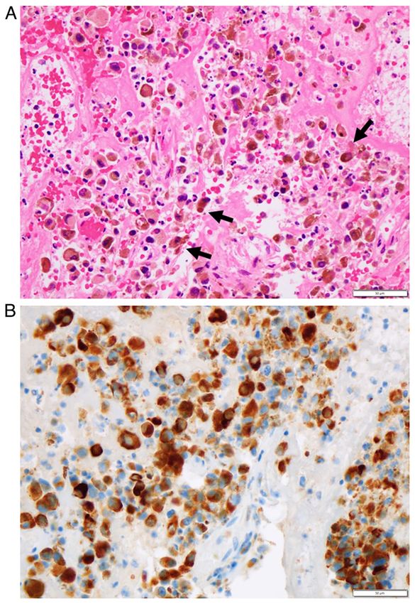
2                      MATSUO et al: PROTON BEAM THERAPY FOR CONJUNCTIVAL MALIGNANT MELANOMA

a common sequela to the initial excision of conjunctival mela‑
noma. Orbital exenteration is a final surgical option to achieve
the local control in the advanced stages when conjunctival
melanoma shows infiltration into the surrounding tissue such
as the eyelid skin and the sclera of the eyeball. It would be
disastrous for patients to lose the vision of one eye abruptly by
the orbital exenteration. Invasive surgery of the orbital exenter‑
ation would also put physical and psychological burdens on the
aged people who are prone to develop conjunctival melanoma.
    In the general belief that melanoma would not respond
sufficiently to external beam radiation, radiotherapy has
played a limited role only in adjunct or palliative treat‑
ment for metastatic diseases of cutaneous melanoma (6,7),
conjunctival melanoma (5) and head and neck mucosal mela‑
noma (8,9). Under the circumstances, proton beam therapy
has been tried as a treatment option for the local control of
conjunctival melanoma and has been indeed shown to be
effective (5,10‑15). In Japan, proton beam therapy for head and
neck cancer including melanoma (16‑18) has been covered by
reimbursement from the national health insurance since April
2018. Within the framework of the national health insurance,
immune checkpoint inhibitors and molecular target drugs such
as MEK inhibitors and BRAF inhibitors can be also used in
the case of unresectable and metastatic malignant melanoma,
based on the genetic testing of BRAF mutations in melanoma
tissues (19,20).
    The present study dealt with an aged patient who showed
giant conjunctival melanoma at the initial presentation and          Figure 1. The mass before and after proton beam therapy. (A) A black, elastic
who decided to choose proton beam therapy as a first‑line            hard, hemorrhage‑prone, thickened mass arising from the ocular surface
therapeutic option for the local control. Furthermore, based on      on the left side which prevents the eyelid from closing at the initial visit in
no BRAF mutation detected in the melanoma tissue, pembro‑            an 80‑year‑old woman. (B and C) The mass has been reduced 2.5 months
                                                                     after the conclusion of proton beam therapy, 5 months from the initial visit
lizumab, PD‑1 immune checkpoint inhibitor (19,20), was               and (D‑F) has almost subsided in half a month, 5.5 months from the initial
introduced as a current standard therapy toward metastatic           visit. (F) Note the transparent cornea to visualize inside the eye globe. (G) In
lung lesions after the proton beam therapy.                          three weeks, spontaneous corneal perforation has been plugged with iris
                                                                     incarceration.
Case report

An 80‑year‑old woman noticed injection and hemorrhage in             standardized uptake value, SUVmax=14.04) and had no
the left eye one year previously and she removed the painless        abnormal uptake in other sites of the body. Surface biopsy
ocular surface scab by herself frequently. One month previ‑          of the mass and the neutral formalin‑fixed paraffin sections
ously, the black mass grew out of the lid fissure rapidly and        demonstrated anomalous melanin‑containing cells in fibrin
she could not close the left eye (Fig. 1A). She visited a local      and hemorrhage (Fig. 3A). Immunostaining at the in‑house
hospital and was referred to Okayama University Hospital. At         pathology laboratory showed that anomalous cells were
the initial visit, the best‑corrected visual acuity was 1.2 in the   positive for cocktail‑mix antibodies against tyrosinase,
right eye and light perception in the left eye. The intraocular      melanoma antigen recognized by T cells‑1 and human mela‑
pressure in the right eye was 12 mmHg and the optic nerve            noma black‑45 (Fig. 3B), leading to pathological diagnosis of
disc had glaucomatous cupping as a cup/disc ratio of 0.9.            malignant melanoma.
Otherwise, the right eye had nothing notable. She had under‑             The patient underwent proton beam therapy one month
gone cataract surgery in the left eye four years previously. She     after the initial visit, at the total dose of 70.4 Gy (relative
had no other medical history and took no medication. A black,        biological effectiveness) in 32 fractions (~10 min each)
elastic hard, hemorrhage‑prone, thickened mass in the size of        for one and a half months. One month after the conclu‑
30x40 mm with a presumed wide stalk covered the total area           sion of proton beam therapy and three and a half months
of the lid fissure on the left side (Fig. 1A) and the mass moved     from the initial visit, the patient was found by computed
slightly with eye movement, indicative of the tumor origin on        tomographic scan to have multiple metastatic lesions in
the ocular surface.                                                  bilateral lung fields (Fig. 2C). Excisional biopsy of residual
    Magnetic resonance imaging showed the intact eye ball on         melanotic lesions in the upper eyelid skin on the left side
the left side and no infiltration deeply into the orbit (Fig. 2A).   (Fig. 1B and D) showed melanin‑containing epithelioid
Whole‑body 2‑[18F]fluoro‑2‑deoxy‑D‑glucose positron emis‑            cells with large nuclei and apparent nucleoli (Fig. 4A and B)
sion tomography (FDG‑PET) showed high uptake in the                  arranged in alveolar pattern, which confirmed the patho‑
eyelid area with the mass on the left side (Fig. 2B, maximum         logical diagnosis of malignant melanoma at the in‑house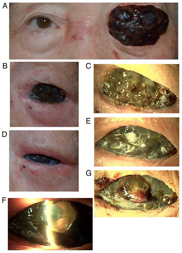
MOLECULAR AND CLINICAL ONCOLOGY 16: 12, 2022 3
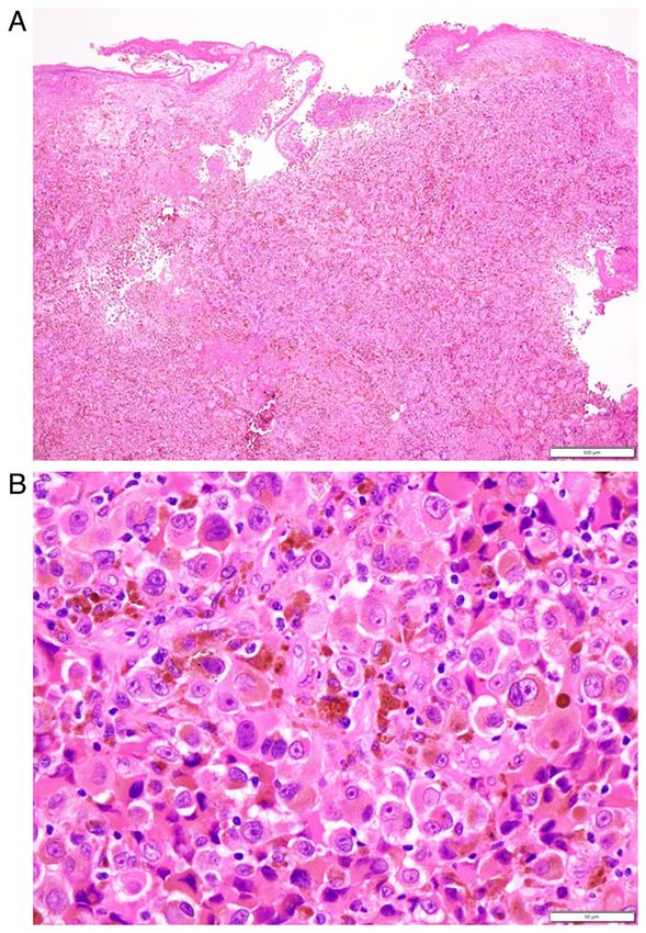
Figure 3. (A) Biopsy specimen in hematoxylin‑eosin stain of the ocular sur‑
Figure 2. Images of the mass before and after proton beam therapy. face mass at the initial visit. Note anomalous epithelioid cells (arrows) with
(A) T1‑weighted magnetic resonance image at the initial visit in an melanin pigments which are dispersed in fibrin and hemorrhage. (B) The
80‑year‑old woman, showing lobulated ocular surface mass with contrast anomalous cells were positive for cocktail‑mix antibodies against tyrosinase,
enhancement on the left side (arrowed). (B) Abnormal uptake in the area melanoma antigen recognized by T cells‑1 and human melanoma black‑45,
of the left eyelid (maximum standardized uptake value, SUVmax=14.04) in indicative of malignant melanoma. Scale bar, 50 µm.
whole‑body 2‑[18F]fluoro‑2‑deoxy‑D‑glucose positron emission tomography
at the initial visit. (C) CT scan, showing multiple metastatic nodules in
bilateral lung fields one month after the conclusion of proton beam therapy,
3.5 months from the initial visit. (D) CT scan at the same time, showing 0.25 µg daily. She died suddenly of unknown cause 7.5 months
the reduced ocular surface mass with contrast enhancement (arrowed).
from the initial visit.
CT, Computed tomography.
Discussion
pathology laboratory. BRAF mutations (V600E and V600K) The clinical question in the present study is that proton beam
were tested by real‑time PCR in the DNA sample extracted therapy might be a treatment option in the standard of care for
from the neutral formalin‑fixed paraffin sections and were advanced conjunctival malignant melanoma in the era when
shown to be absent at an external diagnostic laboratory proton beam therapy is included in the reimbursement of the
(LSI Medience Corporation). The patient then, underwent national health insurance in Japan. Orbital exenteration was
intravenous administration of pembrolizumab 77.2 mg every initially indicated for the local control of malignancy as the
three weeks five times in total. standard of care in this patient. However, the patient and her
At three months after the conclusion of proton beam family wished to avoid radical surgery and the clinicians, also
therapy, five and a half months from the initial visit, ocular wished to search for the other options because of the patient's
surface melanoma almost subsided (Fig. 1D and E) and the advanced age. Proton beam therapy was thus chosen as the
clear cornea allowed visualization of the intraocular lens first‑line treatment for unresectable melanoma in the head and
inside the eye (Fig. 1F). In three weeks, spontaneous corneal neck according to the rule of the national health insurance
perforation was plugged with iris incarceration (Fig. 1G). in Japan (16‑18). The patient had satisfactory outcome and
Around the same time, the patient felt malaise and was found regained better quality of life: She had covered the left‑side
to have hypothyroidism which was evidenced by low free T3 at tumor with gauze eye patch for cosmetic reasons before the
1.75 pg/ml, low free T4 at 0.90 ng/dl, high TSH at 39.9 µU/ml, therapy while she could blink and close the eye with the eyelid
high anti‑thyroglobulin antibody (TgAb) at 20,664 IU/ml and on the left side after the therapy.
high anti‑thyroid peroxidase antibody (TPOAb) at 2,580 IU/ml. On the initial presentation, biopsy could only obtain super‑
The patient started to take levothyroxine sodium hydrate ficial hemorrhage‑prone tissue of the tumor and pathological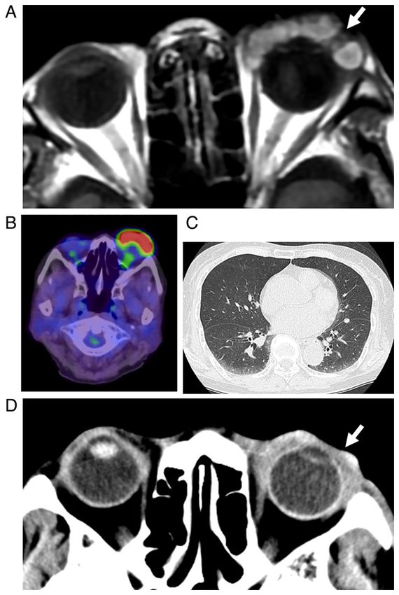
4                            MATSUO et al: PROTON BEAM THERAPY FOR CONJUNCTIVAL MALIGNANT MELANOMA

                                                                                      was taking oral thyroid hormone. Autoimmune diseases,
                                                                                      such as thyroiditis, pneumonitis and uveitis, should be
                                                                                      kept in mind in patients who receive immune checkpoint
                                                                                      inhibitors (21). The sudden mortality might be attributed
                                                                                      to possible carditis or pneumonitis as a manifestation of
                                                                                      pembrolizumab‑induced autoimmune diseases, but the exact
                                                                                      situation was not confirmed clinically in the patient. Until
                                                                                      the sudden mortality, the patient was healthy and had normal
                                                                                      activities of daily life.
                                                                                          A major limitation in this case report is that the patient's
                                                                                      presentation was unusual and that the sequence of event was
                                                                                      thus out of the standard. First, only the surface biopsy could
                                                                                      be done in the large protruding hemorrhage‑prone melanoma
                                                                                      instead of standard resection. Secondly, proton beam therapy
                                                                                      was chosen as the first‑line treatment for this giant conjunc‑
                                                                                      tival melanoma. The aim of therapeutic strategy at the initial
                                                                                      presentation was to enhance the quality of life in this aged
                                                                                      woman and the corneal melt as a sequel to proton beam
                                                                                      therapy was anticipated in advance. On the initial phase
                                                                                      of the patient's presentation, it was planned to use immune
                                                                                      checkpoint inhibitors or molecular target drugs as an adjunct
                                                                                      therapy since distant metastases to lung or liver would be
                                                                                      unavoidable at the stage of the disease. In this context, it was
                                                                                      not expected the complete response to proton beam therapy
                                                                                      alone and residual melanoma lesions in the eyelid skin could
                                                                                      be used as the biopsy site for examining the BRAF mutation
                                                                                      to determine the appropriate adjunct therapy. It should be
                                                                                      emphasized that the present case would not be recognized
                                                                                      as the standard for local control of conjunctival melanoma at
Figure 4. Biopsy of melanotic lesions in the upper eyelid skin on the left side one
month after the conclusion of proton beam therapy, 3.5 months from the initial        the current time.
visit. Note melanin‑containing epithelioid cells with large nuclei and apparent           In conclusion, proton beam therapy could be placed as a
nucleoli in alveolar pattern, confirming pathological diagnosis of malignant          treatment option for local control of unresectable conjunctival
melanoma. Hematoxylin‑eosin stain. (A) Scale bar, 500 µm and (B) 50 µm.               malignant melanoma. Proton beam therapy as the first‑line
                                                                                      treatment, followed by adjunct therapy with immune check‑
                                                                                      point inhibitors, could become the standard of care in near
diagnosis was based on immunocytochemical examinations.                               future for unresectable giant conjunctival melanoma. This
Following the proton beam therapy, excisional biopsy of the                           conclusion, based on a single patient, naturally has a limitation
residual eyelid lesion was done again to confirm the patho‑                           to being generalized to the other patients with conjunctival
logical diagnosis of malignant melanoma and also to examine                           malignant melanoma.
BRAF mutations in the melanoma tissue. Around the same
time, the patient was found to have multiple metastatic lesions                       Acknowledgements
in bilateral lung fields. According to the standard protocol
for unresectable and metastatic malignant melanoma (19,20),                           Not applicable.
pembrolizumab was introduced, instead of the combination of
MEK inhibitor and BRAF inhibitor, based on the absence of                             Funding
the BRAF mutation in the melanoma tissue. The sequence of
events in this patient, namely, proton beam therapy for local                         No funding was received.
control of unresectable conjunctival malignant melanoma,
followed later by the administration of immune checkpoint                             Availability of data and materials
inhibitors or the combination of MEK inhibitor and BRAF
inhibitor, could become the standard of care in the modern                            The datasets used and/or analyzed during the current study are
era. Indeed, in the present patient, pembrolizumab appeared                           available from the corresponding author on reasonable request.
to have an effect on the reduction of malignant melanoma in
the original and metastatic lesions, but actual evaluation of the                     Authors' contributions
effect could not be accomplished since the patient suddenly
succumbed.                                                                            TM, as an ophthalmologist and OY, as a dermatologist,
    The patient was reported to have succumbed suddenly                               followed and treated the patient, TT, as a pathologist, made
to unknown causes by the emergency transportation to a                                the pathological diagnosis and KK and TW, as radiologists,
local hospital. The patient had hypothyroidism, probably                              treated the patient. TM wrote the manuscript and OY, TT,
induced by the administration of pembrolizumab, and thus                              KK and TW did critical review of the manuscript. All authors
MOLECULAR AND CLINICAL ONCOLOGY 16: 12, 2022 5
confirm the authenticity of all the raw data and approved the 10. Wuestemeyer H, Sauerwein W, Meller D, Chauvel P, Schueler A,
Steuhl KP, Bornfeld N and Anastassiou G: Proton radiotherapy
final version of the manuscript. as an alternative to exenteration in the management of extended
conjunctival melanoma. Graefes Arch Clin Exp Ophthalmol 244:
Ethics approval and consent to participate 438‑446, 2006.
11. Westekemper H, Anastassiou G, Sauerwein W, Chauvel P,
Bornfeld N, Steuhl KP and Meller D: Analysis of ocular
Ethics committee review was not applicable to case reports, surface alterations following proton beam radiation in eyes
based on the Ethical Guidelines for Medical and Health with conjunctival malignant melanoma. Ophthalmologe 103:
588‑595, 2006 (In German).
Research Involving Human Subjects, issued by the Government 12. Krause L, Mladenova A, Bechrakis NE, Kreusel KM, Plath T,
of Japan. Moser L and Foerster M: Treatment modalities for conjunctival
melanoma. Klin Monbl Augenheilkd 226: 1012‑1016, 2009
(In German).
Patient consent for publication 13. Maschi‑Cayla C, Doyen J, Gastaud P and Caujolle JP: Conjunctival
melanomas and proton beam therapy. Acta Ophthalmol 91: e647,
Verbal informed consent was obtained from the patient for her 2013.
14. Scholz SL, Hérault J, Stang A, Griewank KG, Meller D, Thariat J,
anonymized information to be published in this article. Steuhl KP, Westekemper H and Sauerwein W: Proton radio‑
therapy in advanced malignant melanoma of the conjunctiva.
Competing interests Graefes Arch Clin Exp Ophthalmol 257: 1309‑1318, 2019.
15. Thariat J, Salleron J, Maschi C, Fevrier E, Lassalle S, Gastaud L,
Baillif S, Claren A, Baumard J, Herault J and Caujolle JP:
The authors declare that they have no competing interests Oncologic and visual outcomes after postoperative proton therapy
of localized conjunctival melanomas. Radiation Oncol 14: 239,
2019.
References 16. Zenda S, Kawashima M, Nishio T, Kohno R, Nihei K, Onozawa M,
Arahira S and Ogino T: Proton beam therapy as a nonsurgical
1. Matsuo T, Ogino Y, Ichimura K, Tanaka T and Kaji M: approach to mucosal melanoma of the head and neck: A pilot
Clinicopathological correlation for the role of fluorodeoxy‑ study. Int J Radiat Oncol Biol Phys 81: 135‑139, 2011.
glucose positron emission tomography computed tomography in 17. Fuji H, Yoshikawa S, Kasami M, Murayama S, Onitsuka T,
detection of choroidal malignant melanoma. Int J Clin Oncol 19: Kashiwagi H and Kiyohara Y: High‑dose proton beam therapy
230‑239, 2014. for sinonasal mucosal malignant melanoma. Radiat Oncol 9: 162,
2. Matsuo T, Tanaka T and Yamasaki O: Lacrimal sac malignant 2014.
melanoma in 15 Japanese patients: Case report and literature review. 18. Sakurai H, Ishikawa H and Okumura T: Proton beam therapy in
J Investig Med High Impact Case Rep 7: 2324709619888052, 2019. Japan: Current and future status. Jpn J Clin Oncol 46: 885‑892,
3. Seregard S: Conjunctival melanoma. Surv Ophthalmol 42: 2016.
321‑350, 1998. 19. Larsen AC, Dahmcke CM, Dahl C, Siersma VD, Toft PB,
4. Vora GK, Demirci H, Marr B and Mruthyunjaya P: Advances in Coupland SE, Prause JU, Guldberg P and Heegaard S: A
the management of conjunctival melanoma. Surv Ophthalmol 62: retrospective review of conjunctival melanoma: Presentation,
26‑42, 2017. treatment and outcome and an investigation of features associated
5. Jain P, Finger PT, Fili M, Damato B, Coupland SE, Heimann H, with BRAF mutations. JAMA Ophthalmol 133: 1295‑1303, 2015.
Kenawy N, Brouwer NJ, Marinkovic M, Van Duinen SG, et al: 20. Kiyohara T, Tanimura H, Miyamoto M, Shijimaya T,
Conjunctival melanoma treatment outcomes in 288 patients: A Nagano N, Nakamaru S, Makimura K and Iwai H: Two cases
multicentre international data‑sharing study. Br J Ophthalmol 105: of BRAF‑mutated, bulbar conjunctival melanoma, and review of
1358‑1364, 2021. the published literature. Clin Exp Dermatol 45: 207‑211, 2020.
6. Ballo MT and Ang KK: Radiotherapy for cutaneous malignant 21. Matsuo T and Yamasaki O: Vogt‑Koyanagi‑Harada disease‑like
melanoma: Rationale and indications. Oncology (Williston posterior uveitis in the course of nivolumab (anti‑PD‑1 antibody),
Park) 18: 99‑110, 113‑114, 2004. interposed by vemurafenib (BRAF inhibitor), for metastatic
7. Gorayski P, Burmeister B and Foote M: Radiotherapy for cutaneous malignant melanoma. Clin Case Rep 5: 694‑700, 2017.
cutaneous melanoma: Current and future applications. Future
Oncol 11: 525‑534, 2015.
8. López F, Rodrigo JP, Cardesa A, Triantafyllou A, Devaney KO, This work is licensed under a Creative Commons
Mendenhall WM, Haigentz M Jr, Strojan P, Pellitteri PK, Attribution-NonCommercial-NoDerivatives 4.0
Bradford CR, et al: Update on primary head and neck mucosal International (CC BY-NC-ND 4.0) License.
melanoma. Head Neck 38: 147‑155, 2016.
9. Grant‑Freemantle MC, O'Neill BL and Clover AJP: The effec‑
tiveness of radiotherapy in the treatment of head and neck
mucosal melanoma: Systematic review and meta‑analysis. Head
Neck 43: 323‑333, 2021.You can also read