Progression of aortic intramural hematoma with associated penetrating aortic ulcers with medical management requiring surgical management case report
←
→
Page content transcription
If your browser does not render page correctly, please read the page content below
Case Report
                                                                                                                                             Page 1 of 6

Progression of aortic intramural hematoma with associated
penetrating aortic ulcers with medical management requiring
surgical management case report
Koral Shah1^, Hamna Ahmad1,2, Jonathan E. Wilson1,3, Mala Goyal1,2, Sarah Dubin1,2
1
    School of Medicine, University of Missouri-Kansas City, Kansas, MO, USA; 2Department of Pulmonary, Critical Care, and Sleep Medicine,
University of Missouri-Kansas City, Kansas, MO, USA; 3Department of Vascular Surgery, University of Missouri-Kansas City, Kansas, MO, USA
Correspondence to: Koral Shah. School of Medicine, University of Missouri-Kansas City, Kansas, MO, USA. Email: koralushah@gmail.com.

                  Abstract: Penetrating ulcers of the aorta, aortic dissections, and intramural hematomas (IMH) all fall
                  under acute aortic syndromes (AAS) and have important similarities and differences. We present a case of
                  an asymptomatic patient with uncontrolled hypertension who was found to have a unique combination of
                  penetrating aortic ulcers (PAUs) with an associated IMH. Furthermore, the patient had PAUs located in
                  the aortic arch, which is an uncommon since the majority are located in the descending thoracic aorta. His
                  PAUs and IMH progressed despite medical management and subsequently required thoracic endovascular
                  aortic repair (TEVAR). The treatment of IMHs and PAUs is less well known compared to the classic
                  aortic dissection. Often, they may not be treated as an AAS or may be treated as an aortic dissection. This
                  case report addresses this challenge clinicians face with unclear delineation of treatment between different
                  AAS. This case demonstrates how a type B IMH, when associated with penetrating ulcers, may follow a
                  more malignant course, and should be considered for early surgical intervention. This case illustrates the
                  importance of understanding the distinction between the AAS and how treatment differs based on Stanford
                  classification and risk factors of progression.

                  Keywords: Case report; penetrating aortic ulcer (PAU); intramural hematoma (IMH); acute aortic syndrome
                  (AAS); thoracic endovascular aortic repair (TEVAR)

                  Received: 30 October 2020; Accepted: 17 January 2021; Published: 25 July 2021.
                  doi: 10.21037/jeccm-20-153
                  View this article at: http://dx.doi.org/10.21037/jeccm-20-153

Introduction                                                                 based on the location of the lesion and classifies lesions in
                                                                             the ascending aorta as type A and the rest as type B (3). In
Acute aortic syndrome (AAS) is an umbrella term for aortic
                                                                             this case, the patient presents with a unique mix of different
pathologies encompassing the classic aortic dissection,
intramural hematoma (IMH), contained aortic rupture,                         AAS which progressed despite medical management,
and penetrating aortic ulcer (PAU) (1). A PAU refers to an                   eventually requiring surgical intervention. The distinction
ulceration which penetrates the media of the aortic wall.                    between an IMH and aortic dissection in treatment is
The Debakey and The Stanford classification are two                          not widely recognized, nor is the treatment of PAUs. We
widely recognized classification systems for AAS (2,3). Out                  present the following case in accordance with the CARE
of the two, the Stanford classification is more commonly                     reporting checklist (available at http://dx.doi.org/10.21037/
used in most clinical settings. The Stanford classification is               jeccm-20-153).

^ ORCID: 0000-0002-4868-8276.

© Journal of Emergency and Critical Care Medicine. All rights reserved.           J Emerg Crit Care Med 2021;5:25 | http://dx.doi.org/10.21037/jeccm-20-153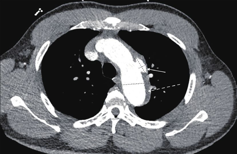
Page 2 of 6                                                                        Journal of Emergency and Critical Care Medicine, 2021

                                                                          and a negative UDS. A Chest X-ray showed prominent
                                                                          atherosclerotic thoracic aorta that prompted a computed
                                            15.6 mm                       tomography angiography (CTA) of chest was per the
                                       6.6 mm                             “Dissection Protocol”. CTA revealed a penetrating lateral
                                40.1 mm                                   aortic arch ulcer measuring 2 cm by 9 mm in transverse
                                                                          dimension and 1.5 cm craniocaudad dimension. A second,
                                                                          smaller penetrating ulcer closely adjacent, along the inferior
                                                                          margin of the aortic arch measuring 6.6 mm ×15.6 mm
                                                                          ×8 mm was also seen. CTA also showed an IMH 9 mm in
Figure 1 Initial computed tomography angiography showed IMH               thickness along the aortic arch beginning just past the origin
originating distal to subclavian artery and PAU. Dotted arrow             of the left subclavian artery and extending to the distal aspect
indicates IMH and solid arrow indicates PAU. PAU, penetrating             of the aortic arch (Figure 1). The CTA abdomen/pelvis per
aortic ulcer; IMH, intramural hematoma.                                   “Dissection Protocol” showed no flow-limiting stenosis,
                                                                          dissection, or aneurysm of the aorta. EKG shows prolonged
                                                                          QTc interval of 505 ms with left ventricular hypertrophy. The
Case presentation                                                         patient was immediately treated for hypertensive emergency
A 50-year-old African American male with a history of                     and started on an esmolol and nicardipine drip with a target
                                                                          decrease of mean arterial pressure by 20% in the first hour,
uncontrolled hypertension, not currently taking any
                                                                          followed by 10% over the next 23 hours. Vascular surgery
medications for the past month, presented to the emergency
                                                                          was consulted and recommended medical management with
room with shortness of air for the past 2 weeks with
                                                                          a repeat CTA of chest after 48 hours. Fortunately, the patient
headache and blurry vision. He was found to be severely
                                                                          did not face any diagnostic challenges due to socioeconomic
hypertensive with pressure readings no less than 250
                                                                          determinants of health. The patient’s hypertension was very
/150 mmHg, a heart rate of 77 beats per minute, and an
                                                                          resistant and necessitated titrating up to maximum doses
oxygen saturation of 97% on room air. Patient had tested
                                                                          of multiple oral medications in order to wean patient off
negative for COVID-19 one week prior. Upon evaluation
                                                                          the intra-venous anti-hypertensives. The patient’s blood
by the ICU team, patient denied headache, dizziness, vision
                                                                          pressure was controlled on oral carvedilol 25 mg twice a day,
changes, chest pain, palpitations, syncope, abdominal pain,
                                                                          nifedipine 120 mg once a day, clonidine 0.3 three times a day
nausea, vomiting, cough, or sweats. Past medical history
                                                                          with the addition of IV labetalol 10 mg and IV hydralazine
was significant only for hypertension and a family history
                                                                          10 mg to used as needed. The patient continued to remain
of hypertension, CVA, and end-stage renal disease in his
                                                                          completely asymptomatic. The repeat CTA of chest 48 hours
father. He had no prior relevant interventions. Social                    showed an increase in size of the largest multi-lobulated
history was significant for use of cigars once a month with               irregular penetrating ulcer to 24.6 mm ×10.9 mm ×1.7 mm
no other illicit substance use. Patient was active at baseline            and the smaller adjacent ulcer to 10.3 mm ×21 mm ×8 mm.
with either running or walking miles with his dogs daily. He              There was also re-demonstration of liquefying IMH along
worked as a barber with no heavy lifting or increased stress              the aortic arch (Figure 2) and proximal descending thoracic
at the work place. Patient took no medications. His entire                aorta with associated aneurysmal dilation of the same areas.
physical exam including cardiovascular and respiratory                    Extension of the hematoma to the origin of the left common
auscultation was unremarkable. Patient was transferred to                 carotid artery was difficult to exclude. Vascular surgery
ICU care. All procedures performed in studies involving                   performed thoracic endovascular aortic repair (TEVAR)
human participants were in accordance with the ethical                    on hospital day 4. Gore C-TAG active control device was
standards of the institutional and national research                      used for the TEVAR. Patient continued to be asymptomatic
committees and with the Helsinki Declaration (as revised                  after surgery with controlled blood pressures. Work-up for
in 2013). Consent to writing and publishing this case report              secondary hypertension was underway prior to transfer out
was obtained from the patient both verbally and through                   of the ICU. Patient plans to follow up with vascular surgery
written consent.                                                          outpatient, along with a new primary care provider and
   Initial workup showed an elevated creatinine of 3.74 mg/dL             nephrology.
with an unknown baseline, negative troponin-T x1,                            As for follow-up, a chest radiograph was done for

© Journal of Emergency and Critical Care Medicine. All rights reserved.      J Emerg Crit Care Med 2021;5:25 | http://dx.doi.org/10.21037/jeccm-20-153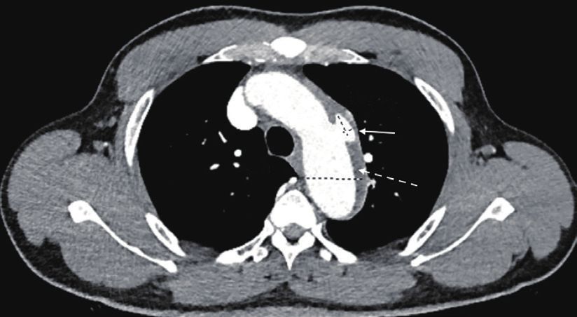
Journal of Emergency and Critical Care Medicine, 2021 Page 3 of 6
type B dissection. Type A acute dissection are repaired
emergently using open surgical techniques.
Acute IMH was first described by Krukenberg in 1920 (5)
24.6 mm with the main difference being there is an absence of tunica
10.9 mm intima disruption and hematomas are likely due to vaso-
43.3 mm vasorum injury. Per literature review, the appropriate
management of an IMH is not as well defined nor as
understood as that of classic dissection primarily because
hematomas can stabilize, regress, resolve, or progress.
PAUs are typically associated with extensive vascular
Figure 2 Repeat computed tomography angiography showed
atherosclerosis disease and hypertension, caused by
increase in size of IMH and PAU. Dotted arrow indicates IMH
ulceration of an aortic atherosclerotic lesion which
and solid arrow indicates PAU. PAU, penetrating aortic ulcer;
penetrates the internal elastic media into the media (6).
IMH, intramural hematoma.
PAUs may be associated with a hematoma within the media
and may progress to perforation or aortic dissection.
A meta-analysis by Maraj et al. found that 143 IMHs
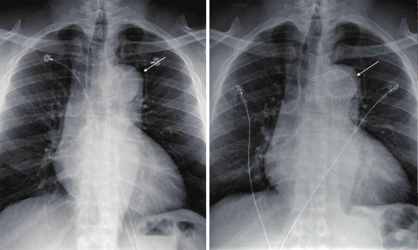
fever a week post-op which showed postsurgical changes showed that 57% were type A and 43% were type B, and
of endovascular stent graft involving the aortic arch and 94% had a non-traumatic cause (7). According to the IRAD
proximal descending thoracic aorta with stable appearance review, IMH is more common in the descending aorta
of the thoracic aorta (Figure 3). At a 20-day post-operative compared to aortic dissection, 60% vs. 35% respectively (8).
visit, vascular surgery requested a CTA chest 6 months The distinction between an IMH and aortic dissection
after TEVAR procedure. Of recently, patient’s blood is paramount as both differ in treatment and clinical
complications. Type B IMH treated similarly to a type B
pressures are at goal with carvedilol, amlodipine, clonidine,
dissection is generally acceptable except when the IMH is
furosemide, and lifestyle modifications. This is assessed
associated with a factor of progression or is a complicated
with follow-up visits and blood pressure logs. Patient has
course, for which earlier intervention may be needed to
been diagnosed with CKD Stage IV and follows with
prevent complications. Type B IMH progress to aortic
nephrology. Routine lab workup for secondary hypertension
rupture, hematoma expansion, or dissection in 8–16%
was negative. Patient follows with Sleep Clinic to evaluate
of patients. One of the predictors of progression are the
for obstructive sleep apnea. There have been no adverse
presence of PAUs (8,9). Other predictors of progression,
or unanticipated events thus far. Over a recent phone call,
despite adequate medical management, include maximum
patient reports that he is very appreciative of the care he
aortic diameter ≥40 mm, maximum aortic thickness
received in the ICU and that he feels much improved. He is
≥10 mm on CT scan, >70 years of age, and presence of
motivated to stay on top of his outpatient appointments.
a PAU (10). Sebastià et al. believe signs of a complicated
course can be presence of continuous chest pain despite
Discussion medical treatment, hemodynamic instability, signs of aortic
rupture, presence of large ulcer like projections >10 mm,
Differentiating between the aortic syndromes, especially maximum aortic diameter >55 mm, or rapid aortic growth
IMH and aortic dissection has been controversial which while in the hospital (11). These patients would undergo
can lead to confusion in approaching treatment in the real- endovascular treatment or surgical treatment if the former
world setting. was contraindicated. Timperley and Banning show that an
Aortic dissection is a tear in the aortic intima which IMH associated with a PAU has a higher rate of progression
allows blood between the layers of the vessel wall, thus with medical therapy. This is illustrated in their series of
creating an intimal flap and divides the aorta into a true and 65 patients with IMH where 31 of them had type B IMH
false lumen (4). It is common clinical practice that any of with associated PAU. Progression with medical therapy
the type B aortic dissection are initially treated with medical occurred significantly more often in those with associated
management while endovascular intervention is reserved PAU than those who did not (48% vs. 8%) (12). In these
for patients who have complications or progression of the patients, early endovascular intervention should be used
© Journal of Emergency and Critical Care Medicine. All rights reserved. J Emerg Crit Care Med 2021;5:25 | http://dx.doi.org/10.21037/jeccm-20-153Page 4 of 6 Journal of Emergency and Critical Care Medicine, 2021 Figure 3 Chest radiographs pre and postsurgical showed deployed endovascular stent graft in aortic arch and proximal descending thoracic aorta. Solid arrow indicates aortic arch in and deployed stent on right. to treat these lesions and prevent complications. In a small projections (ULP), clinical overlap between the two has series of 26 patients, all had successful endovascular repair provoked confusion regarding prognosis and management. with 3 patients who died within 30 days and 2 had an early Therefore, true prevalence of PAU in literature is not fully leakage of blood around the graft. Overall, the actuarial known since they could have been ULPs in previous studies, survival estimates at 1, 3, and 5 years were 85%, 76%, and misguiding possible management (7). Another limitation 70%, respectively (13). is the constant new flood of guidelines regarding TEVAR In this case report, the patient had a type B aortic IMH intervention in acute aortic disease. The newest guidelines with PAUs located in the aortic arch, which is a unique from October 2020 state briefly that the treatment of PAU presentation since 85–95% of PAUs are located in the is guided by size and symptom, lending towards the option descending thoracic aorta (13). This PAU classification has of surgical intervention being more case by case (14). an increased risk of rupture and generally do not follow The patient’s mixed AAS picture allows this case to be benign courses, requiring earlier intervention. The patient a prime example of what may occur when there is a lack in this case had a type B IMH from non-traumatic cause of distinction between treatment of the different AAS. likely to be from his uncontrolled resistant hypertension. This case portrays the progression of the patient’s IMH Furthermore, this patient with type B IMH was initially and PAU when the components are not treated separate of treated for hypertensive emergency with gradual pressure aortic dissection. Firstly, it is important that the literature reduction likely due to the unclear nature of how to treat and knowledge regarding management of IMH be easily IMH once dissection had been ruled out. Then the patient accessible and clear. Secondly, the association of IMH was transitioned to medical management of a type B and penetrating ulcer implies a more malignant course, dissection. This is generally acceptable unless patient has necessitating early endovascular or surgical intervention. increased risk of IMH progression, which this patient had. It is important to not treat a type B IMH as a type B Due to the size of his PAU and his young age placing him dissection without accounting for the total clinical picture at high risk for developing complications later on, patient and factors of progression. This case report corroborates underwent endovascular intervention around hospital day the belief that early endovascular intervention is needed for 4 at which point there was already progression of his acute type B IMH with associated PAUs due to the increased risk aortic pathologies. of progression. With medical management, this patient had Limitations of this case report include the varying progression of his IMH where it could not be excluded that opinions per author when it comes to the defining AAS and it had extended to the origin of the left common carotid also the treatment. PAUs are strikingly similar to ulcer-like artery from the left subclavian artery. He also had widening © Journal of Emergency and Critical Care Medicine. All rights reserved. J Emerg Crit Care Med 2021;5:25 | http://dx.doi.org/10.21037/jeccm-20-153
Journal of Emergency and Critical Care Medicine, 2021                                                                                     Page 5 of 6

of both transverse arch PAUs, all within 48 hours along with              both verbally and through written consent
no changes in his asymptomatic clinical status. This patient
showed definite increase in size of his PAUs while trying to              Open Access Statement: This is an Open Access article
optimize blood pressure with medical management, which                    distributed in accordance with the Creative Commons
may increase the risk of rupture.                                         Attribution-NonCommercial-NoDerivs 4.0 International
   This case report also addresses the challenge clinicians               License (CC BY-NC-ND 4.0), which permits the non-
face with unclear delineation of treatment between different              commercial replication and distribution of the article with
AAS. It is natural for physicians to treat all the AAS similar            the strict proviso that no changes or edits are made and the
to aortic dissection since there are not well-known clinical              original work is properly cited (including links to both the
guidelines to refer to. Also, for medicine and emergency                  formal publication through the relevant DOI and the license).
physicians, it is important to realize that early involvement             See: https://creativecommons.org/licenses/by-nc-nd/4.0/.
of vascular surgeons is of paramount importance. This case
study also emphasizes the fact that association of an IMH
                                                                          References
with a PAU has increased risk to progress to dissection,
hematoma expansion, or rupture. We present the less                       1.  Vilacosta I, Aragoncillo P, Cañadas V, et al. Acute aortic
well known AAS such as aortic IMH and PAUs. This case                         syndrome: A new look at an old conundrum. Postgrad
demonstrates how a type B IMH, when associated with                           Med J 2010;86:52-61.
penetrating ulcers, may follow a more malignant course,                   2. Debakey ME, Henly WS, Cooley DA, et al. Surgical
and should be considered for early surgical intervention.                     Management of Dissecting Aneurysms of the Aorta. J
This case illustrates the importance of understanding the                     Thorac Cardiovasc Surg 1965;49:130-49.
distinction between all of the AAS and how treatment                      3. Daily PO, Trueblood HW, Stinson EB, et al.
differs based on Stanford classification and risk factors of                  Management of acute aortic dissections. Ann Thorac
progression.                                                                  Surg 1970;10:237-47.
                                                                          4. Teo EP, Isselbacher EM. Essential Echocardiography.
                                                                              In: Valvular Heart Disease: A Companion to Braunwald’s
Acknowledgments
                                                                              Heart Disease. Amsterdam: Elsevier, 2019;354-68.
Funding: None.                                                            5. Krukenberg E. Beitrage zur frage des aneurysma dissecans.
                                                                              Beitr Pathol Anat Allg Pathol 1920;67:329-51.
                                                                          6. Coady MA, Rizzo JA, Elefteriades JA. Pathological
Footnote
                                                                              variants of thoracic aortic dissections. Penetrating
Reporting Checklist: The authors have completed the CARE                      atherosclerotic ulcers and intramural hematomas. Cardiol
reporting checklist. Available at http://dx.doi.org/10.21037/                 Clin 1999;17:637-57.
jeccm-20-153                                                              7. Maraj R, Rerkpattanapipat P, Jacobs LE, et al. Meta-
                                                                              analysis of 143 reported cases of aortic intramural
Conflicts of Interest: All authors have completed the ICMJE                   hematoma. Am J Card 2000;86:664-8.
uniform disclosure form (available at http://dx.doi.                      8. Evangelista A, Mukherjee D, Mehta RH, et al.
org/10.21037/jeccm-20-153). The authors have no conflicts                     International Registry of Aortic Dissection (IRAD)
of interest to declare.                                                       Investigators. Acute intramural hematoma of the aorta: a
                                                                              mystery in evolution. Circulation 2005;111:1063-70.
Ethical Statement: The authors are accountable for all                    9. Evangelista A, Czerny M, Nienaber C, et al.
aspects of the work in ensuring that questions related                        Interdisciplinary expert consensus on management of type
to the accuracy or integrity of any part of the work are                      B intramural haematoma and penetrating aortic ulcer. Eur
appropriately investigated and resolved. All procedures                       J Cardiothorac Surg 2015;47:209-17.
performed in studies involving human participants were in                 10. Cambria R. Analysis of predictive factors for progression
accordance with the ethical standards of the institutional                    of type B aortic intramural hematoma with computed
and national research committees and with the Helsinki                        tomography. J Vasc Surg 2002;35:1295-6.
Declaration (as revised in 2013). Consent to writing and                  11. Sebastià C, Pallisa E, Quiroga S, et al. Aortic Dissection:
publishing this case report was obtained from the patient                     Diagnosis and Follow-up with Helical CT. Radiographics

© Journal of Emergency and Critical Care Medicine. All rights reserved.        J Emerg Crit Care Med 2021;5:25 | http://dx.doi.org/10.21037/jeccm-20-153
Page 6 of 6 Journal of Emergency and Critical Care Medicine, 2021
1999;19:45-60. and recommendations for the use of thoracic endovascular
12. Timperley J, Banning, AP. Prognosis of Aortic Intramural aortic repair in acute and chronic thoracic aortic disease:
Hematoma with and Without Penetrating Atherosclerotic an expert consensus document of the European Society
Ulcer: A Clinical and Radiological Analysis. Circulation for Cardiology (ESC) Working Group of Cardiovascular
2003;107:e63. Surgery, the ESC Working Group on Aorta and
13. Demers P, Miller DC, Mitchell RS, et al. Stent-graft repair Peripheral Vascular Diseases, the European Association of
of penetrating atherosclerotic ulcers in the descending Percutaneous Cardiovascular Interventions (EAPCI) of the
thoracic aorta: Mid-term results. Ann Thorac Surg ESC and the European Association for Cardio-Thoracic
2004;77:81-6. Surgery (EACTS). Eur J Cardiothorac Surg 2021;59:74-9.
14. Czerny M, Pacini D, Aboyans V, et al. Current options
doi: 10.21037/jeccm-20-153
Cite this article as: Shah K, Ahmad H, Wilson JE, Goyal
M, Dubin S. Progression of aortic intramural hematoma with
associated penetrating aortic ulcers with medical management
requiring surgical management case report. J Emerg Crit Care
Med 2021;5:25.
© Journal of Emergency and Critical Care Medicine. All rights reserved. J Emerg Crit Care Med 2021;5:25 | http://dx.doi.org/10.21037/jeccm-20-153You can also read