Prevalence of Bacteria and Antimicrobial Resistance Genes in Hospital Water and Surfaces - Cureus
←
→
Page content transcription
If your browser does not render page correctly, please read the page content below
Open Access Original
                            Article                                                       DOI: 10.7759/cureus.18738

                                                Prevalence of Bacteria and Antimicrobial
                                                Resistance Genes in Hospital Water and Surfaces
Review began 09/29/2021
                                                Maira Aleem 1 , Abdul R. Azeem 2 , Sidra Rahmatullah 3 , Sufyan Vohra 3 , Shumyila Nasir 3 , Saadia Andleeb
                                                3
Review ended 10/08/2021
Published 10/13/2021

© Copyright 2021                                1. Biotechnology, Combined Military Hospital (CMH) - Lahore Medical College and Institute of Dentistry, Lahore, PAK
Aleem et al. This is an open access article     2. General Medicine, Combined Military Hospital (CMH), Lahore, PAK 3. Atta-ur-Rahman School of Applied
distributed under the terms of the Creative     Biosciences, National University of Science and Technology, Islamabad, PAK
Commons Attribution License CC-BY 4.0.,
which permits unrestricted use, distribution,
and reproduction in any medium, provided
                                                Corresponding author: Abdul R. Azeem, darazeem7@gmail.com
the original author and source are credited.

                                                Abstract
                                                Purpose
                                                Antimicrobial resistance (AMR) has become a worldwide environmental and public health problem, causing
                                                more than 250,000 deaths per year. Unregulated usage, unsafe hospital practices, and misuse in veterinary
                                                contribute to the development of multidrug resistance in various bacteria. Hospital water was hypothesized
                                                to be a hotspot for AMR transmission because of (1) increased exposure to antibiotic load, (2) poor drainage
                                                and sanitation system, (3) interaction between environmental and clinical microbes. The purpose of the
                                                research was to assess the biodiversity and AMR in hospital tap waters.

                                                Methodology
                                                In this study, the microflora of the hospital tap water and hospital surfaces was observed by obtaining water
                                                samples from the intensive care unit (ICU), surgical wards, and washrooms. These were processed through
                                                membrane filtration and spread on seven different media (Aeromonas Medium, Azide Dextrose Agar,
                                                MacConkey Agar, Mannitol Salt Agar, Pseudomonas Cetrimide Agar, Salmonella Shigella Agar, and
                                                Thiosulfate Citrate Bile Salts Sucrose Agar). Surface samples were collected from the faucet, basin, and drain
                                                and directly spread on the media plates. Isolates were identified using standard bacteriological and
                                                biochemical tests.

                                                Kirby-Bauer disk diffusion method was performed using 21 antibiotic disks from 10 different antibiotic
                                                classes. They included ampicillin (AMP), amoxicillin (AML), piperacillin-tazobactam (TZP), cefipime (FEP),
                                                cefoxitin (FOX), ceftazidime (CAZ), ceftriaxone (CRO), imipenem (IMP), meropenem (MEM), ciprofloxacin
                                                (CIP), moxifloxacin (MXF), levofloxacin (LEV), amikacin (AK), gentamicin (CN), tigecycline (TGC),
                                                aztreonam (ATM), erythromycin (E), clindamycin (DA), rifampicin (RD), colistin (CT), and chloramphenicol
                                                (C). The results were interpreted according to EUCAST guidelines for the antibiogram of the isolates; 38
                                                isolates were selected out of 162 based on different parameters for genotyping and detection of six beta-
                                                lactamase genes (blaSHV, blaTEM, blaCTX-M, blaOXA, blaKPC, blaNDM).

                                                Results
                                                Among these 162 isolates, 82 were obtained from water sources and 80 were collected from surfaces (faucet,
                                                basin, drain). The isolates included a variety of bacteria including Aeromonas spp. (20%), Klebsiella spp.
                                                (13%), Staphylococcus aureus (13%), Pseudomonas spp.(10%), Escherichia coli (9%), Vibrio spp.
                                                (8%), Enterococcus spp. (6%), Shigella spp. (6%), Salmonella spp. (4%), Acinetobacter spp. (3%), Staphylococcus
                                                epidermitis (3%), Streptococci spp. (2%), Proteus spp. (1%), Citrobacter spp. (1%), and Serratia spp. (1%). A
                                                diverse range of microbes were identified including clinically relevant bacteria, which shows that the urban
                                                water cycle is already contaminated with multidrug-resistant microflora of the hospital settings. Macrolide
                                                and lincosamide showed the highest resistance followed by penicillin, monobactam, and cephalosporins.
                                                blaSHV and blaTEM were prevalent in samples. blaNDM was also found which manifests as a real threat
                                                since it causes resistance against carbapenems and colistin, antibiotics reserved as a last resort against
                                                infections.

                                                Conclusions
                                                This study presented the ground reality of antibiotic resistance in Pakistan and how its subsequent spread
                                                poses a great threat to the strides made in the field of medicine and public health. Strict regulations
                                                regarding antibiotic usage, hospital effluent, and urban water sanitation must be imposed to curb the
                                                devastating effects of this increasing phenomenon.

                                                Categories: Infectious Disease, Public Health, Environmental Health
                                                Keywords: colistin resistance, multi-drug resistant bacteria, beta-lactamases, hospital surfaces, hospital tap water,
                                                antibiotic resistance genes, transmission, antimicrobial resistance

                                                Introduction

                             How to cite this article
                             Aleem M, Azeem A R, Rahmatullah S, et al. (October 13, 2021) Prevalence of Bacteria and Antimicrobial Resistance Genes in Hospital Water and
                             Surfaces. Cureus 13(10): e18738. DOI 10.7759/cureus.18738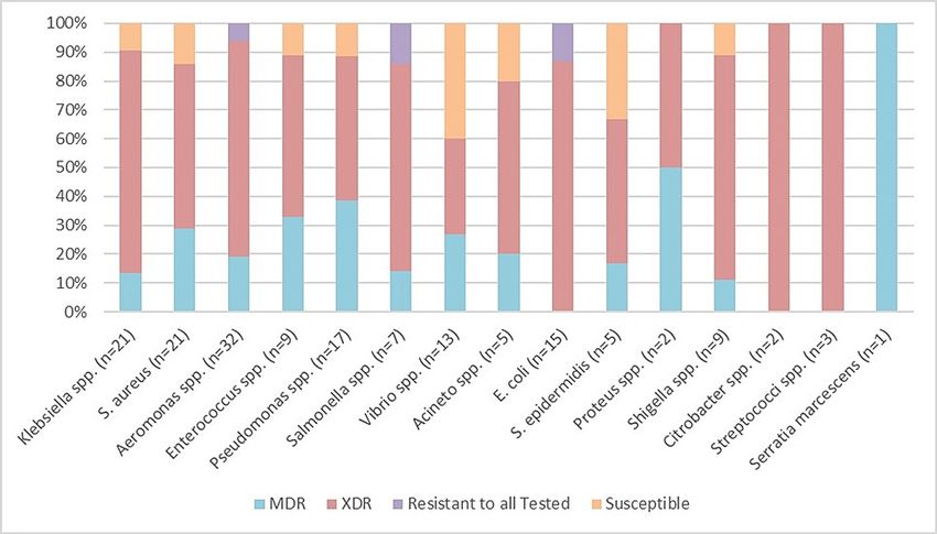
Antimicrobial resistance (AMR) has become a worldwide environmental and public health issue resulting in
                                                  more than 700,000 deaths per year [1]. Water is considered to be the most important vehicle for the
                                                  dissemination of antibiotic resistance in the environment due to its interaction in every compartment and
                                                  its linkage with the human-associated microbiota. This is especially true for low-income countries where
                                                  poor hygiene and sanitation practices further aggravate the problem [2]. The major intersection follows two
                                                  routes, which come together with a full circle. The first one being the discharge of resistant bacteria in the
                                                  environment through different sources and the second one being the presence of such bacteria in our urban
                                                  water system which is consumed by the public.

                                                  The most significant amount of antibiotic residues and resistant bacteria are injected into the aquatic
                                                  ecosystem through hospital water [3]. Hospitals in Pakistan have been estimated to produce 25,000 tons of
                                                  waste each year containing about 1.4 µg/L to 236.6 µg/L of antibiotic residues, which are left untreated and
                                                  added to surface water as such [4]. Untreated hospital effluents, entering into the municipal sewage pose a
                                                  greater threat to the community where mixing of sewage water with drinking water is a common
                                                  phenomenon [5].

                                                  This water ultimately makes its way back as contaminated tap water in hospitals and exposes the already
                                                  ill and immunocompromised patients to several bacteria through ingestion [6]. The water used in hospital
                                                  washbasins and washrooms has been claimed to have caused many nosocomial infections due to easy
                                                  transmission channels between points of contact where patients are exposed to water while bathing,
                                                  washing their hands, exposure to medical equipment, and through health workers and medical personnel
                                                  [7]. The presence of antibiotic resistance bacteria (ARB) in such settings increases the chance of genetic
                                                  transmission between microbes and results in increasing the load of antibiotic resistance and probable
                                                  evolution into multidrug resistance bacteria [8].

                                                  The presence of antibiotic residues, pollutants, and nutrients in the wastewater serves as a selective pressure
                                                  for the microbes to develop resistance and spread it via mutation or horizontal gene transfer [9]. Among
                                                  many genetic determinants, resistance in Gram-negative microbes is mostly attributed to extended-
                                                  spectrum β-lactamases (ESBL). ESBL enzymes have the capability to hydrolyze almost all beta-lactams
                                                  which hinders the first-line defense against many infections. In recent times, blaCTX-M has become the
                                                  most prevalent ESBL and together with blaSHV and blaTEM contribute to resistance against penicillins,
                                                  oxyimino-cephalosporins, and monobactams [10]. Carbapenems and Cephamycins were the next drugs of
                                                  choice against ESBL bacteria but the prevalence of blaOXA, blaKPC, and recently discovered blaNDM have
                                                  also rendered these ineffective against resistant organisms.

                                                  To date, no study had been done in Pakistan on bacteria present in hospital water and their antibiotic
                                                  resistance patterns. Keeping this in view, this study was designed to discern the prevailing issue of increased
                                                  antibiotic resistance and antibiotic-resistant bacteria in the hospital water and surfaces with the aim to
                                                  study their genes and consequent prevalence to better understand the dissemination of these organisms in
                                                  the water and hospital environment.

                                                  Materials And Methods
                                                  Sample collection
                                                  Three governmental tertiary care hospitals located in different cities were targeted for the sample collection
                                                  from both water and surface sources to include and observe a range of microbiota. Intensive care units (ICU),
                                                  surgical wards, and washrooms were selected to check the prevalence of bacteria and antibiotic resistance.
                                                  The water samples were collected in a 50 ml falcon tube after letting the tap run for two to three minutes to
                                                  flush out the cold water. The tubes were then, sealed with the film to prevent contamination.

                                                  Swab samples were obtained through sterile swab sticks at three different sites: faucet, basin, and drain to
                                                  study the bacterial inflow from the water system, the bacterial retention at sinks, and ultimate outflow to
                                                  sewerage through the drainage system. After scrubbing on the respective surfaces, swabs were suspended in
                                                  the transport medium and capped tightly to prevent contamination. All the samples were put in the ice
                                                  container and processed within six hours of sampling with Karachi samples taking a few hours more. The
                                                  experiments were performed in duplicates.

                                                  Sample processing
                                                  The water was passed through the membrane filter of 0.45 µm and then placed gently on the nutrient
                                                  medium with the sterile forceps. This was incubated at 37 °C for 24 hours for the enrichment of microbes.
                                                  The growth was observed the next day and the bacterial suspension was made in 1 ml sterile saline solution.
                                                  This was then, spread onto seven different media (Aeromonas Medium, Azide Dextrose Agar, MacConkey
                                                  Agar, Mannitol Salt Agar, Pseudomonas Cetrimide Agar, Salmonella Shigella Agar, and Thiosulfate Citrate
                                                  Bile Salts Sucrose Agar) to allow the growth of a multitude of microbes according to the nutritional
                                                  specifications and conditions. Likewise, the surface samples were directly spread on the mentioned media on
                                                  the first day of sampling and incubated at 37 °C for 24 hours. Different colonies were picked and identified
                                                  using standard bacteriological analysis protocols and biochemical tests.

                                                  Antibiotic susceptibility assay

2021 Aleem et al. Cureus 13(10): e18738. DOI 10.7759/cureus.18738                                                                                                   2 of 10
This assay was performed using the standard Kirby-Bauer disk diffusion method on Mueller Hinton Agar
                                                   (MHA) with EUCAST guidelines for the antibiogram of the isolates. The inoculum was prepared by
                                                   suspending 24 hour fresh colonies in 1 ml of sterile saline solution equating the turbidity to that of 0.5
                                                   McFarland and swabbed on the media plate. Twenty-one antibiotics disks were dispensed on the surface
                                                   namely including ampicillin (AMP), amoxicillin (AML), piperacillin-tazobactam (TZP), cefipime (FEP),
                                                   cefoxitin (FOX), ceftazidime (CAZ), ceftriaxone (CRO), imipenem (IMP), meropenem (MEM), ciprofloxacin
                                                   (CIP), moxifloxacin (MXF), levofloxacin (LEV), amikacin (AK), gentamicin (CN), tigecycline (TGC),
                                                   aztreonam (ATM), erythromycin (E), clindamycin (DA), rifampicin (RD), colistin (CT), and chloramphenicol
                                                   (C). These agents were chosen on the basis of their importance in treating bacterial infections and for
                                                   diverse representation of different antimicrobial classes. The plate was incubated at 37 °C for 18-24 hours
                                                   and examined. The diameters of the zone of inhibition were measured against each antibiotic including the
                                                   diameter of the disk.

                                                   Isolation of genomic DNA
                                                   Genomic DNA was extracted using the salting-out method where the fresh overnight liquid cultures are used
                                                   for the isolation of DNA. Of the 162 isolates, only 38 (Table 1) were chosen for genotyping based on the
                                                   following criteria: the geographical distribution, the varied sources, and higher antibiotic resistance.

                 Selected AMR isolates (RWP)                         Selected AMR isolates (LH)                      Selected AMR isolates (KH)

                 Isolate   Source      Probable isolate              Isolate   Source      Probable Isolate          Isolate   Source       Probable Isolate

                 R1K1      ICU         Klebsiella spp                L2K1      ICU         Escherichia coli          K3A1      ICU          Aeromonas spp

                 R2K1      ICU         Salmonella spp                L1S2      ICU         Shigella spp              K2K1      ICU          Escherichia coli

                 R3K2      Ward        Escherichia coli              L4M1      Ward        Staphylococcus aureus     K4K1      Ward         Escherichia coli

                 R4M1      Ward        Staphylococcus epidermidis    L3A1      Ward        Aeromonas spp             K5A1      Ward         Aeromonas spp

                 R5A1      Washroom    Aeromonas spp                 L5K1      Washroom    Escherichia coli          K7K1      Washroom     Acinetobacter spp

                 R6K1      Washroom    Klebsiella spp                L5A1      Washroom    Aeromonas spp             K7S1      Washroom     Shigella spp

                 A11       R Faucet    Aeromonas spp                 K42       L Faucet    Salmonella spp            K101      K Faucet     Acinetobacter spp

                 K72       R Faucet    Klebsiella spp                T41       L Faucet    Vibrio spp                S101      K Faucet     Shigella spp

                 S21       R Basin     Escherichia coli              A52       L Basin     Aeromonas spp             P111      K Basin      Pseudomonas spp

                 A22       R Basin     Aeromonas spp                 K51       L Basin     Escherichia coli          D111      K Basin      Enterococcus spp

                 K32       R Drain     Escherichia coli              S62       L Drain     Salmonella spp            D121      K Drain      Enterococcus spp

                 A31       R Drain     Aeromonas spp                 A61       L Drain     Aeromonas spp             S121      K Drain      Escherichia coli

                                                                                                                     K1S1      Sea          Klebsiella spp

                                                                                                                     K1P1      Sea          Pseudomonas spp

                TABLE 1: Selection of isolates: 38 isolates were selected keeping in mind their geographical
                distribution, their source, and their resistance pattern.

                                                   Liquid culture of 1.5 ml was centrifuged at maximum speed for one minute to achieve the pellet cells. This
                                                   was repeated two to three times to get a thick pellet. The pellet was suspended in 600 µl lysis buffer by soft
                                                   pipetting after removing the supernatant. This mixture was incubated at 37 °C for one hour.

                                                   Six hundred microliter of 5M NaCl was then added to the mixture for protein precipitation. This was
                                                   vortexed slowly for 15 seconds before putting it in a centrifuge at 10,000 RPM for 10 minutes. Following the
                                                   step, the upper aqueous layer was carefully transferred to a new tube. This step was repeated until the white
                                                   protein layer completely disappeared.

                                                   For the precipitation of DNA, 2.5 or 3 volumes of absolute chilled ethanol were added to the separated
                                                   aqueous layer and gently mixed. This was refrigerated at −20 °C for 30 minutes followed by centrifugation at
                                                   maximum speed with 4 °C temperature for 15 minutes. After discarding the supernatant, the DNA was
                                                   washed with 1 ml of chilled 70% ethanol, which was centrifuged at the same conditions for two minutes. The
                                                   supernatant was discarded and the DNA pellet was air-dried at room temperature by inverting the tube on a
                                                   paper towel. The DNA was then suspended in 600 µl TE Buffer after drying.

2021 Aleem et al. Cureus 13(10): e18738. DOI 10.7759/cureus.18738                                                                                                   3 of 10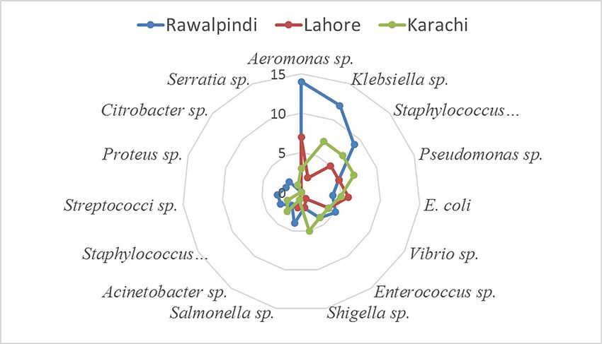
PCR for detection of antibiotic-resistance genes
                                                     Six β lactam genes were subject to be detected in the selected isolates namely: blaSHV, blaTEM, blaCTX-M,
                                                     blaNDM, blaOXA, and blaKPC. The primer sequence and amplicon size are given in Table 2. Each PCR
                                                     reaction consisted of 1× reaction buffer, 1.2 µl of 50 mM MgCl2, 0.5 µl of 0.2 mM dNTPs, 2 µl of 1 mM of each
                                                     primer, 0.2 µl Taq DNA polymerase, and 2 µl of target DNA (final reaction volume of 25 µl). PCR products
                                                     were electrophoresed through 2% agarose and ethidium bromide staining.

                 Primer                           Primer sequence (5’-3’)                                                   Amplicon size

                 blaSHVF                          CGCCATTACCATGAGCGATA
                                                                                                                            86
                 blaSHVR                          CGCAAAAAGGCAGTCAATCC

                 blaTEMF                          AAGTTGCAGGACCACTTCTG
                                                                                                                            202
                 blaTEMR                          GCACCTATCTCAGCGATCTG

                 blaCTX-MF                        CGATGTGCAGTACCAGTAA
                                                                                                                            585
                 blaCTX-MR                        TTAGTGACCAGAATCAGCGG

                 blaNDMF                          GGTTTGGCGATCTGGTTTTC
                                                                                                                            621
                 blaNDMR                          CGGAATGGCTCATCACGATC

                 blaOXAF                          GCGTGGTTAAGGATGAACAC
                                                                                                                            438
                 blaOXAR                          CATCAAGTTCAACCCAACCG

                 blaKPCF                          CGTCTAGTTCTGCTGTCTTG
                                                                                                                            798
                 blaKPCR                          CTTGTCATCCTTGTTAGGCG

                TABLE 2: Amplicon size of detected genes: Six β lactam genes were subject to be detected in the
                selected isolates namely: SHV, TEM, CTXM, NDM, OXA, and KPC.
                 The primer sequences and amplicon size is given in the table.

                                                     Results
                                                     Bacterial diversity
                                                     A total of 162 isolates were obtained from three major cities of Pakistan Rawalpindi (71), Lahore (57), and
                                                     Karachi (34) as shown in Figure 1. Among these 162 isolates, 82 were obtained from water sources and 80
                                                     were collected from surfaces (faucet, basin, and drain). The isolates included a variety of bacteria including
                                                     Aeromonas spp. (20%), Klebsiella spp. (13%), S. aureus (13%), Pseudomonas spp. (10%), E. coli (9%), Vibrio
                                                     spp. (8%), Enterococcus spp. (6%), Shigella spp. (6%), Salmonella spp. (4%), Acinetobacter spp. (3%), S.
                                                     epidermitis (3%), Streptococci spp. (2%), Proteus spp. (1%), Citrobacter spp. (1%), and Serratia spp. (1%).
                                                     Among the three surface sources checked, the drain was the most contaminated which suggests that the
                                                     healthcare workers and patients who wash their hands in the basin are involved in spreading clinical
                                                     bacteria into the drainage, which ultimately leads to sewage lines. The faucet was the least contaminated
                                                     source found (Figure 2).

2021 Aleem et al. Cureus 13(10): e18738. DOI 10.7759/cureus.18738                                                                                                     4 of 10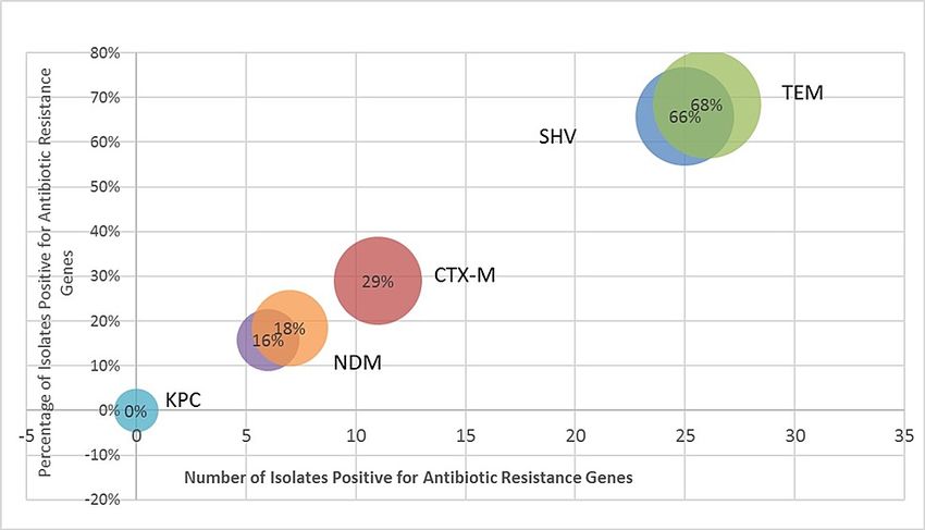
FIGURE 1: Illustrates the variety of species isolated from three different
cities of Pakistan.
FIGURE 2: Illustrates the variety of species isolated from three selected
water and surface sources each.
Antibiotic susceptibility
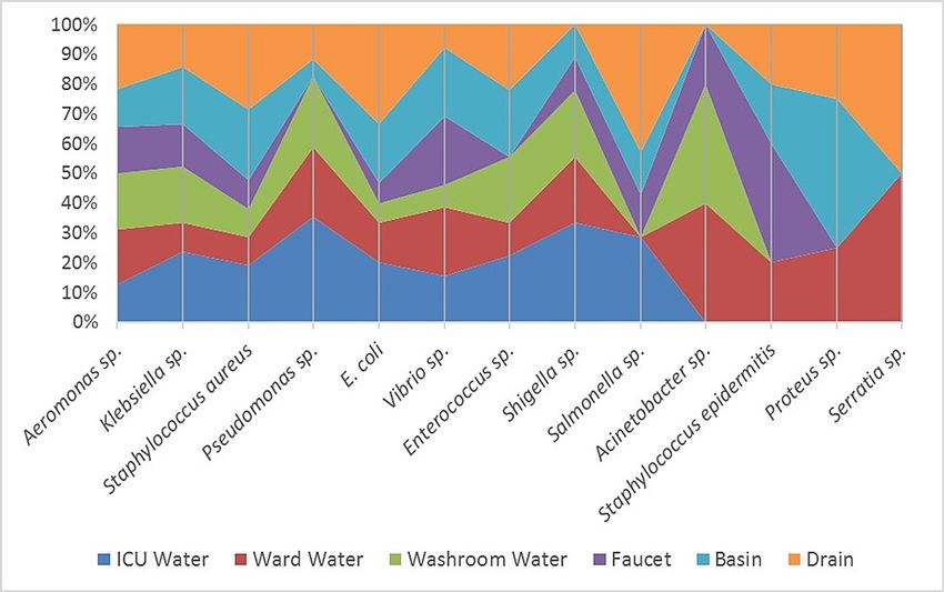
Among the 10 different classes of antibiotics tested, most resistance against macrolides was observed while
resistance against fluoroquinolones was the least with levofloxacin being the most effective in this class as
shown in Figure 3. Overall, colistin was found to be the most effective antibiotic with 52 (32%) resistance
among 162 isolates. Out of the total 162 isolates, about 118 (73%) were extensively drug-resistant (XDR), 28
(17%) were multidrug-resistant (MDR) while only 16 (10%) were susceptible. Among Rawalpindi water
isolates, tap water from washrooms contained the maximum number of XDR bacteria followed by tap water
from ICUs.
2021 Aleem et al. Cureus 13(10): e18738. DOI 10.7759/cureus.18738 5 of 10
FIGURE 3: Illustrates the antibiotic resistance of 162 water and surface
                                                    isolates.
                                                    Ampicillin (AMP), amoxicillin (AML), piperacillin-tazobactam (TZP), cefipime (FEP), cefoxitin (FOX), ceftazidime
                                                    (CAZ), ceftriaxone (CRO), imipenem (IMP), meropenem (MEM), ciprofloxacin (CIP), moxifloxacin (MXF),
                                                    levofloxacin (LEV), amikacin (AK), gentamicin (CN), tigecycline (TGC), aztreonam (ATM), erythromycin (E),
                                                    clindamycin (DA), rifampicin (RD), colistin (CT), and chloramphenicol (C).

                                                  Among the surface isolates, drug resistance was more profound in Rawalpindi drain isolates, in which 23%
                                                  were resistant to all the drugs tested while 92% of the basin surface and 75% of faucet surface isolates were
                                                  also found to be XDR. Isolates from Lahore had 100% XDR bacteria in ICU tap water and washroom tap
                                                  waters. Among the Lahore faucet, basin, and drain surface isolates, XDR bacteria were found to be 100%,
                                                  67%, and 83%, respectively. Among the tap water samples collected from Karachi, ICU water samples
                                                  showed 60% XDR bacteria while ward water isolates showed 67% XDR bacteria. Among the faucet, basin,
                                                  and drain surface isolates, XDR were tested to be 80%, 67%, and 77%, respectively (Figure 4).

                                                    FIGURE 4: Illustrates the antibiotic resistance in all isolates of bacteria.

                                                  All isolates of Streptococci and Citrobacter were tested to be XDR. Klebsiella spp. were found to have 81% XDR
                                                  isolates and 14% MDR isolates as shown in Figure 4. Similarly, S. aureus also had a higher ratio of 1.8 XDR to
                                                  MDR bacteria. E. coli, another common bacteria showed 13% of isolates that were resistant to all the
                                                  antibiotics tested while the remaining 87% of its isolates were found to be XDR. Salmonella spp. showed 14%
                                                  isolates that were resistant to all the antibiotics tested. Enterococcus spp. also had 56% of the isolates
                                                  classified as XDR. Aeromonas, an environmental bacteria and a fish pathogen, had 6% isolates that were
                                                  resistant to all the antibiotics tested, which is a matter of great concern.

2021 Aleem et al. Cureus 13(10): e18738. DOI 10.7759/cureus.18738                                                                                                      6 of 10
Prevalence of beta-lactam genes
The isolates showed a varied presence of the six beta-lactam genes, the most prevalent being blaTEM (68%)
with blaSHV (66%) following closely behind. blaCTX-M was found in 29% with 18% blaNDM and 16%
blaOXA. There was no blaKPC gene detected. The overall prevalence of genes was detected to be higher in
Lahore isolates. The Karachi isolates showed no detection of the blaCTX-M gene, however, it had the highest
frequency of blaSHV (26%). Rawalpindi isolates showed the highest frequency of blaSHV (16%) and blaCTX-
M (16%). Among the species, Aeromonas spp. and E. coli were found to possess all five genes that were
detected while Acinetobacter spp., Salmonella spp., and Klebsiella spp. also harbored four out of five genes.
The highest prevalence of blaNDM was found in Acinetobacter spp. (Figures 5-6).
FIGURE 5: Illustrates the number of genes detected in selected isolates.
FIGURE 6: Illustrates the antibiotic resistance genes in bacterial
isolates.
Discussion
The hospital environment is an intricate ecosystem that has usually been overlooked as a potential reservoir
for bacteria but with the rate of nosocomial infections on the rise; this has become a critical area for the
study of microbes. The transmission of pathogens via surface contamination, lack of proper handwashing
practices among health care workers, and the water system increased the incidence rate of hospital-acquired
infections among the patients [11].
Furthermore, the increase in the antibiotic resistance patterns of the microbes has forced the scientific
community to look into the source of such pathogens and their mechanisms of acquired resistance in
different environmental compartments. Water bodies were also considered to be a reservoir for the antibiotic
resistance, especially because it facilitates the interaction of pathogenic bacteria with the non-pathogenic
2021 Aleem et al. Cureus 13(10): e18738. DOI 10.7759/cureus.18738 7 of 10ones and contribute to the increase in resistance [12].
In the present study, of 162 isolates, 127 were Gram-negative while only 35 isolates were Gram-positive.
Similar ratios were found in other studies that observed the bacterial diversity in hospital settings with the
prevalence of Gram-negative bacteria [13]. In a study conducted on river waters, 81.8% of Aeromonas
samples were found to be multidrug-resistant to the most commonly used antibiotics [14]. The results are in
coherence with our study where 100% resistance was shown towards ampicillin and more than 80% towards
amoxicillin, aminoglycosides, and macrolides. Cephalosporins, which were considered to be suitable drugs
[15] against Aeromonas also showed more than 80% resistance to ceftriaxone whereas susceptibility to
ciprofloxacin has decreased to 44%, concurrent with the study [16].
Similarly, Klebsiella species has also shown an increasing trend of resistance against commonly used
antibiotics with cefotaxime resistance increased from 75% to 94%, ciprofloxacin went from 64% to 84%, and
carbapenems from 2.4% to 52% [17]. The trend in our study is on the higher end of the spectrum with 90%
resistance to ciprofloxacin, cefoxitin, and erythromycin, 80% towards ceftriaxone, and 40% towards
carbapenems. Carbapenem resistance is owed to recently discovered, blaNDM genes and blaCTXM, both of
which were present in our isolates. This leaves colistin as the only suitable option with 70% susceptibility in
Klebsiella isolates coherent with a study done in Europe [18].
Pseudomonas species was the third most common organism isolated in our study with 90% isolates as
multidrug-resistant. More than 80% resistance was shown towards cephalosporins, penicillins, and
macrolides coherent with other studies [19]. The least resistance was observed against ciprofloxacin (30%)
and colistin (10%) supported by a study done in Karachi [20]. This pattern of resistance is mainly due to the
expanded beta-lactam activity of these strains which is contributed to beta-lactamase genes. Our tested
isolates were shown to carry extended-spectrum beta-lactamase genes blaTEM, blaSHV, and blaCTXM. The
frequencies of blaTEM and blaSHV genes in the bacterial isolates were calculated to be 46% and 34%,
respectively [21], in another study which is similar to our findings.
A study conducted on water samples from hospital sources found S. aureus to be the most predominant
organism. Comparatively, it constituted only 13% of total isolates in our study. Different studies have shown
Staphylococcus to be multidrug-resistant with 100% resistance against amoxicillin, streptomycin,
ceftriaxone, and erythromycin, 83% to gentamycin and 78% to cefoxitin and ciprofloxacin [21]. These
findings were supported by our study as more than 80% of resistance was shown towards these drugs classes.
A shifting trend was observed in our study from a study that reported amikacin and levofloxacin as a
susceptible antibiotic against Staphylococcal isolates where resistance has increased to 58% and 52%,
respectively, with 96% sensitivity to imipenem as reported in a study from Peshawar [21]. Staphylococcus
epidermidis also exhibited resistance to macrolides, cephalosporins, and fluoroquinolones as reported in
other studies [22]. Researchers have associated multidrug resistance with blaTEM genes along with blaCTXM
and blaSHV that were prevalent in our Staphylococcal isolates.
Organisms like E. coli, Vibrio, Enterococci, and Shigella have been known to cause water-borne diseases like
cholera and dysentery. Combined, they made 23% of the total isolates. Similar resistance patterns were
observed in isolates of E. coli with 100% resistance towards cephalosporins, more than 90% resistance
against ampicillin, more than 80% resistance towards macrolides, and 68% against fluoroquinolones. This
was also supported by a study that reported 100% resistance towards penicillins, more than 80% towards
first and second-generation cephalosporins, and 20% towards fluoroquinolones and macrolides [23]. On
contrary, Vibrio species showed 90% susceptibility to fluoroquinolones and 80% susceptibility to
carbapenems consistent with findings in China [24] but resistance patterns were similar against penicillins,
erythromycin, and clindamycin as in other isolates in our study. The resistance in both Enterococci and
Shigella was 100% towards erythromycin and ceftriaxone, followed by 90% against ampicillin and cefoxitin
concurrent with other studies [25]. Resistance against ceftriaxone (20%) and ciprofloxacin (12%) in Shigella
has risen in the past seven years to 68% and 58%, respectively, in our study compared to the study done in
Faisalabad. This overall resistance against all these drugs has been conferred to the bacteria’s innate
resistance [26]. blaSHV and blaTEM have been found to confer resistance against many antibiotics and have
been the most prevalent in E. coli, Vibrio, Enterococci, and Shigella. E. coli had the maximum number of
resistance genes including blaCTXM, blaOXA, and blaNDM.
XDR typhi is an extensively drug-resistant strain of Salmonella typhi and is resistant to all the antibiotics
recommended for typhoid fever except azithromycin and carbapenems. The isolates in our study have
exhibited 100% resistance towards ampicillin, ciprofloxacin, aztreonam, clindamycin, and colistin with
similar results reported in other studies [27]. Salmonella isolates were found to possess blaSHV, blaTEM,
blaCTXM, and blaOXA, all of which explain the resistance pattern shown by the isolates. ESBL pattern of
resistance has also been observed in Acinetobacter and Pneumococci isolates with the highest resistance
against penicillins, cephalosporins, and fluoroquinolones consistent with other findings [28]. Recently,
several serotypes of S. pneumoniae have been isolated which show 100% resistance towards penicillins and
cephalosporins with growing resistance towards macrolides and fluoroquinolones. Growing resistance
against chloramphenicol has also been recently discovered where 36% resistance was reported against the
drug [29], but it contradicts our findings of 100% sensitivity towards it. Proteus species on the other hand
has shown 100% resistance towards chloramphenicol as well as penicillins, cephalosporins (all three
generations), and macrolides. It has shown susceptibility towards piperacillin-tazobactam [30] but our
findings are contrary to these as 100% resistance was shown towards this drug with only susceptibility
2021 Aleem et al. Cureus 13(10): e18738. DOI 10.7759/cureus.18738 8 of 10towards imipenem and amikacin.
Conclusions
It was concluded in this study that Gram-negative bacteria have more suited features to be able to survive in
the environment for longer periods of time, which is why they are of concern especially, in the hospital
setting where the patients are more prone to catch an infection. This study suggests that hospital tap water
habitat comprises a diverse range of microbes including the ones that have been identified as clinically
relevant. The study also shows that the urban water cycle is already contaminated with the microflora of the
hospital settings including Aeromonas, Klebsiella, Pseudomonas, Staphylococcus, and Vibrio species in
abundance that are a threat to hospitalized patients, especially immunocompromised ones.
Most resistance was found against macrolides, lincosamides, monobactams, and penicillins. The most
resistant pathogens were found to be in ICU compared to other wards because of more frequent antibiotic
usage and the presence of immunocompromised patients, which call for stringent policies and infection
control programs in the hospitals. Apart from this, many of the organisms in this study were found to be
extended-spectrum beta-lactam (ESBL) producers. This manifests as a real threat, especially with blaNDM
prevalence on the rise, which might result in resistance against carbapenems and colistin, antibiotics
reserved as a last line of defense against infections. Public health measures for clean tap water, clean water
supply in hospitals along with information sharing and stimulation of research in this field shall contribute
towards bridging gaps and a better understanding of this increasing phenomenon. We need more research in
this area involving more hospitals and medical setups around the country.
Additional Information
Disclosures
Human subjects: All authors have confirmed that this study did not involve human participants or tissue.
Animal subjects: All authors have confirmed that this study did not involve animal subjects or tissue.
Conflicts of interest: In compliance with the ICMJE uniform disclosure form, all authors declare the
following: Payment/services info: All authors have declared that no financial support was received from
any organization for the submitted work. Financial relationships: All authors have declared that they have
no financial relationships at present or within the previous three years with any organizations that might
have an interest in the submitted work. Other relationships: All authors have declared that there are no
other relationships or activities that could appear to have influenced the submitted work.
References
1. Kalungia AC, Mwambula H, Munkombwe D, et al.: Antimicrobial stewardship knowledge and perception
among physicians and pharmacists at leading tertiary teaching hospitals in Zambia: implications for future
policy and practice. J Chemother. 2019, 31:378-87. 10.1080/1120009X.2019.1622293
2. Walker GT, Quan J, Higgins SG, et al.: Predicting antibiotic resistance in Gram-negative bacilli from
resistance genes. Antimicrob Agents Chemother. 2019, 63: 10.1128/AAC.02462-18
3. Ahmad M, Khan AU: Global economic impact of antibiotic resistance: a review . J Glob Antimicrob Resist.
2019, 19:313-6. 10.1016/j.jgar.2019.05.024
4. Kumar R, Somrongthong R, Ahmed J: Effect of medical waste management trainings on behavior change
among doctors versus nurses and paramedical staff in Pakistan [internet]. J Ayub Med Coll Abbottabad.
2016, 28:493-6.
5. Almakki A, Jumas-Bilak E, Marchandin H, Licznar-Fajardo P: Antibiotic resistance in urban runoff . Sci Total
Environ. 2019, 667:64-76. 10.1016/j.scitotenv.2019.02.183
6. Ogawara H: Comparison of antibiotic resistance mechanisms in antibiotic-producing and pathogenic
bacteria. Molecules. 2019, 24:3430. 10.3390/molecules24193430
7. Lerminiaux NA, Cameron AD: Horizontal transfer of antibiotic resistance genes in clinical environments .
Can J Microbiol. 2019, 65:34-44. 10.1139/cjm-2018-0275
8. Wencewicz TA: Crossroads of antibiotic resistance and biosynthesis . J Mol Biol. 2019, 431:3370-99.
10.1016/j.jmb.2019.06.033
9. Ben Y, Fu C, Hu M, Liu L, Wong MH, Zheng C: Human health risk assessment of antibiotic resistance
associated with antibiotic residues in the environment: A review. Environ Res. 2019, 169:483-93.
10.1016/j.envres.2018.11.040
10. Chokshi A, Sifri Z, Cennimo D, Horng H: Global contributors to antibiotic resistance. J Glob Infect Dis. 2019,
11:36-42. 10.4103/jgid.jgid_110_18
11. Singh R, Singh AP, Kumar S, Giri BS, Kim KH: Antibiotic resistance in major rivers in the world: A
systematic review on occurrence, emergence, and management strategies. J Clean Prod. 2019, 234:1484-505.
12. Umar M, Roddick F, Fan L: Moving from the traditional paradigm of pathogen inactivation to controlling
antibiotic resistance in water - Role of ultraviolet irradiation. Sci Total Environ. 2019, 662:923-39.
10.1016/j.scitotenv.2019.01.289
13. Tajeddin E, Rashidan M, Razaghi M, et al.: The role of the intensive care unit environment and health-care
workers in the transmission of bacteria associated with hospital acquired infections. J Infect Public Health.
2016, 9:13-23. 10.1016/j.jiph.2015.05.010
14. Harnisz M, Korzeniewska E: The prevalence of multidrug-resistant Aeromonas spp. in the municipal
wastewater system and their dissemination in the environment. Sci Total Environ [Internet]. 2018, 20:377-
83.
15. Ahmed I, Rabbi MB, Sultana S: Antibiotic resistance in Bangladesh: a systematic review . Int J Infect Dis.
2019, 80:54-61. 10.1016/j.ijid.2018.12.017
16. Fernández-Bravo A, Figueras MJ: An update on the genus Aeromonas: taxonomy, epidemiology, and
2021 Aleem et al. Cureus 13(10): e18738. DOI 10.7759/cureus.18738 9 of 10pathogenicity. Microorganisms. 2020, 8:129. 10.3390/microorganisms8010129
17. Martin RM, Bachman MA: Colonization, infection, and the accessory genome of Klebsiella pneumoniae .
Front Cell Infect Microbiol. 2018, 8:4. 10.3389/fcimb.2018.00004
18. Subedi D, Vijay AK, Willcox M: Overview of mechanisms of antibiotic resistance in Pseudomonas
aeruginosa: an ocular perspective. Clin Exp Optom. 2018, 101:162-71. 10.1111/cxo.12621
19. Ebrahimpour M, Nikokar I, Ghasemi Y, Sedigh Ebrahim-Saraie H, Araghian A, Farahbakhsh M, Ghassabi F:
Antibiotic resistance and frequency of class 1 integrons among Pseudomonas aeruginosa isolates obtained
from wastewaters of a burn center in Northern Iran. Ann Ig. 2018, 30:112-9. 10.7416/ai.2018.2202
20. Frequency and susceptibility pattern of Multidrug Resistant Pseudomonas aeruginosa in isolates of patients
from a tertiary care hospital of Karachi, Pakistan [Internet]. (2019). Accessed: October 12, 2021:
https://www.researchgate.net/publication/278730660_Frequency_and_susceptibility_pattern_of_Multidrug_Resistant_Pseudo...
21. Okhonlaye O, Oluwatosin O: Occurrence of multidrug resistant Staphylococcus aureus and Klebsiella
pneumoniae isolated from clinical and environmental samples in Ondo state, Nigeria. J Adv Microbiol
[Internet. 2018, 5:1-10. 10.9734/JAMB/2018/40204
22. Lee JY, Monk IR, Gonçalves da Silva A, et al.: Global spread of three multidrug-resistant lineages of
Staphylococcus epidermidis. Nat Microbiol. 2018, 3:1175-85. 10.1038/s41564-018-0230-7
23. Wang CY, Chen YH, Fang C, et al.: Antibiotic resistance profiles and multidrug resistance patterns of
Streptococcus pneumoniae in pediatrics: a multicenter retrospective study in mainland China. Medicine
(Baltimore). 2019, 98:e15942. 10.1097/MD.0000000000015942
24. Gladkikh AS, Feranchuk SI, Ponomareva AS, Bochalgin NO, Mironova LV: Antibiotic resistance in Vibrio
cholerae El Tor strains isolated during cholera complications in Siberia and the Far East of Russia. Infect
Genet Evol. 2020, 78:104096. 10.1016/j.meegid.2019.104096
25. Tian Y, Yu H, Wang Z: Distribution of acquired antibiotic resistance genes among Enterococcus spp.
isolated from a hospital in Baotou, China. BMC Res Notes. 2019, 12:27. 10.1186/s13104-019-4064-z
26. Okai M, Aoki H, Ishida M, Urano N: Antibiotic-resistance of fecal coliforms at the bottom of the Tama River,
Tokyo. Biocontrol Sci. 2019, 24:173-8. 10.4265/bio.24.173
27. Ranjbar R, Farahani A: Shigella: antibiotic-resistance mechanisms and new horizons for treatment . Infect
Drug Resist. 2019, 12:3137-67. 10.2147/IDR.S219755
28. Mechai A, Debabza M, Thabet R, Sedira H, Fadeleddine S, Mechai A: Occurrence and spread of beta-
lactamases-producing enterobacteriaceae isolated from river receiving treated effluent of wastewater
treatment plant. Desalin Water Treat. 2019, 147:156-63. 10.5004/dwt.2019.23763
29. Ajulo OH, Adetunji OV, Ajulo OM: Antibiotic sensitivity profiles of Salmonella typhimurium and E. coli
O157: H7 isolates from ready to eat chicken meat in Ibadan-Nigeria. Asian Food Sci J. 2019, 10:1-17.
10.9734/afsj/2019/v10i130025
30. Rocha C, Bernal M, Canal E, et al.: First report of New Delhi metallo-β-lactamase carbapenemase-producing
Acinetobacter baumannii in Peru. Am J Trop Med Hyg. 2019, 100:529-31. 10.4269/ajtmh.18-0802
2021 Aleem et al. Cureus 13(10): e18738. DOI 10.7759/cureus.18738 10 of 10You can also read