Preparation and application of chikungunya pseudovirus containing double reporter genes
←
→
Page content transcription
If your browser does not render page correctly, please read the page content below
www.nature.com/scientificreports
OPEN Preparation and application
of chikungunya pseudovirus
containing double reporter genes
Chunyan Su1, Kaiyun Ding1, Jingwen Xu1, Jianchao Wu1, Jiansheng Liu1, Jiayuan Shen2,
Hongning Zhou2 & Hongqi Liu1*
Chikungunya virus (CHIKV), a highly infectious and rapidly spread viral pathogen, is classified as
a pathogenic agent at the biosafety level 3. Operation of live authentic CHIKV needs a specific
laboratory with the P3 or above containment, which greatly confines the CHIKV-associated studies.
To establish an evaluation system of CHIKV that can be utilized in a BSL2 laboratory, we constructed
a pseudovirus (PsV) system of CHIKV containing double reporter genes (ZsGreen1 and luciferase).
The fluorescent ZsGreen1 is a convenient and cheap reporter for monitoring the efficiency of
transfection and titration of PsV. The enzyme luciferase is a sensitive reporter for the application of
PsV to neutralization assay or drug screening. The CHIKV PsV produced in this study, with a titer of
up to 3.16 × 10⁶ TU/ml, was confirmed by Western blotting and transmission electronic microscopy
(TEM). Finally, we developed a microneutralization assay with the CHIKV PsV produced in this study,
which was successfully applied to evaluate neutralizing activities of convalescent sera from CHIKV-
infected patients. In summary, we have established a convenient and sensitive double-reporter CHIKV
pseudovirus system, which provides a safe and effective platform for screening anti-CHIKV drugs and
evaluating vaccines against CHIKV.
Chikungunya, also called chikungunya fever (CHIKF), is an acute febrile complications of red rash, muscle pain,
joint pain and even central nervous system diseases, which is caused by the arthropod-borne chikungunya viruses
(CHIKV)1. Outbreaks of CHIKF have been reported in more than 100 countries all over the world, leading to
about 1 million cases annually2. However, no effective vaccine or drug against CHIKV has been licensed so far3.
Because of high potential of epidemic and unavailable medical countermeasures, CHIKV used to be considered
to be one of the WHO Blueprint priority p athogens2. Moreover, CHIKV is classified as BSL3 pathogen and
operation of live authentic CHIKV should be conducted in P3 or above c ontainment4, which slows down the
research progress of CHIKV.
Pseudovirus (PsV) that is an artificially made recombinant viral particle often possesses the similar conforma-
tion as authentic live virus and can mimic viral infection. Since the nucleic acid inside the pseudovirus cannot
express viral surface membrane proteins, the pseudovirus loses the ability to form a viral particle after the first
round of t ransduction5. PsV particles have been widely used as an alternative to the authentic viral particle for
drug screening, vaccine evaluation, investigation of pathogenesis, and so o n5,6. There are two key advantages of
PsVs over authentic viruses, particularly those being highly pathogenic. Firstly, PsVs of BSL3 or BSL4 viruses
are safer than authentic viruses and can be handled in P2 laboratory. Secondly, reporter genes delivered by PsVs
are convenient for the quantification of interactions between viruses and host cells. In fact, the PsV system
has been widely used in virology studies, including C HIKV7, Lyssaviruses8, Middle East respiratory syndrome
9 10
coronavirus (MERS-COV) , H7N9 influenza viruses , Severe acute respiratory syndrome coronavirus 2 (SARS-
CoV-2)11 and so on.
CHIKV is a single-stranded, positive-sense RNA virus with a viral genome in 11.5 kb, coding four nonstruc-
tural proteins (nsp1-nsp4) and five structural proteins (C-E3-E2-6K-E1). The structural proteins E3-E2-6K-E1
constitutes the envelope of CHIKV. E1 and E2 form a heterodimer that is further assembled into the trimer of
spike glycoprotein on the viral surface. The spike protein mediates viral attachment to host cells and facilitates
viral infection, and thus, is the major player in the induction of neutralizing antibody12,13. E3 mediates the inter-
action between E1 and E2. The 64 amino-acids glycoprotein 6K participates in the translocation of structural
polyproteins and maturation of E214,15. Therefore, these envelope proteins represent critical factors involve in
1
Institute of Medical Biology, Chinese Academy of Medical Sciences and Peking Union Medical College,
Kunming 650118, China. 2Yunnan Provincial Key Laboratory of Vector‑Borne Diseases Control and Research,
Yunnan Institute of Parasitic Diseases, Simao Pu’ er 665000, Yunnan, China. *email: lhq@imbcams.com.cn
Scientific Reports | (2022) 12:9844 | https://doi.org/10.1038/s41598-022-13230-0 1
Vol.:(0123456789)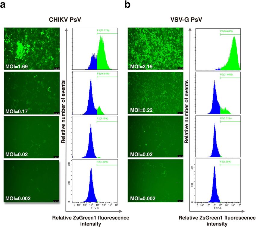
www.nature.com/scientificreports/
the interaction between virus and host cell. In this study, we firstly constructed a three-plasmid packing system,
in which the CHIKV envelope proteins were inset into HIV backbone to form a PsV particle that packed two
reporter genes of ZsGreen1 and luciferase. Then we illustrated the structural features of CHIKV PsVs by electron
microscopy. Finally, we developed a neutralization assay using these PsV particles and successfully quantified
the neutralizing activities of CHIKV-infected human serum samples. Our data indicate that the CHIKV PsV
particle offers a powerful alternative system to study authentic CHIKV, making many CHIKV-associated studies
doable in a P2 laboratory.
Results
Construction of the recombinant plasmid expressing CHIKV envelope proteins. CHIKV PsV
was constructed by HIV-1 lentiviral packaging system (Fig. 1a). Firstly, we performed PCR via specific prim-
ers (described in Methods) to obtain the CHIKV envelope gene and the linearized vector pMD2.G-ΔVSV-G.
Electrophoresis and sequencing showed that the expected fragments with the sizes of 2995 bp and 4295 bp were
successfully amplified (Fig. 1b). Then CHIKV envelope gene was cloned into the linearized pMD2.G-ΔVSV-G
vector. The recombinant plasmid was confirmed by restriction digestion analysis (Fig. 1b) and sequencing. These
results indicated that the recombinant plasmid expressing CHIKV envelope proteins (pMD2.G-CHIKV-env)
was successfully constructed as designated (Fig. 1c).
Production of CHIKV PsV. The three-plasmid system was used to produce CHIKV PsV, including the
CHIKV envelope proteins expressing plasmid pMD2.G-CHIKV-env, the packaging plasmid PsPAX2 and the
transfer plasmid pLVX-IRES-ZsGreen1-Luc. At 24 hours post co-transfection, cells were in good conditions and
70% of them expressed the ZsGreen1 fluorescent protein. At 48 hours post co-transfection, there were about 90%
of cells expressing fluorescent protein (Fig. 2a), suggesting that these plasmids were efficiently transduced into
293T cells. Furthermore, most fluorescent cells began to shrank, fall off and even die with a grape-cluster-like
appearance indicating the release of PsV, which is the possible reason that cells at 24 hpt appeared to be more
fluorescent than those at 48 hpt. The results of immunoblotting analysis confirmed the expression of CHIKV
envelope proteins in the PsV particles (Fig. 2b). Finally, we observed the formation of CHIKV PsV particles with
the size of 80-140 nm and typical enveloped structure under transmission electronic microscope (Fig. 2c). These
results suggest that we have successfully produced CHIKV PsV particles.
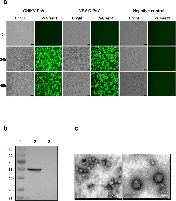
Transduction of CHIKV PsV in 293T cells. The transduction of CHIKV PsV was further evaluated in
293T cells. Expression of the reporter ZsGreen1 was monitored under the fluorescence microscope or by flow
cytometric analysis. The results showed that CHIKV PsV could transduce and release the reporter ZsGreen1 into
293T cells (Fig. 3a). The titer of CHIKV PsV in this study was 3.16×10⁶ TU/ml, similar to that of the positive
control VSV-G PsV (3.29×10⁶ TU/ml) (Fig. 3b).
Development of CHIKV PsV micro‑neutralization assay. To validate the applicability of the con-
structed CHIKV PsV in this study, we developed a microneutralization assay using these PsV and serially diluted
rabbit serum sample that were known to contain anti-CHIKV antibodies. 500 TU of CHIKV PsVs were used for
each neutralization reaction with VSV-G PsV as a control (non-CHIKV envelope proteins control) for the speci-
ficity of neutralization. The activity of luciferase was measured on the microplate reader to evaluate the neutral-
izing ability of antibodies. Compared with the VSV-G PsV, the CHIKV PsV could be specifically neutralized by
the antibody in the known CHIKV serum sample. CHIKV PsV transduction was inhibited by the serum sample
in a dose-dependent manner. The neutralization titer of serum (50% inhibitory dose, ID50) against CHIKV PsV
was 117.4 (Fig. 4), indicating that PsV is potentially applicable in the researches of CHIKV.
CHIKV pseudovirus transduction is inhibited by sera from infected individuals. CHIKV conva-
lescent serum samples were firstly analyzed via ELISA. The human serum samples were then divided into three
groups according to ELISA-OD450 (Table 1). Microneutralization assay was conducted on these samples using
CHIKV PsV produced in this study. Through measurement of luciferase activity, most of the CHIKV convales-
cent human serum samples showed strong neutralization activity in a dose dependent manner (Fig. 5a). Negative
control serum samples showed certain neutralization at the first several concentrations, which decreased rapidly
and kept at a low level. This result suggested that the neutralization is specifically against CHIKV (Fig. 5a). The
CHIKV PsV neutralization titers (PsV NT) were shown in Table 1. In order to analyze the relationship between
neutralizing antibody and total IgG antibodies against CHIKV, we made a plot for these convalescent serum
samples using the values of ELISA-OD450 as x-axis and PsV NTs as y-axis. Through this comparison, we found
there was a significantly positive correlation between neutralizing antibody and total IgG antibodies (Fig. 5b).
Discussion
Adaptive mutation of A226V facilitates CHIKV to infect Ae. Albopictus, contributing to the transmission of
mutant CHIKV to broader region than the prototypic v iruses16. Within last two decades, CHIKV spread rapidly
from African continent to Asia, America and Europe, leading to millions of infectious cases and thousands of
deaths, which becomes a global concern of public h ealth16–19. Therefore, we should make full preparation for this
potential outbreak, including elucidation of pathogenesis, drug screening and vaccine development, and so on.
However, dependence of CHIKV operation on P3 containment hinders the progress of CHIKV researches due
to the limitation of the high-level biosafety facility. In the present study, we constructed CHIKV PsV particles
Scientific Reports | (2022) 12:9844 | https://doi.org/10.1038/s41598-022-13230-0 2
Vol:.(1234567890)
www.nature.com/scientificreports/

                                 Figure 1.  Construction of the CHIKV PsV. (a) The CHIKV PsV construction scheme. Each gene fragment was
                                 indicated by the colored box. (b) Construction and identification of the pMD2.G-CHIKV-env. Primers for PCR
                                 were described in the “Materials and methods” section. 5 ul of each product was analyzed in 0.8% agarose gel.
                                 Lane 1, DNA marker; Lane 2, linearized pMD2.G-ΔVSV-G backbone (4295 bp); Lane 3, CHIKV envelope gene
                                 (2995 bp); Lane 4, undigested pMD2.G-CHIKV-env plasmid (7266 bp); Lane 5, pMD2.G-CHIKV-env plasmid
                                 digested by AscI and Xbal (2967 bp and 4299 bp). (c) Map of the recombinant plasmid pMD2.G-CHIKV-env
                                 that was illustrated by the software SnapGene Viewer (CA, USA).

Scientific Reports |   (2022) 12:9844 |              https://doi.org/10.1038/s41598-022-13230-0                                              3

                                                                                                                                        Vol.:(0123456789)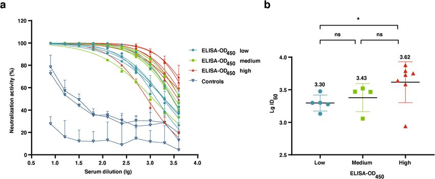
www.nature.com/scientificreports/
Figure 2. Formation of CHIKV PsV particles. (a) Three plasmids (PsPAX2, pLVX-IRES-ZsGreen1-Luc,
and pMD2.G-CHIKV-env or pMD2.G) were co-transfected into 293T cells. At the indicated time points,
fluorescence was observed under the microscope to monitor the transfection efficiency. (b) The PsV particles
concentrated by ultracentrifugation were analyzed by Western blotting using mouse Monoclonal antibody
against E1. 1: 180 kDa protein marker; 2: the lysate of CHIKV PsVs; 3: the lysate of VSV-G PsVs. Unprocessed
images can be found in supplementary information Fig. S1. (c) CHIKV PsV particles was stained with
phosphotungstic acid and observed under transmission electron microscopy. The scale bars represent 500 nm
(left) and 200 nm (right).
containing CHIKV envelope and two reporters. This system is potentially applicable in studying the interaction
between CHIKV and host cell, drug screening, evaluation of vaccine efficacy at the P2 laboratory.
PsV is a kind of virus-like particle with capability of one-time transduction, and made of viral capsid pro-
tein and envelope proteins packing non-viral nucleic acid inside. Therefore, PsV is commonly used as a safe
alternative for authentic live virus, particularly for highly pathogenic viruses, in studies of viral serology 20–22,
anti-viral strategy 13,23,24, and viral pathogenesis24–26. The reporter in PsV is designed for evaluation of the inter-
action between host cell and PsV. The most common reporter is luciferase because it is very sensitive. However,
measurement of luciferase activity is complicate and expensive. Dual reporters (ZsGreen1 and luciferase) have
been increasingly engineered into PsV of this study and other reports27,28. The fluorescent reporter ZsGreen1 in
Scientific Reports | (2022) 12:9844 | https://doi.org/10.1038/s41598-022-13230-0 4
Vol:.(1234567890)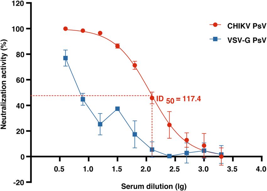
www.nature.com/scientificreports/
Figure 3. Titration of the CHIKV PsV. 293T cells in the 24-well plate were transduced with serial ten-fold
dilutions of PsV particles (a) or VSV-G PsV (b). At 72 h post transduction, cells were firstly observed under
the fluorescence microscope (left panel). Then cells were trypsinized and resuspended for titration via flow
cytometric analysis (right panel). Panels in this figure from top to bottom correspond to 1–1000 times dilutions.
The MOI was calculated for 293T cells in each well according to the PsV titer. Scale bar: 100 μm.
PsV can be used to monitor efficiency of transfection and titration, which is more convenient and cheaper than
the reporter luciferase.
In this study, we constructed PsV particles, where CHIKV envelope was inserted into HIV capsid. Therefore,
the formed PsV particle owned the morphology and size of HIV particle (80–140 nm) (Fig. 2c). It is generally
believed that the capsid is an essential element for CHIKV to form infectious virus particles29–32. Actually, even
though capsid is completely deficient in the mutant CHIKV, infectious virus can still be formed although some
of morphological characteristics are different from naïve C HIKV33.
Prior to the utilization of PsV, it is necessary to determine the titer of viral particle. The copy number of viral
genome or concentration of viral protein (such as p21 of HIV-1) is usually used in the titration experiments.
However, these methods are not only time-consuming and laborious, but also cannot exclude the non-infectious
viral particles, which leads to the higher titer than actual value4,25,34,35. In contrast, PsV constructed in this study
contained the reporter ZsGreen1. Compared with the traditional titration method on basis of the virus particle
itself (qPCR detection of the copy number of viral genome or ELISA measurement of viral protein concentra-
tion), ZsGreen1 is expressed only in infected cells, which effectively excludes the interference of non-infectious
virus particles, making the titration more accurate. In addition, as a fluorescent signal, ZsGreen1 can be easily
captured by flow cytometry, which is easier than the traditional method of titration.
The plaque reduction neutralization test (PRNT) is considered as a gold standard for neutralization assays.
However, PRNT needs the use of hazardous live CHIKV and BSL3 facility, which impedes the application of
PRNT to CHIKV-related studies. In addition, visualization and counting of plaques in PRNT are time-consuming
and laborious procedures. Many studies have demonstrated that the PsV neutralization test could be a good
substitute for the plaque reduction t est27,34. In order to overcome the limitations of PRNT mentioned above, here
we developed a PsV neutralization assay. In this assay, luciferase activity was used to quantify the neutralization
Scientific Reports | (2022) 12:9844 | https://doi.org/10.1038/s41598-022-13230-0 5
Vol.:(0123456789)www.nature.com/scientificreports/
Figure 4. Development of CHIKV PsV micro-neutralization assay. Serially diluted serum from CHIKV-
immunized rabbit was incubated with CHIKV or VSV-G PsV. After one-hour incubation, the mixture was
transferred to 293T cells. Luciferase activity was measured at 72 h post transduction to evaluate the neutralizing
ability of antibody to CHIKV PsV or VSV-G PsV on the microplate reader, followed by the calculation of I D50.
Error bars show the standard deviations (SD) of results from duplicate independent experiments.
Groups Experiment number ELISA-OD450 ID50 (CHIKV PsV NT)
H23 0.46 2991
H24 0.76 1997
ELISA-OD450 low H27 1.00 1988
L23 1.10 1892
H37 1.22 1352
L1 1.45 4399
H25 1.62 3308
ELISA-OD450 medium H1 1.78 2945
H7 1.92 2922
H18 2.11 1141
L8 2.50 6150
H58 2.51 3833
L4 2.65 7548
ELISA-OD450 high H12 2.68 5633
H29 2.80 5339
H30 3.03 4346
L12 3.48 871.2
1 – –
Controls 2 – –
3 – –
Table 1. Summary of CHIKV convalescent human sera (related to Fig. 5b). A total of 20 human serum
samples (17 positive samples from CHIKV convalescent individuals, 3 negative samples from healthy
individuals were used as controls), were divided into three groups according to ELISA-OD450.
activity, which had been considered to be semi-automatic, high throughput, sensitive and a ccurate27,34,36. As
compared with the neutralization activity of VSV-G PsV, our result showed that neutralization assay of CHIKV
PsV is dose dependent and CHIKV specific (Fig. 4). Previous studies have reported that neutralizing antibodies
represent a constant proportion of total a ntibodies34. In this study, we found that the ratio of neutralizing anti-
bodies was positively correlated with the total IgG of CHIKV antibodies from infected individuals. This result
is consistent with the conclusions of previous studies and further illustrated that CHIKV PsV produced in this
study might be a reliable and efficient alternative system for CHIKV research.
In summary, the results of this study demonstrate that CHIKV PsV containing double reporters were suc-
cessfully produced. These PsV, with one-time transduction of 293T cells, could be potentially used to evaluate
Scientific Reports | (2022) 12:9844 | https://doi.org/10.1038/s41598-022-13230-0 6
Vol:.(1234567890)www.nature.com/scientificreports/
Figure 5. Application of CHIKV PsV micro-neutralization assay in human serum samples. CHIKV PsV
neutralization assay was performed with two-fold serial dilutions of CHIKV convalescent human sera.
Luciferase activity was measured at 72 h post-transduction of CHIKV PsV. A total of 20 serum samples were
used, 17 of them were obtained from CHIKV-positive patients, while 3 serum samples were from CHIKV-
negative patients as negative controls. (a) The inhibition curves of human serum samples on CHIKV PsV.
ID50 as neutralizing titer was calculated via the software Prism Graphpad 8.0.2. Error bars show the standard
deviations (SD) of results from three independent experiments. (b) Correlation between ELISA-OD450 groups
and CHIKV PsV L gID50. The statistical significance analysis was performed by Student’s t test. P values of < 0.05
(*) were considered as statistically significant. The mean L gID50 is given on the top.
the interaction between CHIKV and host cells, which provides a safe and effective method for drug screening
and vaccine evaluation in BSL2 laboratories.
Materials and methods
Reagents. The plasmids PsPAX2 (Cat#HG-VMA0649), pLVX-IRES-ZsGreen1-Luc (Cat#HG-VMH1035),
and pMD2.G (Cat#HG-VMA0648) were purchased from Honorgene Company (China, Changsha). The com-
petent Stable cells (Cat#BC118-01) were obtained from AngYuBio Inc. The FUGENE HD Transfection Reagent
(Cat#E2311) and Bright-Glo™ Luciferase Assay System (Cat#E2620) were purchased from Promega (USA), and
mouse anti-CHIKV-E1 monoclonal antibody from R&D (USA, Cat#MAB97792), HRP-conjugated anti-mouse
IgG from Proteintech (USA, Cat#SA00001-1).
Construction of the plasmid pMD2.G‑CHIKV‑env. The gene coding CHIKV envelope proteins (Asia
lineage strain 1151D4f; GenBank accession no. OK316992) was amplified via RT-PCR with primers that con-
tained two enzyme sites AscI and Xbal, CHIKV Env-F 5′-TTGGCGCGCCAAGCCACCATGAGTCTGGCC
ATTCCAGTTAT-3′ and CHIKV Env-R 5′-GCTCTAGAGCTTAGTGCCTGCTAAACGACACG-3′. To add two
enzyme sites AscI and Xbal to the vector pMD2.G, PCR was used to amplify the backbone pMD2.G-ΔVSV-G
with primers, pMD2.G-ΔVSV-G-F 5′-CTAGTCTAGACTAGCCTGCACAACAGATTCTTCATGT-3′ and
pMD2.G-ΔVSV-G-R 5′-TTGGCGCGCCAAACAGATCGATCTCTGTTGAATTCAG-3′. These two fragments
amplified by PCR were digested with two enzyme sites AscI and XbaI, followed by ligation via T4 ligase and
transformation into the competent Stable cells. The recombinant plasmid, named as pMD2.G-CHIKV-env, was
confirmed by specific enzymes and sequencing.
Preparation of CHIKV PsV. Three-plasmid system was used to construct CHIKV PsV, in which the HIV-
based lentivirus packaging vector PsPAX2, which contains lentiviral Gag/Pol, was used to provide the capsid for
PsV. And the plasmid pLVX-IRES-ZsGreen1-Luc that contained two reporter genes, luciferase and ZsGreen1,
was used to provide RNA genome for PsV. The third plasmid pMD2.G-CHIKV-env containing CHIKV envelope
fragment, was used to provide spike glycoprotein for PsV. A schematic diagram of the construction of CHIKV
PsV is shown in Fig. 1a.
These three plasmids were co-transfected into 293T cells (kindly provided by Dr. Longding Liu, the Institute
of Medical Biology, China) using the FuGENE HD Transfection Reagent. The VSV-G PsV system that consists
of three plasmids, PsPAX2, pLVX-IRES-ZsGreen1-Luc and pMD2.G for vesicular stomatitis virus (VSV-G)
spike glycoprotein, was used as a positive control for transfection and production of PsV. The reporter ZsGreen1
was use to evaluate the transfection efficiency. The culture supernatant was harvested at 48 h post transfection,
followed by centrifugation at 2000 g for 5 min. After passing through 0.45 μm membrane, the supernatant PsV
particles was aliquoted and stored at − 80 °C for further analysis.
Scientific Reports | (2022) 12:9844 | https://doi.org/10.1038/s41598-022-13230-0 7
Vol.:(0123456789)www.nature.com/scientificreports/
Identification of CHIKV PsV. The filtered supernatant containing PsV was added to the top of 20% sucrose
solution in the ultracentrifuge tube, followed by ultracentrifugation at 25,000 rpm for 2.5 h with the setting of
acceleration 9 and deceleration 3 (Beckman Coulter, Optima MAX-XP)37–39. After the supernatant was dis-
carded, 1% volume of DMEM was used to resuspend the pellet of PsV particles that was then kept at 4 °C
overnight. Next day, the suspension of PsV particles was ready for further analysis. In western blotting, mouse
anti-CHIKV E1 mAb (0.5 μg/ml) and HRP-conjugated anti-mouse IgG antibody were used to detect CHIKV
envelope protein. Transmission electron microscopy (TEM, HITACHI H-7650) was utilized to confirm the for-
mation of PsV particles. Ten microliter of filtered supernatant was added to the Copper mesh, followed by
absorption for 2 min at room temperature. Excessive water in the Copper mesh was then removed. Phospho-
tungstic acid counterstaining was conducted on samples. After removal of excessive water, stained samples were
kept in a dish for 30 min and observed under TEM.
Titration of PsV particle (flow cytometry). The titer of the PsV was determined by transduction of
HEK 293T with serial ten-fold dilutions of PsV particles. 0.5–1 × 10⁵ cells were seeded per well in a 24-well plate
(500 μl)37. In each well, 500 μl of serially diluted PsV was added in the presence of 8 μg/ml polybrene. After 72 h
of incubation, the percentage of ZsGreen1 positive cells was determined by flow cytometry (Beckman, Cytoflex,
USA). The highest dilution of PsV at which ZsGreen1 positive cells percentage was below 40% was used to cal-
culate the titer as follows, Transduction Units (TU/ml) = (percentage of fluorescent positive cells) × (cell number
per well on the day of transduction) × (PsV dilution factor). On basis of the titer of PsV and number of cells
seeded in each well, the multiplicity of infection (MOI) here can be calculated as follows, Multiplicity of Infec-
tion (MOI) = the volume of CHIKV PsV × the titer of CHIKV PsV/ the number of cells.
ELISA for the antibody IgG against CHIKV. The commercial kit CHIKjj Detect IgG ELISA (Ref#
CHKG-C, WA USA) was used to measure the antibody IgG against CHIKV in human serum samples. ELISA
was performed according to the procedures recommended by the manufacture InBios. The serum sample was
diluted 1/100 in the Sample Dilution Buffer provided. The readout of this assay is the optical density at 450 nm
in a microplate reader.
Micro‑neutralization assay. Neutralization assay was performed in the 96-well plate as described
reviously40. The CHIKV PsV were diluted to 1×10⁴ TU/ml and 50 µl of them were mixed with a 2-fold serial
p
dilution (Fig. 4 and Fig. 5a) of serum samples from CHIKV-immunized rabbits or CHIKV convalescent patients
(kindly provided by Yunnan Provincial Key Laboratory of Vector-borne Diseases Control and Research, Yun-
nan Institute of Parasitic Diseases). The mixture was incubated at 37 ℃ for 1 to 2 h and then added to the cells
(50,000/well) in each well. After 72 h of incubation, the expression of the reporter ZsGreen1 was observed under
the fluorescence microscope (Leica, DMIL LED) and the activity of luciferase was measured via Bright-Glo™
Luciferase Assay System according to the protocol recommended by the manufacture. The neutralization curve
was made on basis of the activity of luciferase. And neutralization titer of serum samples against CHIKV PsV
was expressed as the 50% inhibitory dose (ID50) that was calculated via the inhibition curve.
Statistical data analysis. Determination of ID50 values were performed using the GraphPad Prism
8.0.2 software (La Jolla, CA, USA) as nonlinear regression, which is expressed as log(inhibitor) vs. normalized
response–Variable slope. The statistical significance analysis was performed by Student’s t test. p values ofwww.nature.com/scientificreports/
11. Ou, X. et al. Characterization of spike glycoprotein of SARS-CoV-2 on virus entry and its immune cross-reactivity with SARS-CoV.
Nat. Commun. 11, 1620. https://doi.org/10.1038/s41467-020-15562-9 (2020).
12. Zhou, Q. F. et al. Structural basis of Chikungunya virus inhibition by monoclonal antibodies. Proc. Natl. Acad. Sci. USA 117,
27637–27645. https://doi.org/10.1073/pnas.2008051117 (2020).
13. von Rhein, C. et al. Curcumin and Boswellia serrata gum resin extract inhibit chikungunya and vesicular stomatitis virus infections
in vitro. Antiviral Res. 125, 51–57. https://doi.org/10.1016/j.antiviral.2015.11.007 (2016).
14. Snyder, J. E. et al. Functional characterization of the alphavirus TF protein. J. Virol. 87, 8511–8523. https://doi.org/10.1128/jvi.
00449-13 (2013).
15. Weaver, S. C., Osorio, J. E., Livengood, J. A., Chen, R. & Stinchcomb, D. T. Chikungunya virus and prospects for a vaccine. Expert
Rev. Vaccines 11, 1087–1101. https://doi.org/10.1586/erv.12.84 (2012).
16. Schuffenecker, I. et al. Genome microevolution of chikungunya viruses causing the Indian Ocean outbreak. PLoS Med. 3, e263.
https://doi.org/10.1371/journal.pmed.0030263 (2006).
17. Kumar, N. P., Joseph, R., Kamaraj, T. & Jambulingam, P. A226V mutation in virus during the 2007 chikungunya outbreak in Kerala,
India. J. Gen. Virol. 89, 1945–1948. https://doi.org/10.1099/vir.0.83628-0 (2008).
18. Tsetsarkin, K. A., Vanlandingham, D. L., McGee, C. E. & Higgs, S. A single mutation in chikungunya virus affects vector specificity
and epidemic potential. PLoS Pathog. 3, e201. https://doi.org/10.1371/journal.ppat.0030201 (2007).
19. Zeller, H., Van Bortel, W. & Sudre, B. Chikungunya: Its history in Africa and Asia and its spread to new regions in 2013–2014. J.
Infect. Dis. 214, S436-s440. https://doi.org/10.1093/infdis/jiw391 (2016).
20. Henss, L. et al. Analysis of humoral immune responses in chikungunya virus (CHIKV)-infected patients and individuals vaccinated
with a candidate CHIKV vaccine. J. Infect. Dis. 221, 1713–1723. https://doi.org/10.1093/infdis/jiz658 (2020).
21. Theillet, G. et al. Comparative study of chikungunya Virus-Like Particles and Pseudotyped-Particles used for serological detection
of specific immunoglobulin M. Virology 529, 195–204. https://doi.org/10.1016/j.virol.2019.01.027 (2019).
22. Henss, L. et al. Establishment of an alphavirus-specific neutralization assay to distinguish infections with different members of
the semliki forest complex. Viruses https://doi.org/10.3390/v11010082 (2019).
23. Henß, L. et al. Suramin is a potent inhibitor of Chikungunya and Ebola virus cell entry. Virol. J. 13, 149. https://doi.org/10.1186/
s12985-016-0607-2 (2016).
24. Eleftheriadou, I. et al. Selective transduction of astrocytic and neuronal CNS subpopulations by lentiviral vectors pseudotyped
with Chikungunya virus envelope. Biomaterials 123, 1–14. https://doi.org/10.1016/j.biomaterials.2017.01.023 (2017).
25. Salvador, B., Zhou, Y., Michault, A., Muench, M. O. & Simmons, G. Characterization of Chikungunya pseudotyped viruses:
Identification of refractory cell lines and demonstration of cellular tropism differences mediated by mutations in E1 glycoprotein.
Virology 393, 33–41. https://doi.org/10.1016/j.virol.2009.07.013 (2009).
26. Izumida, M., Hayashi, H., Tanaka, A. & Kubo, Y. Cathepsin B protease facilitates chikungunya virus envelope protein-mediated
infection via endocytosis or macropinocytosis. Viruses https://doi.org/10.3390/v12070722 (2020).
27. Chung, W. C., Hwang, K. Y., Kang, S. J., Kim, J. O. & Song, M. J. Development of a neutralization assay based on the pseudotyped
chikungunya virus of a Korean isolate. J. Microbiol. 58, 46–53. https://doi.org/10.1007/s12275-020-9384-0 (2020).
28. Yang, P. et al. An optimized and robust SARS-CoV-2 pseudovirus system for viral entry research. J. Virol. Methods 295, 114221.
https://doi.org/10.1016/j.jviromet.2021.114221 (2021).
29. Aguilar, P. V., Leung, L. W., Wang, E., Weaver, S. C. & Basler, C. F. A five-amino-acid deletion of the eastern equine encephalitis
virus capsid protein attenuates replication in mammalian systems but not in mosquito cells. J. Virol. 82, 6972–6983. https://doi.
org/10.1128/jvi.01283-07 (2008).
30. Garmashova, N. et al. The Old World and New World alphaviruses use different virus-specific proteins for induction of transcrip-
tional shutoff. J. Virol. 81, 2472–2484. https://doi.org/10.1128/jvi.02073-06 (2007).
31. Aguilar, P. V., Weaver, S. C. & Basler, C. F. Capsid protein of eastern equine encephalitis virus inhibits host cell gene expression. J.
Virol. 81, 3866–3876. https://doi.org/10.1128/jvi.02075-06 (2007).
32. Ni, P. & Cheng Kao, C. Non-encapsidation activities of the capsid proteins of positive-strand RNA viruses. Virology 446, 123–132.
https://doi.org/10.1016/j.virol.2013.07.023 (2013).
33. Zhang, Y. N. et al. Infectious chikungunya virus (CHIKV) with a complete capsid deletion: a new approach for a CHIKV vaccine.
J. Virol. https://doi.org/10.1128/jvi.00504-19 (2019).
34. Kishishita, N., Takeda, N., Anuegoonpipat, A. & Anantapreecha, S. Development of a pseudotyped-lentiviral-vector-based neu-
tralization assay for chikungunya virus infection. J. Clin. Microbiol. 51, 1389–1395. https://doi.org/10.1128/jcm.03109-12 (2013).
35. Wichit, S. et al. Imipramine inhibits chikungunya virus replication in human skin fibroblasts through interference with intracel-
lular cholesterol trafficking. Sci. Rep. 7, 3145. https://doi.org/10.1038/s41598-017-03316-5 (2017).
36. Weber, C., König, R., Niedrig, M., Emmerich, P. & Schnierle, B. S. A neutralization assay for chikungunya virus infections in a
multiplex format. J. Virol. Methods 201, 7–12. https://doi.org/10.1016/j.jviromet.2014.02.001 (2014).
37. Kutner, R. H., Zhang, X. Y. & Reiser, J. Production, concentration and titration of pseudotyped HIV-1-based lentiviral vectors.
Nat. Protoc. 4, 495–505. https://doi.org/10.1038/nprot.2009.22 (2009).
38. Kuroda, H., Marino, M. P., Kutner, R. H. & Reiser, J. Production of lentiviral vectors in protein-free media. Curr. Protoc. Cell Biol.
Chapter 26, Unit 26.28. https://doi.org/10.1002/0471143030.cb2608s50 (2011).
39. Dautzenberg, I. J. C., Rabelink, M. & Hoeben, R. C. The stability of envelope-pseudotyped lentiviral vectors. Gene Ther. 28, 89–104.
https://doi.org/10.1038/s41434-020-00193-y (2021).
40. Ma, J. et al. CpG/Poly (I:C) mixed adjuvant priming enhances the immunogenicity of a DNA vaccine against eastern equine
encephalitis virus in mice. Int. Immunopharmacol. 19, 74–80. https://doi.org/10.1016/j.intimp.2014.01.002 (2014).
Acknowledgements
This work was supported by Yunnan Key R&D project (202103AQ100001), Yunnan Provincial Key Laboratory
of Vector-borne Diseases Control and Research (2015DG037), and Innovation Team Project of Yunnan Sci-
ence and Technology Department (202105AE160020). CAMS Innovation Fund for Medical Sciences (CIFMS,
2021-I2M-1-043).
Author contributions
C.S. contributed to the study design, data collection, data analysis, and drafting of the manuscript. H.L. conceived
of the study and designed the experiments, participated in designing the study and revised manuscript. J.W.
prepared serum samples and identified anti-CHIKV antibodies in them. K.D., J.X. and J.L. contributed to the
study design, and revised of the manuscript. J.S. and H.Z. performed ELISA on human serum samples.
Competing interests
The authors declare no competing interests.
Scientific Reports | (2022) 12:9844 | https://doi.org/10.1038/s41598-022-13230-0 9
Vol.:(0123456789)www.nature.com/scientificreports/
Additional information
Supplementary Information The online version contains supplementary material available at https://doi.org/
10.1038/s41598-022-13230-0.
Correspondence and requests for materials should be addressed to H.L.
Reprints and permissions information is available at www.nature.com/reprints.
Publisher’s note Springer Nature remains neutral with regard to jurisdictional claims in published maps and
institutional affiliations.
Open Access This article is licensed under a Creative Commons Attribution 4.0 International
License, which permits use, sharing, adaptation, distribution and reproduction in any medium or
format, as long as you give appropriate credit to the original author(s) and the source, provide a link to the
Creative Commons licence, and indicate if changes were made. The images or other third party material in this
article are included in the article’s Creative Commons licence, unless indicated otherwise in a credit line to the
material. If material is not included in the article’s Creative Commons licence and your intended use is not
permitted by statutory regulation or exceeds the permitted use, you will need to obtain permission directly from
the copyright holder. To view a copy of this licence, visit http://creativecommons.org/licenses/by/4.0/.
© The Author(s) 2022
Scientific Reports | (2022) 12:9844 | https://doi.org/10.1038/s41598-022-13230-0 10
Vol:.(1234567890)You can also read