Predicting disease progression in advanced non-small cell lung cancer with circulating neutrophil-derived and platelet-derived microparticles
←
→
Page content transcription
If your browser does not render page correctly, please read the page content below
Liu et al. BMC Cancer     (2021) 21:939
https://doi.org/10.1186/s12885-021-08628-4

 RESEARCH                                                                                                                                            Open Access

Predicting disease progression in advanced
non-small cell lung cancer with circulating
neutrophil-derived and platelet-derived
microparticles
Tingting Liu1,2,3†, Jiang Wang1,4†, Tao Li1,2†, Pengfei Cui2, Baicun Hou1,3, Chunxiao Zhuang3, Ge Wei5, Sujie Zhang2,
Hongxia Li3* and Yi Hu2*

  Abstract
  Background: Microparticles (MPs) are extracellular vesicles that are associated with cancer development and
  progression. Advanced non-small cell lung cancer (NSCLC) still shows disease progression after multiple lines of
  treatment. Therefore, the objective of this study was to explore the correlation between circulating MPs and disease
  progression in advanced NSCLC, and to find a new method for concise and rapid determination of disease
  progression.
  Methods: Patients with advanced NSCLC admitted to hospital between October 2019 and October 2020 were
  included and divided into objective remission (OR) and progressive disease (PD) groups. The morphology of MPs
  was observed using transmission electron microscopy. The circulating total MPs, neutrophil MPs (NMPs), and
  platelet MPs (PMPs) before and after treatment were detected by flow cytometry, and a predictive model for
  disease progression in advanced NSCLC was developed.
  Results: Eighty-six patients were included; 60 in the OR group and 26 in the PD group. There was no significant
  difference in total MPs, NMPs, or PMPs at baseline between the two groups. After treatment, total MPs, NMPs, and
  PMPs were significantly higher in the PD than those in the OR group. Multivariate regression analysis showed that
  post-treatment NMPs≥160 events/μL(OR,3.748;95%CI,1.147–12.253,p = 0.029), PMPs≥80 events/μL(OR,10.968;
  95%CI,2.973–40.462,p < 0.0001) and neutrophil/lymphocyte ratio (NLR) ≥3.3 (OR,4.929;95%CI,1.483–16.375,p = 0.009)
  were independently associated with progression of advanced NSCLC. Post-treatment NMPs and PMPs combined
  with NLR were used to build a predictive model for progression of advanced NSCLC. The area under the curve was
  0.825 (95%CI,0.715–0.934, p < 0.0001), optimal cut-off value was 16, sensitivity was 80.8%, and specificity was 88.3%.

* Correspondence: lhxia301@126.com; huyi301zlxb@sina.com
†
 Tingting Liu, Jiang Wang and Tao Li contributed equally to this work.
3
 Department of Pulmonary and Critical Care Medicine, The Second Medical
Center, National Clinical Research Center for Geriatric Diseases, Chinese PLA
General Hospital, Beijing 100853, China
2
 Department of Medical Oncology, The First Medical Center, Chinese PLA
General Hospital, Beijing 100853, China
Full list of author information is available at the end of the article

                                         © The Author(s). 2021 Open Access This article is licensed under a Creative Commons Attribution 4.0 International License,
                                         which permits use, sharing, adaptation, distribution and reproduction in any medium or format, as long as you give
                                         appropriate credit to the original author(s) and the source, provide a link to the Creative Commons licence, and indicate if
                                         changes were made. The images or other third party material in this article are included in the article's Creative Commons
                                         licence, unless indicated otherwise in a credit line to the material. If material is not included in the article's Creative Commons
                                         licence and your intended use is not permitted by statutory regulation or exceeds the permitted use, you will need to obtain
                                         permission directly from the copyright holder. To view a copy of this licence, visit http://creativecommons.org/licenses/by/4.0/.
                                         The Creative Commons Public Domain Dedication waiver (http://creativecommons.org/publicdomain/zero/1.0/) applies to the
                                         data made available in this article, unless otherwise stated in a credit line to the data.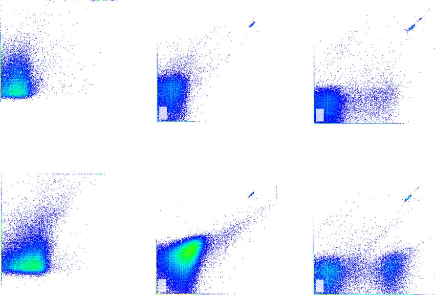
Liu et al. BMC Cancer   (2021) 21:939                                                                        Page 2 of 11

 Conclusion: NMPs and PMPs are associated with progression of advanced NSCLC. The predictive model for
 progression of advanced NSCLC, established combining NMPs, PMPs, and NLR, can screen out 80.8% of patients
 with PD. This is helpful for real-time accurate, concise and rapid assessment of disease progression and timely
 adjustment of drug therapy.
 Trial registration: Chinese Clinical Trial Registry, ChiCTR1800020223. Registered 20 December 2018, http://www.
 chictr.org.cn/index.aspx.
 Keywords: Neutrophil-derived microparticles, Platelet-derived microparticles, Non-small cell lung cancer, Progressive
 disease, Predictive model

Introduction                                                 species, releasing oncostatin M, and remodeling the
Lung cancer is a common malignancy, accounting for           tumor extracellular matrix (ECM) [16, 17]. Neutrophil
11.6% of total cancer incidence and 18.4% of cancer          MPs (NMPs) originate from neutrophils, and we specu-
deaths, and remains the leading cause of cancer death        late that NMPs contain neutrophil-associated signaling
worldwide [1, 2]. Non-small cell lung cancer (NSCLC)         molecules that promote cancer progression. As the
accounts for 85–90% of lung cancers, and radiother-          most-abundant circulating MPs [18], platelet MPs
apy and chemotherapy are still important for treat-          (PMPs) are closely associated with high invasiveness,
ment of advanced NSCLC, but the 5-year survival              metastasis and poor prognosis of cancer [19, 20]. Our
rate is < 15% [3]. In recent years, targeted therapy         previous studies have found that PMPs can independ-
and immunotherapy have been widely used for ad-              ently predict the efficacy of immunotherapy [21]. There-
vanced NSCLC [4–6], but disease progression still oc-        fore, in this study, we investigated the correlation
curs in advanced NSCLC after multiple lines of               between circulating NMPs and PMPs and progression of
treatment. Therefore, exploration of new mechanisms          advanced NSCLC, and established a model for predicting
that promote cancer progression, and rapid and con-          disease progression by combining laboratory indices, to
cise assessment of disease progression to guide and          provide clinicians with a new method for timely identifi-
adjustment of drug therapy are important to prolong          cation of disease progression.
patient survival.
  Microparticles (MPs) are submicron vesicles gener-
                                                             Materials and methods
ated from various types of cells and were initially
                                                             Patients
considered functionless cellular products. However, an
                                                             Patients with advanced NSCLC between October 2019
increasing number of studies have revealed that MPs
                                                             and October 2020 at our hospital were enrolled. Inclu-
are not metabolic waste products of cells and are in-
                                                             sion criteria: patients older than 18 years old; advanced
volved in coagulation, cell signaling, vascular injury,
                                                             non-small cell lung cancer (stage III or IV). Exclusion
and stabilization of the internal environment [7]. In
                                                             criteria: other malignant tumours; severe organ dysfunc-
2005, MPs were defined as cell-derived, extracellular
                                                             tion, such as chronic renal insufficiency (stage III and
vesicles of 0.1–1 μm diameter that lack a nucleus and
                                                             above), liver cirrhosis (Child-Pugh classification C and
the ability to synthesize proteins. They carry parent-
                                                             above); acute infection; autoimmune diseases; and
cell-derived proteins (e.g., signaling proteins, skeletal
                                                             current radiotherapy.
proteins, etc.), nucleic acids (e.g., miRNA, mRNA,
DNA), and lipids, and have high level of membrane
phosphatidylserine [8]. MPs are elevated in a variety        Blood sampling
of diseases, including acute coronary syndromes, sep-        Before and after 20–28 weeks of treatment, the routine
tic shock, and rheumatic immune system diseases [7,          blood, lactate dehydrogenase, and tumor markers were
8]. In cancer, MPs are derived from platelets and            routinely examined, and 3 ml of venous blood from the
endothelial, inflammatory and tumor cells, and are as-       elbow was extracted for the detection of MPs. To early
sociated with cancer staging, thrombosis, angiogenesis,      detect of disease progression and avoid the effects of
proliferation and metastasis [9–12].                         therapeutic drugs on MPs, blood samples were collected
  The peripheral blood neutrophil/lymphocyte ratio           before use of therapeutic drugs on the same day and at
(NLR) has been shown to correlate with the prognosis         least 21 days after the last medication. MPs isolation by
and progression of several cancers [13–15]. Tumor-           differential centrifugation was performed: 3 mL of whole
associated neutrophils (TAMs) promote tumor progres-         blood was centrifuged (2000 g) for 10 min to obtain
sion by producing reactive oxygen and reactive nitrogen      platelet-poor plasma (PPP); then, PPP was ultracentri-
                                                             fuged (13,000 g) for 3 min to collect platelet-free
Liu et al. BMC Cancer (2021) 21:939 Page 3 of 11
plasma (PFP), which was stored in the refrigerator at room temperature, avoiding light. Then, both tubes A
− 80 °C. and B were washed with phosphate-buffered saline
(PBS), transferred to BD absolute counting tubes, and
Microparticle analysis tested on the machine. Furthermore, 0.2 μm, 0.7 μm, and
Observation of MPs morphology by transmission electron 2 μm standard microspheres (invitrogen, carlsbad, CA,
microscopy USA) were used to set the gate and determine the loca-
MPs (20 μl) from patients were placed on copper mesh tion of 0.1–1.0-μm MPs (Fig. 1). To avoid the noise
and incubated for 5 min. We removed the remaining caused by dust and crystallization, the buffer and PBS
liquid with filter paper, and added 20 μl phosphotungstic were filtered through a 0.22-μm filter. MPs were
acid. After standing for 10 min, any remaining liquid was counted in events/μL.
removed with filter paper, and the sample was observed
by transmission electron microscopy.
Therapeutic evaluation
Detection of MPs number by flow cytometry Before and after 20–28 weeks of treatment, chest and ab-
PFP was dissolved at room temperature and then ultra- dominal computed tomography (CT) and head magnetic
centrifuged (4 °C, 21,000 g) for 60 min, and 100 μL of the resonance imaging (MRI) were performed to determine
bottom fraction was obtained as MPs. The number of the stage of cancer and evaluate the therapeutic effect.
MPs was detected by FACSCanto™ II flow cytometer The efficacy of non-immunotreated patients was evalu-
(BD Bioscience, San Jose, CA, USA). The immunofluor- ated by the 2009 Response Evaluation Criteria in solid
escent monoclonal antibodies used were as follows (Bio- tumor (RECIST 1. 1) [22], while the efficacy of immuno-
Legend, San Diego, CA, USA): Annexin V labeled by PE, treated patients was evaluated by the 2017 Immune-
CD66b and CD41 labeled by FITC. CD66b+/Annexin related Response Evaluation Criteria in Solid Tumours
V+ represents NMPs, CD41+/Annexin V+ represents (iRECIST) [23]. The patients were divided into objective
PMPs. Moreover, 25 μL of MPs, 1 μL Annexin V and remission (OR) and progressive disease (PD) groups.
100 μL Annexin V binding buffer were added into two The OR group included patients with complete remis-
Eppendorf tubes A and B, and then incubated for 20 min sion (CR), partial remission (PR) or stable disease (SD).
at room temperature, avoiding light. Then, 1 μL CD66b- PD was defined as a change in the longest diameter of
FITC were added into tube A, and 1 μL CD41-FITC the tumour on CT examination ≥20% or the occurrence
were added into tube B and incubated for 20 min at of new metastatic lesions.
Fig. 1 The setting of gates in the flow cytometer was shown in Fig. 1. The gates in A represented the positions of particles with diameters of
2 μm, 0.7 μm, 0.2 μm respectively, and P2 was the position of particles with diameters of 0.1–1 μm, that was, the position of the MPs. B showed
the flow diagram of MPs on the tester, and most MPs were in the P2 gateLiu et al. BMC Cancer (2021) 21:939 Page 4 of 11
Statistical analysis Karnofsky’s index of performance status (KPS) score,
Count data was compared by chi-square test or Fisher’s number of treatment lines and therapeutic intervention
exact probability test. Quantitative data of normal distri- (p > 0.05, Table 1). Chemotherapy regiments were
butions represented by x ± s were analyzed by t-tests. mainly platinum-containing dual-agent regiments, im-
Quantitative data of nonnormal distributions presented munotherapy included pembrolizumab and nivolumab,
as the medians and interquartile ranges (IQRs) were an- and target therapy included gefitinib, icotinib, erlotinib
alyzed by Mann-Whitney U test. Univariate analysis and and osimertinib. There was no significant difference in
binary multivariate logistic regression analysis (Forward: baseline laboratory indices, total MPs, NMPs and PMPs
LR) were used to find the independent influencing fac- between the two groups (p > 0.05, Table 1).
tors. The scores of independent factors were evaluated
according to the OR value, that is, Score≈OR, and then Comparison of laboratory indices and MPs after
the total score was calculated by adding the scores of treatment between 86 cases of NSCLC in the OR and PD
each factor [24]. We also established a prediction model groups
for disease progression. The sensitivity and specificity of After treatment, there was no significant difference in
the prediction model were tested by receiver operating neutrophils, platelets and squamous cell carcinoma anti-
characteristic (ROC) curve. P < 0.05 were statistically gen between the two groups (p > 0.05, Table 2). NLR,
signifificant. lactate dehydrogenase (LDH), carcinoembryonic antigen,
and cytokeratin 19 fragment (CYFRA 21-1) were signifi-
Result cantly higher in the PD group than those in the OR
MPs morphology group (p < 0.05, Table 2).
MPs have spherical structures with diameters ranging In the OR group, the circulating total MPs, NMPs and
from 100 to 1000 nm, heterogeneity in size, and typical PMPs after treatment were 1975[1090,3120]events/μL,
lipid bilayer structure (Fig. 2). 118[66,229]events/μL, and 56[31,109]events/μL, respectively.
The total MPs, NMPs and PMPs in the OR group were sig-
Comparison of clinical characteristics and baseline MPs nificantly lower than those at baseline (p < 0.05, Fig. 3). In
between 86 cases of NSCLC in the OR and PD groups the PD group, the total MPs, NMPs and PMPs after treat-
There were 86 patients, with 60 in the OR group and 26 ment were 3590[1492,4405]events/μL, 248[116,338] events/
in the PD group. There were no significant differences μL, and 157[93,282]events/μL, respectively. There was no sig-
between the two groups in terms of gender, age, smok- nificant difference in total MPs, NMPs or PMPs in the PD
ing history, pathological staging, tumor stage, metastasis, group compared with the baseline levels (p > 0.05, Fig. 3).
Total MPs, NMPs, and PMPs were significantly higher in the
PD group than those in the OR group after treatment
(p < 0.005, Table 2, Figs. 3 and 4).
Multivariate logistic regression analysis for predicting the
progression of advanced NSCLC
Factors with P < 0.1 in the univariate analysis were included
in the multivariate regression analysis. The results showed
that NMPs≥160events/μL(OR,3.748;95%CI,1.147–12.253,p =
0.029), PMPs≥80events/μL(OR,10.968;95%CI,2.973–40.462,
p < 0.0001), and NLR ≥ 3.3 (OR,4.929;95%CI,1.483–
16.375,p = 0.009) were independently associated with pro-
gression of advanced NSCLC. On the basis of the multivari-
ate regression analysis, significant factors were scored and a
predictive model for progression of advanced NSCLC was
established (Table 3), for which a prediction score > 16 was
considered high risk and ≤ 16 was low risk.
ROC curve to assess the efficacy of the predictive model
The ROC curve for predicting progression of advanced
Fig. 2 The morphology of microparticles (MPs) was observed using NSCLC by prediction score and other related factors
transmission electron microscopy. MPs had spherical structures was plotted, and showed that the area under the curve
under electron microscopy, with diameters ranging from 100 to
for prediction score evaluation for progression of
1000 nm, heterogeneity in size, and typical lipid bilayer structure
advanced NSCLC was 0.825(95%CI,0.715–0.934, P <Liu et al. BMC Cancer (2021) 21:939 Page 5 of 11 Table 1 Baseline variables and micropaticles in OR group and PD group of 86 cases of NSCLC Variables OR (n = 60) PD (n = 26) P Male, gender, n(%) 45 (75.0) 19 (73.1) 0.851* Age, years, x ± s 61.8 ± 10.7 62.6 ± 11.1 0.760§ Current or former smokers, n(%) 31 (51.7) 17 (65.4) 0.239* Histology, n(%) 0.297* Adenocarcinoma 25 (41.7) 14 (53.8) Squamous carcinoma 35 (58.3) 12 (46.2) Disease stage, n(%) 0.274* Stage III 26 (43.3) 8 (30.8) Stage IV 34 (56.7) 18 (69.2) Lines of therapy, n (%) 0.759*
Liu et al. BMC Cancer       (2021) 21:939                                                                                                    Page 6 of 11

Table 2 Micropaticles and laboratory data after treatment in OR group and PD group
Variables                                                  OR (n = 60)                                PD (n = 26)                                P
Neutrophils, ×109/L, median(IQR)                           3.62 [2.70,4.50]                           4.30 [2.93,5.35]                           0.232#
              9
Platelets, ×10 /L, median(IQR)                             205 [154,258]                              196 [160,281]                              0.807#
NLR, median(IQR)                                           2.43 [1.62,3.23]                           3.37 [1.75,7.04]                           0.029#
LDH, U/L, median(IQR)                                      166 [142,200]                              188 [169,240]                              0.004#
CEA, ng/ml, median(IQR)                                    2.85 [1.95,12.76]                          8.72 [3.29,50.13]                          0.026#
CYFRA-211, ng/ml, median(IQR)                              3.15 [2.14,4.08]                           4.54 [3.30,8.11]                           < 0.0001#
SCC, ng/ml, median(IQR)                                    1.10 [0.93,1.58]                           1.05 [0.68,1.93]                           0.491#
Micropaticals, events/μL, median(IQR)
  total MPs                                                1975 [1090,3120]                           3590 [1492,4405]                           0.006#
  NMPs                                                     118 [66,229]                               248 [116,338]                              0.002#
  PMPs                                                     56 [31,109]                                157 [93,282]                                16. This provides a new method                       12 weeks, which does not allow timely detection of
for judging the effect of drug therapy and timely detec-                       disease progression, and identification of pseudo-
tion of disease progression.                                                   progression during immunotherapy is difficult with im-
  NMPs are significantly elevated in advanced NSCLC                            aging [22]. Significant elevations in tumor markers, NLR
when disease progression occurs, suggesting that NMPs                          and LDH suggest poor cancer treatment response, but
may promote cancer progression; however, the relevant                          they have a lag in detection and low specificity. We
mechanisms are unclear. We suggest that the promotion                          found that circulating NMPs and PMPs were independ-
of cancer progression by NMPs might be related to the                          ently associated with disease progression in advanced
proinflammatory effects of NMPs. NMPs can activate                             NSCLC, suggesting that NMPs and PMPs may be novel
Liu et al. BMC Cancer (2021) 21:939 Page 7 of 11
P=0.003 P=0.314
P=0.038 P=0.120 At baseline At baseline
After therapy After therapy
516
6000
276
Total MPs, events/L
NMPs, events/L
4000
181
2000 105
26
0
OR Group PD Group OR Group PD Group
A B
P=0.0001 P=0.777
At baseline
After therapy
234
PMPs, events/L
146
69
13
OR Group PD Group
C
Fig. 3 Box plots for circulating microparticles levels at baseline and after treatment in the OR and PD groups. Top and bottom of box
represented 75th and 25th percentile, respectively; middle bar in box represented the median value; Top and bottom lines extending from box
represented maximum and minimum obtained values, respectively. ° represented mild outliers. In PD group, total MPs, NMPs and PMPs did not
change significantly after treatment (p > 0.05). In OR group, total MPs, NMPs and PMPs were decreased significantly after treatment (p < 0.05).
A showed total MPs, B showed NMPs, C showed PMPsLiu et al. BMC Cancer (2021) 21:939 Page 8 of 11
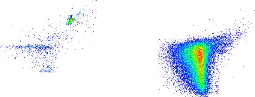
Fig. 4 Flow chart of MPs in the OR and PD groups. A-1 and A-2 showed total MPs in the OR and PD groups after treatment, and total MPs were
significantly higher in the PD group than those in the OR group. B-1 and B-2 showed NMPs in the OR and PD groups after treatment, and NMPs
were significantly higher in the PD group than those in the OR group. C-1 and C-2 showed PMPs in the OR and PD groups after treatment, and
PMPs were significantly higher in the PD group than those in the OR group
mechanisms for promotion of tumor progression. A eliminates the need for repeated imaging, avoids radi-
model using NMPs and PMPs combined with NLR for ation toxicity, and reduces the economic burden on pa-
prediction of disease progression showed a sensitivity of tients and hospitals.
80.8% and specificity of 88.3%. When the prediction According to our literature search, this study was the
score indicates high risk, we can screen out 80.8% of pa- first to analyze the relationship between circulating
tients with disease progression, and we need to actively NMPs and disease progression in advanced NSCLC, and
improve imaging examination to determine disease pro- multivariate regression analysis showed that NMPs and
gression and adjust drug therapy for these patients. PMPs combined with NLR were able to predict disease
When the prediction score for a patient is low, the pa- progression. This study had some shortcomings: first, it
tient has a 91.4% probability of stable disease, which was a single-center study with a small sample size; and
Table 3 Multivariate analyses of factors related with PD in advanced NSCLC
Variables β S.E. Wald df Sig. OR 95% CI for OR Score
Lower Upper
NMPs, ≥160 events/μL 1.321 0.604 4.779 1 0.029 3.748 1.147 12.253 4
PMPs, ≥80 events/μL 2.395 0.666 12.930 1Liu et al. BMC Cancer (2021) 21:939 Page 9 of 11
Table 4 Comparison of Prediction Score and other indicators in predicting the progression of advanced NSCLC
Variables Cutoff AUC 95% CI Sensitivity Specificity PPV NPV P
Prediction Score 16 0.825 (0.715,0.934) 80.8% 88.3% 75.0% 91.4% < 0.0001
total MPs, events/μL 2800 0.686 (0.559,0.813) 69.2% 68.3% 48.6% 83.7% 0.006
NMPs, events/μL 160 0.712 (0.598,0.827) 69.2% 65.0% 46.2% 83.0% 0.002
PMPs, events/μL 80 0.804 (0.706,0.903) 84.6% 68.3% 53.7% 91.1% < 0.0001
NLR 3.3 0.648 (0.513,0.784) 57.7% 78.3% 53.6% 81.0% 0.029
CEA, ng/ml 5.0 0.652 (0.515,0.788) 69.2% 65.0% 46.2% 83.0% 0.026
CYFRA21-1, ng/ml 2.5 0.741 (0.628,0.855) 96.2% 43.3% 42.4% 96.3% 0.058
LDH, U/L 210 0.694 (0.576,0.812) 46.2% 85.0% 57.1% 78.5% 0.004
PPV Positive predictive value, NPV Negative predictive value, MPs microparticals, NMPs neutrophil-derived microparticals, PMPs platelet-derived microparticles, CEA
carcino-embryonic antigen, CYFRA-211 Cytokeratin 19 fragment, NLR neutrophil-lymphocyte ratio, LDH lactate dehydrogenase, CEA carcino-embryonic antigen
second, the mechanism by which MPs promoted cancer for disease progression effectively screened 80.8% of pa-
progression was not further explored. tients with disease progression. We reveal that NMPs
and PMPs may be new factors that promote cancer pro-
gression, and provide a new method for concise, real-
Conclusion time dynamic assessment of advanced NSCLC disease
We found that circulating NMPs and PMPs independ- progression. This could help to adjust drug therapy and
ently predicted disease progression in advanced NSCLC, improve prognosis in a timely manner, and reduce the
and combination with NLR to build a predictive model financial burden on patients.
ROC Curve
1.0
0.8
0.6
Sensitivity
0.4
Prediction Score
Total MPs
PMPs
NMPs
0.2 NLR
LDH
CEA
CYFRA21-1
Reference Line
0.0
0.0 0.2 0.4 0.6 0.8 1.0
1 - Specificity
Diagonal segments are produced by ties.
Fig. 5 Receiver operating characteristic curves (ROC) displayed Prediction Score and other factors for predicting progression in advanced NSCLC.
Area under the curve of Prediction Score was 0.825(95%CI,0.715–0.934, p < 0.0001)Liu et al. BMC Cancer (2021) 21:939 Page 10 of 11
Acknowledgements 9. Litwińska Z, Łuczkowska K, Machaliński B. Extracellular vesicles in
We thank Liwen Bianji, Edanz Editing China, for editing the English text of a hematological malignancies. Leuk Lymphoma. 2019;60(1):29–36. https://doi.
draft of this manuscript. org/10.1080/10428194.2018.1459606.
10. Rousseau A, Van Dreden P, Khaterchi A, et al. Procoagulant microparticles
Authors’ contributions derived from cancer cells have determinant role in the hypercoagulable
HY and LH contributed to the study design. LT (Tingting Liu) and WJ state associated with cancer. Int J Oncol. 2017;51(6):1793–800. https://doi.
completed the isolation and detection of MPs. CP, HB, ZC and WG org/10.3892/ijo.2017.4170.
contributed to the collection of clinical data. LT (Tingting Liu), LT (Tao Li) and 11. Arderiu G, Peña E, Badimon L. Angiogenic microvascular endothelial cells
ZS contributed to the data analysis. LT (Tingting Liu), WJ and LT (Tao Li) release microparticles rich in tissue factor that promotes postischemic
drafted the manuscript. All authors read and approved the final manuscript. collateral vessel formation. Arterioscler Thromb Vasc Biol. 2015;35(2):348–57.
https://doi.org/10.1161/ATVBAHA.114.303927.
Funding 12. Wu K, Xing F, Wu SY, Watabe K. Extracellular vesicles as emerging targets in cancer:
No fundings. recent development from bench to bedside. Biochim Biophys Acta Rev Cancer.
2017;1868(2):538–63. https://doi.org/10.1016/j.bbcan.2017.10.001.
Availability of data and materials 13. Kim TG, Park W, Kim H, Choi DH, Park HC, Kim SH, et al. Baseline neutrophil-
The data used and/or analyzed during the current study are available from lymphocyte ratio and platelet-lymphocyte ratio in rectal cancer patients
the corresponding author on reasonable request. following neoadjuvant chemoradiotherapy. Tumori. 2019;105(5):434–40.
https://doi.org/10.1177/0300891618792476.
Declarations 14. Pirozzolo G, Gisbertz SS, Castoro C, van Berge Henegouwen MI, Scarpa M.
Neutrophil-to-lymphocyte ratio as prognostic marker in esophageal cancer:
Ethics approval and consent to participate a systematic review and meta-analysis. J Thorac Dis. 2019;11(7):3136–45.
All participants in this study signed informed consent forms. The ethics https://doi.org/10.21037/jtd.2019.07.30.
committee of the People’s Liberation Army General Hospital approved this 15. Nakaya A, Kurata T, Yoshioka H, Takeyasu Y, Niki M, Kibata K, et al.
study according to the ethical standards of the Declaration of Helsinki and Neutrophil-to-lymphocyte ratio as an early marker of outcomes in patients
its subsequent amendments (Ethical approval number: S2018–092-01). with advanced non-small-cell lung cancer treated with nivolumab. Int J Clin
Oncol. 2018;23(4):634–40. https://doi.org/10.1007/s10147-018-1250-2.
Consent for publication 16. Triner D, Shah YM. Hypoxic regulation of neutrophils in Cancer. Int J Mol
Not applicable. Sci. 2019;20(17):4189. https://doi.org/10.3390/ijms20174189.
17. Wang X, Qiu L, Li Z, Wang XY, Yi H. Understanding the multifaceted role of
Competing interests neutrophils in Cancer and autoimmune diseases. Front Immunol. 2018;9:
The authors have no conflicts of interest to declare. 2456. https://doi.org/10.3389/fimmu.2018.02456.
18. Burnouf T, Goubran HA, Chou ML, Devos D, Radosevic M. Platelet
Author details microparticles: detection and assessment of their paradoxical functional
1
Medical School of Chinese PLA, Chinese PLA General Hospital, Beijing roles in disease and regenerative medicine. Blood Rev. 2014;28(4):155–66.
100853, China. 2Department of Medical Oncology, The First Medical Center, https://doi.org/10.1016/j.blre.2014.04.002.
Chinese PLA General Hospital, Beijing 100853, China. 3Department of 19. Dovizio M, Bruno A, Contursi A, Grande R, Patrignani P. Platelets and
Pulmonary and Critical Care Medicine, The Second Medical Center, National extracellular vesicles in cancer: diagnostic and therapeutic implications.
Clinical Research Center for Geriatric Diseases, Chinese PLA General Hospital, Cancer Metastasis Rev. 2018;37(2–3):455–67. https://doi.org/10.1007/s10555-
Beijing 100853, China. 4Centre of Pulmonary and Critical Care Medicine, 018-9730-4.
Chinese PLA General Hospital, Beijing 100853, China. 5People Liberation 20. Varon D, Hayon Y, Dashevsky O, Shai E. Involvement of platelet derived
Army Haidian District 17th Retired Cadres Rest Home, Beijing 100143, PR microparticles in tumor metastasis and tissue regeneration. Thromb Res.
China. 2012;130(Suppl 1):S98–9. https://doi.org/10.1016/j.thromres.2012.08.289.
21. Liu T, Wang J, Liu Y, et al. Prediction of the therapeutic effects of
Received: 3 May 2021 Accepted: 26 July 2021 pembrolizumab and nivolumab in advanced non-small cell lung cancer by
platelet-derived microparticles in circulating blood. Technol Cancer Res T.
2021;20:1533033821997817.
References 22. Eisenhauer EA, Therasse P, Bogaerts J, Schwartz LH, Sargent D, Ford R, et al. New
1. Bray F, Ferlay J, Soerjomataram I, Siegel RL, Torre LA, Jemal A. Global cancer response evaluation criteria in solid tumours: revised RECIST guideline (version 1.
statistics 2018: GLOBOCAN estimates of incidence and mortality worldwide 1). Eur J Cancer. 2009;45(2):228–47. https://doi.org/10.1016/j.ejca.2008.10.026.
for 36 cancers in 185 countries. CA Cancer J Clin. 2018;68(6):394–424. 23. Seymour L, Bogaerts J, Perrone A. RECIST working group. iRECIST: guidelines
https://doi.org/10.3322/caac.21492. for response criteria for use in trials testing immunotherapeutics. Lancet
2. Mattiuzzi C, Lippi G. Current Cancer epidemiology. J Epidemiol Glob Health. Oncol. 2017;18(3):e143–52. https://doi.org/10.1016/S1470-2045(17)30074-8.
2019;9(4):217–22. https://doi.org/10.2991/jegh.k.191008.001. 24. LaPar DJ, Likosky DS, Zhang M, et al. Development of a risk prediction
3. Du L, Morgensztern D. Chemotherapy for advanced-stage non-small cell model and clinical risk score for isolated tricuspid valve surgery. Ann Thorac
lung Cancer. Cancer J. 2015;21(5):366–70. https://doi.org/10.1097/PPO. Surg. 2018;106(1):129–36. https://doi.org/10.1016/j.athoracsur.2017.11.077.
0000000000000141. 25. Kanazawa S, Nomura S, Kuwana M, Muramatsu M, Yamaguchi K, Fukuhara S.
4. Singh SS, Dahal A, Shrestha L, Jois SD. Genotype driven therapy for non- Monocyte-derived microparticles may be a sign of vascular complication in
small cell lung Cancer: resistance, Pan inhibitors and immunotherapy. Curr patients with lung cancer. Lung Cancer. 2003;39(2):145–9. https://doi.org/1
Med Chem. 2020;27(32):5274–316. https://doi.org/10.2174/09298673266661 0.1016/S0169-5002(02)00441-5.
90222183219. 26. Najjar F, Alammar M, Al-Massarani G, et al. Circulating endothelial cells and
5. Tan AC. Targeting the PI3K/Akt/mTOR pathway in non-small cell lung microparticles for prediction of tumor progression and outcomes in
cancer (NSCLC). Thorac Cancer. 2020;11(3):511–8. https://doi.org/10.1111/1 advanced non-small cell lung cancer. Cancer Biomark. 2017;20(3):333–43.
759-7714.13328. https://doi.org/10.3233/CBM-170130.
6. Doroshow DB, Herbst RS. Treatment of advanced non-small cell lung 27. Mesri M, Altieri DC. Leukocyte microparticles stimulate endothelial cell
Cancer in 2018. JAMA Oncol. 2018;4(4):569–70. https://doi.org/10.1001/jama cytokine release and tissue factor induction in a JNK1 signaling pathway. J
oncol.2017.5190. Biol Chem. 1999;274(33):23111–8. https://doi.org/10.1074/jbc.274.33.23111.
7. Mackman N. On the trail of microparticles. Circ Res. 2009;104(8):925–7. 28. Mesri M, Altieri DC. Endothelial cell activation by leukocyte microparticles. J
https://doi.org/10.1161/CIRCRESAHA.109.196840. Immunol. 1998;61(8):4382–7.
8. Hargett LA, Bauer NN. On the origin of microparticles: from "platelet dust" 29. Hong Y, Eleftheriou D, Hussain AA, et al. Anti-neutrophil cytoplasmic
to mediators of intercellular communication. Pulm Circ. 2013;3(2):329–40. antibodies stimulate release of neutrophil microparticles. J Am Soc Nephrol.
https://doi.org/10.4103/2045-8932.114760. 2012;23(1):49–62. https://doi.org/10.1681/ASN.2011030298.Liu et al. BMC Cancer (2021) 21:939 Page 11 of 11
30. Anene C, Graham AM, Boyne J, Roberts W. Platelet microparticle delivered
microRNA-let-7a promotes the angiogenic switch. Biochim Biophys Acta Mol basis
Dis. 2018;1864(8):2633–43. https://doi.org/10.1016/j.bbadis.2018.04.013.
31. Liang H, Yan X, Pan Y, et al. MicroRNA-223 delivered by platelet-derived
microvesicles promotes lung cancer cell invasion via targeting tumor
suppressor EPB41L3. Mol Cancer. 2015;11:14–58.
32. Helley D, Banu E, Bouziane A, Banu A, Scotte F, Fischer AM, et al. Platelet
microparticles: a potential predictive factor of survival in hormone-refractory
prostate cancer patients treated with docetaxel-based chemotherapy. Eur
Urol. 2009;56(3):479–84. https://doi.org/10.1016/j.eururo.2008.06.038.
33. Kim HK, Song KS, Park YS, Kang YH, Lee YJ, Lee KR, et al. Elevated levels of
circulating platelet microparticles, VEGF, IL-6 and RANTES in patients with
gastric cancer: possible role of a metastasis predictor. Eur J Cancer. 2003;
39(2):184–91. https://doi.org/10.1016/S0959-8049(02)00596-8.
Publisher’s Note
Springer Nature remains neutral with regard to jurisdictional claims in
published maps and institutional affiliations.You can also read