PR interval as a predictor of syncope in tilt- up testing in adolescents and young adults
←
→
Page content transcription
If your browser does not render page correctly, please read the page content below
Jug et al. The Egyptian Heart Journal   (2021) 73:28
https://doi.org/10.1186/s43044-021-00149-6
                                                                                                                                           The Egyptian Heart
                                                                                                                                                      Journal

 RESEARCH                                                                                                                                               Open Access

PR interval as a predictor of syncope in tilt-
up testing in adolescents and young adults
Juraj Jug1* , Lada Bradić2 , Rea Levicki3                          and Martina Lovrić Benčić1,2

  Abstract
  Background: Syncope, as the most frequent consciousness disorder, is very common in young individuals. The aim
  of this study was to analyze ECG parameters and clinical properties obtained during tilt-up testing in 12 to 30-year-
  old subjects. We enrolled a total of 142 patients from our outpatient clinic (39 males, 103 females) with a true
  positive tilt-up test and analyzed ECG records obtained during tilt-testing. Data were stratified according to the age,
  gender, and type of syncope.
  Results: PR interval shortening preceding syncope was found in all syncope types, irrespective of the gender. All
  types of syncope were more frequent in women (72.5%). Mixed syncope type was found to be the most common
  (47.18%). Male and female subjects differed in initial heart rate (71.56 vs 76.23/min, p=0.05), as well as heart rate
  dynamics during tilt-up testing. A gender difference was also found in systolic blood pressure (116.92 vs 110.44
  mmHg, p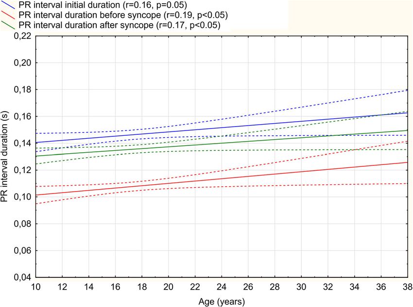
Jug et al. The Egyptian Heart Journal   (2021) 73:28                                                         Page 2 of 8

   Syncope results from the increased vagal tone, which      Tilt-up test protocol
can also reflect in AV node activation, but the shorten-     All patients were tested from 8:00 am to 12:00 am. At
ing of the PR interval duration is the result of sympa-      the beginning of the procedure, patients were positioned
thetic activation. Only several papers associate these       supine, in ambient temperature of 22–24 °C. Each pa-
alterations with the etiopathogenesis of syncope. There      tient was given 250 mL of 5% glucose intravenously.
are few reports on PR interval duration change on ECG        After 20 min in the supine position, patients were
tracing analysis during tilt-up testing, but only in rela-   passively tilted to a 75° head-up position without any
tion to the cardioinhibitory syncope. PR interval reflects   provocation. The test was aborted either due to syncope
conduction through the atria and the AV node [8].            provocation or if 45 min passed without syncope. Nega-
Changes in PR interval duration can result from nervous      tive tests were excluded from the study.
tone alterations or fibrosis in any of the anatomical
structures of the heart, or from pacemaker shift towards     Informed consent
the AV node.                                                 Data was obtained retrospectively from the reports of
   The aim of this study was to investigate PR interval      outpatient tilt-up testing in our clinic. Informed consent
changes during tilt-up testing in patients with a positive   was obtained from all participants. Data analysis has
history for neurally mediated syncope. The focus was         been approved by the clinic’s Ethics committee.
put on the PR interval since many patients with frequent
syncope showed PR interval shortening in ECG during
tilt-up testing.                                             Statistics
                                                             All data are expressed as mean ± standard error. Para-
Methods                                                      metric data were compared using an unpaired t test. For
We enrolled a total of 142 consecutive patients (103 fe-     nonparametric data, we used a Mann-Whitney U test.
males, 39 males), age 12 to 30, admitted to our out-         Graphical data, age comparison, and test results were
patient clinic for a diagnostic workup of syncope. All       analyzed with multifactorial ANOVA. A value of p <
these patients had a positive tilt-up test (with true syn-   0.05 was considered significant. For a significant change
cope), did not take any medication, and were otherwise       in PR interval duration, we have arbitrarily set the value
healthy. We consider the test as positive if the patient     of 25%. All data were analyzed by Statistica 10.0.
experienced true syncope during the testing and met the
criteria for one of the VASIS syncope types. With re-        Results
spect to patient characteristics and to assure a matching    Gender differences
number of patients, test subjects were divided into three    Mean age of male and female subjects was compar-
age subgroups (12–15, 16–18, and 19–30 years). Patients      able (16.8 ± 1.2 vs. 17.6 ± 1.1 years, NS), just as their
over 30 years were excluded because of a small number        age of the first syncope onset (14.7 vs. 14.8 years,
of true syncope during tilt-up testing.                      NS). Most of all enrolled patients were female
  ECG tracings were analyzed in D2 lead manually             (72.5%), with a history of approximately twice as
and automatically in all 12 leads during the entire          many syncope episodes (4.62 vs. 2.79, p
Jug et al. The Egyptian Heart Journal       (2021) 73:28                                                                              Page 3 of 8

    Fig. 1 Heart rate dynamics between gender from supine to the upright position during tilt-up test

VASIS type differences                                                       Age groups analysis
The most common type of vasovagal syncope was a                              PR interval prolongs with aging (r=0.22, p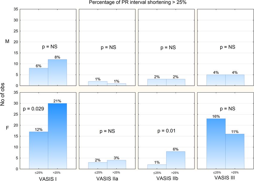
Jug et al. The Egyptian Heart Journal (2021) 73:28 Page 4 of 8
Fig. 2 Number and percentage of patients with PR interval change below and above 25% according to gender and VASIS syncope type. (M =
male, F = female)
Table 3 Variables according to age groups (BS = before syncope, BP = blood pressure, AS = after syncope)
Variable Age groups (years) TOTAL p
12-15 16-18 19–30 12-30 ANOVA
n = 53 n = 54 n = 35 n = 142
Female (%) 37 (69.81%) 40 (74.07%) 26 (74.28%) 103 (72.53%)
PR interval (ms) 142.8 ± 3.2 147.7 ± 2,7 149.4 ± 3.6 146.3 ± 1.8 0.183
PR interval BS (ms) 103.2 ± 2.1 109.1 ± 2.5 112.6 ± 2.5 107.7 ± 1.7 0.046
PR shortening (%) 27.6 ± 1.4 25.8 ± 2.3 24.4 ± 1.7 26.1 ± 0.8 0.155
PR interval AS (ms) 132.6 ± 1.8 135.4 ± 2.1 140.0 ± 2.2 135.5 ± 2.0 0.185
QT interval (ms) 360.6 ± 3.9 362.9 ± 3.5 368.8 ± 3.1 363.5 ± 2.1 0.136
BMI (kg/m2) 21.8 ± 1.2 21.2 ± 0.9 22.5 ± 1.1 21.8 ± 0.6 NS
HR preparation (bpm) 78.5 ± 1.9 73.4 ± 1.5 71.8 ± 1.9 74.9 ± 1.1 0.023
HR orthostatic (bpm) 105 ± 2.2 96.8 ± 1.9 91 ± 2.2 98.7 ± 1.3 0.00003
HR change (bpm) 26.9 ± 1.7 23.4 ± 1.6 19.4 ± 2.2 23.7 ± 1.1 0.008
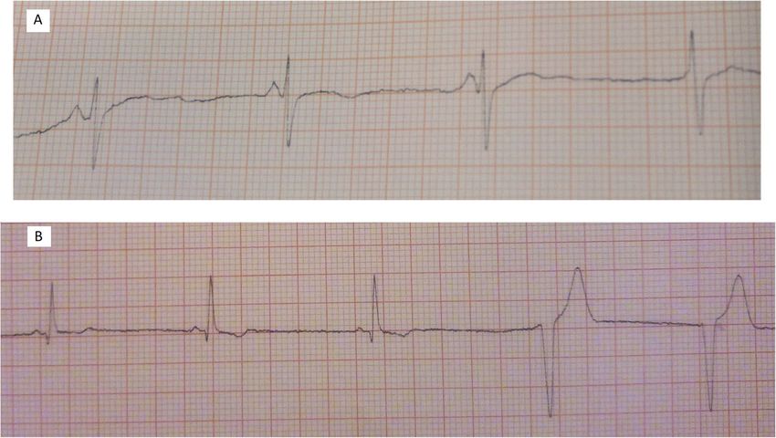
Time of syncope (min) 20.8 ± 1.5 20.5 ± 1.5 16.1 ± 1.6 19.6 ± 0.9 0.037Jug et al. The Egyptian Heart Journal (2021) 73:28 Page 5 of 8 Fig. 3 PR interval duration at the beginning of the test, before and after syncope onset according to age Fig. 4 PR interval duration initially, before and after the syncope in different age groups divided by VASIS types
Jug et al. The Egyptian Heart Journal    (2021) 73:28                                                                     Page 6 of 8

VASIS III type, where nodal rhythm was uncommon in                        injection increased vagal tone in rats, mediated by a high
all groups.                                                               concentration of estrogen receptors in the cardioregula-
                                                                          tory neurons of the brain [15]. In addition, estrogen was
Discussion                                                                found to increase muscarinic receptor concentration on
Thus far, PR changes were described in only a few pub-                    the cell membrane [16, 17]. The hearts of female mice
lished papers and in relation strictly to the cardioinhibi-               were also found to have higher acetylcholine levels than
tory type of syncope (VASIS II). In the pediatric                         males [18]. This is in the agreement with our results,
population, tilt-up testing can be very useful in the iden-               which demonstrated heart rate change in tilt-testing to
tification of pseudo-syncope [9]. Other parameters, not                   be significantly higher in male than in female subjects.
routinely measured during tilting, can also aid in defin-                    Mechanisms underlying PR shortening in patients with
ing properties of vasovagal syncope. One of the pub-                      VASIS II type of syncope could be attributed to SA node
lished papers on PR interval correlation with syncope in                  inhibition by increased vagal tone, as well as a pace-
young subjects was a paper by Makarov et al. [10]. All                    maker shift towards the AV node [11]. Our study sug-
the patients with PR interval shortening had a history of                 gests that all VASIS types of syncope share a common
syncope, while no changes in QT interval duration was                     pathogenesis.
observed. Only one case report was published thus far,                       We found rhythm transition from sinus to nodal in 11
with electrophysiologically demonstrated PR interval                      patients during the tilt-up test procedure, following the
shortening in a young man with cardioinhibitory syn-                      gradual PR shortening. One female patient was found to
cope, showing PR interval shortening from 142 to 121                      have an idioventricular rhythm (Fig. 5).
ms immediately preceding syncope [11]. Another pub-                          Our results point to a potential common mechanism
lished paper on the significance of PR interval changes                   in VASIS I and II syncope, which essentially differ from
by Mehlsen et al. [12] has shown a reduction of a mean                    the mechanisms in VASIS type III (Table 2). The sensi-
PR interval from 159±28 ms supine to 143±23 ms in the                     tivity of muscarinic receptors, as well as SA node to the
upright position preceding syncope. Our study also dem-                   vagal acetylcholine stimulation may be lower in VASIS
onstrated PR interval shortening in all VASIS types with                  III syncope, which explains the frequently encountered
the biggest differences between VASIS I and III group                     constant or increased heart rate response in these types
(Table 2). Seeing the bigger PR shortening, we could                      of syncope. This could account for a difference in syn-
predict that syncope would classify as VASIS I in female                  cope incidence between sexes.
patients, while males had very similar PR shortening ra-                     Autonomic cardiovascular regulation decreases with
tios between VASIS groups. PR interval correlation with                   aging, thus potentially accounting for a decrease in the
VASIS type and gender could be interpreted by in-                         prevalence of VASIS I and II syncope types [3]. In our
creased parasympathetic nervous system activity in                        study, that phenomenon manifested as reduced heart
women (Fig. 2). That was corroborated by several papers                   rate dynamics, as well as PR interval prolongation and
of Saleh et al. [13, 14] where intracerebral estrogen                     reduced PR interval shortening rate with aging (Fig. 3).

 Fig. 5 Transition from sinus rhythm to a nodal rhythm and b idioventricular rhythm
Jug et al. The Egyptian Heart Journal (2021) 73:28 Page 7 of 8
Physiologically, heart rate increases for approximately 30 and editing. MLB: supervision, validation, review, and editing. The authors
bpm in young and 10 bpm in older subjects in an up- have read and approved the manuscript.
right position [19]. It seems that baroreceptor activity
Funding
and sensitivity do not differ between pediatric and young None.
adult patients [20]. However, baroreceptor sensitivity de-
crease can be noticed in adulthood, with increasing age Availability of data and materials
[21]. Changes in PR interval and heart rate variability in The datasets used and/or analyzed during the current study are available
from the corresponding author on reasonable request.
younger population can be attributed to incomplete
autonomic nervous system maturation. The physiology
Declarations
of the autonomic nervous system maturation has not
been entirely elucidated yet and potentially relates to Ethics approval and consent to participate
both mechanical and neural alterations with aging [22]. This study has been approved by the Clinical Hospital Center Zagreb ethic
committee. Class: 8.1-18/24-2. Number: 02/21 AG. Date: January 30, 2018. All
Even though we suggest that PR interval shortening participants have signed a written informed consent or their parents or legal
could be an important predictor of syncope (4.22% of guardians in the case of children under 16 have signed for them.
patients (6/142) did not show PR shortening in ECG), a
tilt-up test should be performed completely because of Consent for publication
Not applicable.
unknown sensibility and specificity for this change. A
control group could not be assessed because if syncope
Competing interests
is absent the PR measurement (done at the moment be- We declare there is no conflict of interest in this study.
fore syncope) is impossible.
Author details
1
University of Zagreb School of Medicine, Zagreb, Croatia. 2University
Conclusion Hospital Zagreb, Zagreb, Croatia. 3General Hospital Požega, Požega, Croatia.
PR interval shortening on ECG tracings during a tilt-up
Received: 7 December 2020 Accepted: 1 March 2021
test can be found in all subtypes of vasovagal syncope,
thereby contrasting previous reports that these changes
are a hallmark of the cardioinhibitory type of syncope. References
Patients with the VASIS III group had the highest num- 1. Krediet CT, van Dijk N, Linzer M, van Lieshout JJ (2002) Management of
ber of syncopal episodes and the longest PR interval dur- vasovagal syncope: controlling or aborting faints by leg crossing and
muscle tensing. Circulation 106:1684–1689
ation, and their PR interval shortening was the smallest 2. White TJ (2000) A review of pathophysiology and therapy of patients with
among all VASIS types. PR interval shortening differed vasovagal syncope. Pharmacotherapy 20:158–165
mostly between VASIS I and VASIS III syncope. We 3. Zaqqa M, Massumi A (2000) Neurally mediated syncope. Tex Heart Inst J 27:
268–272
suggest that PR shortening, if observed during ECG 4. Kapoor W (2000) Syncope. N Engl J Med 343:1856–1862
monitoring, could be a possible predictor of syncope. 5. Pilcher TA, Saarel EV (2019) A teenage fainter (dizziness, syncope, postural
Further investigation is needed to confirm the signifi- orthostatic tachycardia syndrome). Pediatr Clin NA 61(1):29–43
6. Brignole M, Menozzi C, Del R, Costa S, Gaggioli G, Al E (2000) New
cance of these findings. classification of haemodynamics of vasovagal syncope: beyond the VASIS
classification. Analysis of the pre-syncopal phase of the tilt test without and
Study limitations with nitroglycerin challenge. Europace 2(1):66–76
7. Schroeder C, Tank J, Heusser K, Diedrich A, Luft FC, Jordan J (2011)
PR interval changes were measured automatically and Physiological phenomenology of neurally-mediated syncope with
checked manually on ECG tracings of tilt-table tests per- management implications. PLoS One 6(10):e26489
formed on outpatients younger than 30 years in our 8. Ganz L (2016) In: Goldman L, Schafer A (eds) Electrocardiography in
Goldman-Cecil medicine, 25th edn. Elsevier Inc, New York, pp 264–274
clinic because of a very small number of positive results 9. Robinson J, Shivapour J, Snyder C (2017) Tilt table testing to diagnose
in older than 30 years. Specificity and the sensitivity of pseudosyncope in the pediatric population. Congenit Heart Dis 12:411–416
PR interval duration changes in syncope episodes are 10. Makarov L, Kuryleva T, Chuprova S (2003) Shortening of PR interval,
bradycardia and polymorphic ventricular tachycardia - clinico -
unknown because PR interval shortening measurements electrocardiografical syndrome with high risk of sudden death in children.
in the control group are impossible. Kardiologiia 43(7):55–60
11. Avbelj V, Trobec R (2019) Letter to the editor a closer look at
Abbreviations electrocardiographic P waves before and during spontaneous
ECG: Electrocardiogram; AV: Atrioventricular; SA: Sinoatrial; BMI: Body mass cardioinhibitory syncope. Int J Cardiol 166(3):e59–e61
index; HR: Heart rate; BS: Before syncope; BP: Blood pressure; BPM: Beats per 12. Mehlsen J, Kaijer MN, Mehlsen A (2008) Autonomic and
minute electrocardiographic changes in cardioinhibitory syncope. Europace 10:91–
95
Acknowledgements 13. Saleh M, Connell B, Saleh T (2000) Medullary and intrathecal injections of
None. 17β-estradiol in male rats. Brain Res 867(1–2):200–209
14. Saleh T, Connell B (1999) Centrally mediated effect of 17beta-estradiol on
Authors’ contributions parasympathetic tone in male rats. Am J Physiol 276:R474–R481
JJ: investigation, methodology, writing original draft, data curation, formal 15. Dart AM, Du X, Kingwell BA (2002) Gender, sex hormones and autonomic
analysis. LB: conceptualization, methodology, review, and editing. RL: review nervous control of the cardiovascular system. Cardiovasc Res 53:678–687Jug et al. The Egyptian Heart Journal (2021) 73:28 Page 8 of 8
16. Rainbow T, Degroff V, Luine V, McEwen B (1980) Estradiol 17 beta increases
the number of muscarinic receptors in hypothalamic nuclei. Brain Res
198(1):239–243
17. Olsen K, Edwards E, Schechter N, Whalen R (1988) Muscarinic receptors in
preoptic area and hypothalamus: effects of cyclicity, sex and estrogen
treatment. Brain Res 448(2):223–229
18. Nachbar A, B. G-A (1997) Sex dependent electrocardiogram (ECG) changes
in anthracycline-treated mice. Exp Toxicol Pathol 47(1–2):75–77
19. Ingall TJ, McLeod JG, O’Brien PC (1990) The effect of ageing on autonomic
nervous system function. Aust N Z J Med 20:570
20. Alnoor MS, Varner HK, Butler IJ, Zhu L, Numan MT (2019) Baroreceptor
activity and sensitivity: normal values in children and young adults using
the head up tilt test. Pediatr Res 85(6):841–847
21. Pfeifer MA, Weinberg CR, Cook D, Best JD, Reenan A, Halter JB (1983)
Differential changes of autonomic nervous system function with age in
man. Am J Med 75(2):249–258
22. Lenard Z, Studinger P, Beatrix M, Kocsis L, Kollai M (2004) Maturation of
cardiovagal autonomic function from childhood to young adult age.
Circulation 110:2307–2312
Publisher’s Note
Springer Nature remains neutral with regard to jurisdictional claims in
published maps and institutional affiliations.You can also read