Pigment Intensity in Dogs is Associated with a Copy Number Variant Upstream of KITLG - MDPI
←
→
Page content transcription
If your browser does not render page correctly, please read the page content below
G C A T
T A C G
G C A T
           genes
Article
Pigment Intensity in Dogs is Associated with a Copy
Number Variant Upstream of KITLG
Kalie Weich 1 , Verena Affolter 2 , Daniel York 3 , Robert Rebhun 3 , Robert Grahn 4 ,
Angelica Kallenberg 4 and Danika Bannasch 1, *
 1   Department of Population Health and Reproduction, University of California-Davis, Davis, CA 95616, USA;
     kmweich@ucdavis.edu
 2   Department of Pathology, Microbiology, and Immunology, University of California-Davis, Davis, CA 95616,
     USA; vkaffolter@ucdavis.edu
 3   Department of Surgical and Radiological Sciences, University of California-Davis, Davis, CA 95616, USA;
     dyork@ucdavis.edu (D.Y.); rbrebhun@ucdavis.edu (R.R.)
 4   Veterinary Genetics Laboratory, University of California-Davis, Davis, CA 95616, USA;
     ragrahn@ucdavis.edu (R.G.); akallenberg@ucdavis.edu (A.K.)
 *   Correspondence: dlbannasch@ucdavis.edu; Tel.: +1-530-754-8728

 Received: 10 December 2019; Accepted: 8 January 2020; Published: 9 January 2020

 Abstract: Dogs exhibit a wide variety of coat color types, and many genes have been identified that
 control pigment production, appearance, and distribution. Some breeds, such as the Nova Scotia
 Duck Tolling Retriever (NSDTR), exhibit variation in pheomelanin pigment intensity that is not
 explained by known genetic variants. A genome-wide association study comparing light red to dark
 red in the NSDTR identified a significantly associated region on canine chromosome 15 (CFA 15:23
 Mb–38 Mb). Coverage analysis of whole genome sequence data from eight dogs identified a 6 kb copy
 number variant (CNV) 152 kb upstream of KITLG. Genotyping with digital droplet PCR (ddPCR)
 confirmed a significant association between an increased copy number with the dark-red coat color in
 NSDTR (p = 6.1 × 10⁻⁷ ). The copy number of the CNV was also significantly associated with coat
 color variation in both eumelanin and pheomelanin-based Poodles (p = 1.5 × 10⁻⁸ , 4.0 × 10⁻⁹ ) and
 across other breeds. Moreover, the copy number correlated with pigment intensity along the hair
 shaft in both pheomelanin and eumelanin coats. KITLG plays an important role in melanogenesis,
 and variants upstream of KITLG have been associated with coat color variation in mice as well as hair
 color in humans consistent with its role in the domestic dog.

 Keywords: canine; coat color; pheomelanin; eumelanin; dilution

1. Introduction
     The dog, Canis familaris, was domesticated from wolves (Canis lupus), although the exact time and
place are still under scientific debate [1–4]. Evidence for morphological variation between dogs and
wolves is found through the study and dating of archeological remains; however, the colors of these
animals are unknown. Although there is some color variation within wolves, with the majority of
their coat colors being muted to enhance camouflage [5,6]. A black coat color variant was introduced
into wolves from dogs, potentially providing an advantage for hunting at night [5]. Color variants
that lighten or darken the coat have been identified as early as 10,000 years ago in archeological dog
samples, indicating that color variation existed early in the history of dogs [6]. In the mid-16th century,
paintings depicting dogs show the richness of coat colors that had been developed within domesticated
dogs by that time. One example of this is “The Hunters in the Snow” by Pieter Bruegel the Elder
(1565), which shows rich red-, brown-, and black-pigmented hunting dogs. Modern dog breeds were

Genes 2020, 11, 75; doi:10.3390/genes11010075                                      www.mdpi.com/journal/genes
Genes 2020, 11, 75                                                                                   2 of 13

developed in the last couple of hundred years and many breeds exhibit striking pigment intensity
compared to the more muted colors of their ancestors, the wolf.
     The basis of the main coat color types in domestic dogs have been determined [7–10]. Similar to
other mammals, there are yellow (pheomelanin) and black pigments (eumelanin) whose production is
controlled by the pigment-type switching genes encoding melanocortin 1 receptor (MC1R) and agouti
signaling protein (ASIP) [7]. MC1R is a G protein-coupled receptor expressed by melanocytes. Signaling
by its ligand, the α-melanocyte-stimulating hormone (MSH), promotes eumelanin synthesis [11].
Disruption of MC1R signaling occurs when the ASIP protein is expressed, which leads to pheomelanin
synthesis. In wolves, a pulse of ASIP expression occurs during hair growth, leading to banded hair
shafts on the dorsum [8]. Variants that alter either ASIP or MC1R can affect the pheomelanin or
eumelanin production. Solid yellow or red dogs have a loss of function mutation in MC1R that results
in the sole production of pheomelanin pigment [8]. Both a recessive loss-of-function ASIP variant and
a dominant gain-of-function variant in the gene encoding β-defensin 103 (CBD103) can result in black
coat colors in dogs [7,9].
     Genetic variants have been identified that modify the basic coat colors. Breeds with exclusive
pheomelanin production with a white or light cream-colored coat are enriched for a missense variant
in the MFSD12 gene encoding major facilitator superfamily domain-containing 12 [12]. The dilution
of pigment in some dogs can be the result of loss-of-function variants in the MLPH gene encoding
melanophilin, resulting in abnormal clumping of pigment in the hair shaft and keratinocytes [13].
Brown coat color in dogs is an example of eumelanin lightening and is due to recessive alleles at TYRP1,
which disrupt pigment production along the eumelanin pathway [14–16]. Pheomelanin appearance in
dogs can vary from cream to deep red, with identical homozygous loss-of-function alleles at MC1R
and MFSD12 ([12] and results presented here), indicating that there are additional loci that can affect
pigment intensity in dogs.
     In this study, the genetic basis for light and dark red pheomelanin phenotypes was evaluated
using a genome-wide association in the Nova Scotia Duck Tolling Retriever (NSDTR). By analyzing
sequence coverage from whole genome sequence data, a previously identified copy number variant
(CNV) on chromosome 15 upstream of the KITLG coding sequence was found to be associated with the
red color intensity in the NSDTR [17]. Phenotype genotype correlation across a spectrum of coat colors
and breeds demonstrates that this locus governs both eumelanin and pheomelanin pigment intensity
across dog breeds. Quantitative analysis of color along the hair shaft shows that low pigment intensity
leading to lighter color is due to less pigment at the base of the hair. Conversely, high pigment intensity
occurs when the hair is uniformly pigmented.

2. Materials and Methods

2.1. Sample Collection and DNA Extraction
      Blood and saliva samples were collected from privately owned dogs through the University of
California, Davis Veterinary Medical Teaching Hospital (VMTH) or submitted directly to the study
from owners and veterinarians. Collection was performed under the supervision of the UC Davis
Institutional Animal Care and Use Committee (protocol #20356). Genomic DNA from blood samples
was extracted using the Qiagen Gentra Puregene Whole Blood Extraction Kit (QIAGEN, Valencia, CA,
USA). Saliva samples were collected with Performagene Animal Collection swabs (DNA Genotek,
Ottawa, ON, Canada) and extracted with the provided kit and protocol. Breed, date of birth, sex,
and color were provided by owners.

2.2. Genome-Wide Association Study
    Single-nucleotide variant (SNV) genotyping of 35 NSDTR was performed on the Illumina Canine
HD 170 K BeadChip (Illumina, San Diego, CA, USA), which is mapped to the CanFam2.0 reference
genome [18,19]. Dogs were categorized as light (n = 23) or dark (n = 13) based on visual inspection
Genes 2020, 11, 75                                                                               3 of 13

of their coat colors. The dogs were inspected in person or from photographs of the dogs in diffused
lighting and graded by a single individual. Quality control and a Chi-square association analysis
was performed with PLINK 1.9 [20,21]. All samples had a genotyping rate of at least 90%. In total,
63,746 SNVs were excluded due to a minor allele frequency of less than 5%, and 4238 SNVs were
excluded due to a genotyping rate below 90%. Final analysis was performed with 105,678 SNVs,
and the genome-wide significance was evaluated based on a Bonferroni threshold (p ≤ 4.7 × 10⁻⁷ ).
Figures were created with R packages qqman [22], GenAbel [23], and cgmisc [24].

2.3. Whole-Genome Sequencing
     Whole-genome sequencing of 100 canine genomes was performed as described by Brown et al. [25].
Thirteen dogs were NSDTR (6 light, 6 medium, and 1 dark red) and 1 was an Irish Setter (dark red).
The sequencing depth was an average 8.7× coverage per sample, and reads were aligned to the canine
reference genome (CanFam 3.1) [26]. Variants from CanFam3.1 chr15: 23–38 Mb were screened for
homozygous segregation with a light or dark coat color. Variants were screened for conservation across
human, mice, and rats with the 4-Way Multiz Alignment and Conservation track on the UCSC genome
browser CanFam2 (https://genome.ucsc.edu/index.html) [27–30]. Variant prediction for biological
consequences was performed with the Ensembl Variant Effect Predictor (VEP) [31,32]. Coverage
analysis across the region surrounding the CNV (chr15:29,800,000–29,850,000) was performed with
igvtools in sliding 2 kb windows for each sample [33]. The average coverage excluding the 12 kb of the
CNV was then used to normalize each sample to the reference genome and graphed in Microsoft Excel.

2.4. Digital Droplet PCR Genotyping
     Copy number quantification of samples was performed with a digital droplet PCR (ddPCR) assay
on a BioRad QX200 Droplet Digital System (BioRad, Hercules, CA, USA). Primers for the KITLG CNV
were designed from the CanFam3.1 reference genome (F: GAGTAGGTGTAATTTACCGGACA, R:
AGCTATTTGCACAGGCTTTTT, probe: CCCATCCATCTTTACCTTCAGAAACA) with a 50 6-FAM
(fluorescein) dye and quenched with 30 Zen Iowa Black dye as designed by Integrated DNA Technologies
(IDT, Coralville, IA, USA). A control gene assay for ETS proto-Oncogene 1 (ETS1) was designed
by BioRad with HEX (hexachloro-fluorescein) dye to a region conserved across mammalian Taxa
chr5:6,166,572–6,166,747 (BioRad Assay ID: 10042961). Prior to PCR, DNA was digested with New
England Biosciences AvaII restriction enzyme (Product #R0153L, NEB, Ipswich, MA, USA). The copy
number was calculated using the BioRad QX200 software standard protocol as a ratio of positive
KITLG droplets to positive ETS1 droplets over all accepted droplets. Sample and primer concentrations
were optimized to produce single templates per droplets. Statistical analysis of the results and
generation of figures was performed with GraphPad Prism Version 8 (GraphPad, San Diego, CA,
USA). A Mann-Whitney test was used to evaluate the differences between dark and light coat colors in
NSDTRs and Poodles.

2.5. Quantitative Hair Pigment Analysis
     In the Poodle, coat color information was provided by the owners (i.e., red, apricot, cream,
white, black, silver, etc.). For breeds like the NSDTR, whose color variation is not recognized in the
registered color of the dog, further quantification of the hair color from root to tip was performed by
photographing the dogs’ coats under diffused light using a WhiBal G7 photography gray card (Michael
Tapes Design, Melbourne, AUS). The photos were then color corrected and analyzed for the mean
color depth at the root and tip of the hair shaft using the GNU Image Manipulation Program (GIMP
version 2.10.8). The difference from root to tip was used to compare high and low copy number dogs
and evaluated for statistical significance with a linear regression test in GraphPad Prism.
Genes 2020, 11, 75                                                                                                        4 of 13

3. Results

3.1. Genome-Wide Association Study in Light and Dark Red NSDTR
      Genes 2020, 11, 75                                                                                            4 of 13
      The NSDTR breed comes in a spectrum of coat colors ranging from a light golden red to a dark
coppery red The(Figure
                 NSDTR1A        andcomes
                            breed     AKCinNSDTR        breed
                                               a spectrum       standard).
                                                            of coat         None of
                                                                    colors ranging     thea previously
                                                                                    from    light goldenidentified     canine
                                                                                                           red to a dark
coat color
      coppery red (Figure 1A and AKC NSDTR breed standard). None of the previously identified canine in
             variants    explain    this phenotypic      variation.  A  GWAS    was   performed      to identify  a region
the genome     thatvariants
      coat color     is associated
                              explainwith    this variation
                                       this phenotypic        in coatAcolor
                                                          variation.   GWAS using
                                                                               was23   light redtoNSDTR
                                                                                    performed        identifyand  13 dark
                                                                                                              a region in red
NSDTR.      All dogsthat
      the genome       were    wild typewith
                           is associated    for the
                                                 this previously
                                                      variation in identified  variant
                                                                   coat color using      at MFSD12
                                                                                     23 light red NSDTR (orthologous
                                                                                                             and 13 darkto the
human redvariant
            NSDTR.    All dogs were
                   rs751,585,493        wild type for the previously
                                      (ENST00000355415:                  identified
                                                               Chr19:3,557,253      > A (C >
                                                                                  G variant  at TMFSD12    (orthologous
                                                                                                   in dogs);  p.(Arg51Cys)))
      to  the  human     variant   rs751,585,493    (ENST00000355415:     Chr19:3,557,253
associated with lighter pheomelanin coat color [12]. One SNV was genome-wide significant     G   >  A  (C >  T in dogs);
                                                                                                                       with a
      p.(Arg51Cys)))
pBonferroni of 0.8 with     mostto
                                         respect   associated    SNV (Figure
                                                      the most associated  SNV1C).
                                                                                 (Figure 1C).

      FigureFigure   1. Genome-wide
               1. Genome-wide          association
                                   association  coatcoat color
                                                     color      in the
                                                            in the     NSDTR.
                                                                    NSDTR.   (A)(A)  RepresentativeNSDTR
                                                                                 Representative       NSDTRpictures
                                                                                                               picturesfrom
            from light red (left) and dark red (right) coat-colored dogs. (B) Quantile-quantile plot from NSDTR
      light red (left) and dark red (right) coat-colored dogs. (B) Quantile-quantile plot from NSDTR GWAS
            GWAS showing a genomic inflation, λ, of 1.03. Chromosome 15 SNVs are highlighted in red. (C)
      showing a genomic inflation, λ, of 1.03. Chromosome 15 SNVs are highlighted in red. (C) Manhattan
            Manhattan plot. A single locus on chromosome 15 (in red) reached genome-wide significance after a
      plot. A  single locus on chromosome 15 (in red) reached genome-wide significance after a Bonferroni
            Bonferroni correction (CanFam2 chr15: 32,383,555, praw = 3.09 × 10⁻⁸, pBonferroni = 0.0033). Solid line
      correction   (CanFam2
            indicates            chr15:
                       genome-wide       32,383,555,
                                      significance    prawon=Bonferroni
                                                   based       3.09 × 10⁻⁸ , pBonferroni
                                                                         multiple        = correction.
                                                                                   testing  0.0033). Solid   line indicates
                                                                                                       (D) SNVs    from
      genome-wide       significance  based   on Bonferroni    multiple  testing  correction.    (D) SNVs
            a 30 Mb region surrounding the most associated SNV (indicated by *). SNVs are color coded by the from   a 30
                                                                                                                      r2 Mb
      regionvalue
              surrounding                                                                                         2
                                                                                                            the r value to
                    to show the   mostdisequilibrium
                              linkage    associated SNV in (indicated
                                                           the region. by *). dashed
                                                                        Red   SNVs are linecolor codedthe
                                                                                             indicates  by Bonferroni
      showthreshold.
             linkage disequilibrium in the region. Red dashed line indicates the Bonferroni threshold.

      3.2. Whole Genome Sequence Variant and Coverage Analysis
Genes 2020, 11, 75 5 of 13
3.2. Whole
Genes Genome
2020, 11, 75 Sequence Variant and Coverage Analysis 5 of 13
In an initial screen for candidate causative variants, paired-end whole genome sequences of six
In an initial screen for candidate causative variants, paired-end whole genome sequences of six
light red NSDTR, one dark red NSDTR, and one dark red Irish Setter were investigated for variants
light red NSDTR, one dark red NSDTR, and one dark red Irish Setter were investigated for variants
ininthe
thecritical
criticalregion
regionsurrounding
surrounding the the best
best associated SNV. There
associated SNV. There were
were 81,560
81,560 SNVSNVandand53,277
53,277small
small
insertion/deletion (indel) variants identified, and 112 of those variants segregated
insertion/deletion (indel) variants identified, and 112 of those variants segregated with coat color with coat color
intensity (90 SNVs and 22 indels). None of these 112 variants were predicted to
intensity (90 SNVs and 22 indels). None of these 112 variants were predicted to have protein coding have protein coding
effects,
effects,and
andnone
noneof ofthe
thevariants
variantswerewere in in highly
highly conserved
conserved regions
regions ofof the genome. This
the genome. Thisregion
regionwaswas
also visually inspected on alignment files to screen for large indels or structural
also visually inspected on alignment files to screen for large indels or structural changes that were changes that were
not
notidentified
identifiedby bythe
thevariant
variant calling
calling software,
software, andand aa literature search was
literature search was performed
performedto toidentify
identifyany
any
known
knownlarge
largestructural
structural variants
variants inin the
the region.
region. Preliminary analysis of
Preliminary analysis of the
the aligned
alignedsequences
sequencesofofthe the
abovementioned eight dogs plus an additional six medium red NSDTR showed
abovementioned eight dogs plus an additional six medium red NSDTR showed that coverage over a that coverage over
apreviously
previouslyidentified
identified 6 kb
6 kb CNV CNV at CanFam3.1
at CanFam3.1 chr15:29,821,450–29,832,950
chr15:29,821,450–29,832,950 segregated
segregated with coatwith coat
color,
color, with dark red dogs showing an increase in the relative copy number
with dark red dogs showing an increase in the relative copy number compared to light red dogscompared to light red dogs
(Figure
(Figure2A).
2A).Note
Notethat
thatthe
thereference
referencegenome
genome shows
shows two tandem copies of of the
the CNV
CNVspanning
spanningaaregion
region
ofofapproximately
approximately12 12kb.
kb. The
The CNV
CNV is is located in an intergenic
intergenic region
region about
about 152152kbkbupstream
upstreamfrom fromthe
the
closest gene,KITLG.
closestgene, KITLG.
Figure2.2.Relative
Figure Relativecoverage
coverageacross
acrossthe
theregion
regionofofthethe KITLG
KITLG CNV.
CNV. (A)(A) Relative
Relative coverage
coverage fromfrom paired-
paired-end
end whole-genome sequencing is plotted on the y-axis, and CanFam3.1 chr15 base
whole-genome sequencing is plotted on the y-axis, and CanFam3.1 chr15 base pair position is plotted pair position is
plotted
on on the
the x-axis. x-axis.
The region The
of region
the CNV of is
the CNV is
marked bymarked
the red bybarthe red x-axis.
on the bar on Each
the x-axis. Each linea
line represents
represents
single a single
individual individual
dog, and thedog,
colorand the line
of the colorrepresents
of the linethe
represents theof
coat color coat
thatcolor
dog.ofThe
thatsolid
dog. lines
The
solid lines
represent represent
NSDTR andNSDTR and lines
the dashed the dashed lines
represent represent
Brittanys andBrittanys
an Irishand an Irish
Setter. Setter. (B)
(B) NSDTR NSDTR
comparison
comparison
between light between
and darklight
dogsand dark
using dogs
digital usingPCR
droplet digital droplet the
to estimate PCRcopy
to estimate
number. the copy number.
Comparison using
a Mann–Whitney test. p = 6.1 × 10 . Dark red, n = 32. Light red, n = 26. **** = highly significant.
Comparison using a Mann–Whitney −7 test. p = 6.1 × 10 −7. Dark red, n = 32. Light red, n = 26. **** = highly
significant.
3.3. Validation of the KITLG CNV in Pheomelanin-Based Coat Colors
3.3. Validation of the KITLG CNV in Pheomelanin-Based Coat Colors
To validate the association of the high copy number at the KITLG CNV with the dark red coat
color in
Tothe NSDTR,
validate the 26 dark redof
association andthe32 light
high red number
copy dogs confirmed to beCNV
at the KITLG homozygous for the
with the dark redloss
coatof
function
color in alleles at MC1R
the NSDTR, and wild
26 dark type32
red and forlight
the red
cream-associated
dogs confirmed allele
to be MFSD12 werefor
at homozygous genotyped
the loss ofat
function
the CNV withalleles at MC1R
a digital and wild
droplet PCR type for the
(ddPCR) cream-associated
assay. Dark red NSDTR allele(median = 5 genomic
at MFSD12 were genotyped
copies) hadat
athe CNV withhigher
significantly a digital droplet
copy number PCRof(ddPCR)
the CNVassay. Dark to
compared redlight
NSDTR (median(median
red NSDTR = 2 genomic
= 5 genomic copies)
copies, = 6.1 × 10⁻⁷higher
had a psignificantly copy
) (Figure 2B).number of the CNV
After verifying compared in
an association to the
lightNSDTR,
red NSDTR
the CNV(median = 2
was then
genomic copies,
genotyped p = 6.1 × 10
in additional with a fixed dark red phenotype. Irish Setters (n = 50), a breed
−7) (Figure 2B). After verifying an association in the NSDTR, the CNV was
breeds
then genotyped
characterized in additional
by intense dark redbreeds with ahad
pigment, fixed dark red
a median phenotype.
genotype Irishgenomic
of eight Setters (ncopies.
= 50), aAnother
breed
characterized
breed with dark byred
intense dark the
pigment, redBrittany
pigment,(nhad = 4),
a median genotype
had a median of eightof
genotype genomic copies. Another
seven genomic copies.
breed with dark red pigment, the Brittany (n = 4), had a median genotype of seven genomic copies.
In order to confirm the segregation of the high copy number with dark red coats, comparisons
were made of cream- and red-colored Poodles (Figure 3A). the comparison of cream Poodles (n = 26)
to red Poodles (n = 26) showed a significant increase in the copy number in red Poodles (median = 8Genes 2020, 11, 75 6 of 13
In order to confirm the segregation of the high copy number with dark red coats, comparisons
Genes 2020,
were made 11, 75
of cream- and red-colored Poodles (Figure 3A). the comparison of cream Poodles 6 of 13
(n = 26) to red Poodles (n = 26) showed a significant increase in the copy number in red Poodles
genomic copies) compared to cream (median = 3 genomic copies, p = 4.0 × 10⁻⁹) (Figure 3B). Cream
(median = 8 genomic copies) compared to cream (median = 3 genomic copies, p = 4.0 × 10⁻⁹ ) (Figure 3B).
dogs with a homozygous genotype for pheomelanin dilution at MFSD12 (orthologous to human
Cream dogs with a homozygous genotype for pheomelanin dilution at MFSD12 (orthologous to human
rs751,585,493: G > A) were not included in the analysis. Two other phenomenalistic breeds with non-
rs751,585,493: G > A) were not included in the analysis. Two other phenomenalistic breeds with
functional MC1R that show coat color variation are the Golden Retriever (GR) and the yellow
non-functional MC1R that show coat color variation are the Golden Retriever (GR) and the yellow
Labrador Retriever (LR). Their variation is not explained by the variant identified at MFSD12 [12].
Labrador Retriever (LR). Their variation is not explained by the variant identified at MFSD12 [12].
Neither of these breeds showed a significant association with the copy number and light or dark coat
Neither of these breeds showed a significant association with the copy number and light or dark coat
color (GR, p = 0.9977, light n = 8, medium n = 19, dark n = 8; LR, p = 0.7377, light n = 8, dark n = 8). The
color (GR, p = 0.9977, light n = 8, medium n = 19, dark n = 8; LR, p = 0.7377, light n = 8, dark n = 8).
breeds and number of dogs of each coat color are shown in Table S1.
The breeds and number of dogs of each coat color are shown in Table S1.
Figure 3. Eumelanin and pheomelanin-based Poodle coat colors and CNV genotypes. (A) Representative
Figure 3. Eumelanin and pheomelanin-based Poodle coat colors and CNV genotypes. (A)
Poodle hair samples from eumelanin low intensity silver (top left), eumelanin high intensity black (top
Representative Poodle hair samples from eumelanin low intensity silver (top left), eumelanin high
right), pheomelanin low intensity cream (bottom left), and pheomelanin high intensity red (bottom
intensity black (top right), pheomelanin low intensity cream (bottom left), and pheomelanin high
right). (B) Genomic copy number measured with digital droplet PCR from high and low intensity
intensity red (bottom right). (B) Genomic copy number measured with digital droplet PCR from high
Poodles with pheomelanin and eumelanin-based coat colors. Silver compared to black (p = 1.5 × 10⁻⁸ ).
and low intensity Poodles with pheomelanin and eumelanin-based coat colors. Silver compared to
Cream compared to red (p = 4.0 × 10⁻⁹ ). Sample numbers from left to right are as follows: n = 22,
black (p = 1.5 × 10⁻⁸). Cream compared to red (p = 4.0 × 10⁻⁹). Sample numbers from left to right are as
n = 25, n = 26, and n = 26. **** = highly significant.
follows: n = 22, n = 25, n = 26, and n = 26. **** = highly significant.
3.4. Validation of the KITLG CNV in Eumelanin-Based Coat Colors
3.4. Validation of the KITLG CNV in Eumelanin-Based Coat Colors
In the Poodle breed, pigment intensity differences occur in both pheomelanin and eumelanin-based
In the (Figure
coat colors Poodle 3A).
breed, pigment intensity
Eumelanin-based differences
colors range fromoccursilver
in both pheomelanin
or light and eumelanin-
grey to black, and these
basedevaluated
were coat colorsfor (Figure
the CNV3A). Eumelanin-based
upstream colors
of KITLG. Black range (n
Poodles = 25)
from silver
had or light grey tohigher
a significantly black,copy
and
these were
number (median = 6 genomic
evaluated for the CNV upstream
copies) compared of to
KITLG.
light Black Poodles =(n2 =genomic
grey (median 25) had copies, n = 22)
a significantly
higher
dogs = 1.5number
(p copy (median3B).
× 10−8 ) (Figure = 6 genomic copies)
Breeds fixed for compared
light grey or to light
blackgrey
coat(median
color were= 2 also
genomic copies,
genotyped,
n = 22) dogs
including light(pgrey
= 1.5 × 10like
breeds −8 ) (Figure 3B). Breeds
the Bearded fixed for= light
Collie (median 4 genomic or blackn =
grey copies, coat color
5) and were
Old also
English
Sheepdog (median = 2 genomic copies, n = 7), and black breeds like the Border Collie (BC, median =
genotyped, including light grey breeds like the Bearded Collie (median = 4 genomic copies, n = 5) and
4,
n = 19), the Flat-Coated Retriever (FCR, median = 7, n = 25), and the Rottweiler (median = 7, n = 17).
Old English Sheepdog (median = 2 genomic copies, n = 7), and black breeds like the Border Collie
(BC, median grey
Additionally, = 4, wolves
n = 19),with
the unknown
Flat-Coated Retriever
coat colors from(FCR, median
North n == 25),
= 7, (n
America and (n
2), Asia = 1),
the Rottweiler
Europe
(n = 1), and Africa (n = 1) were genotyped and all were found to have a very low copy number
(median = 7, n = 17). Additionally, grey wolves with unknown coat colors from North America (n =
2), Asia (n = 1), Europe (n = 1), and Africa
(approximately two genomic copies) (Figure 4). (n = 1) were genotyped and all were found to have a very
low copy number (approximately two genomic copies) (Figure 4).Genes 2020, 11, 75 7 of 13
Genes 2020, 11, 75 7 of 13
Figure 4. Survey of the digital droplet-estimated genomic copy number number in common dog breeds and
wolves. Breeds
Breeds known
knowntotosegregate
segregatelight and
light dark
and coatcoat
dark colors in eumelanin
colors and/or
in eumelanin pheomelanin-based
and/or pheomelanin-
colors colors
based were separated by color.
were separated BreedsBreeds
by color. are color
are coded by an by
color coded approximation of theof
an approximation overall coat color,
the overall coat
and sample numbers are provided in Table S1. * Breeds whose copy number estimates did
color, and sample numbers are provided in Table S1. * Breeds whose copy number estimates did not not seem to
match their coat colors.
seem to match their coat colors.
3.5. Comparison of the Hair Shaft in High and Low Copy Number Dogs
3.5. Comparison of the Hair Shaft in High and Low Copy Number Dogs
Two breeds did not fit the association of the copy number with pigment intensity. The black and
Two breeds did not fit the association of the copy number with pigment intensity. The black and
white BC did not have a high copy number as expected, and NSDTR with a low copy number were not
white BC did not have a high copy number as expected, and NSDTR with a low copy number were
as light colored as low copy number pheomelanin-based Poodles. Poodles have mutations that alter
not as light colored as low copy number pheomelanin-based Poodles. Poodles have mutations that
their hair length, which functions by extending the anagen phase of the hair growth cycle. This led
alter their hair length, which functions by extending the anagen phase of the hair growth cycle. This
us to hypothesize that the pigment intensity might be due to changes along the length of the hair in
led us to hypothesize that the pigment intensity might be due to changes along the length of the hair
dog breeds with more typical anagen phase growth. Changes in the pigment color along the hair
in dog breeds with more typical anagen phase growth. Changes in the pigment color along the hair
shaft were evaluated quantitatively using high-resolution photographs and compared between high
shaft were evaluated quantitatively using high-resolution photographs and compared between high
and low copy number dogs. Low copy number dogs appear to have less pigmentation at the root
and low copy number dogs. Low copy number dogs appear to have less pigmentation at the root
compared to high copy number dogs (Figure 5A). Linear regression analysis of 17 NSDTR with the
compared to high copy number dogs (Figure 5A). Linear regression analysis of 17 NSDTR with the
mean color difference and estimated ddPCR genomic copy number identified a significant association
mean color difference and estimated ddPCR genomic copy number identified a significant
of the high copy number with a low mean color difference (p = 0.0035) (Figure 5B). Additionally, low
association of the high copy number with a low mean color difference (p = 0.0035) (Figure 5B).
copy number eumelanin-based breeds showed a higher mean color difference compared to high copy
Additionally, low copy number eumelanin-based breeds showed a higher mean color difference
number eumelanin-based breeds, such as the FCR (p = 4.3 × 10⁻⁸ ) (Figure 5C).
compared to high copy number eumelanin-based breeds, such as the FCR (p = 4.3 × 10⁻⁸) (Figure 5C).Genes 2020, 11, 75 8 of 13
Genes 2020, 11, 75 8 of 13
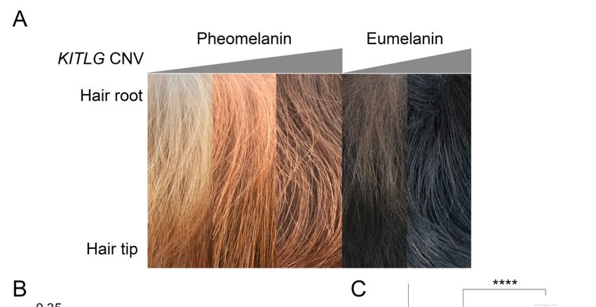
Figure Hair
Figure5. 5. sample
Hair quantification
sample from pheomelanin
quantification and eumelanin-based
from pheomelanin breeds. (A) Representative
and eumelanin-based breeds. (A)
hair samples from
Representative (leftsamples
hair to right)from
low, (left
medium, and low,
to right) high medium,
copy number
and NSDTR,
high copy and low and
number high copy
NSDTR, and
number eumelanin-based dogs (BC and FCR). (B) Linear regression analyses
low and high copy number eumelanin-based dogs (BC and FCR). (B) Linear regression analyses revealed a significant
association between anassociation
revealed a significant increased between
copy number and lower
an increased copymean color
number difference
and lower mean between root and
color difference
tip hair color in NSDTR (p = 0.00345, n = 17). (C) FCRs (n = 25) have a significantly
between root and tip hair color in NSDTR (p = 0.00345, n = 17). (C) FCRs (n = 25) have a significantly higher copy
number in comparison
higher copy number in BC (n = 19) in
to comparison toaBCddPCR assay
(n = 19) = 4.3 × assay
in a(pddPCR
−8
10 ). (p
The graph
= 4.3 × 10color represents
−8). The the
graph color
approximate hair color at the root of the hair shaft. **** = highly significant.
represents the approximate hair color at the root of the hair shaft. **** = highly significant.
4. Discussion
4. Discussion
Segregation of pheomelanin intensity in the NSDTR enabled identification of a region on CFA
Segregation of pheomelanin intensity in the NSDTR enabled identification of a region on CFA
15 associated with pigment intensity in dogs. Variant analysis identified a 6 kb CNV upstream of
15 associated with pigment intensity in dogs. Variant analysis identified a 6 kb CNV upstream of
KITLG that was highly associated with pheomelanin intensity within the NSDTR (p < 0.0001) and
KITLG that was highly associated with pheomelanin intensity within the NSDTR (p < 0.0001) and in
in other breeds. The copy number of this CNV is significantly associated with pheomelanin and
other breeds. The copy number of this CNV is significantly associated with pheomelanin and
eumelanin intensity in the Poodle and across breeds. All tested wolves had two copies of this element,
eumelanin intensity in the Poodle and across breeds. All tested wolves had two copies of this element,
indicating that chromosomes with higher copy numbers and high pigment intensity represent a derived
indicating that chromosomes with higher copy numbers and high pigment intensity represent a
state. Pigment intensity is due to the differing distribution of pigment along the hair shaft, with low
derived state. Pigment intensity is due to the differing distribution of pigment along the hair shaft,
copy number individuals having lighter hair at the root while more intensely pigmented individuals
with low copy number individuals having lighter hair at the root while more intensely pigmented
have no difference in pigment intensity from the root to hair tip. In the Poodle breed, the hair is
individuals have no difference in pigment intensity from the root to hair tip. In the Poodle breed, the
hair is continuously growing, such that the difference in colors associated with the different CNV
alleles are particularly striking.Genes 2020, 11, 75 9 of 13
continuously growing, such that the difference in colors associated with the different CNV alleles are
particularly striking.
KITLG and its receptor, KIT, have previously been shown to function in hematopoiesis,
melanogenesis, and gametogenesis [34]. Pleiotropic effects of genes responsible for coat color
variation are common in mammals [35]. In this case, this ligand receptor pair is essential for stem
cell populations to migrate and proliferate during development [36,37]. These stem cell populations
include the primordial germ cells, hematopoietic progenitor cells, and melanoblasts. In addition
to this early developmental function in melanoblast migration, KITLG is important for postnatal
cutaneous melanogenesis [38]. Specifically, KITLG expression is essential in hair follicle epithelial cells
for melanocyte terminal differentiation [39]. The essential role that KITLG plays in melanogenesis,
both developmentally and in the hair shaft, is consistent with its identification as a pigmentation
intensity locus in dogs.
Variants of KITLG and its receptor KIT cause pigmentation alternations in humans. Coding
sequence variants in KITLG have been shown to cause familial progressive hyperpigmentation
with or without hypopigmentation [40] as well as Waardenburg syndrome type 2 (with pigmentary
abnormalities) [41]. Additionally, coding variants within the receptor KIT have been associated with
classic autosomal dominant piebaldism characterized by congenital patches of skin with no melanocytes
present [42–45]. However, regulatory variants have been shown to affect pigment intensity. Sulem et al.
(2007) found that a variant upstream of KITLG was associated with hair color in a genome-wide
association scan in Icelanders and Dutch [46]. This variant likely lies in linkage disequilibrium, with
an extended haplotype upstream of KITLG showing a strong signature of selection in humans [46–48].
Guenther et al. (2014) found that this region drove expression exclusively in hair follicles using reporter
constructs in mice [49]. KITLG has diverse important developmental functions; however, the alternation
of just hair follicle expression may be a means to limit the effects to pigmentation phenotypes.
Regulatory variants of KITLG (or Kitl in the mouse) have been identified as causing pigment
differences in other animal species. The original Kitl mouse mutants were called steel due to their
uniformly diluted coat colors [50]. The steel panda allele is caused by an inversion that disrupts the
upstream regulatory region of Kitl, which also leads to diluted pigmentation [49,51]. In both goats and
mink, KITLG expression is lower in lighter-colored animals as compared to more darkly pigmented
animals [52,53]. In this work, expression levels of KITLG were not evaluated due to challenges in
obtaining skin samples from healthy pet dogs; however, based on the work done in other species,
the CNV likely alters expression levels of KITLG in the hair follicle.
While many dog breeds tested had a pigment intensity correlated to the KITLG CNV, some
breeds with pheomelanin variation did not. Notably, the Golden Retriever and Labrador Retriever
had variability at the CNV, but it did not correlate to their coat colors, indicating that there are still
additional pigment intensity loci or variants to be identified in dogs. It is not surprising that many loci
affect pigmentation since dogs are under such strong artificial selection for the variety in coat colors.
The canine CNV upstream of KITLG was previously associated with susceptibility for digital
squamous cell carcinoma (DSCC) in darkly colored dogs [54]. The analysis was performed in darkly
pigmented cases of DSCC and control dogs, and a high copy number of this CNV was identified as the
susceptibility locus; however, this effect was only found in eumelanistic cases with functional MC1R.
KITLG variants have been implicated in melanoma, testicular cancer, colorectal cancer, and general
cancer risk in humans [55–58]. The risk allele for DSCC in dogs had >four copies of the CNV
and homozygous animals were more likely to have DSCC [54]. Black-pigmented dogs are highly
overrepresented for DSCC, as 92% of samples were from black-coated dogs [59]. However, there are
breeds like the Flat-Coated Retrievers that have a high copy number, black hair coat, and are not
predisposed to this type of tumor [59]. It is possible that a high copy number at the KITLG CNV
is another risk factor for breeds that are already more susceptible to this particular tumor type.
Alternatively, some black dogs with high KITLG copy numbers may also have protective alleles.Genes 2020, 11, 75 10 of 13
Regulatory variants of human KITLG that affect hair color are located in a similar region to the CNV
identified in dogs. Conservation tracks within the CNV are 86.1% identical to the human sequence.
The human homologous region to the canine CNV is located 200 kb upstream of human KITLG.
The strongest selective signature for KITLG in humans lies 218 kb upstream of the coding sequence [60]
while the SNVs associated with light pigmentation are located 355 kb upstream of KITLG [47]. In dogs,
there is also evidence that KITLG is under selective pressure as it was one of the top 20 regions of
the genome to have a signature of selection within domestic dogs [61,62]. Interestingly, in canines,
the selection by humans is for darker pigmentation over the wild-type wolf colors. Vivid and rich
pigmentation within domestic dogs is one way that they may have been distinguished from wolves.
Wolves have just a single copy of this 6 kb region, indicating that amplification of the region occurred
within domestic dogs. It is interesting to speculate that this may have been one useful means of
distinguishing partner proto-dogs from their wild ancestors.
5. Conclusions
The KITLG CNV is associated with coat pigment intensity in the domestic dog. An increase in the
copy number is associated with a darker, more intense appearance to the coat and a more uniform
pigment intensity across the hair shaft in many breeds. Wolves do not show copy number variation at
this locus, and previous genome-wide screens have identified the KITLG CNV region as being under
selection in domestic dogs. KITLG plays an important role in melanogenesis, both in development
and in the adult hair follicle, and genetic variants upstream of KITLG in both mice and humans are
associated with coat and hair color variation. This research suggests that the KITLG CNV is a new
intensity locus in domestic dogs.
Supplementary Materials: The following are available online at http://www.mdpi.com/2073-4425/11/1/75/s1,
Table S1: Number of dogs genotyped for KITLG CNV copy number on ddPCR.
Author Contributions: Conceptualization, D.B.; Data curation, K.W., D.Y., R.G., A.K. and D.B.; Formal analysis,
K.W. and D.B.; Funding acquisition, D.B.; Investigation, K.W., V.A., D.Y., R.R., R.G., A.K. and D.B.; Project
administration, D.B.; Resources, V.A., R.R., R.G. and D.B.; Visualization, K.W.; Writing—original draft, K.W. and
D.B.; Writing—review and editing, K.W. and D.B. All authors have read and agreed to the published version of
the manuscript.
Funding: This research was funded in part by the Maxine Adler Endowed Chair Funds, the Maxine Adler
Graduate Fellowship, and the Center for Companion Animal Health, grant number CCAH 2019-8-F.
Acknowledgments: We would like to acknowledge and thank the owners who donated samples from their dogs
for this study.
Conflicts of Interest: The authors declare no conflict of interest.
References
1. Larson, G.; Karlsson, E.K.; Perri, A.; Webster, M.T.; Ho, S.Y.W.; Peters, J.; Stahl, P.W.; Piper, P.J.; Lingaas, F.;
Fredholm, M.; et al. Rethinking dog domestication by integrating genetics, archeology, and biogeography.
Proc. Natl. Acad. Sci. USA 2012, 109, 8878–8883. [CrossRef] [PubMed]
2. Shannon, L.M.; Boyko, R.H.; Castelhano, M.; Corey, E.; Hayward, J.J.; McLean, C.; White, M.E.; Abi Said, M.;
Anita, B.A.; Bondjengo, N.I.; et al. Genetic structure in village dogs reveals a Central Asian domestication
origin. Proc. Natl. Acad. Sci. USA 2015, 112, 13639–13644. [CrossRef] [PubMed]
3. Wang, G.-D.; Peng, M.-S.; Yang, H.-C.; Savolainen, P.; Zhang, Y.-P. Questioning the evidence for a Central
Asian domestication origin of dogs. Proc. Natl. Acad. Sci. USA 2016, 113, E2554–E2555. [CrossRef] [PubMed]
4. Botigue, L.R.; Song, S.; Scheu, A.; Gopalan, S.; Pendleton, A.L.; Oetjens, M.; Taravella, A.M.; Seregely, T.;
Zeeb-Lanz, A.; Arbogast, R.M.; et al. Ancient European dog genomes reveal continuity since the Early
Neolithic. Nat. Commun. 2017, 8, 16082. [CrossRef]
5. Anderson, T.M.; vonHoldt, B.M.; Candille, S.I.; Musiani, M.; Greco, C.; Stahler, D.R.; Smith, D.W.;
Padhukasahasram, B.; Randi, E.; Leonard, J.A.; et al. Molecular and evolutionary history of melanism in
North American gray wolves. Science (New York N.Y.) 2009, 323, 1339–1343. [CrossRef]Genes 2020, 11, 75 11 of 13
6. Ollivier, M.; Tresset, A.; Hitte, C.; Petit, C.; Hughes, S.; Gillet, B.; Duffraisse, M.; Pionnier-Capitan, M.;
Lagoutte, L.; Arbogast, R.-M.; et al. Evidence of Coat Color Variation Sheds New Light on Ancient Canids.
PLoS ONE 2013, 8, e75110. [CrossRef]
7. Kerns, J.A.; Newton, J.; Berryere, T.G.; Rubin, E.M.; Cheng, J.F.; Schmutz, S.M.; Barsh, G.S. Characterization
of the dog Agouti gene and a nonagoutimutation in German Shepherd Dogs. Mamm. Genome 2004, 15,
798–808. [CrossRef]
8. Newton, J.M.; Wilkie, A.L.; He, L.; Jordan, S.A.; Metallinos, D.L.; Holmes, N.G.; Jackson, I.J.; Barsh, G.S.
Melanocortin 1 receptor variation in the domestic dog. Mamm. Genome 2000, 11, 24–30. [CrossRef]
9. Candille, S.I.; Kaelin, C.B.; Cattanach, B.M.; Yu, B.; Thompson, D.A.; Nix, M.A.; Kerns, J.A.; Schmutz, S.M.;
Millhauser, G.L.; Barsh, G.S. A -defensin mutation causes black coat color in domestic dogs. Science (New
York N.Y.) 2007, 318, 1418–1423. [CrossRef]
10. Berryere, T.G.; Kerns, J.A.; Barsh, G.S.; Schmutz, S.M. Association of an Agouti allele with fawn or sable coat
color in domestic dogs. Mamm. Genome 2005, 16, 262–272. [CrossRef]
11. Robbins, L.S.; Nadeau, J.H.; Johnson, K.R.; Kelly, M.A.; Rosellirehfuss, L.; Baack, E.; Mountjoy, K.G.;
Cone, R.D. Pigmentation phenotypes of variant extension locus alleles result from point mutations that alter
msh receptor function. Cell 1993, 72, 827–834. [CrossRef]
12. Hedan, B.; Cadieu, E.; Botherel, N.; Dufaure de Citres, C.; Letko, A.; Rimbault, M.; Drogemuller, C.;
Jagannathan, V.; Derrien, T.; Schmutz, S.; et al. Identification of a Missense Variant in MFSD12 Involved in
Dilution of Phaeomelanin Leading to White or Cream Coat Color in Dogs. Genes 2019, 10, 386. [CrossRef]
[PubMed]
13. Philipp, U.; Hamann, H.; Mecklenburg, L.; Nishino, S.; Mignot, E.; Gunzel-Apel, A.R.; Schmutz, S.M.; Leeb, T.
Polymorphisms within the canine MLPH gene are associated with dilute coat color in dogs. BMC Genet.
2005, 6, 34. [CrossRef]
14. Schmutz, S.M.; Berryere, T.G.; Goldfinch, A.D. TYRP1 and MC1R genotypes and their effects on coat color in
dogs. Mamm. Genome 2002, 13, 380–387. [CrossRef] [PubMed]
15. Cargill, E.J.; Famula, T.R.; Schnabel, R.D.; Strain, G.M.; Murphy, K.E. The color of a Dalmatian’s spots:
Linkage evidence to support the TYRP1gene. BMC Vet. Res. 2005, 1, 1. [CrossRef] [PubMed]
16. Hrckova Turnova, E.; Majchrakova, Z.; Bielikova, M.; Soltys, K.; Turna, J.; Dudas, A. A novel mutation in
the TYRP1 gene associated with brown coat colour in the Australian Shepherd Dog Breed. Anim. Genet.
2017, 48, 626. [CrossRef]
17. Nicholas, T.J.; Cheng, Z.; Ventura, M.; Mealey, K.; Eichler, E.E.; Akey, J.M. The genomic architecture of
segmental duplications and associated copy number variants in dogs. Genome Res. 2009, 19, 491–499.
[CrossRef]
18. Lindblad-Toh, K.; Wade, C.M.; Mikkelsen, T.S.; Karlsson, E.K.; Jaffe, D.B.; Kamal, M.; Clamp, M.; Chang, J.L.;
Kulbokas, E.J., III; Zody, M.C.; et al. Genome sequence, comparative analysis and haplotype structure of the
domestic dog. Nature 2005, 438, 803–819. [CrossRef]
19. Kim, K.S.; Lee, S.E.; Jeong, H.W.; Ha, J.H. The complete nucleotide sequence of the domestic dog (Canis
familiaris) mitochondrial genome. Mol. Phylogenet. Evol. 1998, 10, 210–220. [CrossRef]
20. Chang, C.C.; Chow, C.C.; Tellier, L.C.A.M.; Vattikuti, S.; Purcell, S.M.; Lee, J.J. Second-generation PLINK:
Rising to the challenge of larger and richer datasets. GigaScience 2015, 4. [CrossRef]
21. Steiß, V.; Letschert, T.; Schäfer, H.; Pahl, R. PERMORY-MPI: A program for high-speed parallel permutation
testing in genome-wide association studies. Bioinformatics (Oxf. Engl.) 2012, 28, 1168–1169. [CrossRef]
[PubMed]
22. Turner, S.D. qqman: An R package for visualizing GWAS results using Q-Q and manhattan plots. bioRxiv 2014.
[CrossRef]
23. Karssen, L.C.; van Duijn, C.M.; Aulchenko, Y.S. The GenABEL Project for statistical genomics. F1000Res
2016, 5, 914. [CrossRef] [PubMed]
24. Kierczak, M.; Jablonska, J.; Forsberg, S.K.; Bianchi, M.; Tengvall, K.; Pettersson, M.; Scholz, V.; Meadows, J.R.;
Jern, P.; Carlborg, O.; et al. cgmisc: Enhanced genome-wide association analyses and visualization.
Bioinformatics (Oxf. Engl. ) 2015, 31, 3830–3831. [CrossRef] [PubMed]
25. Brown, E.A.; Dickinson, P.J.; Mansour, T.; Sturges, B.K.; Aguilar, M.; Young, A.E.; Korff, C.; Lind, J.;
Ettinger, C.L.; Varon, S.; et al. FGF4 retrogene on CFA12 is responsible for chondrodystrophy and
intervertebral disc disease in dogs. Proc. Natl. Acad. Sci. USA 2017, 114, 11476–11481. [CrossRef] [PubMed]Genes 2020, 11, 75 12 of 13
26. Hoeppner, M.P.; Lundquist, A.; Pirun, M.; Meadows, J.R.S.; Zamani, N.; Johnson, J.; Sundström, G.; Cook, A.;
FitzGerald, M.G.; Swofford, R.; et al. An Improved Canine Genome and a Comprehensive Catalogue of
Coding Genes and Non-Coding Transcripts. PLoS ONE 2014, 9, e91172. [CrossRef] [PubMed]
27. Kent, W.J.; Sugnet, C.W.; Furey, T.S.; Roskin, K.M.; Pringle, T.H.; Zahler, A.M.; Haussler, D. The human
genome browser at UCSC. Genome Res. 2002, 12, 996–1006. [CrossRef]
28. Kent, W.J.; Baertsch, R.; Hinrichs, A.; Miller, W.; Haussler, D. Evolution’s cauldron: Duplication, deletion,
and rearrangement in the mouse and human genomes. Proc. Natl. Acad. Sci. USA 2003, 100, 11484–11489.
[CrossRef]
29. Chiaromonte, F.; Yap, V.B.; Miller, W. Scoring pairwise genomic sequence alignments. Pac. Symp. Biocomput.
2002, 115–126. [CrossRef]
30. Blanchette, M.; Kent, W.J.; Riemer, C.; Elnitski, L.; Smit, A.F.; Roskin, K.M.; Baertsch, R.; Rosenbloom, K.;
Clawson, H.; Green, E.D.; et al. Aligning multiple genomic sequences with the threaded blockset aligner.
Genome Res. 2004, 14, 708–715. [CrossRef]
31. McLaren, W.; Gil, L.; Hunt, S.E.; Riat, H.S.; Ritchie, G.R.S.; Thormann, A.; Flicek, P.; Cunningham, F.
The Ensembl Variant Effect Predictor. Genome Biol. 2016, 17, 122. [CrossRef] [PubMed]
32. McLaren, W.; Pritchard, B.; Rios, D.; Chen, Y.; Flicek, P.; Cunningham, F. Deriving the consequences of
genomic variants with the Ensembl API and SNP Effect Predictor. Bioinformatics (Oxf. Engl.) 2010, 26,
2069–2070. [CrossRef] [PubMed]
33. Robinson, J.T.; Thorvaldsdóttir, H.; Winckler, W.; Guttman, M.; Lander, E.S.; Getz, G.; Mesirov, J.P. Integrative
genomics viewer. Nat. Biotechnol. 2011, 29, 24. [CrossRef] [PubMed]
34. Besmer, P. The kit ligand encoded at the murine Steel locus: A pleiotropic growth and differentiation factor.
Curr. Opin. Cell Biol. 1991, 3, 939–946. [CrossRef]
35. Reissmann, M.; Ludwig, A. Pleiotropic effects of coat colour-associated mutations in humans, mice and other
mammals. Semin. Cell Dev. Biol. 2013, 24, 576–586. [CrossRef]
36. Geissler, E.N.; Ryan, M.A.; Housman, D.E. The dominant-white spotting (W) locus of the mouse encodes the
c-kit proto-oncogene. Cell 1988, 55, 185–192. [CrossRef]
37. Brannan, C.I.; Lyman, S.D.; Williams, D.E.; Eisenman, J.; Anderson, D.M.; Cosman, D.; Bedell, M.A.;
Jenkins, N.A.; Copeland, N.G. Steel-Dickie mutation encodes a c-kit ligand lacking transmembrane and
cytoplasmic domains. Proc. Natl. Acad. Sci. USA 1991, 88, 4671–4674. [CrossRef]
38. Grichnik, J.M.; Burch, J.A.; Burchette, J.; Shea, C.R. The SCF/KIT pathway plays a critical role in the control of
normal human melanocyte homeostasis. J. Investig. Dermatol. 1998, 111, 233–238. [CrossRef]
39. Liao, C.P.; Booker, R.C.; Morrison, S.J.; Le, L.Q. Identification of hair shaft progenitors that create a niche for
hair pigmentation. Genes Dev. 2017, 31, 744–756. [CrossRef]
40. Wang, Z.Q.; Si, L.; Tang, Q.; Lin, D.; Fu, Z.; Zhang, J.; Cui, B.; Zhu, Y.; Kong, X.; Deng, M.; et al. Gain-of-function
mutation of KIT ligand on melanin synthesis causes familial progressive hyperpigmentation. Am. J.
Hum. Genet. 2009, 84, 672–677. [CrossRef]
41. Zazo Seco, C.; Serrao de Castro, L.; van Nierop, J.W.; Morin, M.; Jhangiani, S.; Verver, E.J.; Schraders, M.;
Maiwald, N.; Wesdorp, M.; Venselaar, H.; et al. Allelic Mutations of KITLG, Encoding KIT Ligand, Cause
Asymmetric and Unilateral Hearing Loss and Waardenburg Syndrome Type 2. Am. J. Hum. Genet. 2015, 97,
647–660. [CrossRef] [PubMed]
42. Giebel, L.B.; Spritz, R.A. Mutation of the KIT (mast/stem cell growth factor receptor) protooncogene in
human piebaldism. Proc. Natl. Acad. Sci. USA 1991, 88, 8696–8699. [CrossRef] [PubMed]
43. Yang, Y.J.; Zhao, R.; He, X.Y.; Li, L.P.; Wang, K.W.; Zhao, L.; Tu, M.; Tang, J.S.; Xie, Z.G.; Zhu, Y.M. A novel
splicing mutation of KIT results in piebaldism and auburn hair color in a Chinese family. BioMed Res. Int.
2013, 2013, 689756. [CrossRef] [PubMed]
44. Richards, K.A.; Fukai, K.; Oiso, N.; Paller, A.S. A novel KIT mutation results in piebaldism with progressive
depigmentation. J. Am. Acad. Dermatol. 2001, 44, 288–292. [CrossRef]
45. Murakami, T.; Fukai, K.; Oiso, N.; Hosomi, N.; Kato, A.; Garganta, C.; Barnicoat, A.; Poppelaars, F.;
Aquaron, R.; Paller, A.S.; et al. New KIT mutations in patients with piebaldism. J. Dermatol. Sci. 2004, 35,
29–33. [CrossRef]
46. Sulem, P.; Gudbjartsson, D.F.; Stacey, S.N.; Helgason, A.; Rafnar, T.; Magnusson, K.P.; Manolescu, A.;
Karason, A.; Palsson, A.; Thorleifsson, G.; et al. Genetic determinants of hair, eye and skin pigmentation in
Europeans. Nat. Genet. 2007, 39, 1443–1452. [CrossRef]Genes 2020, 11, 75 13 of 13
47. Miller, C.T.; Beleza, S.; Pollen, A.A.; Schluter, D.; Kittles, R.A.; Shriver, M.D.; Kingsley, D.M. cis-Regulatory
changes in Kit ligand expression and parallel evolution of pigmentation in sticklebacks and humans. Cell
2007, 131, 1179–1189. [CrossRef]
48. Yang, Z.; Shi, H.; Ma, P.; Zhao, S.; Kong, Q.; Bian, T.; Gong, C.; Zhao, Q.; Liu, Y.; Qi, X.; et al. Darwinian
positive selection on the pleiotropic effects of KITLG explain skin pigmentation and winter temperature
adaptation in Eurasians. Mol. Biol. Evol. 2018. [CrossRef]
49. Guenther, C.A.; Tasic, B.; Luo, L.; Bedell, M.A.; Kingsley, D.M. A molecular basis for classic blond hair color
in Europeans. Nat. Genet. 2014, 46, 748–752. [CrossRef]
50. Sarvella, P.A.; Russell, L.B. Steel, a new dominant gene in the house mouse. J. Hered. 1956, 47, 123–128.
[CrossRef]
51. Bedell, M.A.; Brannan, C.I.; Evans, E.P.; Copeland, N.G.; Jenkins, N.A.; Donovan, P.J. DNA rearrangements
located over 100 kb 5’ of the Steel (Sl)-coding region in Steel-panda and Steel-contrasted mice deregulate Sl
expression and cause female sterility by disrupting ovarian follicle development. Genes Dev. 1995, 9, 455–470.
[CrossRef]
52. Song, X.; Xu, C.; Liu, Z.; Yue, Z.; Liu, L.; Yang, T.; Cong, B.; Yang, F. Comparative Transcriptome Analysis of
Mink (Neovison vison) Skin Reveals the Key Genes Involved in the Melanogenesis of Black and White Coat
Colour. Sci. Rep. 2017, 7, 12461. [CrossRef] [PubMed]
53. Wu, S.; Li, J.; Ma, T.; Li, J.; Li, Y.; Jiang, H.; Zhang, Q. MiR-27a regulates WNT3A and KITLG expression in
Cashmere goats with different coat colors. Anim. Biotechnol. 2019. [CrossRef]
54. Karyadi, D.M.; Karlins, E.; Decker, B.; vonHoldt, B.M.; Carpintero-Ramirez, G.; Parker, H.G.; Wayne, R.K.;
Ostrander, E.A. A copy number variant at the KITLG locus likely confers risk for canine squamous cell
carcinoma of the digit. PLoS Genet. 2013, 9, e1003409. [CrossRef] [PubMed]
55. Duffy, D.L.; Zhu, G.; Li, X.; Sanna, M.; Iles, M.M.; Jacobs, L.C.; Evans, D.M.; Yazar, S.; Beesley, J.; Law, M.H.;
et al. Novel pleiotropic risk loci for melanoma and nevus density implicate multiple biological pathways.
Nat. Commun. 2018, 9, 4774. [CrossRef] [PubMed]
56. Landero-Huerta, D.A.; Vigueras-Villasenor, R.M.; Yokoyama-Rebollar, E.; Arechaga-Ocampo, E.;
Rojas-Castaneda, J.C.; Jimenez-Trejo, F.; Chavez-Saldana, M. Epigenetic and risk factors of testicular
germ cell tumors: A brief review. Front. Biosci. (Landmark. Ed.) 2017, 22, 1073–1098. [PubMed]
57. Yang, S.; Li, W.S.; Dong, F.; Sun, H.M.; Wu, B.; Tan, J.; Zou, W.J.; Zhou, D.S. KITLG is a novel target of miR-34c
that is associated with the inhibition of growth and invasion in colorectal cancer cells. J. Cell. Mol. Med.
2014, 18, 2092–2102. [CrossRef]
58. Zeron-Medina, J.; Wang, X.; Repapi, E.; Campbell, M.R.; Su, D.; Castro-Giner, F.; Davies, B.; Peterse, E.F.;
Sacilotto, N.; Walker, G.J.; et al. A polymorphic p53 response element in KIT ligand influences cancer risk
and has undergone natural selection. Cell 2013, 155, 410–422. [CrossRef]
59. Belluco, S.; Brisebard, E.; Watrelot, D.; Pillet, E.; Marchal, T.; Ponce, F. Digital squamous cell carcinoma
in dogs: Epidemiological, histological, and immunohistochemical study. Vet. Pathol. 2013, 50, 1078–1082.
[CrossRef]
60. Williamson, S.H.; Hubisz, M.J.; Clark, A.G.; Payseur, B.A.; Bustamante, C.D.; Nielsen, R. Localizing recent
adaptive evolution in the human genome. PLoS Genet. 2007, 3, e90. [CrossRef]
61. Boyko, A.R.; Quignon, P.; Li, L.; Schoenebeck, J.J.; Degenhardt, J.D.; Lohmueller, K.E.; Zhao, K.; Brisbin, A.;
Parker, H.G.; vonHoldt, B.M.; et al. A simple genetic architecture underlies morphological variation in dogs.
PLoS Biol. 2010, 8, e1000451. [CrossRef] [PubMed]
62. Vaysse, A.; Ratnakumar, A.; Derrien, T.; Axelsson, E.; Rosengren Pielberg, G.; Sigurdsson, S.; Fall, T.;
Seppala, E.H.; Hansen, M.S.; Lawley, C.T.; et al. Identification of genomic regions associated with phenotypic
variation between dog breeds using selection mapping. PLoS Genet. 2011, 7, e1002316. [CrossRef] [PubMed]
© 2020 by the authors. Licensee MDPI, Basel, Switzerland. This article is an open access
article distributed under the terms and conditions of the Creative Commons Attribution
(CC BY) license (http://creativecommons.org/licenses/by/4.0/).You can also read