ORAL AND RADIOGRAPHIC FEATURES OF MAJEWSKI OSTEODYSPLASTIC DWARFISM: A CASE REPORT - The South African Radiographer
←
→
Page content transcription
If your browser does not render page correctly, please read the page content below
THE SOUTH AFRICAN RADIOGRAPHER       Volume 60 Number 1 | MAY 2022

Peer Reviewed Case Report

ORAL AND RADIOGRAPHIC FEATURES OF MAJEWSKI
OSTEODYSPLASTIC DWARFISM: A CASE REPORT
Ntaoleng J. Mohapi1 NDip: Rad (D), BTech: Rad (D), Cert. Mammography | Mzubanzi Mabongo2 BDS, MChD, FCFS
Wits Oral Health Centre, School of Oral Health Sciences, University of Witwatersrand
1

School of Dentistry, Department of Maxillo-Facial and Oral Surgery, University of Pretoria
2

https://doi.org/10.54450/saradio.2022.60.1.641

    ABSTRACT
    Majewski osteodyplastic primordial dwarfism type II (MOPD II) is a rare, autosomal recessive disorder characterised by
    severe intrauterine and postnatal growth retardation. A case report with a distinct rare oral and radiographic feature
    with MOPD II is presented. It differs from the description of primordial dwarfism with characteristics of small head
    diameter at birth, which progresses to severe microcephaly and mild mental retardation.
    Keywords: Autosomal, panorex, microdontia, small roots

    LAY ABSTRACT
    A description of a rare genetic disorder in a teenage male is provided with unique radiographic features that differ from
    what were previously documented in literature.

CASE REPORT
A 16-year-old male patient presented with a complaint of
loose, painful teeth. He is the only child of healthy non-con-
sanguineous parents. His mother had a normal pregnancy,
but had a caesarean section at 32 weeks. He was on treat-
ment for hypothyroidism and has been taking growth hor-
mones from the age of four years.
On extra-oral examination, his stature was smaller than ex-
pected relative to his age. His complexion was darker than
normal. He had a prominent nose, nasal bridge and a high-
pitched voice. He seemed shy at first until he was comfort-
able after which he interacted well with the healthcare pro-
fessionals that examined him.
Intra-oral examination of the soft tissue revealed no ab-
normality. His teeth were however smaller than one would
expect for his age. Some teeth were mobile and teeth 55
and 65 were retained. Generalised caries was noted. He was
referred by the department of human genetics and under-
went testing which confirmed that he has Majewski osteod-
ysplastic primordial dwarfism (MOPD) type II on molecular
diagnosis.
Radiographic examinations were performed to assist with
further management of his disorder. A lateral cephalomet-
ric projection of the face (Figure 1) showed proportional
growth of both jaws with a class 1 occlusion. A widening of
the sella turcica was noted. A calcified deposit was identi-
fied in the region of the sella turcica. The cervical vertebral
                                                                  Figure 1. Lateral cephalometric projection of the skull and face
bodies maturation stage of C4, C5, C6 and C7 were not typ-        shows. Class 1 occlusion (green arrow). Widened sella turcica and
ical for his age. A panorex radiograph of the maxilla and         calcified deposit (red box). Concave cervical bodies (blue arrows).

    32   www.sorsa.org.za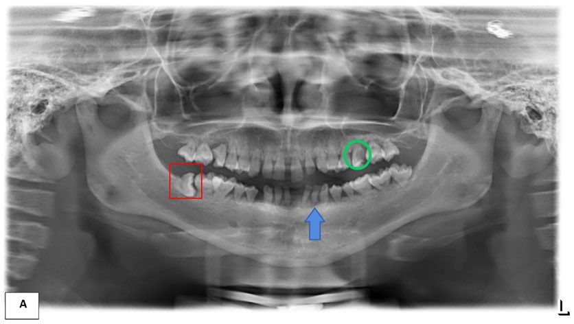
MAY 2022 | Volume 60 Number 1 THE SOUTH AFRICAN RADIOGRAPHER
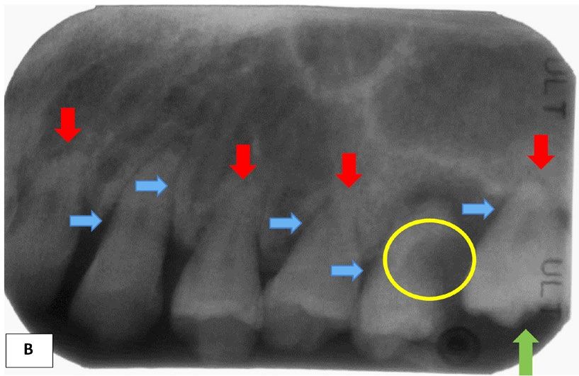
Figure 2. Figure A demonstrates a smaller maxilla and mandible compared to that of a normal 16-year-old in
Figure B. Microdontia teeth with short roots of permanent dentition were noted with an example pointed out (blue
arrow). There is horizontal impaction of the right lower third wisdom teeth (red box). The upper right molar demon-
strates a carious lesion with periapical radiolucency (green circle).
mandible was performed (Figure 2a). The maxilla and man- epiphysis, are atypical. A full-mouth series was performed.
dible were smaller than expected for his age in comparison The periapical radiographs are shown in Figures 4a and b.
to a panorex of a normal 16-year-old. Permanent dentition A combination of microdontia, shortness of roots and rela-
showed microdontia with small roots (Figure 2a). There was tively thick lamina dura were demonstrated in some areas.
horizontal impaction of the right lower third wisdom tooth.
The upper first molars had a huge carious lesion with peri- DISCUSSION
apical radiolucency.
Majewski osteodyplastic primordial dwarfism type II (MOPD
A dorsi-palmer projection of the hand (Figure 3) demon- II) is a rare, autosomal recessive disorder caused by muta-
strated ossification of a sesamoid bone in the thumb region, tions in the pericentrin gene in some individuals.[1] It be-
which is prevalent in post-puberty. The radiolucencies in the longs to a heterogeneous group of disorders of primordial
diaphysis-epiphysis of metacarpals are atypical. The fusions dwarfism characterised by severe intrauterine and post-
of the bases of the 2nd to 5th phalanges and distal ulnar natal growth retardation.[2] Features of this syndrome in-
epiphysis, as well as the capping noted at the distal radial clude severe proportionate intrauterine growth retardation
www.sorsa.org.za 33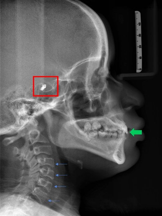
THE SOUTH AFRICAN RADIOGRAPHER                Volume 60 Number 1 | MAY 2022

                                                                        (IUGR), poor postnatal growth with adult stature of approx-
                                                                        imately 100 cm, microcephaly which becomes progressively
                                                                        disproportionate, skeletal dysplasia, and characteristic fa-
                                                                        cial features, such as a prominent nose and small jaw.[1,3]
                                                                        The patient in this case report presented with short stature
                                                                        and small body frame. Galasso described this characteristic
                                                                        as small frame.[2] Short stature is believed to be caused by
                                                                        multiple molecular defects, including intracellular signaling,
                                                                        extracellular matrix, and paracrine and endocrine regula-
                                                                        tion.[4] The approach in the past to short stature primarily
                                                                        focused on clinical manifestations: for example, primordial
                                                                        dwarfism, syndromic short stature or skeletal dysplasia to
                                                                        categorise them by similar clinical features. Currently, the
                                                                        combination of the clinical approach and improved genetic
                                                                        diagnosis are advancing our understanding of congenital
                                                                        growth disorders and have helped further expand the un-
                                                                        derstanding of the clinical variability and genetic heteroge-
                                                                        neity of short stature syndrome.[4]
                                                                        Cranial presentation of poor postnatal growth has been de-
                                                                        scribed as microcephaly[1] and micrognathia.[2] The cranium
                                                                        of the patient in this case report had proportional micro-
                                                                        cephaly. It is defined as a head circumference more than
                                                                        two standard deviations (SD) below the mean for gender
                                                                        and age, and it affects 2% of the population. The patient
                                                                        in this case report did not have micrognathia: his mandible
                                                                        demonstrated proportional growth (Figure 1).[5] Widening
                                                                        of his sella turcica was noted. This feature is described by
                                                                        Terlemez et al.[6]
Figure 3. Ossification of sesamoid bone of the thumb (blue circle).
                                                                        Literature highlights that a patient with primordial dwarfism
Fusion of the 2nd to 5th phalange bases (red octagons). Capping of
the distal radial epiphysis (green arrow). Fusion of the distal ulnar   has a small head diameter at birth, which progresses to se-
epiphysis (purple box).                                                 vere microcephaly and mild mental retardation.[4] However,

            Figure 4a. A full mouth series shows a combination of microdontia and shortness of roots.

 34      www.sorsa.org.za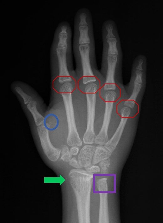
MAY 2022 | Volume 60 Number 1          THE SOUTH AFRICAN RADIOGRAPHER

          Figure 4b. Periapical radiography of the upper left maxilla i.e. premolar and molar, indicating thick lamina (blue
          arrows), short roots (red arrows), microdontia (i.e., small tooth) (green arrow) and a carious lesion with periapical radio-
          lucency (yellow circle).

the intellectual capacity of the patient in this case report was          panorex radiographs that very low birthweight in children
above average, based on his performance at school. Most                   tends to result in a delay in dental maturation compared to
individuals affected by MOPD II are outgoing, talkative and               normal birthweight children. Dental development can also
have good social skills.[7] This was the case for this patient            be advanced in a patient with systemic syndromes; the ad-
and it could also be attributed to relatively good postnatal              ministration of growth hormones had no effect.[10] In clinical
cranial growth. Distinctive facial features with mild, down               practice when determination of skeletal age is done, skele-
slanting palpebral fissures, prominent nose, hypoplasia of                tal development is an important maturity indicator and is
the alae nasi, microdontia, micrognathia, and low-set, dys-               helpful in the diagnosis of disorders of growth and develop-
plastic ears have been associated with MOPD II.[2] According              ment. Skeletal maturation in the vertebral column and hand
to Lui et al.[8] severe micrognathia and mandibular hypoplas-             and wrist regions are reliable anatomical sites for maturity
ia can be diagnosed prenatally by ultrasound. Low-set ears                determination.[10] There are several indications of atypical
are usually associated with micrognathia involving the man-               and premature maturity in the patient discussed in our
dible.[2] Since there was a proportional postnatal growth of              case. The dorsi-palmer hand projection demonstrated pre-
the cranium, the appearance of low-set ear was not noted                  mature ossification of the sesamoid bone in the thumb re-
in the patient in our case.[6] The combination of hypoplastic             gion, fusion of the distal ulnar epiphysis and capping of the
alveolar processes, microdontia, short-rooted incisors, and               distal radial epiphysis (Figure 3). The panorex radiograph
rootless molar teeth has been described in MOPD.[4,6] Ter-                showed a fully erupted wisdom tooth, which is another indi-
lemez et al.[6] reported a female patient with MOPD II with               cation of early maturity (Figure 2). Considering the maturity
microdontia in the primary dentition.[6] According to Kanta-              of the cervical vertebrae (Figure 1) the inferior borders of
putra et al.,[9] dental anomalies can be striking and present             cervical vertebrae 4, 5 and 6 (C4, C5 & C6) were concave
as severe microdontia, opalescent and rootless molars, and                and C7 was nearly square-shaped. These features indicate
an unerupted tooth.[9] The alveolar process can also be se-               that 5% to 10% adolescent growth can be expected in him
verely hypoplastic and mandibular premolars are unusually                 relative to his age.[11]
small and malformed, comprising many cusps.[9] The patient
in this case report had microdontia, short roots and mobile               CONCLUSION
teeth in his permanent dentition. This suggests that both
                                                                          The patient in this case presented with most of the symp-
primary and secondary dentition were affected by micro-
                                                                          toms associated with MOPD II. However, the symptoms rel-
dontia. Since both dentitions were affected and this implies
                                                                          ative to the clinical presentation suggest that his symptoms
that the genetic defect occurs before odontogenesis; before
                                                                          are mild. Our case report contributes to the literature as
the four weeks of embryonic life.[7]
                                                                          it showed severe microdontia, normal intellectual capacity
Farman[10] found when determining dental maturity from                    and premature eruption of a wisdom tooth.

                                                                                                                     www.sorsa.org.za    35
THE SOUTH AFRICAN RADIOGRAPHER Volume 60 Number 1 | MAY 2022
CONFLICT OF INTEREST America. 2017; 46(2):259-281. [cited 2022 January 19]. Available
from: http://dx.doi.org/10.1016/j.ecl.2017.01.001.
None to declare.
5. Hanzlik E, Gigante J. Microcephaly. Children. 2017; 4(6):1-
8. [cited 2022 February 28]. Available from: https://dx.doi.
ETHICS APPROVAL org/10.3390%2Fchildren4060047.
Informed consent to use records and to publish was ob- 6. Terlemez A, Altunsoy M, Çelebi H. Majewski osteodyplastic
primordial dwarfism type II: clinical findings and dental manage-
tained from the patient. Ethics approval was granted by
ment of a child patient. Journal of Istanbul University Faculty of
Human Research Ethics Committee of Witwatersrand Dentistry. 2015; 49(1):41-46. [cited 2022 January 19]. Available
[M1910101]. from: http://dx.doi.org/10.17096/jiufd.73283.
7. Alqerban A, Jacobs R, Fieuws S, Willems G. Comparison of two
CONTRIBUTIONS OF AUTHORS cone beam computed tomographic systems versus panoram-
ic imaging for localization of impacted maxillary canines and
NJM and MM both contributed to the conceptualisation and detection of root resorption. European Journal of Orthodontics.
writing of this paper. 2011; 33(1):93-102. [cited 2022 January 16]. Available from:
https://doi.org/10.1093/ejo/cjq034.
8. Liu X, Sun W, Wang J, Chu G, He R, Zhang B, Zhao Y. Prenatal
diagnosis of auriculocondylar syndrome with a novel missense
REFERENCES variant of GNAI3: a case report. BMC Pregnancy and Childbirth.
1. Bober MB, Khan N, Kaplan J, Lewis K, Feinstein JA, Scott CI 2021; 21(780):1-7. [cited 2022 February 24]. Available from:
Jr, Steinberg GK. Majewski osteodysplastic primordial dwarf- https://doi.org/10.1186/s12884-021-04238-x.
ism type II (MOPD II): expanding the vascular phenotype. 9. Kantaputra PN, Tanpaiboon P, Unachak K, Praphanphoj V. Mi-
American Journal of Medical Genetics. 2010; 152A(4):960- crocephalic osteodysplastic primordial dwarfism with severe mi-
965. [cited 2022 January 16]. Available from: https://doi. crodontia and skin anomalies: confirmation of a new syndrome.
org/10.1002/ajmg.a.33252. American Journal of Medical Genetics. 2004; 130A(2):181-190.
2. Galasso C, Lo-Castro A, Lalli C, Cerminara C, Curatolo P. [cited 2022 February 22]. Available from: https://onlinelibrary.
Neurologic aspects of microcephalic osteodysplastic primor- wiley.com/doi/pdf/10.1002/ajmg.a.30079.
dial dwarfism type II. Paediatric Neurology. 2008; 38(6):435- 10. Farman AG. Assessing growth and development with panoramic
438. [cited 2022 January 29]. Available from: https://doi. radiographs and cephalometric attachments: a critical tool for
org/10.1016/j.pediatrneurol.2008.02.011. dental diagnosis and treatment planning. Panoramic Imaging
3. Majewski F, Goecke T, Opitz JM. Studies of microcephalic News. 2004; 4(4):1-11. [cited 2022 February 22]. Available from:
osteodysplastic primordial dwarfism I: approach to a deline- http://www.pancorp.com/images/files/upload/1289257666Pan_
ation of the seckel syndrome. American Journal of Medical Imaging_News_V4_I4_(10-04).pdf.
Genetics. 1982; 12(1):7-21. [cited 2022 January 16]. Availa- 11. Hassel B, Farman AG. Skeletal maturation evaluation using cervi-
ble from: https://onlinelibrary.wiley.com/doi/epdf/10.1002/ cal vertebrae. American Journal of Orthodontics and Dentofacial
ajmg.1320120103. Orthopaedics, 1995;107(1):58-66. [cited 2022 February 28].
4. Jee YH, Andrade AC, Baron J, Nilsson O. Genetics of short Available from: https://doi.org/10.1016/S0889-5406(95)70157-5.
stature. Endocrinology and Metabolism Clinics of North
36 www.sorsa.org.zaYou can also read