OPTIMIZED BEAM SHAPING ASSEMBLY FOR A 2.1 MEV PROTON ACCELERATOR BASED NEUTRON SOURCE FOR BORON NEUTRON CAPTURE THERAPY
←
→
Page content transcription
If your browser does not render page correctly, please read the page content below
www.nature.com/scientificreports
OPEN Optimized beam shaping
assembly for a 2.1‑MeV
proton‑accelerator‑based neutron
source for boron neutron capture
therapy
Pablo Torres‑Sánchez, Ignacio Porras*, Nataliya Ramos‑Chernenko,
Fernando Arias de Saavedra & Javier Praena
Boron Neutron Capture Therapy (BNCT) is facing a new era where different projects based on
accelerators instead of reactors are under development. The new facilities can be placed at hospitals
and will increase the number of clinical trials. The therapeutic effect of BNCT can be improved if a
optimized epithermal neutron spectrum is obtained, for which the beam shape assembly is a key
ingredient. In this paper we propose an optimal beam shaping assembly suited for an affordable
low energy accelerator. The beam obtained with the device proposed accomplishes all the IAEA
recommendations for proton energies between 2.0 and 2.1 MeV. In addition, there is an overall
improvement of the figures of merit with respect to BNCT facilities and previous proposals of new
accelerator-based facilities.
Boron Neutron Capture Therapy (BNCT) is an experimental form of radiotherapy which is selective at the cellular
level. In recent years BNCT is back in limelight due to the most recent successful clinical trials for very malignant
diseases like brain tumors and recurrent head and neck c ancers1. In addition, with the advances in accelerator
technologies, now it is possible to obtain neutron beams suitable for BNCT in facilities that can be built inside
hospitals, and different projects had been started for this purpose throughout the w orld2,3.
The first accelerator-based BNCT facility suitable for clinical trials (C-BENS) has been developed in the Kyoto
Research Reactor Institute with a 30-MeV cyclotron of Sumitomo Heavy Industries, and clinical trials have been
recently started4. This pioneering facility may boost the development of new centers. Most of the other projects
make use of lower energy proton beams on targets of 7 Li or 9 Be, in the aim of producing lower energy neutrons,
thus requiring less moderation in order to reach the optimal energies for BNCT, which are known as epithermal
energies (up to 10 keV).
The International Atomic Energy Agency (IAEA), in the Technical Document of 19905 establishes some
recommendations for the beam quality for BNCT treatments. These recommendations limit the dose due to fast
neutrons and gamma contamination, as well as the thermal neutron flux below certain upper bounds, provide a
condition on the divergence of the beam and a lower bound for the epithermal neutron flux. Fulfilling all these
recommendations from an accelerator-based neutron source is a challenging task. The moderation of the high-
energy neutrons produced at the target to the epithermal energies for reducing the fast neutron dose increases
the thermal neutron ratio and decreases the total neutron flux, so the selection of materials and their dimensions
is critical for achieving all the recommendations.
In this work, within the frame of the NeMeSis Project6, we present a design of a beam shaping assembly (BSA)
that accomplishes all these recommendations and which produces a very well defined beam for BNCT with low
proton energies (between 2.0 and 2.1 MeV) on a Li target. This is a very interesting possibility as the accelerator
required can be substantially cheaper than those required for other options at higher energies. In addition, the
in-phantom figures of merit, which are essential for the suitability as a therapeutic beam for deep seated tumors,
obtained from our design compare very well with other proposals published in the literature.
Departamento de Física Atómica, Molecular y Nuclear, Facultad de Ciencias, Universidad de Granada, 18071 Granada,
Spain. *email: porras@ugr.es
Scientific Reports | (2021) 11:7576 | https://doi.org/10.1038/s41598-021-87305-9 1
Vol.:(0123456789)
www.nature.com/scientificreports/
Figure 1. Optimization of the moderator thickness. The optimal value is found by keeping the epithermal flux
(blue line) above the IAEA minimum recommendation (black line), while the fast dose per epithermal flux (red
line) is below the IAEA maximum value recommended (same black line). This happens for a thickness of 21.8
cm.
This paper is structured as follows. “Results and discussion” section shows the results from the optimization
of the BSA and discusses the beam quality against other facilities of its kind. The conclusions derived from this
research are highlighted in “Conclusions” section. “Methods” section describes the choice of target and the
simulations carried out for the optimization of the BSA, together with the set beam parameters and of figures of
merit used in the beam quality assessment.
Results and discussion
The design of the geometry and the material choice was established following a optimization to meet the IAEA
recommendations for BNCT5. In addition, it was paramount that the neutron spectrum peaked around 2–9 keV,
matching the most adequate neutron energies for reaching deep-seated tumors.
BSA design. The use of low energy proton beams profit from a less extended high energy tail in the neu-
tron spectrum. In these conditions, 7 Li is a best suited target option, provided its less energetic reaction yield
compared to 9 Be or spallation targets as Ta, W or Pb. Indeed, the pursuit to stick to low energy proton beams
imposes a further dim of neutron production. This aspect has to be compensated with a higher intensity particle
accelerator. In the following, we assume a 30 mA proton beam onto a metallic Li target. The proton energy range
from the threshold to 2.3 MeV was studied. This choice limits the use of the broad 2.25 MeV resonance but also
avoids the population of the first excited state of 7 Be at 431 keV, whose threshold is found at 2.373 MeV, even
considering that the neutron yield from this channel is less than 10% below 5 MeV7. The aim of this work is to
explore for the first time the possibility of producing a neutron beam suitable for BNCT at the lowest energy
possible, with the aim of strongly restricting the high energy tail due to kinematics. For example, for protons
at 2.1 MeV onto 7 Li , no neutrons are produced above 350 keV, which can reduce the effects on normal tissues
due to the fast neutrons. Working at that low energies also avoids a region (400 keV to 1 MeV) where, for the
inelastic scattering of 19 F (a main component of the moderator), there are discrepancies between experimental
and evaluated data for the cross sections. Also the inelastic scattering with the Mg isotopes is avoided, as the first
excited state of their isotopes occurs at 585 keV.
In the reaction between the protons below 2.3 MeV and the 7 Li , neutrons of up to few hundreds of keV are
generated in the forward direction. Therefore, a neutron moderator with low absorption at these energies is
fundamental for achieving an intense enough neutron beam for treatments. MgF2 has been considered exten-
sively for the moderation of neutrons from accelerator-based sources8. The reason is the low capture cross-
sections and complementary location of resonances of both fluorine and the magnesium isotopes. In our case,
special emphasis comes near 100 keV, around the mean energy of neutrons produced at the 7 Li source at 2.1
MeV. In that region, a pair of resonances partially overlap, giving a broad range for continuous neutron energy
reduction. The MgF2 also surrounds the 7 Li source in the backward direction in a cylindrical shape in order
to recover medium-energy back-propagated neutrons. This structure is fully covered by a Pb shield that works
as a neutron reflector. In the forward direction, the neutron moderation is completed by means of an Al layer,
that in addition effectively filters the remaining neutrons by its resonances at 35 and 87.5 keV. Then, a LiF sheet
is placed to reduce the over-moderated thermal neutrons so as to increase the beam penetrability. Finally, a Bi
layer efficiently attenuates the gamma radiation generated in the previous steps, without a manifest effect in the
neutron flux unlike Pb, profiting from its lower elastic cross-section. For making the design more realistic, a 1
mm layer of water for cooling the lithium t arget9 and an air gap of 1 mm around the accelerator tube have been
considered for the simulations.
The dimensions of the elements in the BSA have been chosen optimizing the figures of merit of the in-air
IAEA recommendations, by means of a compromise between making the epithermal flux as high as possible
while keeping the fast dose below the recommended value. This is illustrated as an example in Figs. 1 and 2 for
the most critical dimensions (moderator length and radius).
Once the neutron spectrum achieved is adequate, it is key to shape and collimate the beam to achieve a satis-
factory divergence and fairy low neutron and gamma contamination off-beam. The beam shaping and focusing
Scientific Reports | (2021) 11:7576 | https://doi.org/10.1038/s41598-021-87305-9 2
Vol:.(1234567890)www.nature.com/scientificreports/
Figure 2. Optimization of the moderator radius for the optimal thickness. The optimal value is found by
keeping the epithermal flux (blue line) as high as possible while the fast dose per epithermal flux (red line) is
minimized, which happens at a radius of 25 cm.
is managed by means of a tapered geometry towards the beam aperture. Pb is used to this aim, benefiting from its
reflector behavior and low moderating effect. For the off-beam contamination suppression, lithiated polyethylene
blocks, combined with LiF and Pb layers accomplish this goal.
The optimization of the BSA for the choice of 2.1 MeV proton energy, brought in a selection of dimensions
that meet the IAEA recommendations and serve adequately for a BNCT treatment. The direct along-the-axis
thickness of the MgF2 core moderator (W2) is 21.8 cm. The total diameter of the moderator (ø1) is 50 cm. The
successive Al, LiF and Bi filters account for 1.0 cm, 0.2 cm and 1.0 cm each, respectively. The surrounding Pb
reflector has an outer diameter (ø2) of 120 cm. The distance from the Bi layer and the neutron beam aperture
(W1) is 19.8 cm, and the beam aperture diameter (A) is 14 cm. All materials have been considered as pure,
with a density of 3.148 g/cm3 for MgF2, 2.365 g/cm3 for LiF and 1.06 g/cm3 for lithiated polyethylene. A 100%
enrichment was assumed for 6 LiF, and a 7.56% mass content of 6 Li in lithiated polyethylene. Figure 3 (upper
panel) illustrates the BSA final configuration.
Concerning the proton energy for the neutron production onto 7 Li , the optimal energy was found to be 2.1
MeV. Table 1 shows the in-air beam parameters that are achieved with our configuration of the BSA at different
energies in the range 1.95 to 2.3 MeV. In addition, Fig. 4 explicitly shows the dependence of epithermal flux and
fast dose with proton energy, pointing that 2.1 MeV corresponds to the energy of the best compromise among
both criteria.
In‑air IAEA beam parameters. In Table 1 we compare the in free-air parameters with the IAEA
recommendations5. The neutron flux, divergence, and contamination from gammas, thermal and fast neutrons
were evaluated at the beam aperture in free air conditions. It is clear from the results that all IAEA recommenda-
tions are fulfilled at 2.1 MeV, while only the epithermal flux is below 109 n/cm2 /s at 2.0 MeV. Despite this, it is
above the minimum recommendation of 5 · 108 n/cm2 /s. This indicates that energies below 2.1 MeV down to
2.0 MeV are also valid for use with this BSA. With this geometry, more energetic proton beams would result in
a higher energy neutron yield, specially considering the close reaction resonance at 2.25 MeV. That would no
longer compensate the fast neutron dose contribution. On the other hand, proton beams below 2.0 MeV could
not sustain a neutron flux high enough to operate the beam for treatments.
Figure 3 illustrates the neutron (middle panel) and gamma (lower panel) radiation flux distribution throughout
the BSA, in free air conditions.
Apart from the epithermal flux, the thermal and current to flux ratios are well managed, found adequately
within the recommendations. The gamma radiation dose contamination is well attained below the safe margins,
even including the contributions from the 7 Li source and the Al activation. The fast neutron dose contamina-
tion is properly settled just at the limit of the IAEA when defining the epithermal range from 0.5 eV to 10 keV.
This gives rise to a more detailed remark on this point. This recommendation for the fast neutron component is
currently under revision, as there has been suggested that neutrons slightly above 10 keV can also be useful for
therapy21. Table 2 shows the performance of the BSA both at 2.0 and 2.1 MeV, each considering an epithermal
upper limit at 10 and 20 keV. If one considers the epithermal range up to 20 keV, then the fast neutron dose
reduces down to less than a half. Only 51% of the fast neutron flux corresponds to neutrons above 20 keV. Neu-
trons above 40 keV are only 32% of the total fast neutron flux. This is due to the fact that the starting neutron
spectrum at the source has a much shorter energy tail than in usual beams. This underlines the advantageous
properties of the beam in terms of contamination. For completeness, the results at a proton energy of 2.0 MeV
turn out to be slightly more adequate in terms of beam contamination, with the drawback of a less intense neu-
tron flux. Considering all these, in the following, all the remaining results will be given for the BSA solution at
a proton energy of 2.1 MeV.
When comparing the results from this BSA and previous designs for BNCT, one has to mention the features
of previous and current reactor-based BNCT facilities, such as FiR-1 in F inland10,11, KURRI in J apan12 or cur-
rently THOR in T aiwan13. KURRI in epithermal mode and THOR share high fast neutron contamination (6.1
and 2.8, in units of 10−13 Gy cm2 per epithermal neutron), and also a high thermal neutron ratio (0.12) in the case
of THOR. However, they had convenient beam intensity (9.1 · 108 and 1.7 · 109 n/cm2 s at KURRI and THOR,
respectively) and divergence (0.81 in the case of THOR). Concerning accelerator-based facilities (AB-BNCT),
Scientific Reports | (2021) 11:7576 | https://doi.org/10.1038/s41598-021-87305-9 3
Vol.:(0123456789)www.nature.com/scientificreports/
Figure 3. Upper panel: Layout of the BSA design, including general sizes and material specification. Middle and
lower panel: Neutron and gamma radiation flux distribution throughout the BSA. The color key illustrates the
neutron and photon intensity relative to the total neutrons generated in the target.
Scientific Reports | (2021) 11:7576 | https://doi.org/10.1038/s41598-021-87305-9 4
Vol:.(1234567890)www.nature.com/scientificreports/
Proton energy (MeV)
IAEA recommendation 1.95 2.0 2.1 2.2 2.3
φepi (n/cm2 s) > 5 · 108 3.124 · 108 5.459 · 108 1.019 · 109 1.681 · 109 2.706 · 109
φth /φepi < 0.05 0.0385 0.0378 0.0372 0.0364 0.0349
Jn /φn > 0.7 0.7138 0.7128 0.7120 0.7109 0.7099
Dfast /φepi (Gy cm2 ) < 2 · 10−13 1.42 · 10−13 1.82 · 10−13 1.97 · 10−13 2.18 · 10−13 2.81 · 10−13
Dγ /φepi (Gy cm2 ) < 2 · 10−13 1.05 · 10−13 1.01 · 10−13 0.99 · 10−13 0.95 · 10−13 0.96 · 10−13
Table 1. Free-air figures of merit for the conformed beam, for different incident proton energies from 1.95 to
2.3 MeV, compared to with the IAEA recommendations. The figures for Ep = 2.1 MeV and Ep = 2.0 MeV are
the only ones that satisfy all criteria at once.
Figure 4. Detail of the Epithermal Flux and Fast Dose as a function of the proton energy incident on the
7 Li target, compared with the IAEA recommendation figures. The value of 2.1 MeV is marked as both the
epithermal flux and the fast dose are attained above and below their criterion, respectively.
Our proposal (2.0 MeV) Our proposal (2.1 MeV)
Epith. limits Epith. limits Epith. limits Epith. limits
IAEA recommendation 0.5 eV–10 keV 0.5 eV–20 keV 0.5 eV–10 keV 0.5 eV–20 keV
φepi (n/cm2 s) > 5 · 108 5.459 · 108 5.783 · 108 1.019 · 109 1.081 · 109
φth /φepi < 0.05 0.0378 0.0357 0.0372 0.0351
Jn /φn > 0.7 0.7128 0.7130 0.7120 0.7119
Dfast /φepi (Gy cm2 ) < 2 · 10−13 1.82 · 10−13 0.94 · 10−13 1.97 · 10−13 1.09 · 10−13
Dγ /φepi (Gy cm2 ) < 2 · 10−13 1.01 · 10−13 0.92 · 10−13 0.99 · 10−13 0.97 · 10−13
Table 2. In-air beam parameters compared to the IAEA recommendations, for proton accelerator energies of
2.0 and 2.1 MeV. The results considering the epithermal upper limit at 10 and 20 keV are included.
peration14. It has a high intensity (1.2 · 109 n/cm2 s) and low thermal neu-
C-BENS is the first facility already in o
tron contamination (0.04), yet the fast neutron dose exceeds the maximum (5.8 · 10−13 Gy cm2) even considering
an extended epithermal range to 40 keV14. Other projects and designs achieve desirable beam intensities and low
thermal contamination, though most of them suffer from high fast neutron c ontamination15–17, while others do
not achieve a tight collimation and low d ivergence18,19. A design in Italy achieves high epithermal flux, but doses
from fast neutrons and gamma radiation remain higher than recommendations20.
Neutron spectrum and lateral out‑of‑field flux profiles. In the following, let us overview the neutron
spectrum obtained at the BSA aperture. Figure 5 shows the spectrum from our BSA design compared to previ-
ous reactor-based infrastructures as FiR-1, and C-BENS. The use of 7 Li and a proton energy of 2.1 MeV gener-
ates neutrons with a maximum energy of 350 keV. The further moderation makes the neutron intensity vanish
for energies above 200 keV. This is capital in the aim of reducing the fast neutron dose and its preeminence in
superficial tissue. In the same way, the low energy tail of the spectrum is well suppressed, as in the other facilities.
Moreover, the neutron spectrum is satisfactorily peaked at around 2–3 keV, which lies within the most suitable
neutron energy range for deep-seated tumor treatments in BNCT, considered as the 2–9 keV r ange21.
In order to go more into detail in the neutron spectrum shaping process, it is worth to mention the optimal
sizing of the moderator. Figure 6 shows the progress of the moderating and beam shaping. An intermediate
Scientific Reports | (2021) 11:7576 | https://doi.org/10.1038/s41598-021-87305-9 5
Vol.:(0123456789)www.nature.com/scientificreports/
Figure 5. Spectrum at the BSA aperture (UGR) compared to others (FiR-1 and C-BENS) in log-lin scale (upper
panel) and log-log scale (lower panel). The epithermal range from 0.5 eV to 10 keV is marked in gold and the
extension from 10 to 20 keV in pale orange. The 2 keV energy is marked with a vertical dotted line.
Figure 6. Neutron spectra at different positions of the BSA from the Lithium target to the aperture. The
spectrum shaping effects from each material are exhibited. Also, an intermediate scoring plane in MgF2 is
shown. The epithermal range from 0.5 eV to 10 keV is marked in gold and the extension from 10 to 20 keV in
pale orange.
moderating step is registered to emphasize the need of an adequate moderating thickness. MgF2 is a satisfac-
tory material in the role of moderation, productively shifting the neutron energy to the epithermal range. An
increased moderator size reduces the total neutron flux available as shown when comparing the spectra at the
intermediate and last MgF2 layers in Fig. 6. When the neutron spectrum is close to the optimal, the addition of a
small layer of Al produces a relevant reduction of flux near its resonances above the epithermal range, complet-
ing definitely the moderation. The width of the Bi is reduced to the extent possible in order to limit the neutron
beam degradation, but serving to attenuate the gamma contamination.
At a more detailed level, the out-of-beam performance of the BSA is discussed below. A convenient sup-
pression of neutrons and gamma radiation is achieved and the beam edges are well defined. The arrangement
of several layers of fully-thermalizing and neutron absorbing materials together with Pb for gamma radiation
attenuation effectively reduces the out-of-beam contamination. Figure 7 presents the lateral flux profiles. The
neutron flux reduces by 2 orders of magnitude within the first 15 cm for epithermal neutrons, whereas this
suppression is much sharper for thermal neutrons, namely 5 cm. The gamma radiation is well attenuated at
safe margins throughout the entire irradiation area, even considering that off-beam suppression is less shaped
compared to the neutron profiles. For instance, these improve the remarkable results of out-of-field flux profiles
shown in the multiple room design at Southern Tohoku BNCT Research Center, in J apan22.
Scientific Reports | (2021) 11:7576 | https://doi.org/10.1038/s41598-021-87305-9 6
Vol:.(1234567890)www.nature.com/scientificreports/
Figure 7. Lateral flux beam profile at the BSA aperture. Beam collimation and contamination suppression are
attained.
In order to show the front shielding performance, the neutron and gamma radiation flux distribution are
displayed in Fig. 3. The neutron flux is greatly weakened outside the beam. The adjustment of the collimation
angle plays a determinant role in producing a well-peaked forward beam with reduced lateral spreading off the
axis. Neutrons exiting the BSA in improper directions are efficiently suppressed. Subsequent generation and
shielding of gamma radiation from hydrogen captures is also noticeable.
In‑phantom Figures of Merit. After discussing the full compliance of the BSA design based on the in-air
IAEA recommendations, we will focus on its clinical adequacy for BNCT treatments in relation to in-phantom
dose simulations.
Several Figures of Merit (FOM) have been used to characterize the quality of the beam: Advantage Depth
(AD), the depth where the dose to the tumor equals the maximum dose to the normal tissue; Advantage Depth
Dose Rate (ADDR) is the maximum delivered dose rate to normal tissue; Treatable Depth (TD) defines the depth
where the tumor dose falls below twice of the maximum dose to normal tissue; Maximum Treatment dose Ratio
(TR) is the maximum ratio between the maximum delivered dose rate to normal and tumor tissue; Treatment
time (TT) can be estimated as the time to reach the maximum allowable dose to the healthy tissue, namely 12.5
Gy; Average treatment dose Ratio (AR) is the ratio between the total tumor and normal tissue dose, each one
integrated from the tissue surface to the AD.
The depth and lateral dose profiles for a Snyder head phantom and a phantom of standard 4-component ICRU
(ICRU33) soft t issue23 are shown in Fig. 8. The most relevant FOMs are also included in the Figure. For Snyder
head phantom, AD is 9.74 cm with ADDR of 0.331 Gy-Eq/min; TD is 7.85 cm; The TR and AR ratios reach the
values of 6.19 and 5.78, respectively. For standard tissue ICRU33 phantom, AD reaches the 8.95 cm, with ADDR
of 0.301 Gy-Eq/min; TD is 6.80 cm; Accordingly, TR and AR ratios are 4.72 and 4.43. In both cases the maximum
TT is less than 1 h, with 38 min for brain and 42 min for standard ICRU33 tissue, approximately. It is important
to note that the real clinical treatment time can be greatly reduced taking into account the optimal tumor dose24
and depending of the tumor depth location.
Analyzing the reactor-based BNCT facilities as FiR-1, THOR and the Studsvik’s R2-0 in S weden25, in all cases
TD is less or around 7 cm, In our case, TD reaches almost 8 cm. Regarding AD, our result is one of the best
among those mentioned, reaching 9.74 cm in contrast to the closest one of 9.7 cm of the Studsvik R2-0 reactor.
The ADDR is 0.331 Gy/min in our case, compared with 0.45 Gy/min at FiR-1 and 0.50 Gy/min at THOR in
compliance with the total neutron flux at each facility. These results highlight the therapeutic goodness of the
beam, remarkably for deep-seated tumors.
Moving forward to KURRI we will compare our results with the reactor-based KUR-HWNIF and C-BENS
neutron sources. For both facilities, the maximum AD value ( ≃ 10 cm) is only achieved when assuming
tumor:normal tissue ratio of 4.5 and 50 ppm boron concentration24. We achieve almost the same AD (9.74 cm)
with a lower boron concentration ratio and less fast neutron c ontamination14.
Focusing on accelerator-based facilities, different projects have been in development in California (USA)26,
Obninsk (Russia)18, Korea27 or A rgentina28. None of them achieve AD higher than 9.5 cm, in comparison with
our result (9.74 cm). The most recent work in Osaka (Japan)15 has good results in TT (24.5 min), although with
AD of 9.1 cm. Another proposal was made in Novosibirsk (Russia)19, with similar AD (9.7 cm) result, but with
less TD (7.52 cm) and TR (5.38).
It is worth to mention that in the previous comparisons, not in all cases the parameters reported from the
different designs are obtained by using the same evaluation standard than in this work. In the design of the R2-0
at Studsvik, there is a difference in boron concentration, where 25 ppm of B were considered instead of the 18
ppm used at brain phantoms in this study. In the THOR beam design, the in-phantom parameters are obtained
for a head located at 10 cm from the beam exit. Therefore, these comparisons have to be considered with caution.
Also, we mention that the comparisons have been performed with published data, that in some cases are old.
New updated data of some reactor beams could affect those comparisons.
The contribution of the different dose components in normal brain and soft tissue are illustrated in Fig. 9.
In addition to the total gamma dose (which includes the primary gammas from the beam and those produced
by neutron captures by hydrogen), the contribution from the beam contamination, remarkably low, has been
explicitly displayed.
Scientific Reports | (2021) 11:7576 | https://doi.org/10.1038/s41598-021-87305-9 7
Vol.:(0123456789)www.nature.com/scientificreports/
Figure 8. Dose profiles for brain (Snyder head model, left) and for standard ICRU33 tissue in a cylindrical
phantom (right). The upper panels show the in-depth dose rate profiles along the beam axis. These panels include
the relevant FOMs for each case, namely ADDR, AD, TD, TR and AR. The lower panels show the lateral dose
distributions (normalized to the maximum dose in normal tissue) at four depths, namely 1.5, 2.5, 5.0 and 7.5
cm.
Figure 9. Dose rate components at the brain tissue for a model irradiation of the Snyder phantom (left panel)
and for the ICRU33 standard tissue cylinder (right panel). Thermal, fast and total gamma dose rates are given.
The primary gamma dose, directly impinging to the phantom from the beam is also shown separately. This
remarks the low gamma contamination of the designed beam.
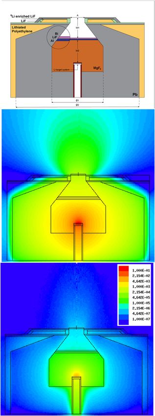
Finally, just in order to illustrate the capabilities of the beam to cover a wide volume of tissue for BNCT
therapies, an example of a one-field and a two-field irradiation are shown in Fig. 10. The color map illustrates
the equivalent dose at tumor relative to the maximum dose in normal brain tissue. The maximum dose rate in
normal tissues, delivered at scalp, even considering the higher data for the boron concentration (1.5 times the
concentration in brain) and CBE at skin (2.5), is 0.36 Gy-eq/min, close to the maximum dose delivered at normal
brain (0.331 Gy-eq/min), for the single beam case. As the tolerable dose is much higher (24 Gy-eq. respect to
12.5 Gy-eq for brain), the skin dose is not a problem. The dose at skull is much smaller. As one could notice, a
Scientific Reports | (2021) 11:7576 | https://doi.org/10.1038/s41598-021-87305-9 8
Vol:.(1234567890)www.nature.com/scientificreports/
Figure 10. Dose map at brain (Snyder head phantom) for two irradiation procedures, with one and two fields.
The one-field irradiation is frontal, the fields for the two-field irradiation are symmetrically rotated by 60◦ each
with respect to the head axial symmetry axis. Dashed-dotted lines indicate the beam axis for each irradiation
field. The color scale is normalized to the maximum dose in normal tissue.
simplistic two-field irradiation almost covers the full brain region, with a tumor- to-normal dose ratio greater
than 2. Although none corresponds to an actual treatment, it suggests that a multi-field irradiation with a desir-
able angle-positioning combination may reach any tumor location in brain with a therapeutic dose.
Conclusions
A design of a beam shaping assembly for an accelerator-based BNCT facility has been presented. It improves in
terms of both in-air and in-phantom figures of merit compared to previously proposed devices. The well defined
neutron beam allows a safe clinical operation within a hospital, and as a novelty, requires a low energy proton
accelerator (up to 2.1 MeV). The features of this design allow, according to Monte Carlo simulations, to treat
tumors with one or two beams located in a wide range of tissue, in particular, for brain tumors in any part to the
brain, provided the tumor uptake of the boron compound.
Scientific Reports | (2021) 11:7576 | https://doi.org/10.1038/s41598-021-87305-9 9
Vol.:(0123456789)www.nature.com/scientificreports/
It is worth to mention that the physics of the neutron production and moderation at these proton energies
(below the inelastic threshold) are very well known which guarantees that the Monte Carlo simulations will give
a realistic estimation of the performance of this device.
Methods
Beam shaping assembly design. In the following, the process of designing the BSA is described.
The 7 Li(p, n)7 Be thick-target yield near the reaction threshold was computed according to Lee and Zhou29.
The process to determine the most appropriate materials and sizes was based on the search for isotopes where
the elastic interaction dominates.
The presence of few resonances above the epithermal range was considered as a key factor. In addition, mean-
free-path and moderation estimations were computed to estimate the sizes of the main components. Once this
process derived in a reasonable solution, an optimization based on perturbing all the dimensions and sizes was
followed. That procedure kept ongoing until a good compromise was reached among the best suited sizes for all
IAEA recommendations, in-phantom FOMs and beam shaping needs.
The neutron transport simulations were carried out using the Monte Carlo code MCNP630.
Independent simulations were run to determine also the influence of the gamma radiation production at 7 Li
in the total gamma contamination at the beam aperture. An isotropic, 478 keV photon source was used, whose
yield was normalized with respect to the neutron production following the tabulation at Lee31. In addition,
gamma emission from activation was evaluated in the case of 27 Al due to the low half-life of 28 Al as in previous
studies26. An upper bound for delayed gamma emission from decays was included in the gamma dose contami-
nation computations via these MCNP6 simulations.
Analysis of the beam properties. The fast neutron and gamma dose contamination were derived inte-
grating the flux with the corresponding kerma factors. Neutrons in the range between 0.5 eV and 10 keV are
considered epithermal.
In order to evaluate the therapeutic effectiveness of the neutron beam, in-phantom dose profiles simula-
tions were performed. A cylinder phantom of ICRU standard tissue denoted as I CRU3323 and the Snyder head
phantom for skull and brain32 were used.
The total photon-equivalent dose (Eq. 1), in grays-equivalent (Gy.-Eq.), was estimated as separate contribu-
tions of thermal neutron dose (Dt ), fast neutron dose (Df ), boron (DB) and gamma dose (Dγ ), weighted to each
compound factor (CP).
DT = wt · Dt + wf · Df + wB · DB + wγ · Dγ (1)
The weighting factors reflect the relative biological effectiveness (RBE) of each component, with the follow-
ing values: 3.2 for thermal and fast neutrons; 3.8 and 1.3 for boron contribution in tumor and normal tissue,
respectively; 1 for gamma contribution33. The tumor and normal tissue 10 B concentration for standard tissue
(ICRU33) was set at 35 and 10 ppm, respectively. It has been reported that for the most severe brain tumors
(i.e. glioblastoma) the 10 B concentration ratio (tumor:normal) is greater than 3.5:134,35. Therefore, in Snyder
phantom the 10 B concentrations were assumed to be 65 and 18 ppm for tumor and normal brain tissue, as in
previous studies13,18,36.
Received: 2 November 2020; Accepted: 25 March 2021
References
1. Barth, R. F. et al. Current status of boron neutron capture therapy of high grade gliomas and recurrent head and neck cancer.
Radiat. Oncol. 7, 1–21. https://doi.org/10.1186/1748-717X-7-146 (2012).
2. Kreiner, A. J. et al. Present status of accelerator-based BNCT. Rep. Pract. Oncol. Radiother. 21, 95–101. https://doi.org/10.1016/j.
rpor.2014.11.004 (2016) (7th Young BNCT meeting).
3. Porras, I. et al. Proceedings of the 15th International Conference on Nuclear Reaction Mechanisms, chap. Perspectives on Neutron
Capture Therapy of Cancer, 295–304 (CERN, Geneva, 2018). https://doi.org/10.23727/CERN-Proceedings-2019-001.
4. Sakurai, Y. Progress in Reactor and accelerator based BNCT at Kyoto University Research Reactor Institute. Proceedings of Science
INPC2016, 127 (2017). https://doi.org/10.22323/1.281.0127.
5. Current Status of Neutron Capture Therapy. No. 1223 in TECDOC Series (International Atomic Energy Agency, 2001). https://
www.iaea.org/publications/6168/current-status-of-neutron-capture-therapy.
6. Porras, I. et al. BNCT research activities at the Granada group and the project NeMeSis: Neutrons for medicine and sciences,
towards an accelerator-based facility for new BNCT therapies, medical isotope production and other scientific neutron applica-
tions. Appl. Radiat. Isot. 165, 109247. https://doi.org/10.1016/j.apradiso.2020.109247 (2020).
7. Liskien, H. & Paulsen, A. Neutron production cross sections and energies for the reactions 7 Li(p, n)7 Be and 7 Li(p, n)7 Be∗. Atom.
Data Nucl. Data Tables 15, 57–84. https://doi.org/10.1016/0092-640X(75)90004-2 (1975).
8. Kiyanagi, Y. Accelerator-based neutron source for boron neutron capture therapy. Ther. Radiol. Oncol. 2 (2018). http://tro.amegr
oups.com/article/view/4713.
9. Mastinu, P. et al. Micro-channel-based high specific power lithium target. Il Nuovo Cimento C 38, 1–7. https://doi.org/10.1393/
ncc/i2015-15193-y (2015).
10. Seppälä, T. FIR 1 Epithermal neutron beam model and dose calculation for treatment planning in Neutron Capture Therapy. Ph.D.
thesis, University of Helsinki (2002).
11. Koivunoro, H. Dosimetry and dose planning in boron neutron capture therapy: Monte Carlo studies. Ph.D. thesis, University of
Helsinki (2012).
12. Sakurai, Y. et al. Advances in boron neutron capture therapy (BNCT) at Kyoto University—From reactor-based BNCT to acceler-
ator-based BNCT. J. Korean Phys. Soc. 67, 76–81. https://doi.org/10.3938/jkps.67.76 (2015).
Scientific Reports | (2021) 11:7576 | https://doi.org/10.1038/s41598-021-87305-9 10
Vol:.(1234567890)www.nature.com/scientificreports/
13. Liu, Y.-W., Huang, T., Jiang, S. & Liu, H. Renovation of epithermal neutron beam for BNCT at THOR. Appl. Radiat. Isot. 61,
1039–1043. https://doi.org/10.1016/j.apradiso.2004.05.042 (2004) (Topics in Neutron Capture Therapy: Proceedings of the
Eleventh World Congress on Neutron Capture Therapy (ISNCT-11)).
14. Tanaka, H. et al. Experimental verification of beam characteristics for cyclotron-based epithermal neutron source (C-BENS). Appl.
Radiat. Isot. 69, 1642–1645. https://doi.org/10.1016/j.apradiso.2011.03.020 (2011) (Special Issue: 14th International Conference
on Neutron Capture Therapy).
15. Koay, H. et al. Feasibility study of compact accelerator-based neutron generator for multi-port BNCT system. Nucl. Instrum.
Methods Phys. Res. Sect. A Accel. Spectrom. Detect. Assoc. Equip. 899, 65–72. https://doi.org/10.1016/j.nima.2018.05.025 (2018).
16. Kiyanagi, Y. et al. A project of boron neutron capture therapy system based on a proton Linac neutron source. Physics Procedia 26,
223–230. https://doi.org/10.1016/j.phpro.2012.03.029 (2012) (Proceedings of the first two meetings of the Union of Compact
Accelerator-Driven Neutron Sources).
17. Masuda, A. et al. Neutron spectral fluence measurements using a Bonner sphere spectrometer in the development of the iBNCT
accelerator-based neutron source. Appl. Radiat. Isot. 127, 47–51. https://doi.org/10.1016/j.apradiso.2017.05.010 (2017).
18. Kononov, O. et al. Optimization of an accelerator-based epithermal neutron source for neutron capture therapy. Appl. Radiat. Isot.
61, 1009–1013. https://doi.org/10.1016/j.apradiso.2004.05.028 (2004) (Topics in Neutron Capture Therapy: Proceedings of the
Eleventh World Congress on Neutron Capture Therapy (ISNCT-11)).
19. Zaidi, L., Belgaid, M., Taskaev, S. & Khelifi, R. Beam shaping assembly design of 7 Li(p, n)7 Be neutron source for boron neutron
capture therapy of deep-seated tumor. Appl. Radiat. Isot. 139, 316–324. https://doi.org/10.1016/j.apradiso.2018.05.029 (2018).
20. Postuma, I. Clinical application of accelerator-based Boron Neutron Capture Therapy. Ph.D. thesis, University of Pavia. https://doi.
org/10.13140/RG.2.1.3539.3680 (2015).
21. Torres-Sánchez, P., Arias de Saavedra, F., Sabariego, M. & Praena, J. On the upper limit for the energy of epithermal neutrons for
Boron Neutron Capture Therapy. Radiat. Phys. Chem. 156, 240–244. https://doi.org/10.1016/j.radphyschem.2018.11.015 (2019).
22. Kato, T. et al. Design and construction of an accelerator-based boron neutron capture therapy (AB-BNCT) facility with multiple
treatment rooms at the Southern Tohoku BNCT Research Center. Appl. Radiat. Isot. 156, 108961. https://doi.org/10.1016/j.aprad
iso.2019.108961 (2020).
23. Report 46: Photon, Electron, Proton and Neutron Interaction Data for Body Tissues. Journal of the International Commission on
Radiation Units and Measurements, Bethesda, MD. (1992). https://inis.iaea.org/search/search.aspx?orig_q=RN:23082971.
24. Tanaka, H. et al. Characteristics comparison between a cyclotron-based neutron source and KUR-HWNIF for boron neutron
capture therapy. Nucl. Instrum. Methods Phys. Res. Sect. B Beam Interact. Mater. Atoms 267, 1970–1977. https://doi.org/10.1016/j.
nimb.2009.03.095 (2009).
25. Giusti, V., Munck af Rosenschöl, P. M., SköldSköld, K., Montagnini, B. & Capala, J. Monte Carlo model of the Studsvik BNCT
clinical beam: Description and validation. Med. Phys. 30, 3107–3117. https://doi.org/10.1118/1.1626120 (2003).
26. Bleuel, D. L., Donahue, R. J., Ludewigt, B. A. & Vujic, J. Designing accelerator-based epithermal neutron beams for boron neutron
capture therapy. Med. Phys. 25, 1725–1734. https://doi.org/10.1118/1.598353 (1998).
27. Kim, K.-O., Kim, J. K. & Kim, S. Y. Optimized therapeutic neutron beam for accelerator-based BNCT by analyzing the neutron
angular distribution from 7 li(p, n)7 be reaction. Appl. Radiat. Isot. 67, 1173–1179. https://doi.org/10.1016/j.apradiso.2009.02.004
(2009) (6th International Conference on Isotope).
28. Minsky, D. & Kreiner, A. Beam shaping assembly optimization for 7 li(p, n)7 be accelerator based BNCT. Appl. Radiat. Isot. 88,
233–237. https://doi.org/10.1016/j.apradiso.2013.11.088 (2014) (15th International Congress on Neutron Capture Therapy
Impact of a new radiotherapy against cancer).
29. Lee, C. & Zhou, X.-L. Thick target neutron yields for the 7 Li(p, n)7 Be reaction near threshold. Nucl. Instrum. Methods Phys. Res.
Sect. B Beam Interact. Mater. Atoms 152, 1–11. https://doi.org/10.1016/S0168-583X(99)00026-9 (1999).
30. Werner, C. J. et al. MCNP6.2 Release Notes—report LA-UR-18-20808 (Los Alamos National Laboratory (LANL), 2018).
31. Lee, C. L., Zhou, X.-L., Kudchadker, R. J. & Harmon, F. A Monte Carlo dosimetry-based evaluation of the reaction near threshold
for accelerator boron neutron capture therapy. Med. Phys. 27, 192–202 https://doi.org/10.1118/1.598884 (2000).
32. Goorley, T. MCNP Medical Physics Geometry Database. Tech. Rep. (2008). https://laws.lanl.gov/vhosts/mcnp.lanl.gov/pdf_files/
la-ur-08-2468.pdf.
33. Coderre, J. A. & Morris, G. M. The radiation biology of boron neutron capture therapy. Radiat. Res. 151, 1–18. https://doi.org/10.
2307/3579742 (1999).
34. Savolainen, S. et al. Boron neutron capture therapy (BNCT) in Finland: Technological and physical prospects after 20 years of
experiences. Phys. Med. 29, 233–248. https://doi.org/10.1016/j.ejmp.2012.04.008 (2013).
35. Coderre, J. A. et al. Biodistribution of boronophenylalanine in patients with glioblastoma multiforme: Boron concentration cor-
relates with tumor cellularity. Radiat. Res. 149, 163–170. https://doi.org/10.2307/3579926 (1998).
36. Binns, P., Riley, K. & Harling, O. Dosimetric comparison of six epithermal neutron beams using an ellipsoidal water phantom. In
Research and development in Neutron Capture Therapy, 405–409 (eds Sauerwein, W. et al.) (Monduzzi Editore, 2002). Proceedings
Paper: 10th International Congress on Neutron Capture Therapy.
Acknowledgements
We acknowledge partial financial support for this work from the Junta de Andalucía (Andalusian Regional
Government, Programa Operativo FEDER Andalucia 2014–2020 under contract A-FQM-371-UGR18) and
the donors of the University Chair “Neutrons for Medicine”: Spanish Fundación ACS, Capitán Antonio, La
Kuadrilla de Iznalloz and Sonriendo se Puede Ganar. N. R. acknowledges support from the Junta de Andalucía
(Andalusian Regional Government) and European Social Fund (ESF), under contract of Iniciativa de Garantía
Juvenil. P. T. acknowledges support from the Spanish Ministry of Science, Innovation and Universities under
the FPU grant FPU17/02305.
Author contributions
All authors have contributed substantially to this work. I.P, J.P. and P.T. conceived the idea. J.P. and F.A. performed
the neutron production calculations. P.T. and N.R. performed the Monte Carlo simulations. All authors contrib-
uted to the analysis of data and optimization. P.T. and I.P. wrote the main manuscript text. Figures were made
by P.T. and N.R. All authors reviewed the manuscript.
Competing interests
The results of this work are subject to a patent application of the University of Granada, in which all authors
(P.T., I.P., N.R., F.A. and J.P.) are inventors.
Scientific Reports | (2021) 11:7576 | https://doi.org/10.1038/s41598-021-87305-9 11
Vol.:(0123456789)www.nature.com/scientificreports/
Additional information
Correspondence and requests for materials should be addressed to I.P.
Reprints and permissions information is available at www.nature.com/reprints.
Publisher’s note Springer Nature remains neutral with regard to jurisdictional claims in published maps and
institutional affiliations.
Open Access This article is licensed under a Creative Commons Attribution 4.0 International
License, which permits use, sharing, adaptation, distribution and reproduction in any medium or
format, as long as you give appropriate credit to the original author(s) and the source, provide a link to the
Creative Commons licence, and indicate if changes were made. The images or other third party material in this
article are included in the article’s Creative Commons licence, unless indicated otherwise in a credit line to the
material. If material is not included in the article’s Creative Commons licence and your intended use is not
permitted by statutory regulation or exceeds the permitted use, you will need to obtain permission directly from
the copyright holder. To view a copy of this licence, visit http://creativecommons.org/licenses/by/4.0/.
© The Author(s) 2021
Scientific Reports | (2021) 11:7576 | https://doi.org/10.1038/s41598-021-87305-9 12
Vol:.(1234567890)You can also read