NOTICE WARNING CONCERNING COPYRIGHT RESTRICTIONS - Bear Lab
←
→
Page content transcription
If your browser does not render page correctly, please read the page content below
NOTICE
                WARNING CONCERNING
               COPYRIGHT RESTRICTIONS

   The copyright law of the United States [Title 17, United States
Code] governs the making of photocopies or other reproductions
of copyrighted material
    Under certain conditions specified in the law, libraries and archives
are authorized to furnish a photocopy or other reproduction.One of
these specified conditions is that the reproduction is not to be
used for any purpose other than private study, scholarship, or research.
If a user makes a request for, or later uses, a photocopy or repro-
duction for purposes in excess of "fair use," that use may be liable
for copyright infringement.
   This institution reserves the right to refuse to accept a copying
order if, in its judgement, fullfillment of the order would involve
violation of copyright law. No further reproduction and distribution
of this copy is permitted by transmission or any other means.
Vision Research 183 (2021) 53–60
Contents lists available at ScienceDirect
Vision Research
journal homepage: www.elsevier.com/locate/visres
The ups and downs of sensory eye balance: Monocular deprivation has a
biphasic effect on interocular dominance
Mahalakshmi Ramamurthy a, b, *, Erik Blaser a
a
Department of Psychology, University of Massachusetts Boston, United States
b
Department of Developmental-behavioral Pediatrics, School of Medicine and Graduate School of Education, Stanford University, United States
A R T I C L E I N F O A B S T R A C T
Keywords: Classic studies of ocular dominance plasticity in early development showed that monocular deprivation sup
Monocular deprivation presses the neural representation and visual function of the deprived eye. However, recent studies have shown
Contrast gain control that a short period of monocular deprivation (
M. Ramamurthy and E. Blaser                                                                                                           Vision Research 183 (2021) 53–60

to emerge: throughout the lifespan, to deprive an eye is to shift balance            participation, participants performed a sighting test2 to determine their
toward the non-deprived eye (for a review, see Dahlhaus and Levelt                   preferred eye. All subjects gave informed consent and were compensated
(2010)).                                                                             for their time as approved by the Institutional Review Board of the
    This made it all the more surprising when recent work in adult                   University of Massachusetts Boston.
humans showed that short-term (
M. Ramamurthy and E. Blaser Vision Research 183 (2021) 53–60
4. Results
4.1. Deprivation duration effects on dichoptic global motion coherence
thresholds (GMC)
We compared GMC thresholds from the deprived and the non-
deprived eye at six different timepoints. The resulting laminar, well-
separated functions for the non-deprived and deprived eye indicate a
robust effect (Fig. 2a). A repeated measures ANOVA on these data with
deprivation duration (0, 30, 150, 300, 450, and 600 min) and deprivation
state (deprived or non-deprived) as factors showed a significant main
effect of duration [F(5,30) = 3.090; p = 0.023; η2 = 0.034] and state [F
(1,6) = 17.150; p = 0.006; η2 = 0.42] and a significant interaction [F
(5,30) = 7.004; p < 0.001; η2 = 0.08]. To facilitate comparison between
measures, and to previous work (Ramamurthy & Blaser, 2018; Zhou,
Clavagnier, et al., 2013), we then calculated normalized threshold ra
tios. For each participant and deprivation duration, an interocular ratio
of thresholds was calculated (non-deprived eye/deprived eye), then
normalized by the respective pre-deprivation ratio (Fig. 2b). This shows
a net shift in interocular balance away from the non-deprived eye as a
function of deprivation duration, with a turning point at 300 min (5 h) of
deprivation. Normalized ratios from two participants that participated
in the re-test experiment are shown for reference.
5. Deprivation duration effects on monocular contrast
sensitivity
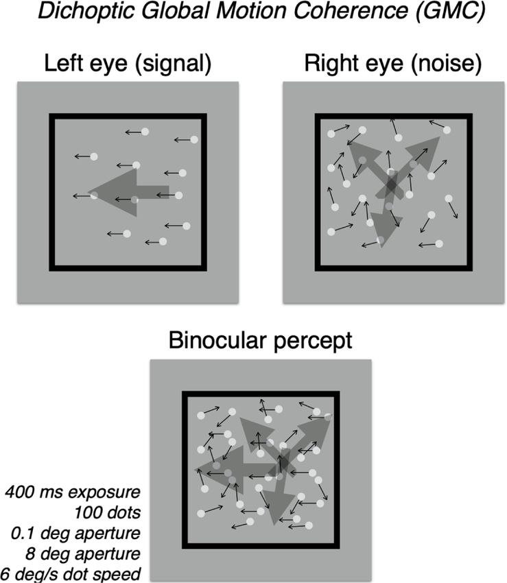
Fig. 1. Schematic of Global Motion Coherence (GMC) tests. In these tests, one
We then measured monocular contrast sensitivity functions (CSF) of
eye receives a small set of coherently moving dots (signal, shown here in left the deprived and the non-deprived eye. We extracted three parameters
eye) and the other, a large set of random-vector dots (noise, shown here in right from the qCSF (Lesmes et al., 2010): peak contrast sensitivity, area under
eye). Images were interleaved dichoptically at 120 Hz. Which eye received the log CSF (AULCSF), and spatial frequency at peak sensitivity.
signal or noise was randomized, trial to trial, as was the direction, left versus For peak contrast, the laminar, well-separated functions for the non-
right, of the signal dots. The participant was instructed to indicate the direction deprived and deprived eye indicate a robust effect (Fig. 2c). A repeated
of this motion with a keypress. An adaptive staircase was used to determine measures ANOVA on these data with deprivation duration (0, 30, 150,
coherence thresholds (i.e. the proportion of signal dots required to achieve 75% 300, 450, and 600 min) and deprivation state (deprived or non-deprived)
correct direction judgements). as factors showed a significant main effect of duration [F(5,30) = 2.87;
p = 0.031; η2 = 0.098] and state [F(1,6) = 74.2; p < 0.001; η2 = 0.251]
peak sensitivity (‘gain’), area under the log CSF (AULCSF), and peak and a significant interaction [F(5,30) = 4.98; p = 0.002; η2 = 0.115]. For
frequency (i.e. the spatial frequency at which there was peak sensi each participant and deprivation duration, an interocular ratio was
tivity). Both eyes’ thresholds were tested in mixed blocks. A block took calculated (non-deprived eye/deprived eye) then normalized by the
approximately 6 min to complete. respective pre-deprivation ratio (Fig. 2d). This shows a net shift in
interocular balance away from the non-deprived eye as a function of
3. Experimental procedures deprivation duration, with a turning point at 300 min. Normalized ratios
from two participants that participated in the re-test experiment are
Each participant was scheduled for a 10-hour session of deprivation shown for reference.
during which participants wore a light-tight patch on their preferred For AULCSF, the laminar, well-separated functions for the non-
eye. While many short-term deprivation studies have not shown any deprived and deprived eye indicate a robust effect (Fig. 2e). A
interaction with eye preference (Ramamurthy & Blaser, 2018; Zhou repeated measures ANOVA on these data with deprivation duration (0,
et al., 2014), some have shown larger effect sizes with deprivation of the 30, 150, 300, 450, and 600 min) and deprivation state (deprived or non-
preferred eye (Lunghi et al., 2011). Participants could engage in deprived) as factors showed a significant main effect of duration [F
everyday activities during deprivation, but were restricted to the labo (5,30) = 4.79; p = 0.002; η2 = 0.125] and state [F(1,6) = 79.8; p <
ratory suite to ensure compliance and facilitate periodic testing. The 0.001; η2 = 0.181] and a significant interaction [F(5,30) = 15.70; p <
main experiment consisted of a pre-deprivation baseline measure of 0.001; η2 = 0.087]. Normalized interocular ratios showed a net shift
ocular balance, followed by five intermittent testing points at 30, 150, away from the non-deprived eye as a function of deprivation duration,
300, 450, and 600 min. The testing points consisted of the dichoptic with a turning point at approximately 150 min (Fig. 2e). Normalized
GMC task followed by monocular qCSF tests, for a total of approximately ratios from two participants that participated in the re-test experiment
12 min. are shown for reference.
Several days after the main experiment, two participants returned for We did not observe any shifts in spatial frequency tuning as a func
testing. All procedures were the same as in the main experiments, except tion of deprivation duration. A repeated measures ANOVA on these data
there were only two testing points (as opposed to six), the pre- with deprivation duration and deprivation state as factors showed no sig
deprivation baseline and a post-deprivation testing point after the full nificant effect of duration [F(5,30) = 0.617; p = 0.688 η2 = 0.022].
10 h of deprivation. This re-test was conceived both as a modest internal There was a significant effect of state [F(1,6) = 18.9; p = 0.005; η2 =
replication, and as a way to sidestep any potential influence of the 0.096], but no interaction [F(5,30) = 1.72; p = 0.160; η2 = 0.056]. Since
intermittent testing used in the main experiment. the difference in spatial frequency tuning between the eyes (i.e., state)
was present pre-deprivation, and not influenced by deprivation dura
tion, it is likely unrelated to our experimental manipulations. It is worth
55M. Ramamurthy and E. Blaser Vision Research 183 (2021) 53–60 Fig. 2. Interocular balance changes as a function of monocular deprivation duration. The three panels on the left show thresholds (for GMC (a), CSF peak sensitivity (c), and AULCSF (e) tests, respectively) following monocular deprivation of up to 10 h, for both the non-deprived (open symbols) and deprived eyes (light-tight patched; filled symbols). Data points represent average values for all participants in the main experiment (N = 7; for separate participant plots, see Supplemental Materials Fig. 1). Error bars represent SEM. Within a particular test, we compared thresholds at each deprivation duration and for each eye (corrected for multiple comparisons) to pre-deprivation values (p < 0.05 (*),
M. Ramamurthy and E. Blaser Vision Research 183 (2021) 53–60
“dose–response” models (Baldi & Bucherelli, 2005; Calabrese & Bald test was not appropriate, so we conducted an AIC (Akaike’s Information
win, 2001; Tuček et al., 2002). A bell-shaped regression has seven pa Criterion) model comparison. The probability that the exponential
rameters, but four of these could be fixed, leaving three free parameters. plateau model was correct was 30.6%, while that for the bell-shaped
The starting plateau was fixed at 1.0 to indicate a balanced interocular model was 69.4%, yielding a ratio of 2.27 in favor of the bell-shaped
ratio, pre-deprivation. The asymptotic plateau was set to zero, to indicate model.
a theoretical expectation of total dominance of the non-deprived eye, in Given that the bell-shaped model was preferred, we then used it as
the limit. The two slope parameters, which determine the steepness of planned to generate estimates, for each of the three measures, of two
the underlying excitatory and inhibitory responses (reflecting the two points of interest: a balance point (an estimate of the number of hours of
processes underlying the short- and long-term deprivation effects, deprivation until binocular balance is restored, i.e., an interocular ratio
respectively), respectively, were both fixed to a value of − 1, a standard of 1.0), and a reversed-shift point (an estimate of the number of hours of
default for bell-shaped data (Motulsky, 2007). This left three free pa deprivation until interocular balance shifts substantially away from the
rameters. Two of these (EC50_1 and EC50_2) modulate the time point at deprived eye which we specified to be an interocular ratio of 0.5,
which the underlying excitatory and inhibitory processes, i.e. the short- commensurate, but opposite, to an interocular ratio of 2.0). For GMC
and long-term deprivation effects, respectively, are at half-height. The threshold measures, the model predicted the balance point would be
final, dip parameter modulates the peak of the bell response. reached at 35.7 h (95% CI: 15.8 to 98.1) (Fig. 3). In other words, it is
Our goal was to fit the three data sets simultaneously, with EC50_1 predicted that it would take approximately 1.5 days of continuous
and EC50_2 treated as shared parameters, and with the dip parameter left deprivation for the long-term effect to cancel out the short-term effect.
to vary between data sets (thereby allowing different measures to be The model predicted a reversed-shift of 84.0 h (CI: 23.6 to 224.3), or
differentially sensitive to deprivation). The regression considered each approximately 3.5 days of continuous deprivation, to observe a robust
replicate as an individual point (N = 42 for GMC thresholds, peak shift in interocular balance away from the deprived eye. For CSF peak
sensitivity, and AULCSF data, respectively; N = 126 overall). As a first sensitivity, the model predicted a balance point of 53.5 h (2.2 days) (CI:
step, then, we tested whether the sharing of EC50_1 and EC50_2 was 20.2 to 140.9) and a reverse-shift of 118.9 h (5.0 days) (CI: 28.5 to 308).
warranted. As expected, the extra-sum-of-squares F test could not reject For the AULCSF, the balance point was 17.1 h (CI: 8.4 to 52.7) and
the null hypothesis that these parameters were the same for all data sets reverse-shift was 47.5 h (2.0 days) (CI: 16.4 to 135.5).
(F(4,117) = 0.321; p = 0.864). In the final fit (Se = 0.80), the values for
the two shared parameters were EC50_1 = 390 and EC50_2 = 391. The 8. Discussion
unshared dip parameters for GMC threshold, peak sensitivity, and
AULCSF were 1434, 2036, and 800, respectively (the differences in the We used (up to) 10 h of light-tight monocular deprivation to induce
dip parameter reflects the underlying differences in the sensitivity of the shifts in interocular balance. We found a biphasic effect as a function of
measures to deprivation). deprivation duration on both dichoptic global motion coherence
We then compared this model to a null hypothesis that monocular thresholds (GMC) and monocular contrast sensitivity (we did not
deprivation had a monotonic effect on the interocular ratio (i.e., that observe any deprivation-induced effects on spatial frequency tuning).
there were no separate short- and long-term processes, but just one Consistent with recent reports of short-term deprivation effects (Lunghi
process that shifts balance away from the non-deprived eye, asymptot et al., 2011; Ramamurthy & Blaser, 2018; Zhou, Clavagnier, et al., 2013;
ically, as a function of deprivation duration), which we modeled as J. Zhou et al., 2015), we initially observed a post-deprivation shift in
plateaued exponential growth. This model had three parameters, Y0 (the interocular balance away from the open, non-deprived eye. This effect
starting point of growth, which was fixed at an interocular ratio of 1.0), k increased as a function of deprivation duration over a period of
(controlling growth rate, which was a shared parameter for all three approximately 5 h. We then observed a turning point: more deprivation
data sets), and YM (the plateau of growth) which was left as a free time did not serve to further increase the effect, but reduce it. To our
parameter to allow for the three data sets to have different asymptotes. knowledge, this is the first time a biphasic effect of monocular depri
After fitting, k = 0.0121, and YM was equal to 2.08, 2.73, and 1.39 for vation on ocular dominance plasticity has been observed in typical adult
GMC, peak CSF, and AULCSF, respectively. Since the bell-shaped and humans.
exponential plateau models were not nested, an extra-sum-of-squares F However, nearly 50 years ago, there was a group that tested the
Fig. 3. Biphasic regression model for GMC thresholds, peak contrast sensitivity, and AULCSF as a function of deprivation duration. All participants’ data (N = 7)
from all three measures (replotted here from Fig. 2 b/d/f) were fit (Se = 0.80) by a common bell-shaped dose response model (the null hypothesis of a monotonic,
plateaued exponential was rejected) with one free parameter for each measure, modulating the peak effect. As an exploratory analysis, extrapolated fits were used to
estimate two points of interest: a balance point, reflecting a prediction of when interocular balance would be restored, and a reversed-shift point, a prediction of the
deprivation duration required for a robust reversal (to 0.5) of the interocular ratio.
57M. Ramamurthy and E. Blaser Vision Research 183 (2021) 53–60
effect of up to two weeks of monocular deprivation on monocular critical detection thresholds (in either eye), there was a greater increase in
flicker frequency thresholds. Consistent with our results, Zubek & Bross plasticity, i.e. a relative strengthening, of the non-deprived eye, versus
(1972), Zubek and Bross (1973a) found an initial decrease in the critical the deprived eye (as measured by changes in contrast detection
flicker frequency (CFF) threshold in the non-deprived eye (i.e., a thresholds in a perceptual learning regime). After two days of monocular
weakening), followed by an increase in the CFF in that eye (estimating deprivation, Lou et al. (2011) concluded that cortical excitability, as
from the two studies, the total delta from decrease to increase was measured by the perception of TMS-induced phosphenes, was reduced in
approximately 3–4 flash/s, or just under a 10% change from a starting both hemispheres symmetrically. In their critical flicker frequency tests,
CFF of 40–41 flash/s). The transition from weakening to strengthening following days or weeks-long deprivation, Zubek & Bross (1972), Zubek
of the non-deprived eye occurred at the 6–9 h mark, not far off from our and Bross (1973a) found an improvement in the non-deprived, but not
present finding of 5 h. Zubek’s work though was controversial: coercive the deprived, eye.) There has been some related work in long-term
to its subjects, and seen as facilitating the development of sensory monocular deprivation in adult, animal models. In general, the animal
deprivation as a form of torture (Raz, 2013). After publication of back- literature tracks very closely to the human literature in terms of the
to-back studies in Science (Zubek & Bross, 1972) and Nature (Zubek and basics of ocular dominance plasticity, including the neural and behav
Bross, 1973a), this line of inquiry was abandoned by the field, and Zubek ioral consequences of deprivation and the interaction with development
died soon after, of a probable suicide (Raz, 2013). While this work has (Dahlhaus & Levelt, 2010). After maturation of the visual system in
not been cited in recent studies on monocular deprivation, it certainly adult animals, as in humans, the effects of long-term (several days)
presages it. That said, these studies did have critical limitations, deprivation are more modest, and with evidence of reciprocal effects on
including testing with only a monocular measurement (as opposed to eyes, but here too some evidence for bias can be found, with more
including inherently interocular measures such as dichoptic GMC, pronounced effects in the non-deprived eye (Hofer et al., 2006; Sato &
binocular phase combination, and rivalry), and the fact that they did not Stryker, 2008; Tschetter et al., 2013).
observe any effects with ‘diffuser’ deprivation (i.e. flooding an eye with It is difficult to determine the source of eye-bias in deprivation effects
unpatterned light, usually through fitting a translucent lens) (Zubek and (short-term, or long-term) currently, since they have been inconsistent
Bross, 1973b). This latter finding is incompatible with recent work in previous work, and there has been no direct attempt to characterize
showing both that diffuser deprivation (Lunghi et al., 2011; Zhou, them. We can only speculate that biases may be exposed, or induced, by
Clavagnier, et al., 2013) and many other forms of deprivation - even the particulars of the deprivation duration and type (e.g. light-tight or
those that preserve gross luminance, and color such as low-pass filtering translucent), and the particulars of the testing session itself, including
(Zhou et al., 2014) or kaleidoscopic image fractionation (Ramamurthy & the type of tests (e.g., measuring effects on perceived, suprathreshold
Blaser, 2018) – induce effects comparable to light-tight patching. contrast stimuli versus contrast thresholds, or measuring effects on a full
CSF versus effects at a particular frequency), how long after deprivation
9. Deprivation effects in the deprived versus non-deprived eye tests are initiated, and how long they take. In animal models as well, this
complex interplay between affected eyes (and the interaction with
Here we found a significant deprivation effect in the non-deprived development) is not yet well understood (Sawtell et al., 2003), but is
eye, for all three measures, but no significant effect in the deprived thought to reflect the action of opposing, mutually-regulating Hebbian
eye. While our previous work showed evidence for reciprocal effects, in and homeostatic mechanisms in visual cortex (Keck et al., 2017; Mrsic-
monocular CSF tests, effects were smaller in the deprived eye (Ram Flogel et al., 2007; Turrigiano & Nelson, 2000, 2004; Whitt et al., 2014).
amurthy & Blaser, 2018). It is challenging to fully evaluate eye-specific
effects in the literature, because most studies either report only inter 10. Future work with yet longer-term deprivation
ocular ratios, or employ only inherently push–pull tests (such as rivalry
or binocular phase combination). In either case, it is ambiguous whether The maximum deprivation duration of 10 h in the present study was
an observed shift in balance is due to a change in the deprived eye, the sufficient to see a turning point in ocular dominance plasticity, but not a
non-deprived eye, or both. There is very little data from purely restoration of balance (or, further, a reversal of the interocular ratio).
monocular tests following deprivation, and these results are mixed. Since we could not observe them directly, we extrapolated a biphasic
Zhou, Thompson, et al. (2013), Zhou et al. (2017), Zhou, Clavagnier, (bell-shaped) dose-response model of our data to estimate these points.
et al. (2013) have measured monocular contrast thresholds in a handful Our predictions indicated that for dichoptic global motion coherence,
of observers (following 150 min of deprivation) and observed a for instance, the short-term and long-term responses should cancel out,
threshold increase in the non-deprived eye and a reciprocal decrease in restoring interocular balance, after about 36 h of deprivation. A robust
the deprived, but, while effects were present in both eyes, there was no reversal, away from the deprived eye, was estimated at 84 h (3.5 days) of
direct comparison, so it is difficult to assess bias. Lunghi et al. (2011, deprivation.
supplemental materials) measured contrast detection thresholds It is important to be clear about the limitations of this exercise. While
(following 150 min of deprivation) and found an increase in threshold in providing testable predictions for future work, our estimates are
the non-deprived eye, and no effect in the deprived. Taking a different underspecified by the data, as evidenced by large confidence intervals,
tack, Chadnova, et al. (2017) measured monocular deprivation effects and should be treated with caution. Even with a dedicated study that
based on MEG power (of frequency tagged signals in primary visual could extend the deprivation times sufficiently, recovery or reversal may
cortex) and found reciprocal effects, but with a much more robust effect never be reached. Given the only modest effects of even two weeks of
in the deprived eye (effects in the non-deprived eye only reached sig deprivation from Zubek & Bross’ controversial work (1973a), and the
nificance at one of four post-deprivation testing points, in a light-tight conventional wisdom that patching interventions for amblyopia in adult
patch condition, and did not reach significance at any time point in a humans may have only limited effect (for a review, see Holmes & Levi
translucent patch condition). Taken together then, there is only one (2018)), there is reason to be pessimistic. Additionally, while the pre
thing that can be said with certainty: short-term monocular deprivation dictions for the balance and reversal points differed for the three mea
weakens the non-deprived eye and/or strengthens the deprived. sures, confidence intervals were so large that it is not possible to reject
In long-term monocular deprivation with adult humans, while we see the (more parsimonious) null hypothesis that these points may actually
the expected, complementary pattern (i.e., that long-term monocular be common across the measures. This would, in fact, be our hypothesis
patching strengthens the non-deprived eye and/or weakens the for future work: while effect sizes would certainly vary depending on
deprived), again there is little work that bears on bias. Shibata, et al. how sensitive a particular psychophysical measure is to interocular
(2012) found that after 3 days of deprivation, while there were no balance, the turning, balance, and reversed-shift points would be shared.
immediately evident deprivation effects on monocular contrast This seems reasonable, as the underlying mechanism that drives these
58M. Ramamurthy and E. Blaser Vision Research 183 (2021) 53–60
effects (assumedly, interocular gain control) is common, independent of Proceedings of the National Academy of Sciences of the United States of America, 98(25),
14698–14701.
the particular test used to assess it. Finally, while the bell-shaped
Brainard, D. H. (1997). The Psychophysics Toolbox. Spatial Vision, 10(4), 433–436.
regression model has biological plausibility (Baldi & Bucherelli, 2005; Calabrese, E. J., & Baldwin, L. A. (2001). U-shaped dose-responses in biology, toxicology,
Calabrese & Baldwin, 2001; Tuček et al., 2002), we employed it pri and public health. Annual Review of Public Health, 22(1), 15–33.
marily as a data flitting method. We fully expect a future, dedicated Chadnova, E., Reynaud, A., Clavagnier, S., & Hess, R. F. (2017). Short-term monocular
occlusion produces changes in ocular dominance by a reciprocal modulation of
treatment to supplant it with a model that incorporates processes interocular inhibition. Scientific Reports, 7, 41747.
thought to be involved in short- and long-term deprivation effects, likely Dahlhaus, M., & Levelt, C. N. (2010). Structure and function relationships during ocular
emerging from formal models of interocular gain control (Ding et al., dominance plasticity in the visual cortex. Reviews in the Neurosciences, 21(3),
223–237.
2013; Ding & Sperling, 2006). Ding, J., Klein, S. A., & Levi, D. M. (2013). Binocular combination of phase and contrast
Ultimately, we argue here that the biphasic nature of interocular explained by a gain-control and gain-enhancement model. Journal of Vision, 13(2),
balance shifts as a function of deprivation reflect two opposing processes 13. https://doi.org/10.1167/13.2.13.
Ding, J., & Levi, D. M. (2011). Recovery of stereopsis through perceptual learning in
that act at different timescales. Upon deprivation, when the visual in human adults with abnormal binocular vision. Proceedings of the National Academy of
formation from one eye is removed (for instance through light-tight Sciences of the United States of America, 108(37), E733–E741.
patching as in the present work), reduced (through, for instance, Ding, J., & Sperling, G. (2006). A gain-control theory of binocular combination.
Proceedings of the National Academy of Sciences of the United States of America,
diffuser deprivation), or manipulated in a way that leads to its sup 103(4), 1141–1146.
pression (for instance, reducing its usefulness to active vision, as in Doshi, N. R., & Rodriguez, M. L. F. (2007). Amblyopia. American Family Physician, 75(3),
kaleidoscopic deprivation (Ramamurthy & Blaser, 2018)), the visual 361–367.
Epelbaum, M., Milleret, C., Buisseret, P., & Duffer, J. L. (1993). The sensitive period for
system leverages interocular gain control mechanisms to downweigh the
strabismic amblyopia in humans. Ophthalmology, 100(3), 323–327.
non-deprived eye and/or potentiate the deprived eye in a homeostatic Fox, K., & Stryker, M. (2017). Integrating Hebbian and homeostatic plasticity:
effort (Fox & Stryker, 2017) to restore interocular balance prior to Introduction. Philosophical Transactions of the Royal Society of London. Series B,
binocular combination. Then, as deprivation continues, a relatively Biological Sciences, 372(1715). https://doi.org/10.1098/rstb.2016.0413.
Glickman, M. E., Rao, S. R., & Schultz, M. R. (2014). False discovery rate control is a
sluggish system, noting the persistent lack of utility of one monocular recommended alternative to Bonferroni-type adjustments in health studies. Journal
stream, begins a process of downweighting it and/or potentiating the of Clinical Epidemiology, 67(8), 850–857.
non-deprived eye. During development, this latter process leads to Hensch, T. K., & Quinlan, E. M. (2018). Critical periods in amblyopia. Visual
Neuroscience, 35, E014.
extreme outcomes, including permanent changes to neural representa Hofer, S. B., Mrsic-Flogel, T. D., Bonhoeffer, T., & Hübener, M. (2006). Prior experience
tion and visual processing (Sengpiel & Blakemore, 1996). In an adult enhances plasticity in adult visual cortex. Nature Neuroscience, 9(1), 127–132.
system, consequences are likely more modest (Morishita & Hensch, Holmes, J. M., & Levi, D. M. (2018). Treatment of amblyopia as a function of age. Visual
Neuroscience, 35, E015.
2008; Sato & Stryker, 2008), but further work with yet longer-term Huang, C.-B., Zhou, Y., & Lu, Z.-L. (2008). Broad bandwidth of perceptual learning in the
deprivation is necessary to reveal the full trajectory of the ocular visual system of adults with anisometropic amblyopia. Proceedings of the National
dominance shift. Academy of Sciences of the United States of America, 105(10), 4068–4073.
Hubel, D. H., & Wiesel, T. N. (1970). The period of susceptibility to the physiological
effects of unilateral eye closure in kittens. The Journal of Physiology, 206(2),
CRediT authorship contribution statement 419–436.
Hubel, D. H., Wiesel, T. N., & LeVay, S. (1977). Plasticity of ocular dominance columns in
monkey striate cortex. Philosophical Transactions of the Royal Society of London. Series
Mahalakshmi Ramamurthyi: Conceptualization, Methodology,
B, Biological Sciences, 278(961), 377–409.
Software, Validation, Formal analysis, Data curation, Writing - original Karmarkar, U. R., & Dan, Y. (2006). Experience-dependent plasticity in adult visual
draft, Visualization, Investigation. Erik Blaser: Supervision, Resources, cortex. Neuron, 52(4), 577–585.
Karni, A., & Bertini, G. (1997). Learning perceptual skills: Behavioral probes into adult
Project administration, Funding acquisition, Writing - review & editing.
cortical plasticity. Current Opinion in Neurobiology, 7(4), 530–535.
Keck, T., Toyoizumi, T., Chen, L., Doiron, B., Feldman, D. E., Fox, K., … van
Declaration of Competing Interest Rossum, M. C. (2017). Integrating Hebbian and homeostatic plasticity: The current
state of the field and future research directions. Philosophical Transactions of the Royal
Society of London. Series B, Biological Sciences, 372(1715). https://doi.org/10.1098/
The authors declare that they have no known competing financial rstb.2016.0158.
interests or personal relationships that could have appeared to influence Kim, H.-W., Kim, C.-Y., & Blake, R. (2017). Monocular Perceptual Deprivation from
the work reported in this paper. Interocular Suppression Temporarily Imbalances Ocular Dominance. Current Biology:
CB, 27(6), 884–889.
Kiorpes, L., & Daw, N. (2018). Cortical correlates of amblyopia. Visual Neuroscience, 35,
Acknowledgment E016.
Kleiner, M., Brainard, D., Pelli, D., Ingling, A., & Murray, R. (2007). What’s new in
Psychtoolbox-3. Perception. http://www.kyb.mpg.de/publications/attach
This work was supported by a Doctoral Dissertation Research grant ments/ECVP2007-Kleiner-slides_5490[0].pdf.
to Dr. Ramamurthy from the University of Massachusetts Boston. Lesmes, L. A., Lu, Z.-L., Baek, J., & Albright, T. D. (2010). Bayesian adaptive estimation
of the contrast sensitivity function: The quick CSF method. Journal of Vision, 10(3),
17, 1–21.
Appendix A. Supplementary data Levi, D. M. (2006). Visual processing in amblyopia: Human studies. Strabismus, 14(1),
11–19.
Supplementary data to this article can be found online at https://doi. Li, R. W., Ngo, C., Nguyen, J., Levi, D. M., & Fahle, M. (2011). Video-Game Play Induces
Plasticity in the Visual System of Adults with Amblyopia. PLoS Biology, 9(8),
org/10.1016/j.visres.2021.01.010. e1001135. https://doi.org/10.1371/journal.pbio.1001135.
Li, X., & Lu, Z.-L. (2012). Enabling high grayscale resolution displays and accurate
References response time measurements on conventional computers. Journal of Visualized
Experiments: JoVE, 60. https://doi.org/10.3791/3312.
Lou, A. R., Madsen, K. H., Paulson, O. B., Julian, H. O., Prause, J. U., Siebner, H. R., &
Baldi, E., & Bucherelli, C. (2005). The Inverted “U-Shaped” Dose-Effect Relationships in
Kjaer, T. W. (2011). Monocular visual deprivation suppresses excitability in adult
Learning and Memory: Modulation of Arousal and Consolidation. Nonlinearity in
human visual cortex. Cerebral Cortex, 21(12), 2876–2882.
Biology, Toxicology, Medicine, 3(1), nonlin.003.01.002.
Lunghi, C., Burr, D. C., & Morrone, C. (2011). Brief periods of monocular deprivation
Bavelier, D., Levi, D. M., Li, R. W., Dan, Y., & Hensch, T. K. (2010). Removing brakes on
disrupt ocular balance in human adult visual cortex. Current Biology: CB, 21(14),
adult brain plasticity: From molecular to behavioral interventions. The Journal of
R538–R539.
Neuroscience: The Official Journal of the Society for Neuroscience, 30(45),
Lunghi, C., Burr, D. C., & Morrone, M. C. (2013). Long-term effects of monocular
14964–14971.
deprivation revealed with binocular rivalry gratings modulated in luminance and in
Benjamini, Y., Krieger, A. M., & Yekutieli, D. (2006). Adaptive linear step-up procedures
color. Journal of Vision, 13(6). 10.1167/13.6.1.
that control the false discovery rate. Biometrika, 93(3), 491–507.
Lunghi, C., Emir, U., Morrone, M., & Bridge, H. (2015). Short-term monocular
Berardi, N., Pizzorusso, T., & Maffei, L. (2000). Critical periods during sensory
deprivation alters GABA in the adult human visual cortex. Current Biology: CB, 25
development. Current Opinion in Neurobiology, 10(1), 138–145.
(11), 1496–1501.
Boroojerdi, B., Battaglia, F., Muellbacher, W., & Cohen, L. G. (2001). Mechanisms
underlying rapid experience-dependent plasticity in the human visual cortex.
59M. Ramamurthy and E. Blaser Vision Research 183 (2021) 53–60
Mansouri, B., Thompson, B., & Hess, R. F. (2008). Measurement of suprathreshold Archive for Clinical and Experimental Ophthalmology = Albrecht von Graefes Archiv Fur
binocular interactions in amblyopia. Vision Research, 48(28), 2775–2784. Klinische Und Experimentelle Ophthalmologie, 257(2), 379–389.
Maurer, D., & McKee, S. P. (2018). Classification and diversity of amblyopia. Visual Sperling, G., & Ding, J. (2010). An early gain-control mechanism in binocular
Neuroscience, 35, E012. combination. Journal of Vision, 6(6), 832–832.
Min, S. H., Baldwin, A. S., & Hess, R. F. (2019). Ocular dominance plasticity: A binocular Spiegel, D. P., Baldwin, A. S., & Hess, R. F. (2017). Ocular dominance plasticity:
combination task finds no cumulative effect with repeated patching. Vision Research, Inhibitory interactions and contrast equivalence. Scientific Reports, 7, 39913.
161, 36–42. Stewart, C. E., Moseley, M. J., Stephens, D. A., & Fielder, A. R. (2004). Treatment dose-
Min, S. H., Baldwin, A. S., Reynaud, A., & Hess, R. F. (2018). The shift in ocular response in amblyopia therapy: The Monitored Occlusion Treatment of Amblyopia
dominance from short-term monocular deprivation exhibits no dependence on Study (MOTAS). Investigative Ophthalmology & Visual Science, 45(9), 3048–3054.
duration of deprivation. Scientific Reports, 8(1), 17083. Tschetter, W. W., Alam, N. M., Yee, C. W., Gorz, M., Douglas, R. M., Sagdullaev, B., &
Moradi, F., & Heeger, D. J. (2009). Inter-ocular contrast normalization in human visual Prusky, G. T. (2013). Experience-enabled enhancement of adult visual cortex
cortex. Journal of Vision, 9(3), 13.1–22. function. The Journal of Neuroscience: The Official Journal of the Society for
Morishita, H., & Hensch, T. K. (2008). Critical period revisited: Impact on vision. Current Neuroscience, 33(12), 5362–5366.
Opinion in Neurobiology, 18(1), 101–107. Tuček, S., Michal, P., & Vlachová, V. (2002). Modelling the consequences of receptor–G-
Motulsky, H. J. (2007). Prism 5 Statistics Guide. 2007. GraphPad Software Inc., San protein promiscuity. Trends in Pharmacological Sciences, 23(4), 171–176.
Diego CA. https://studylib.net/doc/8334374/graphpad-prism-statistics-guide. Turrigiano, G. G., & Nelson, S. B. (2000). Hebb and homeostasis in neuronal plasticity.
Motulsky, H. J., & Christopoulos, A. (2004). Fitting Models to Biological Data Using Linear Current Opinion in Neurobiology, 10(3), 358–364.
and Nonlinear Regression: A Practical Guide to Curve Fitting. Oxford University Press. Turrigiano, G. G., & Nelson, S. B. (2004). Homeostatic plasticity in the developing
Mrsic-Flogel, T. D., Hofer, S. B., Ohki, K., Reid, R. C., Bonhoeffer, T., & Hübener, M. nervous system. Nature Reviews. Neuroscience, 5(2), 97–107.
(2007). Homeostatic regulation of eye-specific responses in visual cortex during Whitt, J. L., Petrus, E., & Lee, H.-K. (2014). Experience-dependent homeostatic synaptic
ocular dominance plasticity. Neuron, 54(6), 961–972. plasticity in neocortex. Neuropharmacology, 78, 45–54.
Ooi, T. L., & He, Z. J. (2020). Sensory Eye Dominance: Relationship Between Eye and WICK, BRUCE, WINGARD, MICHAEL, COTTER, SUSAN, & SCHEIMAN, MITCHELL
Brain. Eye and Brain, 12, 25–31. (1992). Anisometropic amblyopia: Is the patient ever too old to treat? Optometry and
Pascual-Leone, A., Amedi, A., Fregni, F., & Merabet, L. B. (2005). The plastic human Vision Science: Official Publication of the American Academy of Optometry, 69(11),
brain cortex. Annual Review of Neuroscience, 28(1), 377–401. 866–878.
Pelli, D. G. (1997). The VideoToolbox software for visual psychophysics: Transforming Wiesel, T. N. (1982). Postnatal development of the visual cortex and the influence of
numbers into movies. Spatial Vision, 10(4), 437–442. environment. Nature, 299(5884), 583–591.
Polat, U., Ma-Naim, T., Belkin, M., & Sagi, D. (2004). Improving vision in adult Zhou, J., Baker, D. H., Simard, M., Saint-Amour, D., & Hess, R. F. (2015). Short-term
amblyopia by perceptual learning. Proceedings of the National Academy of Sciences of monocular patching boosts the patched eye’s response in visual cortex. Restorative
the United States of America, 101(17), 6692–6697. Neurology and Neuroscience (Vol., 33(3), 381–387. https://doi.org/10.3233/rnn-
Prins, N. (2012). The Adaptive Psi Method and the Lapse Rate. Journal of Vision, 12(9), 140472.
322–322. Zhou, J., Clavagnier, S., & Hess, R. F. (2013). Short-term monocular deprivation
Ramamurthy, M., & Blaser, E. (2018). Assessing the kaleidoscope of monocular strengthens the patched eye’s contribution to binocular combination. Journal of
deprivation effects. Journal of Vision, 18(13), 14. https://doi.org/10.1167/18.13.14. Vision, 13(5). 10.1167/13.5.12.
Raz, M. (2013). Alone again: John Zubek and the troubled history of sensory deprivation Zhou, J., Reynaud, A., & Hess, R. F. (2014). Real-time modulation of perceptual eye
research. Journal of the History of the Behavioral Sciences, 49(4), 379–395. dominance in humans. Proceedings. Biological Sciences / The Royal Society, 281(1795).
Rosenbach, O. (1903). On monocular prevalence in binocular vision. Med Wochenschrift, https://doi.org/10.1098/rspb.2014.1717.
30, 1290–1292. Zhou, J., Reynaud, A., Kim, Y. J., Mullen, K. T., & Hess, R. F. (2017). Chromatic and
Sato, M., & Stryker, M. P. (2008). Distinctive features of adult ocular dominance achromatic monocular deprivation produce separable changes of eye dominance in
plasticity. The Journal of Neuroscience: The Official Journal of the Society for adults. Proceedings. Biological Sciences / The Royal Society, 284(1867). https://doi.
Neuroscience, 28(41), 10278–10286. org/10.1098/rspb.2017.1669.
Sawtell, N. B., Frenkel, M. Y., Philpot, B. D., Nakazawa, K., Tonegawa, S., & Bear, M. F. Zhou, J., Thompson, B., & Hess, R. F. (2013). A new form of rapid binocular plasticity in
(2003). NMDA receptor-dependent ocular dominance plasticity in adult visual adult with amblyopia. Scientific Reports, 3, 2638.
cortex. Neuron, 38(6), 977–985. Zhou, Y., Huang, C., Xu, P., Tao, L., Qiu, Z., Li, X., & Lu, Z.-L. (2006). Perceptual learning
Scheiman, M. M., Hertle, R. W., Beck, R. W., Edwards, A. R., Birch, E., Cotter, S. A., improves contrast sensitivity and visual acuity in adults with anisometropic
Crouch, E. R., Jr, Cruz, O. A., Davitt, B. V., Donahue, S., Holmes, J. M., Lyon, D. W., amblyopia. Vision Research, 46(5), 739–750.
Repka, M. X., Sala, N. A., Silbert, D. I., Suh, D. W., Tamkins, S. M., & Pediatric Eye Zilles, K. (1992). Neuronal plasticity as an adaptive property of the central nervous
Disease Investigator Group. (2005). Randomized trial of treatment of amblyopia in system. Annals of Anatomy = Anatomischer Anzeiger. Official Organ of the Anatomische
children aged 7 to 17 years. Archives of Ophthalmology, 123(4), 437–447. Gesellschaft, 174(5), 383–391.
Sengpiel, F., & Blakemore, C. (1996). The neural basis of suppression and amblyopia in Zubek, J. P., & Bross, M. (1972). Depression and later enhancement of the critical flicker
strabismus. Eye, 10(2), 250–258. frequency during prolonged monocular deprivation. Science, 176(4038), 1045–1047.
Shapley, R., & Enroth-Cugell, C. (1984). Visual adaptation and retinal gain controls. Zubek, J. P., & Bross, M. (1973). Changes in critical flicker frequency during and after
Progress in Retinal and Eye Research, 3, 263–346. fourteen days of monocular deprivation. Nature, 241(5387), 288–290.
Shibata, K., Kawato, M., Watanabe, T., & Sasaki, Y. (2012). Monocular deprivation boosts Zubek, J. P., & Bross, M. (1973). Effect of prolonged monocular deprivation
long-term visual plasticity. Current Biology: CB, 22(9), R291–R292. (homogeneous illumination) on the CFF of the nonoccluded and occluded eye.
Simonsz-Tóth, B., Joosse, M. V., & Besch, D. (2019). Refractive adaptation and efficacy of Perception & Psychophysics, 13(3), 499–501.
occlusion therapy in untreated amblyopic patients aged 12 to 40 years. Graefe’s
60You can also read