Molecular characterization of direct interactions between MPP1 and flotillins - Nature
←
→
Page content transcription
If your browser does not render page correctly, please read the page content below
www.nature.com/scientificreports
OPEN Molecular characterization of direct
interactions between MPP1
and flotillins
Agnieszka Biernatowska1,5, Paulina Olszewska1,5, Krzysztof Grzymajło2, Dominik Drabik1,
Sebastian Kraszewski3, Aleksander F. Sikorski4 & Aleksander Czogalla1*
Flotillins are the major structural proteins in erythroid raft domains. We have shown previously that
the dynamic nanoscale organization of raft domains in erythroid cells may depend on flotillin-MPP1
interactions. Here, by using molecular dynamic simulations and a surface plasmon resonance-based
approach we determined that high-affinity complexes of MPP1 and flotillins are formed via a so far
unidentified region within the D5 domain of MPP1. Significantly, this particular “flotillin binding
motif” is of key physiological importance, as overexpression of peptides containing this motif
inhibited endogenous MPP1-flotillin interaction in erythroid precursor cells, thereby causing lateral
disorganization of raft domains. This was reflected by both reduction in the plasma membrane order
and markedly decreased activation of signal transduction via the raft-dependent insulin receptor
pathway. Our data highlight new molecular details concerning the mechanism whereby MPP1
functionally links flotillins to exert their physiological role in raft domain formation.
Subcompartmentalization is a key feature of cellular membranes. Small and dynamic assemblies called mem-
brane rafts form functional platforms involved in a wide array of cellular processes1. Their potential role in
signaling and sorting is of particular interest in the context of cancer and new targets for anticancer t herapies2.
Membrane rafts differ in composition and biophysical properties from the bulk membrane as a result of prefer-
ential associations between sphingolipids (and/or saturated glycerophospholipids) and cholesterol. These are the
driving forces for the formation of more ordered domains to which certain proteins and lipids are r ecruited3,4.
Among a few peripheral proteins which are considered as markers of plasma membrane rafts are flotillins5,6.
These proteins form multiprotein complexes at the cytosolic site of the plasma membrane comprising among
others proteins of the MAGUK (membrane-associated guanylate kinases) superfamily, and thus contribute to
raft domain assembly and dynamics.
On the other hand, widely expressed, peripheral scaffolding MAGUK proteins are specialized in organizing
multi-protein complexes at the plasma m embrane7. One of the essential features of MAGUKs is their ability to
interact with proteins via highly conserved domains, arranged sequentially into a PDZ-SH3-GUK tandem. Such
characteristic architecture allows them to act as molecular scaffolders, thereby enabling formation and clustering
of numerous protein complexes at the cytosolic side of the plasma membrane that are crucial for maintaining the
architecture of the plasma membrane or controlling specific signaling p athways7,8. MAGUK-based complexes
have been implicated in numerous cellular processes such as maintaining cell p olarity9–11, cell adhesion and
intracellular signaling t ransduction12,13, synaptic plasticity and d
evelopment14–16. Importantly, it has been shown
that mutation in genes encoding MAGUKs or their target proteins are directly linked with numerous diseases,
including cancer17, indicating therefore the importance of the MAGUK-driven contribution to the structural
specialization of the plasma membrane and cell physiology.
Human erythroid MPP1(p55) belongs to the MAGUK MPP (membrane palmitoylated protein) s ubfamily18,
and was originally identified in red blood cells (RBCs) as a major palmitoylated protein19. Sharing a character-
istic, single PDZ-SH3-GUK module and additional D5 domain (Fig. 1), MPP1 was initially characterized as a
key organizer of the junctional complex attaching a spectrin-actin-based skeleton to the RBC membrane lipid
1
Department of Cytobiochemistry, Faculty of Biotechnology, University of Wrocław, 50‑383 Wrocław,
Poland. 2Department of Biochemistry and Molecular Biology, Faculty of Veterinary Medicine, Wrocław University
of Environmental and Life Sciences, Norwida 25, 50‑375 Wrocław, Poland. 3Laboratory for the Biophysics
of Macromolecular Aggregates, Department of Biomedical Engineering, Wroclaw University of Technology,
50‑370 Wrocław, Poland. 4Research and Development Center, Regional Specialist Hospital, Kamieńskiego
73a, 51‑154 Wrocław, Poland. 5These authors contributed equally: Agnieszka Biernatowska and Paulina
Olszewska. *email: aleksander.czogalla@uwr.edu.pl
Scientific Reports | (2021) 11:14751 | https://doi.org/10.1038/s41598-021-93982-3 1
Vol.:(0123456789)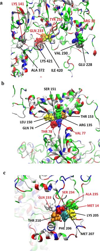
www.nature.com/scientificreports/
Figure 1. Structure of MPP1. Schematic representation (a) of the overall domain composition and ribbon
model (b) of the structure of MPP1. PDZ blue, SH3 green, D5 light blue, GUK red. Domain boundaries were
marked based on Quinn et al.11. Structural model redrawn according to Listowski et al.44.
bilayer via integral proteins. In this case the N-terminal PDZ domain of MPP1 binds to the membrane protein,
glycophorin C20, and the central D5 domain of MPP1 interacts with 4.1R p rotein21,22, thus forming a complex
which is critical for maintaining the stability and mechanical properties of erythrocyte membrane. Significantly,
our recent study performed on erythroid cells showed a novel physiological role of MPP1 in organizing func-
tional raft domains. Using MPP1 knockdown erythroid precursor HEL (human erythroleukemia) cells and giant
plasma membrane vesicles (GPMVs) derived from them, we demonstrated that the marked decrease of MPP1
protein expression (or inhibition of its palmitoylation) is directly associated with significant changes in phys-
icochemical properties of the plasma membrane monitored as an increase in membrane fluidity parameters and
phase-separation properties23,24. This, in turn was correlated with noticeable loss in isolation of DRM (detergent
resistance membranes) and marked reduction in activation of raft-dependent receptors and their downstream
signaling pathways25,26. Detailed characterization of the molecular mechanism underlying the phenomenon of
MPP1-dependent raft domain formation led us to identify the raft-marker proteins flotillin 1 and flotillin 2 as a
new, direct MPP1-binding partners in RBC plasma m embrane23. Importantly, these MPP1-flotillin interactions
were shown to be physiologically relevant and independent from well-established, aforementioned interactions of
MPP1 with 4.1 and glycophorin C, indicating a new role of the MPP1-flotillin linkage in stabilization of plasma
membrane lateral heterogeneity in native R BC23. In fact, flotillins are important scaffolding components of the
raft domains, playing a structural role in their o rganization6,27. The membrane-organizing capacity assigned to
ligomers28 that serve as active assembly sites controlling numerous dif-
flotillins is due to their ability to form o
ferent cellular processes such as s ignaling29–32, endocytosis33,34, and cell a dhesion35. These features emphasized
flotillins as preferable molecular candidates for interacting with MPP1 in the context of plasma membrane lateral
organization. Thus, based on these data, we proposed a concept where MPP1 and flotillins act as a driving force
for functional raft domain formation in living cells. Our hypothesis assumes that MPP1 binds the pre-existing
flotillins-nanoclusters/unstable rafts elements and therefore induces their fusion into larger nanodomains and
stabilizes them as membrane rafts domains which become functional. Such rearrangement is connected to a
change in membrane-lipid properties resembling formation of ordered domains and their separation from the
bulk membrane. This hypothesis is in agreement with others36, however emphasize the major role of MPP1 in
promoting oligomerization of flotillins, which in turn triggers co-assembly of flotillin-based oligomers and facili-
tates raft domain formation23. However, to build a comprehensive picture of this interesting novel mechanism, we
decided to dissect the direct interaction between MPP1 and flotillins and precisely define the molecular details
concerning their mutual binding capacity in vitro. Here, we demonstrated in vitro high affinity interactions
between MPP1 and flotillin 1 or 2 and provided molecular details of this interaction by identifying a hitherto
unknown “flotillin binding motif ”’. Moreover, a recombinant protein corresponding to this domain via inhibit-
ing this interaction reduced membrane order and markedly decreased activation of signal transduction via the
raft-dependent insulin receptor pathway.
Materials and methods
Cloning, expression and purification of recombinant proteins. All the sequences of primers for the
construction of plasmids used in this study are summarized in the Supplementary Table 1. The MPP1-truncated
mutants (MPP1-Mut1-5) were subcloned into the pGEX-6p1 vector and expressed as soluble GST tagged pro-
teins in Escherichia coli BL21 (DE3) or LEMO (DE) cells. Full length MPP1-GST and its truncated mutants were
isolated in native conditions based on HBS buffers (10 mM HEPES, 150 mM NaCl, pH 7,4) and immobilized on
Scientific Reports | (2021) 11:14751 | https://doi.org/10.1038/s41598-021-93982-3 2
Vol:.(1234567890)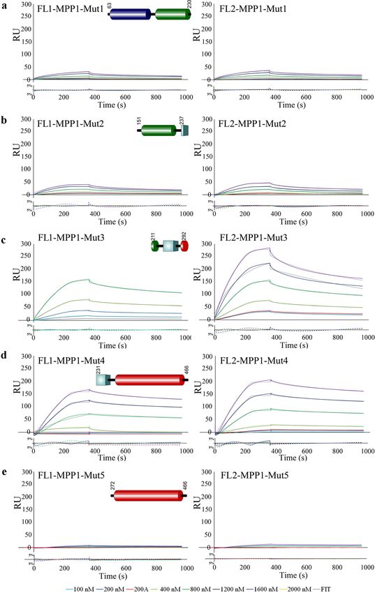
www.nature.com/scientificreports/
Figure 2. Recombinant MPP1 binds recombinant flotillin 1 or flotillin 2 with equilibrium dissociation constant,
KD, values in nanomolar range. HIS-tagged flotillins were immobilized on the Ni–NTA chip and its interaction
with full-length recombinant, untagged MPP1 (series of concentration 0–2000 nM) was analyzed by SPR using
a BIAcore T200. Black dotted lines represent fitted 1:1 Langmuir binding model. Residual plots beneath every
sensorgram series were evaluated for systematic divergences from the fitting curves. Other details in “Materials
and methods” section.
glutathione-Sepharose 4B beads (GE Healthcare). The GST tag was cleaved off on the column with the PreScis-
sion protease (Sigma) according to the manufacturer’s protocol. Purified recombinant proteins were validated
by SDS-PAGE and Coomassie Blue staining (see Fig. S2). His-tagged flotillin 1 and 2 were purified under dena-
turation conditions as described previously23. After purification recombinant proteins were dialyzed into HBS-T
(HBS-0.05% Tween-20) buffer and subsequently used for SPR binding study. For mammalian cell experiments
the MPP1-Mut4-FLAG construct was additionally cloned into the p3XFLAG-CMV-10 vector (Sigma, St. Louis,
MO).
Real‑time interaction analysis by surface plasmon resonance. The binding of full length MPP1
(Fig. 2) and its truncated mutants (Fig. 5) to recombinant His tagged flotillin 1 or flotillin 2 immobilized on
Ni–NTA sensor chips (Series S sensor chip NTA; GE Healthcare) was analyzed by SPR using a BIAcore T200
(GE Healthcare) as described elsewhere37. Briefly, purified recombinant flotillin 1 or flotillin 2 was bound to the
prepared surface via the His tag to a final level of approximately 4000 RU. To determine affinity of all analytes to
flotillin 1 or flotillin 2, at least five different concentrations of each analyte (range from 50 to 2000 nM), as well as
a sample buffer blank, were passed over the ligand-immobilized-(association phase—360 s) or the control empty
chip surface followed by dissociation with running buffer (600 s). The resulting sensorgrams were obtained by
subtracting the buffer blank from sample curves (recorded for the interactions of flotillins with MPP1 or its
mutants) followed by substraction of sensorgrams obtained from empty chip surface. The equilibrium constants
(KD) defined as a kon/koff ratio were determined using BIAevaluation 3.1 software using global fitting and a 1:1
Langmuir binding model with an included mass transport step. Residuals were evaluated for systematic diver-
gences from the fitting algorithms as a measure of the appropriateness of the binding model.
Cell lines. HEL cells were kindly provided by Prof. M. Majka from Jagiellonian University of School Medi-
cine. Cells were grown in RPMI 1640 medium supplemented with 10% fetal calf serum, 2 mM glutamine, 100
units/mL penicillin, and 100 μg/mL streptomycin at 37 °C in a humidified atmosphere of 5% C
O2.
Cell transfection and activation. Transient transfections of HEL cells were performed by CLB (Lonza,
Basel, Switzerland) electroporation. Briefly, 2 × 10⁶ cells were transfected with empty p3XFLAG-CMV-10 vec-
tor (Sigma, St. Louis, MO) (FLAG-control) or MPP1-Mut4 plasmid respectively. 24 h post transfection cells
were serum-starved for 20 h before treatment with human recombinant insulin (Gibco) [1 μg/ml] for 5 min, at
37 °C in a humidified atmosphere of 5% CO2. For immunoblotting, the stimulated cells were harvested, washed
with ice-cold PBS and lysed for 30 min on ice in the lysis buffer (50 mM HEPES, pH 7.5, 100 mM NaCl, 1 mM
EDTA, 10% glycerol, 1% NP-40) supplemented with 100 μM PMSF, protease inhibitor cocktail (Sigma-Aldrich)
and phosphatase inhibitor cocktail (Santa Cruz). Proteins were separated by SDS-PAGE, followed by Western
blot analysis. Immunodetection was performed using antibodies against phospho ERK (p-ERK 1/2), total ERK
1/2, and FLAG. Blots were quantified and normalized to appropriate loading controls using ImageJ software. All
immunoblotting data presented here are representative of four independent experiments.
FLIM analysis. FLIM was used to measure fluorescence lifetime values of a membrane-order sensitive
probe, di-4-ANEPPDHQ (Life Technologies). After 48 h cells transfected with appropriate plasmids (FLAG
transfected/control; MPP1-Mut4 transfected) were stained with 2 μM di-4 for 15 min at room temperature (RT),
subsequently washed with HBSS buffer supplemented with 10 mM HEPES, pH 7.4 (Gibco) and transferred
onto poly-l-lysine coated lab-TEK chambers and left for 20 min at RT. Such conditions resulted in labeling not
Scientific Reports | (2021) 11:14751 | https://doi.org/10.1038/s41598-021-93982-3 3
Vol.:(0123456789)
www.nature.com/scientificreports/
only plasma membrane but also intracellular membranes, as shown in Fig. S8 and reported elsewhere38,39. FLIM
measurements were performed at 23 °C using an LSM 510 META microscope (Carl Zeiss GmbH, Germany)
upgraded with FLIM and FCS capabilities (PicoQuant GmbH, Germany) as described previously23,25. Briefly,
470 nm pulsed-laser with 40 MHz repetition rate was used for di-4 excitation and emission was detected with the
use of long pass 505 nm emission filter. Average photon rate was approximately at the level of 1 04–105. Acquisi-
tion time was dependent on the intensity of the samples. Lifetime calculations of di-4 probe was measured only
from the plasma membrane region (ROI; region of interest) as described previously38,39. Decay curves were fit-
ted in the range up to 20 ns for all the experiments and the FLIM raw data were processed using SymPhoTime
software (PicoQuant GmbH, Germany). Statistical analysis was performed using two-tailed unpaired t-test.
CD analysis. Circular dichroism (CD) measurements were performed using a JASCO J-1500 spectrometer
in a temperature-controlled cell (0.1 or 0.01 cm path length). All proteins were analyzed in HEPES-based buffer
with addition of 0.1% Tween-20. Raw data were converted into molar ellipticity per residue using the equation:
CDunits
θ = 10∗n∗p∗c , where CD units were in [mdeg] n-number of residues, p- path length in [cm], c- concentration in
[ mol
l ].
Flotillin 1 and flotillin 2 protein modelling. The sequences for flotillin 1 (O75955-1) and flotillin 2
(Q14254) proteins were obtained in FASTA format from UniProt. As no single complete protein structures
of both flotillin 1 and 2 have been published, a model was built using I-TASSER software40. The obtained AA
sequences of flotillin 1 and 2 were given as a query. The web server operated at its default setting, that is, no
specification of templates was given. Also, no assignment of contact or distance restraints was given. The server
was also set to not exclude any homologous or specific templates. Since the full structure of flotillin 1 remains
unknown, the top template which was selected by the server was the SPFH domain of flotillin 2 (PDB: 1WIN).
For flotillin 2 a protein from the liver vault was selected as a template (PDB: 4V60). To validate the correctness
of the server selection, known fragments of flotillins (SPFH domains) were compared to those of 1WIN, 4FVF,
4FVJ, 4FVG and 3BK6 using the TM-align online tool41. They were found to be structurally similar (TM-score
0.72, 0.75, 0.88, 0.91 and 0.97 for flotillin 1; TM-score 0.95, 0.78, 0.82, 0.84, 0.64 for flotillin 2). Additionally,
C-score and total energy were calculated. The top-ranked model constructed by the server was used in further
in silico experiments.
Molecular dynamics simulation. The full-atomistic molecular dynamics simulations were performed
using NAMD 2.1342 software with CHARMM36 force fi eld43 under NPT conditions (constant: number of parti-
cles, pressure, and temperature). Several systems were created for both stabilization of investigated proteins (flo-
tillin 1 and flotillin 2) and investigation of interactions between them and the MPP1 protein m odel44. All systems
were hydrated with TIP3P water molecules and ionized with 0.15 M NaCl. Stabilization of investigated systems
was carried out for at least 100 ns for each of the investigated protein models. Simulations involving studying the
interactions between the flotillin 1 and MPP1 proteins took, in total, 115 ns. For flotillin 2 they took 56 ns as the
bindings occurred faster. Three dimensional periodic boundary conditions were applied in order to deal with
potential energy disruption due to the origin cell discontinuity. More detailed descriptions of individual simula-
tions are presented in Supplementary Information. To establish the binding between the flotillins and MPP1 a
simple approach was used. For each simulation step possible binding sites were flagged if at least three MPP1/
flotillin atoms from the amino acid group were within 3 Å of flotillin/MPP1 atoms.
Results
MPP1 binds flotillin 1 (FL1) and 2 (FL2) with high affinity in vitro. In our previous study we dem-
onstrated that the newly identified linkage between MPP1 and flotillins contributes to the lateral plasma mem-
brane organization in native R BCs23. Here, we decided to characterize the molecular details concerning MPP1-
flotillin interaction and more precisely define their kinetic parameters and binding capacity in vitro using an
SPR approach. Therefore, to examine the nature of individual interactions first, recombinant His-tagged flotillin
1 or flotillin 2 was immobilized on Ni–NTA biosensors and a series of concentrations of full length recombi-
nant MPP1 was used as an analyte. The double-referenced sensorgrams were fit globally to a 1:1 kinetic binding
model (Fig. 2), and calculated kon, koff and KD values, which represent the association rate, dissociation rate and
equilibrium disassociation constant, respectively, are shown in Table 1.
Interestingly, the kinetic analysis revealed that both recombinant flotillin 1 and flotillin 2 interact with MPP1
with KD in the nanomolar range where the interaction of MPP1 and flotillin 1 exhibited a K D value of approxi-
mately 22.8 nM, while with flotillin 2 it showed a K D of 31 nM (Fig. 2, Table 1). Significantly, the obtained K D
values were independent of analyte concentration, as presented in Fig. S1, and global calculations for K D are
similar to those calculated from individual curves for different analyte concentrations. Moreover, these particular
data indicate a relatively high global association rate of binding MPP1 ~ 4.5 × 10⁴ 1/Ms for both proteins (see
Table 1). We have to stress that the results of single concentration series were in good agreement with the data
obtained by calculating the averages from all experiments for both flotillins. Thus, the results of the systematic
approach presented here document the interaction parameters between MPP1 and flotillin 1 or flotillin 2 with
high consistency, indicating that these proteins are able to form high-affinity complexes in vitro.
Molecular dynamic (MD) simulations of MPP1‑flotillin interactions. In order to provide in-depth
information about the potential binding site(s) involved in mutual MPP1-flotillin interactions MD simulations
were carried out. For the purpose of these simulations both flotillin models were built from the sequence and
rotein44 to each of the flotillins was ana-
tested (see Supplementary pdb files). Binding of the full length MPP1 p
Scientific Reports | (2021) 11:14751 | https://doi.org/10.1038/s41598-021-93982-3 4
Vol:.(1234567890)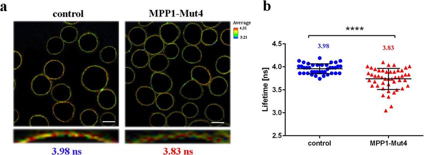
www.nature.com/scientificreports/
Kinetic parameters
Interaction kon (1/Ms) koff (1/s) KD (M)
FL1-MPP1 4,50E+04 1,03E−03 2,28E−08
SD 3,80E+03 9,36E−05 5,48E−10
FL1-MPP1-Mut1 n/a n/a n/a
FL1-MPP1-Mut2 1,68E+03 1,18E−03 7,02E−07
SD 2,98E+02 2,29E−04 4,38E−08
FL1-MPP1-Mut3 3,75E+03 5,15E−04 1,37E−07
SD 5,73E+02 3,99E−05 3,46E−08
FL1-MPP1-Mut4 8,13E+03 3,19E−04 3,92E−08
SD 2,13E−02 3,04E−05 5,21E−09
FL1-MPP1-Mut5 n/a n/a n/a
FL2-MPP1 4,13E+04 1,28 E−03 3,10E−08
SD 8,64E+03 2,35 E−04 2,89E−09
FL2-MPP1-Mut1 n/a n/a n/a
FL2-MPP1-Mut2 1,63E+03 1,60E−03 9,82E−07
SD 4,04E+02 4,74E−04 8,51E−08
FL2-MPP1-Mut3 4,17E+03 8,28E−04 1,99E−07
SD 5,74E+02 1,52E−05 7,77E−09
FL2-MPP1-Mut4 5,60E+03 2,81E−04 5,02E−08
SD 9,71E+02 4,79E−05 7,30E−09
FL2-MPP1-Mut5 n/a n/a n/a
Table 1. Kinetic parameters for the interaction of full length recombinant MPP1 or its truncated mutants with
flotillin 1 and 2. Obtained KD values are averages of 3 independent series of experiments presented in Figs. 2
and 5 and were found to be independent of ligand concentration (see Figure S1).
lyzed and several simulations were performed with different orientations in the simulation space (see Fig. S4
and S5). In the case of MPP1-flotillin 1 five potential binding sites were detected during experiments (Table S2);
however, some of them exhibited relatively low electrostatic binding force strength (lower than ten percent of the
highest values), and were considered unstable. The other, highly probable binding sites in molecular simulations
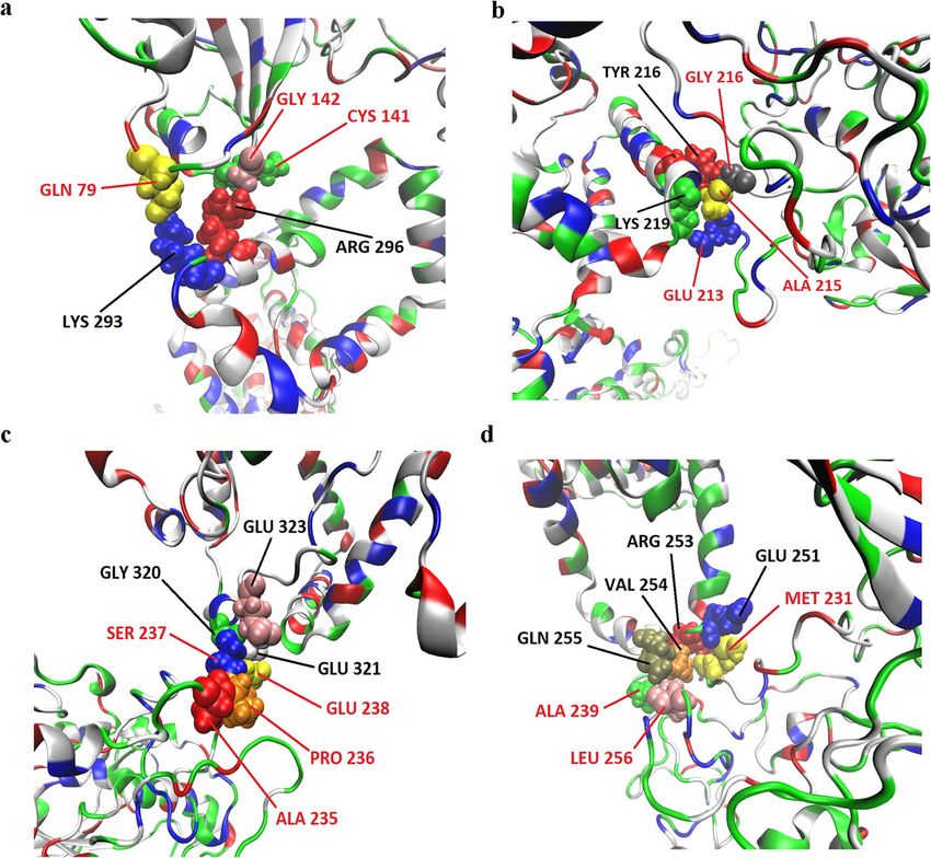
involved Gln79/Cys141/Gly142 on MPP1 and Lys293/Arg296 on flotillin 1 (Fig. 3a), Glu213/Ala215/Gly216 on
MPP1 and Tyr216/Lys219 on flotillin 1 (Fig. 3b), Ala235/Pro236/Ser237/Glu238 on MPP1 and Gly320/Glu321/
Glu323 on flotillin 1 (Fig. 3c) and Met231/Ala239/Leu256 on MPP1 and Glu251/Arg253/Val254/Gln255 on
flotillin 1 (Fig. 3d). In the case of MPP1-flotillin 2 simulations revealed four binding sites (Table S1) from
which three were considered as stable binding sites; those were as follows: Arg32/Lys141/Gln233/Tyr255 on
MPP1 and Asn228/Val230/Ala372/Ile420/Lys421 (Fig. 4a), Val77/Thr78 on MPP1 and Gln74/Thr135/Leu150/
Ser151/Thr153 of flotillin 2 molecule (Fig. 4b) and Gln233/Ser234/Ala235/Met14 on MPP1 and Lys205/Phe206/
Met207/Thr201 on flotillin 2 molecule (Fig. 4c).
Mapping of a direct MPP1 interacting site for flotillins with SPR analysis. To evaluate the results
obtained from our MD simulations, and check which of the above-mentioned potential binding sites might
occur in vitro, we designed a series of MPP1-truncated mutants (Fig. 5), which were further used as analytes in
SPR measurements to assess their individual binding with both flotillins. Purity of these recombinant proteins
and their CD spectra are shown in Fig. S2 and S3. As shown in Table 1 and Fig. 5, from all analyzed MPP1
mutants, the highest affinity for flotillins was observed for MPP1-Mut4 (residues 231–466) (Fig. 5d). The cal-
culated KD value was 39.2 nM for flotillin 1, and 50.2 nM for flotillin 2 (Table 1). On the other hand, we also
observed significant interaction between MPP1-Mut3 (residues 211–292) and MPP1-Mut2 (residues 151–237)
with each of the flotillins. Here, the obtained K D values of MPP1-Mut3 were approximately 137 nM for flotil-
lin 1 and 199 nM for flotillin 2 (Fig. 5c, Table 1), while in the case of MPP1-Mut2 the binding was weaker, but
still observable, and the K D values were 702 nM and 686 nM, respectively (Fig. 5b, Table 1). The obtained K D
values were highly reproducible and in all cases independent of analyte concentration (Fig. S1). Importantly, our
kinetic analysis of MPP1-Mut1 (residues 63–230) and MPP1-Mut5 (residues 272–466) virtually excluded the
involvement of both the N-terminal and C-terminal regions of MPP1 in binding of flotillins, as no significant
interactions were observed (Fig. 5a,e, Table 1). Notably, the sensorgram response curves revealed a very slow
association even in high analyte concentrations, and therefore seemed to be the effect of non-specific binding
in our experimental conditions rather than real interaction. Altogether, the above-presented kinetic parameters
for MPP1 recombinant mutants highlighted that the central region of MPP1 is directly involved in flotillin 1 and
2 binding. Being more precisely based on the MPP1-mutant sequences and MD simulation data, we indicate
the involvement of key amino acid residues in the MPP1 molecule, in particular in positions 231–238 and 256,
which serve as critical flotillin binding sites. This particular region corresponds to the D5 domain of MPP1.
Scientific Reports | (2021) 11:14751 | https://doi.org/10.1038/s41598-021-93982-3 5
Vol.:(0123456789)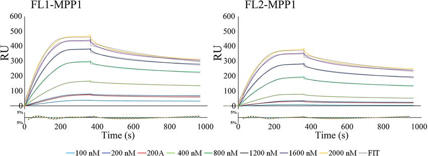
www.nature.com/scientificreports/
Figure 3. Visualization of MPP1-flotillin 1 binding sites mapped by molecular dynamics simulations. (a–d)
Binding sites of MPP1 and FL1. MPP1 amino acid residues are marked in red, FL1 in black. Water molecules
were removed for clarity.
MPP1‑Mut4 affects plasma membrane order in living cells and modulates the raft‑dependent
signaling pathway. To assess the physiological significance of the newly identified “flotillin binding motif ”
we decided to test whether the MPP1-Mut4 has any effect on endogenous MPP1-flotillin interaction in living
cells. Here, we took advantage of our well-described model system, the HEL cell line. Decreasing the level of
MPP1 in these cells was previously shown to significantly affect raft domain formation, observed as a marked
increase in plasma membrane and GPMVs fluidity parameters24,25. Therefore, to evaluate the possible biologi-
cal impact of this fragment, HEL cells were transiently transfected with MPP1-Mut4-FLAG plasmid and con-
trol cells were transfected with “empty” FLAG plasmid. Subsequently, cells were labeled with the lipid bilayer
order-sensing fluorescent dye di-4-ANEPPDHQ (di-4)38, the fluorescence lifetime of which is sensitive to mem-
brane fluidity and analyzed in FLIM (fluorescence lifetime imaging microscopy). As shown in Fig. 6, FLIM data
revealed statistically significant reduction in the lifetime value of the di-4 dye (~ 0.15 ns shift), in plasma mem-
brane (ROI) of MPP1-Mut4 transfected cells (~ 3.83 ns) compared to the control (~ 3.98 ns). Such fluorescence
lifetime shift of the di-4 probe is associated with an increase in membrane fl uidity38. In this particular context
it might be understood as partial disruption of MPP1-dependent raft domains due to the competitive binding
of MPP1-Mut4 to endogenous flotillins, which eliminates the latter from interactions with endogenous MPP1.
As the disorganization of functional raft domains was reported previously to be correlated with impaired
signal transduction from the activated raft-dependent insulin receptor (IR)25,26, to cross-check the “competi-
tive” effect of MPP1-Mut4 on MPP1-flotillin interaction, the activation of IR with insulin was performed on
transfected cells. As only the ERK1/2 signaling cascade downstream of the activated IR was shown to be MPP1-
dependent in HEL c ells26, the level of phosphorylated ERK1/2 (pERK1/2) was then monitored from whole-
cell lysates after stimulation. Interestingly, we observed a marked reduction of pERK1/2 level in MPP1-Mut4
transfected cells (Fig. 7). The difference in activation level was approximately 46%, which may imply a partial
competition of exogenously expressed MPP1-Mut4 and hence partial displacement of endogenous MPP1 from
Scientific Reports | (2021) 11:14751 | https://doi.org/10.1038/s41598-021-93982-3 6
Vol:.(1234567890)www.nature.com/scientificreports/
Figure 4. Visualization of MPP1-flotillin 2 binding sites mapped by molecular dynamics simulations. (a–c)
Binding sites of MPP1 and FL2. MPP1 amino acid residues are marked in red, FL2 in black. Water molecules
were removed for clarity.
Scientific Reports | (2021) 11:14751 | https://doi.org/10.1038/s41598-021-93982-3 7
Vol.:(0123456789)www.nature.com/scientificreports/
Figure 5. The D5 domain of MPP1 is of key importance for binding of flotillins. (a–e) HIS-tagged flotillins
were immobilized on the Ni–NTA chip and their interactions with untagged MPP1 truncated mutants (MPP1-
Mut1-5) in a series of concentrations 0–1600 nM were analyzed using a BIAcore T200. Other details as in Fig. 2
legend.
Scientific Reports | (2021) 11:14751 | https://doi.org/10.1038/s41598-021-93982-3 8
Vol:.(1234567890)www.nature.com/scientificreports/
Figure 6. MPP1-Mut4 affects membrane order in HEL cells. Representative FLIM images (a) and quantitative
analysis (b) of di-4 lifetime distribution collected from plasma membrane (ROI) of control (FLAG-transfected)
and MPP1-Mut4-FLAG transfected HEL cells indicating significant (p < 0.0001) reduction in membrane
order parameters after inhibition of endogenous MPP1-flotillin interactions. Each dot on a graph represents a
single image of control (n = 38) and MPP1-Mut4-FLAG transfected cells (n = 42). Uncropped images, lifetime
histograms as well as decay curves for representative images are shown in Fig. S8. Statistical analysis was
performed using two-tailed unpaired t-test. Scale bar, 5 µm.
Figure 7. MPP1-Mut4 inhibits signal transduction from raft-dependent IR receptor in HEL cells. Control
(FLAG-transfected) and MPP1-Mut4 transfected cells were treated with insulin and the whole-cell extracts were
subjected to Western blot analysis and probed with antibodies against indicated proteins (a). Quantification of
the relative phosphorylation levels of ERK1/2 in control and MPP1-Mut4 transfected cells (average ± S.D. from
four independent experiments; statistical analysis was performed using non-parametric t-test p < 0.05) were
performed (b). Uncropped blots are shown in Fig. S9.
the flotillin-binding complex. Notably, these data are consistent with our previously described effect of MPP1
knockdown on IR receptor activation and its downstream signaling in HEL c ells26.
Discussion
Our previous study showed that direct interaction between MPP1 and flotillins exists in the plasma membrane
of RBC and functionally contributes to the organization of membrane rafts in living erythroid cells23,25. Namely,
we showed that the decrease in functional MPP1 is correlated with di-4 lifetime in both plasma membranes of
living cells and GPMVs derived from erythroid cells. Silencing of the MPP1 gene also led to a dramatic decrease
in the DRM fraction25, but on the other hand, no changes in major lipid classes (including cholesterol and sphin-
gomyelins) for plasma membrane-derived GPMVs could be observed24. Moreover, macroscopic phase separation
in GPMVs obtained from MPP1-knockdown cells was observable approx. up to 15 °C, whereas control vesicles
remained phase separated up to 17 °C24 confirming that MPP1 stabilizes more ordered phases and membrane
phase coexistence. Next, flotillins, membrane raft marker proteins were found to be a direct binding partners
for MPP1 in RBC plasma membrane23. These data opened a novel outlook in our understanding of the role of
the MPP1 molecule as a critical regulatory and structural partner of raft-associated proteins23. The physiologi-
cal relevance of formation of the MPP1-flotillin complexes in plasma membrane raised an important question
about the molecular details underlying this phenomenon. Therefore, in this study we decided to dissect these
interactions at the molecular level and precisely define the flotillins binding capacity of MPP1 and identify flotillin
Scientific Reports | (2021) 11:14751 | https://doi.org/10.1038/s41598-021-93982-3 9
Vol.:(0123456789)www.nature.com/scientificreports/
binding site(s) in vitro. Using a system of recombinant proteins and SPR-based kinetic analysis, we demonstrated
for the first time the quantitative parameters of binding of recombinant MPP1 to both recombinant flotillin 1
and 2. Such an approach is particularly important, as so far no kinetic data have been available, although several
flotillin-binding partners are known5. Interestingly, our data showed that the values of obtained equilibrium
dissociation constants as well as the association and dissociation constants (kon and koff ) were similar in the case
of both flotillins (Fig. 2, Table 1). This may be associated with a high degree of homology for flotillin 1 and 2,
which share ~ 50% amino acid sequence identity45,46, which in turn might suggest that MPP1 probably binds
homologous regions within flotillins (see below). It should be emphasized that the obtained K D values were highly
reproducible and in all cases independent of analyte concentration (Fig. S1), indicating that MPP1 and each of the
flotillins form high-affinity complexes in vitro. Being interested which regions of MPP1 are directly involved in
this binding, we performed the MD simulations of full length MPP1 and each flotillin. Structural models of the
latter were built based on their sequences. This analysis revealed four, in the case of MPP1-flotillin 1 and three for
MPP1-flotillin 2, presumable binding sites (Figs. 3, 4). Based on these data, a series of MPP1-truncated mutants
were designed and their binding capacity towards each flotillin was further evaluated in SPR experiments. Such
an approach enabled us to experimentally verify and more precisely characterize the “flotillin binding motif ”
within the MPP1 molecule. In particular, we found that three MPP1 mutants whose sequences share a common
central region starting from residue 231 showed significant binding of flotillins in our SPR experiments. The
highest affinity was observed for MPP1-Mut4 (231–466) (Fig. 5d, Table 1), and these values were close to those
obtained for the full length recombinant MPP1 with each flotillin (Fig. 2). Over three times higher KD values were
observed for MPP1-Mut3, whose sequence included the range of residue 211–292 (Fig. 5c, Table 1). The weakest,
although still detectable, interaction with flotillins was observed for MPP1-Mut2 (residues 151–237) (Fig. 5b,
Table 1). This result is particularly interesting, as it suggests that the sequence of 7 amino acid residues between
231 and 237 have a considerable contribution to the affinity of MPP1 for flotillins (Fig. 5, Table 1). Notably, as
no interaction with flotillins was detected for mutants comprising the N-terminus (up to residue 230) (MPP1-
Mut1) or C-terminus (starting from residue 272) (MPP1-Mut5), we concluded that the “flotillin binding motif ” is
located between residues 231–271. Most precisely, together with MD simulation data we ascertained that amino
acid residues 231, 235, 236, 237, 238 in the MPP1 molecule are engaged in stable binding with flotillin 1 (Fig. 3).
In the case of flotillin 2 such binding is found for residues 233, 234, 235 and 256 in the MPP1 molecule (Fig. 4).
Thus, we found that these key amino acid residues within the D5 domain of MPP1 are essential for high affinity
interaction with flotillins. Moreover, further experiments with MPP1-Mut4 performed on the well-established
erythroid HEL cell line (erythroblastoma) showed the biological relevance of this characteristic “flotillin bind-
ing motif ”, which acts as an “endogenous competitor” for naturally occurring MPP1-flotillins complexes that
can be observed primarily at plasma membrane of HEL cells, as determined in proximity ligation assay (PLA)
(see Fig. S7 and supplementary movies). Such specific inhibition capability of MPP1-Mut4 was manifested as
significant loss of membrane ordering parameters of the di-4 probe (~ 0.15 ns) compared to the control cells
(Fig. 6). Importantly, these data are in line with our studies performed on RBC23 or HEL MPP124,25. Further-
more, together with the changes in plasma membrane order, we also observed significant modulation of signal
transduction via raft-dependent IR receptor signaling in cells transfected with MPP1-Mut4, where the level of
the downstream activated pERK1/2 was approximately 50% lower compared to control cells (Fig. 7). Moreover,
we also confirmed the inhibitory effect of recombinant MPP1-Mut4 on recombinant full-length MPP1-flotillin 1
interaction using competitive ELISA assay and bacterially expressed proteins (Fig. S6). Here, the inhibition effect
of MPP1-Mut4 was approximately 15% when compared to MPP1-Mut1, and the magnitude of the competition
effect was statistically significant. Taken together with the high affinity of MPP1-Mut4 for flotillins, it might imply
that this mutant interferes with the endogenous MPP1 which forms complexes with flotillins and results in the
disorganization of MPP1-dependent raft domains. Such competition leads to a significant decrease in plasma
membrane ordering parameters and, as a consequence, affects raft-dependent signaling pathways.
The localization of such a newly mapped “flotillin binding motif ”’ in MPP1 is particularly interesting, as so
far the MPP1-D5 domain has been characterized as a main binding site for 4.1R protein in R BCs21,22, defining
its primary role in maintaining the mechanical properties of RBCs. The high affinity of both 4.1R (70 nM22) and
flotillins to the same region in the MPP1 molecule thus suggests its bifunctional involvement in different cel-
lular processes. The question remains whether these molecules compete for binding to the D5 domain of MPP1
or rather such complexes can be formed independently of each other. Notably, our recent study indicated that
the interaction of MPP1 with flotillins is independent of 4.1R binding23, which strongly suggests that MPP1
may simultaneously form two important types of complexes which independently control specialized functions
within plasma membranes. Binding and stabilizing protein–protein complexes is a primary assigned function
for the MPP subfamily. To fulfil this role, the conserved PDZ-SH3-GUK has been suggested to play key roles in
multiple interactions with other molecules13. In erythroid cell membrane the role of the D5 domain is crucial. Of
note, the D5 domain has been found in the structure of five (MPP1, MPP2, MPP5, MPP6, MPP7) out of seven
members of this subfamily18. Such an additional domain might functionally distinguish these members from
the others, allowing them to act as more versatile multifaceted organizers. However, when aligning amino acid
sequences of the D5 domain of MPP members, particularly with respect to the putative “flotillin binding motifs”,
we could not find any similarities. This, in turn, emphasizes that the “flotillin binding motif ” in the D5 domain
of MPP1 is unique. Therefore, the molecular characterization of direct interaction between MPP1 and flotillins
brings us closer to defining key aspects of membrane organization and identifying novel potential therapeutic
targets. This would be of particular interest in therapies of several tumors in which flotillin-dependent domains
were shown to be closely associated with progression, development and m etastasis47. Further systematic studies
should consider experimental identification of the MPP1-binding site in flotillins; however, our MD simulation
data indicate that C-terminal domains (flotillin domain) of each flotillin are involved in MPP1 binding (Figs. 3,
4). Given that the N-terminal region of flotillins mediates membrane binding and the C-terminal flotillin domain
Scientific Reports | (2021) 11:14751 | https://doi.org/10.1038/s41598-021-93982-3 10
Vol:.(1234567890)www.nature.com/scientificreports/
is responsible for the oligomerization of flotillins in living c ells5,6, binding of MPP1 close to the C-terminal region
could therefore mediate the latter process.
Although the high-affinity interactions between MPP1 and flotillins were observed in solution, the impact
of lipid-bilayer cannot be excluded, since both proteins operate within or at the vicinity of plasma membrane
of living cells (see Fig. S7). This fact is directly linked with the observed effects of the loss of MPP1 or MPP1-
flotillins interactions in RBCs and HEL cells on the lateral organization of plasma membrane. Based on the model
proposed by o thers36, and the fact that that functional raft domains are formed and stabilized temporarily upon
internal factors (like oligomerization), we proposed a hypothesis which assumed the major role of MPP1 in
oligomerization of flotillins assemblies into larger stable functional domains. Such specific MPP1-based seques-
tering/clustering process, may trigger the local changes in the organization of surrounding lipids, and attracting
other molecules, resulting in physicochemical changes in plasma m embrane23,26. It might be expected that the
reported here high-affinity MPP1-flotillins interactions could be further strengthened at the cytoplasmic surface
of plasma membrane due to multiple factors, including direct interactions of MPP1 and/or flotillins with lipids
which may lead to reduced dimensionality. Indeed, results of some studies show that many protein–protein
interactions can experience increases of effective affinities due to membrane localization48. This hypothesis should
definitely point at further research directions in this field, which would require establishing new experimental
models, such as MPP1 and/or flotillins reconstituted in proteoliposomes and immobilization-free technology
to measure interactions.
To the best of our knowledge, this is the first report showing molecular details underlying formation of the
MPP1-flotillin complex that opens a new outlook in our understanding of the involvement of MAGUK-scaffold-
ing molecules and raft-marker proteins, flotillins, in the mechanism that governs the organization of functional
raft domains. In fact, high affinity interaction of MPP1 to flotillins explains at least in part the biological ability of
the formed complexes to maintain and modulate the properties of the plasma membrane, i.e. lateral membrane
organization and its homeostasis. In other words, it becomes evident that these interactions are endogenous
factors regulating raft domain formation in living cells.
Data availability
Further information and requests for resources and reagents should be directed to and will be fulfilled by the
Lead Contact, Aleksander Czogalla (aleksander.czogalla@uwr.edu.pl). Materials and plasmids generated in this
study are available upon request from the Lead Contact.
Received: 10 January 2021; Accepted: 28 June 2021
References
1. Lingwood, D. & Simons, K. Lipid rafts as a membrane-organizing principle. Science 327(5961), 46–50 (2010).
2. Hryniewicz-Jankowska, A. et al. Membrane rafts as a novel target in cancer therapy. Biochim. Biophys. Acta 1845(2), 155–165
(2014).
3. Sezgin, E. et al. The mystery of membrane organization: composition, regulation and roles of lipid rafts. Nat. Rev. Mol. Cell Biol.
18(6), 361–374 (2017).
4. Simons, K. & Ikonen, E. Functional rafts in cell membranes. Nature 387(6633), 569–572 (1997).
5. Kwiatkowska, K. et al. Flotillins: At the intersection of protein S-palmitoylation and lipid-mediated signaling. Int. J. Mol. Sci. 21(7),
2283 (2020).
6. Langhorst, M. F., Reuter, A. & Stuermer, C. A. Scaffolding microdomains and beyond: the function of reggie/flotillin proteins. Cell
Mol Life Sci. 62(19–20), 2228–2240 (2005).
7. Dimitratos, S. D. et al. Signaling pathways are focused at specialized regions of the plasma membrane by scaffolding proteins of
the MAGUK family. BioEssays 21(11), 912–921 (1999).
8. Ye, F., Zeng, M. & Zhang, M. Mechanisms of MAGUK-mediated cellular junctional complex organization. Curr. Opin. Struct. Biol.
48, 6–15 (2018).
9. Gosens, I. et al. MPP1 links the Usher protein network and the Crumbs protein complex in the retina. Hum. Mol. Genet. 16(16),
1993–2003 (2007).
10. Kim, G. et al. Membrane palmitoylated protein 2 is a synaptic scaffold protein required for synaptic SK2-containing channel func-
tion. Elife 5, e12637 (2016).
11. Quinn, B. J. et al. Erythrocyte scaffolding protein p55/MPP1 functions as an essential regulator of neutrophil polarity. Proc. Natl.
Acad. Sci. USA 106(47), 19842–19847 (2009).
12. Funke, L., Dakoji, S. & Bredt, D. S. Membrane-associated guanylate kinases regulate adhesion and plasticity at cell junctions. Annu.
Rev. Biochem. 74, 219–245 (2005).
13. Gonzalez-Mariscal, L., Betanzos, A. & Avila-Flores, A. MAGUK proteins: structure and role in the tight junction. Semin. Cell Dev.
Biol. 11(4), 315–324 (2000).
14. Oliva, C. et al. Role of the MAGUK protein family in synapse formation and function. Dev. Neurobiol. 72(1), 57–72 (2012).
15. Won, S. et al. MAGUKs: multifaceted synaptic organizers. Curr. Opin. Neurobiol. 43, 94–101 (2017).
16. Zheng, C. Y. et al. MAGUKs, synaptic development, and synaptic plasticity. Neuroscientist 17(5), 493–512 (2011).
17. Zhu, J., Shang, Y. & Zhang, M. Mechanistic basis of MAGUK-organized complexes in synaptic development and signalling. Nat
Rev Neurosci 17(4), 209–223 (2016).
18. Chytla, A. et al. Not just another scaffolding protein family: The multifaceted MPPs. Molecules 25(21), 4954 (2020).
19. Ruff, P., Speicher, D. W. & Husain-Chishti, A. Molecular identification of a major palmitoylated erythrocyte membrane protein
containing the src homology 3 motif. Proc. Natl. Acad. Sci. USA 88(15), 6595–6599 (1991).
20. Marfatia, S. M. et al. The PDZ domain of human erythrocyte p55 mediates its binding to the cytoplasmic carboxyl terminus of
glycophorin C. Analysis of the binding interface by in vitro mutagenesis. J. Biol. Chem. 272(39), 24191–24197 (1997).
21. Hemming, N. J. et al. Identification of the membrane attachment sites for protein 4.1 in the human erythrocyte. J. Biol. Chem.
270(10), 5360–5366 (1995).
22. Seo, P. S. et al. Alternatively spliced exon 5 of the FERM domain of protein 4.1R encodes a novel binding site for erythrocyte p55
and is critical for membrane targeting in epithelial cells. Biochim. Biophys. Acta 1793(2), 281–289 (2009).
Scientific Reports | (2021) 11:14751 | https://doi.org/10.1038/s41598-021-93982-3 11
Vol.:(0123456789)www.nature.com/scientificreports/
23. Biernatowska, A. et al. MPP1 directly interacts with flotillins in erythrocyte membrane: Possible mechanism of raft domain forma-
tion. Biochim. Biophys. Acta Biomembr. 1859(11), 2203–2212 (2017).
24. Podkalicka, J. et al. MPP1 as a factor regulating phase separation in giant plasma membrane-derived vesicles. Biophys. J. 108(9),
2201–2211 (2015).
25. Biernatowska, A. et al. The role of MPP1/p55 and its palmitoylation in resting state raft organization in HEL cells. Biochim. Biophys.
Acta 1833(8), 1876–1884 (2013).
26. Podkalicka, J. et al. The microdomain-organizing protein MPP1 is required for insulin-stimulated activation of H-Ras. Oncotarget
9(26), 18410–18421 (2018).
27. Sikorski, A. F. et al. Membrane rafts in the erythrocyte membrane: a novel role of MPP1p55. Adv. Exp. Med. Biol. 842, 61–78 (2015).
28. Solis, G. P. et al. Reggie/flotillin proteins are organized into stable tetramers in membrane microdomains. Biochem. J. 403(2),
313–322 (2007).
29. Amaddii, M. et al. Flotillin-1/reggie-2 protein plays dual role in activation of receptor-tyrosine kinase/mitogen-activated protein
kinase signaling. J. Biol. Chem. 287(10), 7265–7278 (2012).
30. Neumann-Giesen, C. et al. Role of EGF-induced tyrosine phosphorylation of reggie-1/flotillin-2 in cell spreading and signaling
to the actin cytoskeleton. J. Cell. Sci. 120(Pt 3), 395–406 (2007).
31. Sugawara, Y. et al. The lipid raft proteins flotillins/reggies interact with Galphaq and are involved in Gq-mediated p38 mitogen-
activated protein kinase activation through tyrosine kinase. Cell Signal 19(6), 1301–1308 (2007).
32. Koh, M. et al. A novel role for flotillin-1 in H-Ras-regulated breast cancer aggressiveness. Int. J. Cancer 138(5), 1232–1245 (2016).
33. Glebov, O. O., Bright, N. A. & Nichols, B. J. Flotillin-1 defines a clathrin-independent endocytic pathway in mammalian cells. Nat.
Cell Biol. 8(1), 46–54 (2006).
34. Redpath, G. M. I. et al. Flotillins promote T cell receptor sorting through a fast Rab5-Rab11 endocytic recycling axis. Nat. Com-
mun. 10(1), 4392 (2019).
35. Guillaume, E. et al. Flotillin microdomains stabilize cadherins at cell-cell junctions. J. Cell Sci. 126(Pt 22), 5293–5304 (2013).
36. Hancock, J. F. Lipid rafts: contentious only from simplistic standpoints. Nat. Rev. Mol. Cell Biol. 7(6), 456–462 (2006).
37. Grzymajlo, K. et al. The novel type 1 fimbriae FimH receptor calreticulin plays a role in salmonella host specificity. Front Cell Infect.
Microbiol. 7, 326 (2017).
38. Owen, D. M. et al. Fluorescence lifetime imaging provides enhanced contrast when imaging the phase-sensitive dye di-4-ANEP-
PDHQ in model membranes and live cells. Biophys. J. 90(11), L80–L82 (2006).
39. Owen, D. M. et al. High plasma membrane lipid order imaged at the immunological synapse periphery in live T cells. Mol. Membr.
Biol. 27(4–6), 178–189 (2010).
40. Zhang, Y. I-TASSER server for protein 3D structure prediction. BMC Bioinformatics 9, 40 (2008).
41. Zhang, Y. & Skolnick, J. TM-align: A protein structure alignment algorithm based on the TM-score. Nucleic Acids Res. 33(7),
2302–2309 (2005).
42. Phillips, J. C. et al. Scalable molecular dynamics with NAMD. J. Comput. Chem. 26(16), 1781–1802 (2005).
43. Vanommeslaeghe, K., Raman, E. P. & MacKerell, A. D. Jr. Automation of the CHARMM General Force Field (CGenFF) II: assign-
ment of bonded parameters and partial atomic charges. J. Chem. Inf. Model 52(12), 3155–3168 (2012).
44. Listowski, M. A. et al. Cholesterol interaction with the MAGUK protein family member, MPP1, via CRAC and CRAC-like motifs:
an in silico docking analysis. PLoS ONE 10(7), e0133141 (2015).
45. Otto, G. P. & Nichols, B. J. The roles of flotillin microdomains–endocytosis and beyond. J. Cell Sci. 124(Pt 23), 3933–3940 (2011).
46. Rivera-Milla, E., Stuermer, C. A. & Malaga-Trillo, E. Ancient origin of reggie (flotillin), reggie-like, and other lipid-raft proteins:
convergent evolution of the SPFH domain. Cell Mol. Life Sci. 63(3), 343–357 (2006).
47. Liu, X. X. et al. Roles of flotillins in tumors. J. Zhejiang Univ. Sci. B 19(3), 171–182 (2018).
48. Yogurtcu, O. N. & Johnson, M. E. Cytosolic proteins can exploit membrane localization to trigger functional assembly. PLoS
Comput Biol 14(3), e1006031 (2018).
Acknowledgements
We thank Weronika Gajdzik-Nowak and Agnieszka Chytła for help with protein expression and purification.
Author contributions
The manuscript was written through contributions of all authors. A.B. designed and performed experiments and
wrote the paper. P.O. performed kinetic experiments and ELISA assay. K.G. designed, performed and analyzed
kinetic experiments. D.D. performed MD simulations and analysis. S.K. modelled flotillins from the sequence.
A.C. performed CD experiments and was responsible for funding acquisition. A.F.S. and A.C. conceptualized
and supervised the project, analyzed the data and wrote the paper. All authors have given approval for the final
version of the manuscript.
Funding
This work was supported by the National Science Centre, Poland, 2016/21/B/NZ1/02821. Numerical resources
for molecular dynamics simulations were granted by Wroclaw Centre of Networking and Supercomputing, grant
no. 274. Publication of this article was financially supported by the Excellence Initiative-Research University
(IDUB) program for the University of Wroclaw.
Competing interests
The authors declare no competing interests.
Additional information
Supplementary Information The online version contains supplementary material available at https://doi.org/
10.1038/s41598-021-93982-3.
Correspondence and requests for materials should be addressed to A.C.
Reprints and permissions information is available at www.nature.com/reprints.
Publisher’s note Springer Nature remains neutral with regard to jurisdictional claims in published maps and
institutional affiliations.
Scientific Reports | (2021) 11:14751 | https://doi.org/10.1038/s41598-021-93982-3 12
Vol:.(1234567890)www.nature.com/scientificreports/
Open Access This article is licensed under a Creative Commons Attribution 4.0 International
License, which permits use, sharing, adaptation, distribution and reproduction in any medium or
format, as long as you give appropriate credit to the original author(s) and the source, provide a link to the
Creative Commons licence, and indicate if changes were made. The images or other third party material in this
article are included in the article’s Creative Commons licence, unless indicated otherwise in a credit line to the
material. If material is not included in the article’s Creative Commons licence and your intended use is not
permitted by statutory regulation or exceeds the permitted use, you will need to obtain permission directly from
the copyright holder. To view a copy of this licence, visit http://creativecommons.org/licenses/by/4.0/.
© The Author(s) 2021
Scientific Reports | (2021) 11:14751 | https://doi.org/10.1038/s41598-021-93982-3 13
Vol.:(0123456789)You can also read


