Molecular and clinical profiles of syndecan-1 in solid and hematological cancer for prognosis and precision medicine
←
→
Page content transcription
If your browser does not render page correctly, please read the page content below
www.impactjournals.com/oncotarget/                                                                     Oncotarget, Vol. 6, No. 30

  Molecular and clinical profiles of syndecan-1 in solid and
  hematological cancer for prognosis and precision medicine
  Mohamed R. Akl1, Poonam Nagpal1, Nehad M. Ayoub2, Sathyen A. Prabhu1, Matthew
  Gliksman1, Betty Tai1, Ahmet Hatipoglu1, Andre Goy3 and K. Stephen Suh1
  1
    Genomics and Biomarkers Program, The John Theurer Cancer Center, Hackensack University Medical Center, Hackensack,
  NJ, USA
  2
      Department of Clinical Pharmacy, Faculty of Pharmacy, Jordan University of Science and Technology, Irbid, Jordan
  3
      Lymphoma Division, The John Theurer Cancer Center, Hackensack University Medical Center, Hackensack, NJ, USA
  Correspondence to: K. Stephen Suh, email: ksuh@HackensackUMC.org
  Keywords: syndecan, biomarker, cancer, personalized medicine, CD138
  Received: May 27, 2015                Accepted: July 11, 2015                  Published: July 22, 2015

  This is an open-access article distributed under the terms of the Creative Commons Attribution License, which permits unrestricted use,
  distribution, and reproduction in any medium, provided the original author and source are credited.

  Abstract
                  Syndecan-1 (SDC1, CD138) is a key cell surface adhesion molecule essential for
             maintaining cell morphology and interaction with the surrounding microenvironment.
             Deregulation of SDC1 contributes to cancer progression by promoting cell
             proliferation, metastasis, invasion and angiogenesis, and is associated with relapse
             through chemoresistance. SDC1 expression level is also associated with responses
             to chemotherapy and with prognosis in multiple solid and hematological cancers,
             including multiple myeloma and Hodgkin lymphoma. At the tissue level, the expression
             levels of SDC1 and the released extracellular domain of SDC1 correlate with tumor
             malignancy, phenotype, and metastatic potential for both solid and hematological
             tumors in a tissue-specific manner. The SDC1 expression profile varies among cancer
             types, but the differential expression signatures between normal and cancer cells in
             epithelial and stromal compartments are directly associated with aggressiveness of
             tumors and patient’s clinical outcome and survival. Therefore, relevant biomarkers
             of SDC signaling may be useful for selecting patients that would most likely respond
             to a particular therapy at the time of diagnosis or perhaps for predicting relapse.
             In addition, the reciprocal expression signature of SDC between tumor epithelial
             and stromal compartments may have synergistic value for patient selection and the
             prediction of clinical outcome.

  Introduction                                                               consists of extracellular, transmembrane, and cytoplasmic
                                                                             domains (Figure 1A). The large extracellular domain of
                                                                             syndecan is located on the N-terminus (ectodomain) and is
  Syndecan structure and expression                                          comprised of glycosaminoglycan (GAG) chains (heparan
                                                                             sulfate and chondroitin sulfate) [6]. All syndecans are
                                                                             anchored to the plasma membrane via a 24-25 amino acid
         Syndecans are members of the transmembrane                          hydrophobic transmembrane domain, which is highly
  heparan sulfate proteoglycan (HSPG) family [1].                            conserved among the four syndecans. The cytoplasmic
  Mammals have four syndecan family members, designated                      domain of syndecan contains the C-terminus, which is
  as syndecan-1 (syndecan, SDC1, CD138) [2], syndecan-2                      relatively short and comprised of 28-34 amino acids
  (fibroglycan, SDC2) [3], syndecan-3 (N-syndecan, SDC3)                     (Figure 1A). Importantly, the cytoplasmic domain of
  [4], and syndecan-4 (amphiglycan or ryudocan, SDC4) [5].                   syndecan can be linked to intracellular cytoskeletal
  SDC1 is the most studied and best characterized member                     elements that maintain cell shape and provide support to
  of the syndecan family. The protein structure of syndecan                  the cytoskeleton (Figure 1A) [6-8]. In mammalian cells,

www.impactjournals.com/oncotarget                                   28693                                                       Oncotarget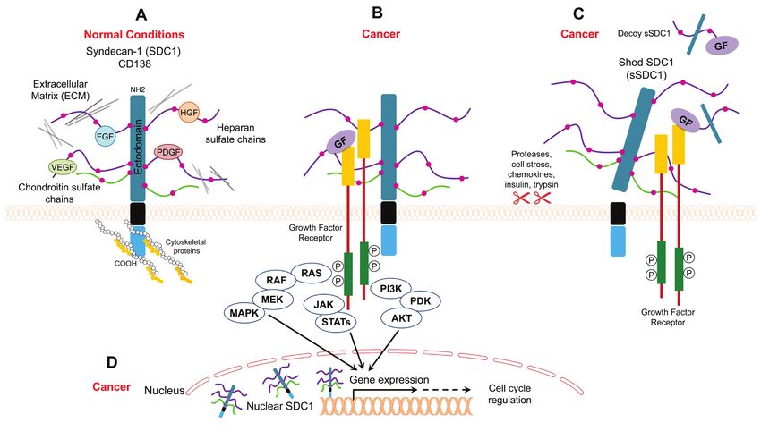
the expression of syndecans is tightly regulated and, in the morphology of epithelial sheets by connecting the
turn, may control many downstream signaling events. extracellular matrix to the intracellular cytoskeleton [15].
Syndecans show different patterns of expression in Syndecan is expressed on the surface of all adherent cells
various tissues. While SDC1 is predominantly expressed and on many non-adherent cells [15]. It is well-established
in epithelial and mesenchymal cells, SDC2 is the most that syndecan serves as coreceptor for various heparin-
abundantly expressed in cells of mesenchymal origin than binding growth factors, such as bFGF/FGF2, vascular
in neuronal and epithelial cells. In hematopoietic tissues, endothelial growth factor (VEGF), TGF-β, and platelet-
SDC1 is predominantly expressed on the cell surface of derived growth factor (PDGF) (Figure 1A, 1B) [16-18].
immature B cells and mature plasma cells [9]. SDC3 is The interaction between syndecan and growth factors is
mainly expressed in neuronal and musculoskeletal tissue, facilitated through heparan sulfate (HS) chains (Figure
whereas SDC4 is ubiquitously expressed [10]. Evidence 1A). In this regard, HS chains serve as templates that
indicates that the expression of SDC1 can be regulated bridge growth factors and their receptors. In the case of
by multiple growth factors, such as tumor growth FGF, syndecan acts as coreceptor to enhance the binding
factor- β (TGF-β) and basic fibroblast growth factor between FGF and the FGF receptor. Such binding lowers
(bFGF or FGF2), in different mammalian cell types [11]. the concentration of FGF required to initiate downstream
Tumor necrosis factor-α (TNF-α) downregulates SDC1 signaling through its receptor and extends the duration
expression in endothelial cells [12], whereas TGF-β2 of receptor signaling (Figure 1B) [19]. In addition to its
downregulates SDC1 expression in epithelial cells [13]. role as a coreceptor, syndecan itself acts as receptor via
In addition, SDC1 expression is highly increased during its HS chains (Figure 1A). Syndecan binds to different
the wound repair process [14]. matrix elements through interactions with heparan-binding
molecules on adjacent cells to potentiate cell-matrix
Syndecan localization and function adhesion (Figure 1A) [20-22]. Examples of extracellular
molecules that commonly bind to syndecan in order to
mediate cell adhesion to the extracellular matrix include
Cell surface syndecan collagens, fibronectin, thrombospondin, and tenascin [20-
Syndecan (originating from the Greek word syndein, 22].
meaning “to bind together”) acts as an anchor to stabilize Unlike HS chains, the biological function of
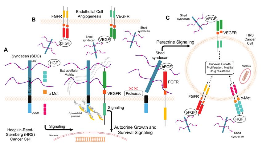
Figure 1: Model for SDC1 function under normal and cancer conditions. A. SDC1 binds to ECM proteins and/or growth
factors through its heparan sulfate chains, and it binds to cytoskeletal proteins for cell anchorage B. SDC1 acts as a coreceptor that facilitates
interaction between growth factors and their receptors and enhances cancer mitogenic signaling C. Shed SDC1 (sSDC1) enhances the
interaction between growth factors and their receptors in cancer or acts as a decoy receptor D. Nuclear SDC1 controls gene expression in
cancer.
www.impactjournals.com/oncotarget 28694 Oncotarget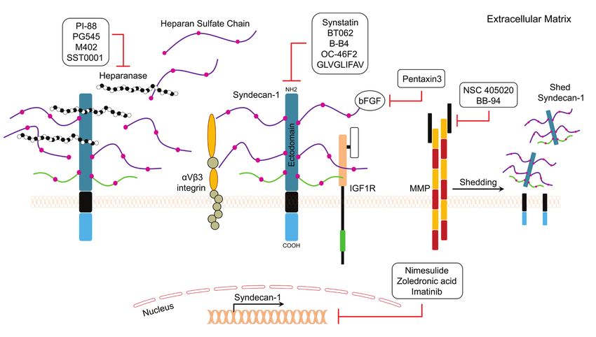
Table 1: Studies evaluating SDC1 as a prognostic biomarker in cancer patients with solid tumors.

www.impactjournals.com/oncotarget                       28695                                        Oncotarget
chondroitin sulfate (CS) chains present in syndecan is               Shed/Soluble syndecan
  not completely understood. A study by Okamoto and                           Syndecan can be proteolytically cleaved at a
  colleagues suggested a cooperative role of CS chains with            juxtamembrane site, which releases the extracellular
  HS chains in binding to the extracellular matrix protein             (ectodomain) core protein bearing both HS and CS chains
  laminin [23]. Although HS chains are major contributors              (Figure 1C) [28]. Cells constitutively shed syndecan at
  to the function of syndecan, recent studies have revealed            low levels, but shedding is accelerated in response to
  that the protein core ectodomains are also engaged in                growth factors, chemokines, heparanase, microbial toxins,
  protein-protein interactions between syndecan and other              insulin, and cellular stress [29, 30]. These stimuli trigger
  peptide molecules [24, 25]. On the intracellular side, the           several signaling pathways that eventually lead to elevated
  cytoplasmic domain binds to several cytoskeletal proteins,           protease activity driving syndecan shedding. The syndecan
  such as ezrin, tubulin, and cortactin, which potentiates             molecules that are shed remain biologically active and
  cell anchorage and stabilizes cell morphology [6-8].                 can bind the same ligands as the intact ectodomain.
  Thus, syndecan plays an important role in the interplay              Accordingly, shed syndecan may act in a paracrine manner
  between target cells and the extracellular matrix. Among             [31] . On the other hand, shed ectodomains may compete
  the different syndecans, SDC1 was first to be identified             for the same ligand as the surface receptor (acting as
  and evaluated and is implicated in the maintenance of                decoy receptors), thus downregulating signal transduction
  epithelial morphology and anchorage-dependent growth                 (Figure 1C) [31].
  [26, 27].

www.impactjournals.com/oncotarget                              28696                                                 Oncotarget
Table 2: Studies evaluating SDC1 as a prognostic biomarker in cancer patients with hematological tumors.

  Syndecan in Cancer                                                       integrin αvβ [46] and Wnt5a [47] signaling in breast
                                                                           cancer and multiple myeloma, respectively (Table 3). SDC
                                                                           increases cell adhesion via activation of focal adhesion
         The expression of SDC1 is dysregulated in
                                                                           kinase (FAK) signaling in lung and colorectal cancers [48,
  cancer, and low expression of SDC1 in epithelial cells
                                                                           49]. Mulitple molecules such as ADAM-10, ADAM-17,
  is associated with poor prognosis and high metastatic
                                                                           MMP-7, MMP-9, MMP-14, and bFGF/FGF2 increase
  potential [32-34]. Tables 1 and 2 summarize the studies to
                                                                           SDC1 shedding in multiple myeloma as well as breast and
  date that have evaluated SDC1 prognostic significance and
                                                                           colon cancers [50-54] (Table 3).
  clinical implications in solid and hematological tumors,
                                                                                 Alterations in the levels of soluble SDC1 have been
  respectively.
                                                                           reported in various cancer types. The levels of soluble
         Studies have shown that SDC1 is involved in
                                                                           SDC1 in the sera of healthy persons are relatively low
  multiple cellular processes, including cell proliferation [24,
                                                                           compared to levels in cancer patients. Heparanase induces
  39-41], migration [42-44], adhesion, and angiogenesis [30,
                                                                           SDC1 shedding, and soluble SDC1 is an independent
  45].In general, the loss of SDC1 expression in carcinoma
                                                                           negative prognostic factor in multiple myeloma [32, 55,
  cells reduces cell adhesion to the extracellular matrix and
                                                                           56]. High levels of heparanase have also been reported
  enhances cell motility and invasion [15]. Alternatively,
                                                                           in the plasma of Hodgkin’s lymphoma (HL) patients
  increased stromal SDC1 expression alters fibronectin
                                                                           and it can be used to evaluate treatment response [57].
  production and extracellular matrix organization [35].
                                                                           Soluble SDC1 is biologically active and can intensify the
  In addition, increased expression of SDC1 in stromal
                                                                           binding between growth factors with their receptors in
  fibroblasts is associated with angiogenesis and cancer
                                                                           tumor stroma [30, 58]. Soluble SDC1 ectodomains bind
  progression [15, 36]. Various signaling molecules function
                                                                           to pro-angiogenic factors, which promotes endothelial cell
  upstream or downstream from SDC1 in cancer (examples
                                                                           invasion (Figure 1C) [30]. Soluble SDC1 also increases
  are listed in Table 3). SDC1 acts as a scaffold that brings
                                                                           fibroblast proliferation and the release of TGF-β [58]. In
  ligands, such as hepatocyte growth factor (HGF), bFGF/
                                                                           addition, soluble SDC1 can act as decoy receptor, and
  FGF2, and VEGF, in close proximity to their cognate
                                                                           thus it may promote cancer progression by sequestering
  receptors. This localization activates downstream signal
                                                                           inhibitory molecules (Figure 1C) [59].
  transduction pathways, such as the “PI3K to Akt” and “Ras
                                                                                 Many reports indicate that Heparan Sulfate
  to MAPK” pathways, which enhances the proliferation
                                                                           Proteoglycans (HSPGs) may localize to the nucleus [60,
  of endothelial cells, cancer cells, and fibroblasts (Figure
                                                                           61]. A recent study showed that SDC1 is present in the
  1B) [15, 37]. For example, the binding of HGF with
                                                                           nucleus of myeloma tumor cells where it activates gene
  SDC1 enhances downstream signaling in myeloma cells,
                                                                           transcription (Figure 1D) [62]. In addition, shed SDC1
  osteoblasts, and stromal cells [15, 38]. SDC activates

www.impactjournals.com/oncotarget                                  28697                                                Oncotarget
Table 3: Examples of proteins associated with the SDC pathway in cancer.
can localize to the nucleus in areas involved in gene However, localization of SDC1 in the membranous or
transcription [63]. cytoplasmic compartments may correlate with the stage
and aggressiveness of urinary bladder cancers.
Syndecan as a prognostic biomarker in solid Breast cancer
tumors
SDC1 has been shown to be expressed at high levels
in breast cancer specimens and was associated with high
Bladder cancer histologic grade, large tumor size, high mitotic count,
and poor prognosis [67]. High SDC1 levels were also
High SDC1 expression was observed in over 60% associated with a higher risk of death in patients treated
of specimens from patients diagnosed with primary with the cyclophosphamide-methotrexate-fluorouracil
non-muscle-invasive bladder cancer, and SDC1 was a chemotherapeutic regimen [67]. High SDC1 expression
significant predictor for recurrence-free survival [64]. In was reported in triple-negative invasive ductal breast
addition, SDC1 was found to be expressed on the cell carcinomas compared to normal breast tissue [68]. SDC1
membrane of normal bladder epithelium and non-muscle- expression was also strongly correlated with patient
invasive bladder cancer cells, but was almost completely overall survival [68]. Evaluation of SDC1 expression
absent in muscle-invasive carcinomas [65]. In contrast, in invasive ductal carcinoma indicated that cytoplasmic
stromal SDC1 as well as serum SDC1 levels were higher in expression of SDC1 was positively correlated with
muscle-invasive compared to non-muscle-invasive bladder WNT1 (a proto-oncogene) and membranous expression
cancer cells. Lymph node-positive cases had the highest of SDC1 was positively correlated with p16 (a tumor-
SDC1 serum concentrations, and SDC1 expression in suppressor protein) [69]. Another study found that SDC1
stromal cells was independently associated with survival. expression was significantly increased in invasive breast
Loss of SDC1 in tumor cells and the simultaneous increase cancer cases, suggesting that it may serve as a useful
of serum SDC1 levels in high-stage, high-grade bladder prognostic biomarker for aggressive breast cancer [33].
cancer cells suggest that SDC1 shedding is associated with A tissue microarray of invasive ductal breast carcinoma
bladder cancer cell aggressiveness [65]. Thus, circulating specimens indicated high expression of SDC1 in the breast
levels of SDC1 may ultimately be a useful prognostic tool epithelium of more than half of the patients, whereas
for identifying patients with lymph node metastases [65]. stromal expression was observed in only one third of the
Assessing urinary SDC1 levels and tumor SDC1 patients. Moreover, a significant correlation was found
expression revealed no significant difference in urinary between the loss of epithelial SDC1 expression in high-
SDC1 levels between cancer and healthy subjects [67]. grade tumors. These findings suggest that lack of SDC1
However, urinary levels of SDC1 were reduced in high- epithelial expression is a strong prognostic marker in
grade disease compared to low-grade disease states breast carcinomas [70]. Tiemann and colleagues studied
[66]. Interestingly, SDC1 predominantly localized to the the role of SDC1 in ductal carcinoma in situ of the breast
cell membrane in normal tissue and low-grade tumors, (DCIS) [71]. Tumor grade was found to be related to
while high-grade tumors exhibited distinct cytoplasmic the proportion of SDC1-positive cells, rather than to the
localization. In these reports, the tumor stage and grade intensity of SDC1 staining. In the same study, estrogen
can change the value of urinary and serum levels of receptor (ER) expression did not affect the staining
SDC1 as prognostic tools in urinary bladder cancers. intensity of SDC1, but negatively correlated with the
www.impactjournals.com/oncotarget 28698 Oncotargetpercentage of SDC1-positive cells. Further findings predict clinical outcome [79].
showed that expression of progesterone receptor (PR) Colorectal cancer
was positively influenced by both the intensity of staining
and the percentage of SDC1-positive cells. These results Recent reports have indicated that colorectal
suggest a potential role of SDC1 in the pathogenesis of patients have higher serum levels of soluble SDC1
DCIS [71]. Additional studies compared expression of compared to healthy adults, which correlates with poor
SDC1 in breast cancer cases with and without distant survival [80]. Patients with high SDC1 serum levels
organ metastasis. SDC1 expression was significantly were also less responsive to 5-fluorouracil, oxaliplatin,
correlated with a higher histological grade. In addition, irinotecan, cisplatin, or paclitaxel chemotherapy
HER2 subtype and triple-negative carcinomas showed treatments [80]. Further studies revealed that SDC1 is
significantly higher SDC1 levels than those of control expressed at the basolateral borders of normal colonic
cases. Importantly, high SDC1 expression had a negative epithelial cells; however, in adenocarcinoma cells,
impact on both overall and disease-free survival rates [72]. SDC1 was found to be present around epithelial cell
In another study, SDC1 expression was detected membranes and in the cytoplasm [81]. In approximately
in approximately 70% of breast cancer cases and found 90% of adenocarcinomas examined, SDC1 expression was
to correlate with tumor grade. The presence of SDC1 in absent, and this correlated with lymph node metastasis.
high-grade tumors was associated with the absence of Stromal SDC1 was expressed in a small fraction of
SDC4 [73]. Further evidence indicated strong staining tumors. These findings emphasize that the loss of tumor
of SDC1 in DCIS tumor samples, which was associated SDC1 may be a potential prognostic biomarker for
with E-cadherin and c-Met expression [46]. In addition, human colon adenocarcinomas [81]. In another study, the
expression of SDC1 and SDC4 was correlated with expression of epithelial SDC1 was observed in over 90%
the Ki-67 mitosis index, suggesting a role in breast of colorectal cancer specimens and was associated with
cancer cell proliferation. In addition, SDC1 and SDC4 lower histological grade and a less advanced clinical stage
expression was correlated with negative ER status and [82]. Expression of stromal SDC1 was observed in 58%
aggressive phenotypes [74]. Strong SDC1 staining was of specimens, but expression did not significantly correlate
observed in more than 80% of neoplastic cells and was with clinical outcome [82]. Taken together, these studies
associated with increased mortality risk. In addition, indicate that SDC1 expression may be a useful biomarker
there was a strong negative correlation between SDC1 for evaluating the stage and grade of colorectal tumors.
expression and extracellular matrix proteins, suggesting The lack of consistency between studies may be related
that SDC1 promotes tumor progression by interacting to patient selection or methodological differences, and
with extracellular matrix components and impacting therefore larger studies are needed to further evaluate the
breast cancer tissue remodeling [75]. In another study, the prognostic impact of SDC1 in patients with colorectal
expression of SDC1 was equivalent in the epithelium and tumors.
stroma of breast tumors, but epithelial SDC1 expression Endometrial cancer
was associated with negative ER status while stromal
In endometrial cancer, epithelial SDC1 expression
SDC1 expression was associated with positive ER status
was significantly lower in advanced stage, high grade, and
[76]. Moreover, loss of epithelial or stromal SDC1
lymph node metastatic disease [83]. In contrast, stromal
expression was associated with a more favorable 10-year
SDC1 expression was significantly higher in high-grade
overall survival rate [76]. These findings indicate that
tumors [83]. Moreover, SDC1 expression was totally
SDC1 is expressed at high levels in breast cancer and its
absent in poorly differentiated endometrial cancer tissues,
expression is associated with aggressive phenotypes and
while it was abundant in normal endometrial and highly
poor clinical outcomes.
differentiated malignant tissues [84].
Cervical cancer
Gallbladder cancer
Most cervical cancer tissues assessed to date have
Epithelial SDC1 was observed in approximately half
been shown to be SDC1-positive, and localization of
of gallbladder cancer cases evaluated, and its expression
SDC1 in the cytoplasm was associated with better patient
was associated with lymph node metastasis. This study
survival. In addition, the change of SDC1 expression
also found that patients with positive SDC1 expression
in cervical cancers was not caused by copy number
had a significantly shorter survival time than patients with
alteration of the gene [77]. The progression of cervical
undetectable expression [85].
intraepithelial neoplasm to early invasive cancer was
found to correlate with reduced levels of SDC1 [78]. In Gastric cancer
another study, biopsies obtained from patients treated for Loss of epithelial SDC1 expression as well as
primary invasive cervical carcinoma showed that SDC1 high stromal SDC1 expression was associated with
expression is associated with histological differentiation unfavorable prognosis in gastric cancer [86]. Additional
grade and squamous histology, but the expression does not studies showed that loss of epithelial SDC1 expression
www.impactjournals.com/oncotarget 28699 Oncotargetwas associated with high stromal SDC1 expression, higher cancers than in moderately or poorly differentiated tumors
tumor grade, poor overall survival, and nodal metastases [94]. Cancers with high SDC1 expression were associated
[87]. Therefore, stromal and epithelial SDC1 expression with more favorable overall survival, suggesting that loss
might have some prognostic impact in gastric cancer. of SDC1 expression occurs as a result of histological
However, these findings are not consistent and require dedifferentiation and that low SDC1 expression is
further investigation. associated with unfavorable outcomes in squamous cell
Glioma carcinoma of the lung [94].
Mesothelioma
Higher gene and protein levels of SDC1 were
detected in glioma tissues compared to controls. Moreover, Studies of the expression of SDC1 protein in
SDC1 expression was increased in high-grade tumors, and mesothelioma tumors and cell lines revealed strong SDC1
the overall survival rate of SDC1 positive patients was expression in epithelial mesotheliomas and in epithelial
significantly lower than that of SDC1 negative patients components of biphasic mesotheliomas, while expression
[88]. was reduced during sarcomatoid differentiation [95].
Laryngeal cancer Moreover, SDC1 expression was associated with longer
overall survival in patients with mesotheliomas compared
SDC1 expression was detected in all laryngeal to patients with no or low SDC1 expression [95]. In
cancer specimens examined by Klatka and colleagues, and another study, SDC1 was detected in pleural effusions, but
expression was significantly correlated with histological not in sera of patients with pleural metastatic disease and
grade and patient survival rate [89]. Additional malignant mesothelioma [67]. These findings distinguish
investigation indicated that tumors with intermediate or malignant and benign diseases and suggest that SDC1
strong staining for SDC1 were associated with higher expression levels may be a prognostic factor that can
overall survival than tumors with no or low SDC1 predict differences in survival [67].
expression [90].
Nasopharyngeal carcinoma and oral cancer
Liver cancer
An analysis of SDC1 and c-Met in samples
In patients with advanced hepatocellular carcinoma from nasopharyngeal carcinoma patients by
(HCC), serum levels of SDC1 were increased compared to immunohistochemical staining indicated that high
those without HCC or with early HCC [91]. High serum coexpression of c-Met and SDC1 was adversely correlated
SDC1 levels were significantly associated with greater with patient survival [96]. Normal oral mucosa has been
risk of tumor recurrence and decreased overall survival shown to express moderate-to-high levels of SDC1, which
in patients with early HCC and with advanced HCC, is reduced or abolished in carcinomas [97]. In another
respectively [91]. Additional studies showed reduced study, SDC1 was found to be mainly expressed in the
expression of the SDC1 gene and protein in metastatic stromal cells, and this pattern was associated with poor
HCC patients compared to those with non-metastatic prognosis of ameloblastomas, keratocystic odontogenic
disease [92]. Thus, the loss of SDC1 expression could tumors [98]. SDC1 expression was significantly higher
be a characteristic feature of HCC with high metastatic in normal controls than in specimens from patients with
potential [92]. mild, moderate, or severe dysplasia as well as invasive
SDC1 protein and gene expression levels were squamous cell carcinoma; however, no significant
also assessed in intrahepatic cholangiocarcinomas difference was found between different tumor grades [99].
and normal bile duct epithelial cells [34]. Intrahepatic In another study, approximately 90% of oral squamous
cholangiocarcinoma cells showed membranous and cell carcinoma cases showed negative or weak SDC1
cytoplasmic expression of SDC1, while normal epithelial staining. Patients with intermediate or strong staining
cells showed restricted basolateral membranous intensity for SDC1 had a significantly better prognosis
expression. In cancerous tissues, the distribution of SDC1 than patients with negative or weak staining intensity
mRNA was similar to that of the protein, suggesting that [100]. SDC1 expression was decreased in more than 80%
SDC1 expression in intrahepatic cholangiocarcinoma is of oral carcinoma cases examined, but positive stromal
regulated at the transcriptional level. Moreover, loss of SDC1 staining proved to be a significant risk factor of
SDC1 expression in carcinoma was associated with poor recurrence and tumor-specific death within a 24-month
differentiation and lymph node metastases [34]. period after surgery, suggesting stromal expression of
Lung cancer SDC1 is a reliable indicator of an adverse prognosis in
oral carcinomas [101]. In another study, SDC1 levels
In lung cancer patients, high serum SDC1 and bFGF
were increased in response to cytostatic treatment, which
levels were associated with poor outcomes at the time
proved to be an important predictive factor and a clear
of diagnosis [93]. In another study, evaluation of SDC1
forecast of a good prognosis [102]. Taken together, these
expression in squamous cell lung carcinoma patients
results suggest that reduced cellular SDC1 or increased
showed higher expression of SDC1 in well-differentiated
stromal SDC1 expression can be useful prognostic factors
www.impactjournals.com/oncotarget 28700 Oncotargetin oral cancers. However, a full understanding of the Squamous cell carcinoma of the head and neck and
contrasting characteristics between epithelial and stromal thyroid cancer
components requires further studies. Analysis of primary squamous cell carcinoma
of the head and neck in patients treated with surgery
Ovarian cancer and post-operative radiotherapy has shown low SDC1
expression [110], which was associated with low grade
Patients with advanced ovarian cancer exhibit of differentiation, large tumor size, increased nodal
significantly lower epithelial SDC1 expression and metastases, high clinical stage, and unfavorable overall
significantly higher stromal SDC1 expression in reciprocal survival [110]. SDC1 expression in these tumors was
pattern compared to normal controls [103]. Additional also associated with higher overall and recurrence-free
studies have evaluated the expression of syndecans in survival compared to no or low SDC1 expression [111]. A
benign and malignant ovarian tumors and found that tissue microarray analysis of SDC1 expression in papillary
SDC2, -3, and -4 are expressed in normal, benign, and carcinomas of the thyroid indicated that SDC1 was mainly
malignant ovarian tissues [104]. In contrast, SDC1 was expressed in the cytoplasm of epithelial cells and stroma
absent in normal ovarian tissues, but present in epithelial of papillary carcinomas of the thyroid [113].
and stromal cells of benign and borderline tumors. In
addition, the expression of stromal SDC1 was a poor SDC1 as a prognostic biomarker in hematological
prognostic factor of overall survival in patients with tumors
ovarian cancer [104].
Pancreatic cancer
Chronic lymphocytic leukemia
Epithelial SDC1 has been observed in most human
Studies assessing the correlation between soluble
pancreatic carcinoma samples evaluated to date, and
SDC1 in plasma and clinical outcome in patients with
expression is predictive of a more favorable prognosis in
chronic lymphocytic leukemia have shown that soluble
patients undergoing curative surgery. In addition, stromal
SDC1 levels were significantly higher in these patients
SDC1 expression was weak or negative in over 60% of the
compared to healthy control subjects. In addition, high
tumors evaluated, and lack of stromal expression predicted
levels of soluble SDC1 were also associated with shorter
a better prognosis in these patients [105].
overall survival [112].
Prostate cancer
Diffuse large B-Cell lymphoma
Early studies have shown that syndecans are
Multiple studies have detected SDC1 in diffuse
expressed in the epithelial cells of prostate cancer patients
large B-cell lymphoma [113, 114]. Tumor biopsies of
[106]. SDC1 showed basolateral membrane localization,
diffuse large B-cell lymphoma patients were examined
whereas SDC2 was preferentially expressed in basal
for SDC1 expression and results tested positive in 30% of
cells. Another study found that the expression patterns
poor overall survival [113, 114], indicating aberrant SDC1
of SDC1 and SDC2 changed to a granular-cytoplasmic
expression correlates with poor clinical outcome.
localization in prostate cancer samples [106]. Moreover,
SDC1 was detected by immunostaining of a tissue Multiple myeloma
microarray in approximately one third of patients with Several studies have shown higher levels of soluble
localized prostatic adenocarcinoma who had been treated SDC1 in multiple myeloma patients compared to healthy
with radical prostatectomy and bilateral lymphadenectomy controls [115-117]. Baseline levels of soluble SDC1
[107]. SDC1 expression was also associated with lymph at the time of diagnosis in patients who responded to
node metastases and aggressive progression after surgery. chemotherapy were lower than non-responders; however,
Further studies showed altered expression of SDC1 protein baseline levels of SDC1 did not predict therapeutic
in specimens obtained from normal, benign, and malignant response in those patients [115]. High levels of soluble
prostate tissues [108]. SDC1 overexpression in human SDC1 and lower expression of cellular SDC1 at the time
prostate cancer was also predictive of early recurrence and of diagnosis are negative prognostic factors for multiple
was associated with tumor-specific survival, high Gleason myeloma [116]. In a cohort of Korean patients diagnosed
grade, the Ki-67 mitosis marker, and Bcl-2 overexpression with multiple myeloma, soluble SDC1 levels correlated
[109]. Together, these findings suggest that expression of with disease stage and characteristics [118]. In addition,
SDC1 can be used as a prognostic marker for patients with high soluble SDC1 levels detected in Korean subjects
localized and advanced prostate cancer. were associated with poor survival [118]. Further studies
showed that soluble SDC1 levels were elevated in the
sera of multiple myeloma patients treated with high-dose
chemotherapy and subsequent autologous transplantation
[119]. In another study, the extent to which soluble
www.impactjournals.com/oncotarget 28701 OncotargetSDC1 levels fell from presentation to the plateau phase SDC1 and HGF
represented a prognostic predictor in multiple myeloma It has been reported that HL patients have increased
patients [56]. In a comparative study of blood dyscrasias, serum levels of HGF, which correlates with advanced
multiple myeloma patients showed higher serum SDC1 stages of the disease [126]. SDC1 binds to HGF, which
levels than patients with plasmocytoma or monoclonal potentiates c-Met downstream signaling by activating the
gammopathy [120]. In addition, serum SDC1 levels PI3K and ERK pathways (Figure 2) [127]. Moreover, it
were diminished in patients who responded well to has been reported that c-Met is expressed by subsets of
chemotherapy, whereas no change was observed in non- Hodgkin Reed Sternberg (HRS) cells, and HGF is secreted
responders [120]. When SDC1 expression was analyzed in the tumor milieu, suggesting an autocrine effect in HL
in normal bone marrow or bone marrow from multiple pathogenesis [128]. In another study, changes in plasma
myeloma and B-cell lymphoma patients, SDC1 was found heparanase levels correlated with the response to treatment
to be expressed predominantly in normal and neoplastic in pediatric patients diagnosed with HL [57]. Heparanase
plasma cells. Moreover, high SDC1 expression was induced HGF expression and shedding of SDC1 through
detected in all multiple myeloma cases examined, whereas the upregulation of matrix metaloprotease-9 (MMP-9) and
all B-cell lymphomas were completely negative. urokinase-type plasminogen activator (uPA) [129]. These
Evaluation of SDC1 levels in the bone marrow of findings suggest that SDC1 or soluble SDC1 binds to HGF
multiple myeloma patients showed much higher levels to facilitate binding and activation of its receptor (Figure
than circulating SDC1 levels in peripheral blood [121]. 2).
Nevertheless, SDC1 blood and bone marrow levels were
positively correlated with microvessel density, HGF SDC1 and VEGF
levels, and reduced survival [121]. Serum SDC1 and Angiogenesis is a crucial process during the
bFGF/FGF2 levels were elevated in multiple myeloma progression of hematological malignancies, including HL
patients before treatment compared to the control group [126]. High serum levels of VEGF were detected in the
[122]. Baseline assessment of SDC1 and bFGF/FGF2 sera of HL patients [126]. Moreover, the levels of VEGF
serum levels showed higher levels of both markers, which and VEGF receptor in HL patients were significantly
was associated with shorter survival than patients with higher than the levels in non-Hodgkin’s lymphoma (NHL)
normal levels [122]. In the same study, myeloma patients patients [130]. In another study, the overexpression of
responding to chemotherapy treatment showed reduced VEGF was approximately 70% of cases of classical HL
SDC1 levels [122]. Bone marrow levels of soluble SDC1 and 30% of nodular lymphocyte predominance HL, and all
and HGF were elevated in multiple myeloma patients neoplastic HRS cells [131]. In a separate study, VEGF-A,
compared to control subjects [117]. In addition, HGF VEGF receptor-1, and VEGF receptor-2 were expressed
existed in a complex form with soluble SDC1 in pleural in HRS cells from patients with classical HL [132]. When
effusions, suggesting an important role of soluble SDC1 heparanase expression was high in the tumor, soluble
as a carrier for HGF in the pathology of myeloma [117]. SDC1 formed a complex with VEGF, which activated
Taken together, various studies have demonstrated SDC1 VEGF receptors on adjacent endothelial cells (Figure
as a potential biomarker for multiple myeloma. Findings 2) [30]. These findings suggest that SDC1 or soluble
from these studies indicate that soluble SDC1 levels may SDC1 enhance VEGF binding to the VEGF receptor, thus
be a prognostic tool in multiple myeloma patients for promoting angiogenesis (Figure 2).
diagnosis, prognosis, and treatment response.
SDC1 and bFGF/FGF2
Putative roles of SDC1 in Hodgkin’s lymphoma Unlike other growth factors, FGFs act with HSPGs
(such as SDC1) to activate FGF receptors and induce
downstream signaling responses [133, 134]. As described
Hodgkin’s Lymphoma (HL) is characterized by above, the binding of bFGF/FGF2 and an HSPG to
the presence of cancerous Hodgkin-Reed-Sternberg the extracellular domain of the FGF receptor induces
(HRS) cells embedded in a background of immune, receptor autophosphorylation. This process leads to the
inflammatory and stromal cells [123]. These cells phosphorylation of docking molecules, such as Shc,
secrete a plethora of cytokines and growth factors phospholipase-Cγ, STAT1, Gab1, and FRS2α, which are
in the tumor microenvironment that lead to tumor regulators of the Ras/MAPK and PI-3K/Akt signaling
growth and dissemination. SDC1 acts to potentiate the pathways (Figure 2) [133]. It has been reported that serum
signaling of cancerous and stromal cells in the tumor levels of bFGF were elevated and correlated with the
microenvironment. Serum levels of SDC1 were higher stage of different hematological malignancies [135]. In
in HL specimens compared to a control group [124]. In addition, the event-free survival rate was higher in NHL
another study, B-cell markers, including SDC1, were patients who had lower bFGF levels [136]. Another study
expressed in 38% of classical HL cases [125]. The showed that high serum levels of bFGF are associated
following sections highlight three potential pathways with a poor outcome in NHL patients [137]. It has been
involving SDC1 in HL pathogenesis.
www.impactjournals.com/oncotarget 28702 Oncotargetreported that HRS cells and stromal cells secrete bFGF, group with a favorable outcome. Strong immunostaining which stimulates fibrosis in the nodular sclerosis (NS) of bFGF and SDC1 was also reported in the poor outcome subtype of HL [123]. In addition, serum bFGF levels HL group [138]. Taken together, these findings suggest were significantly higher in HL patients than in healthy that simultaneous high levels of bFGF and SDC1 correlate individuals and correlated with the clinical outcome of with a poor prognosis in HL patients. HL [126]. Expression levels of FGFs and their receptors were high in HL patient samples, while their expression Syndecan as a therapeutic target in clinical in HL cell line cultures was stimulated in response to settings paracrine factors [133]. The expression of bFGF and SDC1 in HL suggests that they play a role in maintaining the growth of HL cells [138]. The association of high Based on the numerous roles in cancer pathology, serum levels of both SDC1 and bFGF with poor outcome SDC1 is an attractive molecular target for therapeutic in lung cancer has been reported [93]. Furthermore, strategies. Quantification of SDC1 is necessary in basic Kyrtsonis and colleagues demonstrated that patients who discovery research as well as in clinical practice. In vitro had high serum levels of both SDC1 and bFGF had a diagnostics and technologies that allow for the specific shorter overall survival than patients with normal levels, detection and precise quantification of SDC1 continue to and responders to treatment regimens showed reduced evolve. Today, selected clones that produce monoclonal SDC1 levels [122]. In addition, a bioinformatics analysis antibodies can be cultured to produce SDC1-specific showed overexpression of bFGF and SDC1 in HL cell antibodies. This part of the review sheds light on recent lines that were originally derived from primary HRS cells advances in in vitro diagnostics as well as research-use isolated from extranodal sites of refractory or relapsing only diagnostics (Table 4). It also summarizes SDC- HL patients [138]. The expression levels of bFGF and targeting therapeutic modalities (Figure 3; Table 5) and SDC1 protein were significantly elevated in HL patient the progress in clinical trials related to the SDC pathway samples compared to NHL sections and normal lymph (Table 6). node controls [138]. Furthermore, all HL tissue samples Synstatin is a short peptide that mimics the overexpressed FGF2 and SDC1 genes, and the group sequence of the SDC1 extracellular domain [139, 140]. with a poor outcome had a 24-fold higher expression of This peptide antagonizes the SDC1 extracellular domain, FGF2 and 56-fold higher expression of SDC1 than the which is responsible for capturing and activating αvβ3 Figure 2: Model for putative roles of SDC1 in Hodgkin’s lymphoma. A. SDC1 facilitates autocrine interaction between growth factors and their cognate receptors and enhances mitogenic signaling in Hodgkin-Reed-Sternberg (HRS) cancer cells B. Shed SDC1 (sSDC1) binds to growth factor VEGF and bFGF complexes with VEGFR and FGFRs in endothelial cells and promotes angiogenesis C. Shed SDC1 (sSDC1) binds to growth factors to interact with cognate receptors on another HRS cell (paracrine effect). www.impactjournals.com/oncotarget 28703 Oncotarget
Table 4: In vitro diagnostics (IVD) and research use only (RUO) detection methods for SDC1.

  Table 5: Agents targeting SDC1 in cancer.

  or αvβ5 integrins and the insulin-like growth factor-I             A study was conducted to evaluate the effect of BT062
  (IGF-I) receptor (Figure 3). Synstatin competitively               on multiple myeloma patients heavily pretreated with
  displaces the integrin and IGF-I receptor kinase from              revelimid, thalidomide, velcade, or carlfilzomid [141,
  SDC1 and inactivates the complex, which makes it a                 142]. BT062 was well-tolerated in patients and 4%
  promising anti-angiogenic agent [139, 140]. BT062                  achieved partial response, 8% had a minor response,
  (Indatuximab Ravtansine) is an antibody-drug conjugate             while 38% showed stable disease [141]. B-B4 is a
  that is comprised of the anti-SDC1 chimerized monoclonal           monoclonal IgG1 antibody conjugated to cytotoxic drugs
  antibody and the cytotoxic agent DM4. Once bound                   or radioactive isotopes. A phase I/II radioimmunotherapy
  to SDC1 on the cell, the conjugate is internalized and             study using B-B4 conjugated to iodine-131 was conducted
  releases DM4, which consequently leads to cell death.              in refractory multiple myeloma patients [143] and

www.impactjournals.com/oncotarget                            28704                                              Oncotarget
Table 6: Clinical trials related to the SDC1 pathway.

  Figure 3: General mechanisms of action of SDC1 pathway inhibitors are depicted.

www.impactjournals.com/oncotarget                         28705                     Oncotarget
significantly improved clinical outcome than the control               the safety and tolerability of M402 is currently being
  group, suggesting that targeted radioimmunotherapy is                  conducted in patients with metastatic pancreatic cancer
  feasible using an anti-SDC1 monoclonal antibody [143].                 [153]. SST0001 is a modified heparin with anti-heparanase
         OC-46F2 is a fully human recombinant that                       activity that inhibits cancer cell growth and metastasis
  specifically recognizes the SDC1 ectodomain (Figure 3)                 [154]. Results of in vivo studies showed that SST0001
  [144]. OC-46F2 was found to inhibit SDC1 distribution                  effectively inhibited myeloma growth and diminished
  in the tumor milieu, thus preventing vascular maturation               heparanase-induced shedding of SDC1 [154].
  and tumor growth in experimental human melanoma and                           All-trans retinoic acid, an active metabolite of
  ovarian carcinoma models [144]. GLVGLIFAV is an                        retinal, has been shown to exert anticancer activity against
  SDC1-specific peptide that is recognized by cytotoxic                  different cancer cells. It has been reported that benzo(α)
  T lymphocytes generated ex vivo using an HLA-A2-                       pyrene induces accumulation of shed SDC1 in lung cancer
  specific SDC1 epitope against multiple myeloma cells                   [155]. One study examined the level of SDC1 expression
  [145]. The GLVGLIFAV peptide induces antigen-specific                  and the chemopreventive effect of all-trans retinoic acid in
  cytotoxic T lymphocytes, which might be useful for the                 a benzo(α)pyrene-induced lung cancer model in BALB/c
  treatment of multiple myeloma patients with peptide-                   mice. The results indicated that all-trans retinoic acid
  based vaccines or cellular immunotherapy strategies                    inhibited lung tumor development and reduced SDC1
  [145]. Membrane type 1 metalloprotease (MT1-MMP)                       expression in cancer cells [155]. It has been reported that
  is a transmembrane metalloprotease that stimulates                     bFGF/FGF2 induces shedding of SDC1 in cancer cells
  the shedding of several proteoglycans, such as SDC1.                   [156]. Pentraxin-3 is a bFGF antagonist that binds to bFGF
  NSC 405020 is a small molecule inhibitor that inhibits                 with high affinity and prevents the binding of bFGF to its
  MT1-MMP homodimerization, thus blocking its pro-                       receptor [157]. Therefore, pentraxin-3 may be of value to
  tumorigenic activity in vivo [146]. BB-94 (Batimastat)                 inhibit SDC1 shedding induced by bFGF (Figure 3) [156].
  is a potent, broad spectrum small molecule inhibitor of                       Nimesulide is a cyclooxygenase-2 selective,
  MMP [147]. Treatment of cells with BB-94 suppressed                    non-steroidal anti-inflammatory drug [158]. Paul and
  SDC1 shedding and induced accumulation of SDC1 on the                  colleagues reported that nimesulide treatment caused cell
  cell surface [148]. On patients with cytologically positive            cycle arrest in primary effusion lymphoma cell lines, and
  malignant pleural effusions in Phase I study of intrapleural           this effect was accompanied by downregulation of SDC1
  BB-94, BB-94 peaked after 4 h, and remained in plasma                  [159]. Zoledronic acid (Zometa®) is a third-generation
  for up to 12 weeks, indicating that the intrapleural BB-94             bisphosphonate that inhibits SDC1 expression in cancer
  was well tolerated, with evidence of local efficacy [149].             cells in a dose-dependent manner [160]. Moreover,
         PI-88 is a polysaccharide compound with anti-                   zoledronic acid effectively inhibited growth, migration,
  heparanase activity (Figure 3) [52]. In a phase II study in            and adhesion of human breast cancer cells, which was
  hepatocellular carcinoma (HCC) patients, PI-88 treatment               accompanied by downregulation of SDC1 and -2 [161].
  was administered over nine, 4-week treatment cycles,                   Moreover, imatinib (Gleevec®) is a tyrosine kinase
  followed by a 12-week treatment-free period. PI-88 at 160              inhibitor of PDGF receptor, c-Kit, and Bcr-Abl. It exerts a
  mg/day was tolerable and effective as an adjunct therapy               significant inhibitory effect on the expression of SDC2 and
  for post-surgery HCC [150]. In another phase I study                   SDC4 in cancer cells, which leads to suppression of cell
  in patients with advanced solid tumors, the compound                   growth ability, migration, and invasion (Figure 3) [162].
  was found to be well-tolerated when administered for 4                 More recently, a clinical trial was conducted to evaluate
  consecutive days bimonthly or weekly at the recommended                the safety and efficacy of autologous T cells expressing
  dose of 250 mg/day [151]. One melanoma patient had                     an anti-CD138 chimeric antigen receptor (CART138) in
  a partial response, and nine patients maintained a stable              patients with relapsed or refractory multiple myeloma
  disease state for more than six months [151]. M402 is                  [163] and results showed that CART-138 immunotherapy
  another modified heparin compound similar to SST0001,                  was well-tolerated with significant clinical benefits in
  a chemically modified heparin to inhibit myeloma growth                multiple myeloma patients [163].
  [152]. PG545 is a fully sulfated, synthetic tetrasaccharide
  that exerts anti-heparanase activity and has shown                     Conclusions
  promising results against many cancer cells[152]. In
  ovarian cancer cells, PG545 showed synergistic inhibition                    SDC1 is a cell surface adhesion molecule that is
  of growth and migration in combination with paclitaxel                 essential for maintaining cell morphology and interactions
  and cisplatin [152]. M402 is smaller than SST0001 and                  with the microenvironment. SDC1 exerts specific
  has broader activity in binding to growth factors. M402                functional roles by acting as a coreceptor, thus potentiating
  was found to be an effective anticancer agent in different             binding between growth factors and their membrane
  cancer models [152]. M402 inhibits stromal activation and              receptors. Proteolytic activity releases the extracellular
  reduces tumor size in nude mice with human pancreatic                  ectodomain of SDC1, which harbors both the HS and
  cancer cells [153] . An ongoing Phase I/II study evaluating            CS chains, thus resulting in soluble/shed SDC1. Soluble

www.impactjournals.com/oncotarget                                28706                                                  Oncotarget
SDC1 facilitates binding between growth factors and and clinical validation of SDC1 as a new diagnostic
their receptors, or functions as a decoy receptor in other and predictive biomarker will enable individualized
circumstances. SDC1 can be found in the cytoplasmic therapeutic management for poor outcome cancer patients
compartment as well as the nuclear compartment of cells. who are refractory to therapy or under high risk of relapse.
Nuclear SDC1 can activate gene transcription and result in
distinct physiologic activities. In cancer, growing evidence Acknowledgments
indicates that deregulation of SDC1 contributes to the
development and progression of different tumor types. We thank Lisa B. Fishman Foundation and the
The value of SDC1 as a prognostic marker for specific John Theurer Cancer Center and Hackensack University
cancer types has been extensively evaluated in solid and Medical Center for supporting funds and preparation of
hematological cancers. In addition, multiple reports have this manuscript.
examined the prognostic impact of the cellular localization
of SDC1. However, based on the data to date, it is difficult Conflicts of Interest
to directly correlate the levels of SDC1 expression with
tumor characteristics and prognostic significance for all The authors declare no conflicts of interest.
cancers in general and formulate personalized clinical
treatment approaches. However, the concept of precision
medicine can be implicated for specific cancer types, since REFERENCES
higher or lower SDC1 expression is directly associated
1. Tkachenko E, Rhodes JM and Simons M. Syndecans: new
with more aggressive tumors and decreased patient
kids on the signaling block. Circ Res. 2005; 96:488-500.
survival in some cases. Such profiling may be useful
for patient selection at the time of diagnosis or perhaps 2. Saunders S, Jalkanen M, O’Farrell S and Bernfield M.
for relapsing patients. In addition, SDC1 expression is Molecular cloning of syndecan, an integral membrane
associated with a weaker response to chemotherapy for proteoglycan. J Cell Biol. 1989; 108:1547-1556.
numerous solid tumors, including breast, colorectal, 3. Marynen P, Zhang J, Cassiman J-J, Van den Berghe H
and prostate cancers. Therefore, appropriately targeting and David G. Partial primary structure of the 48-and
SDC1 in selected cancers may guide precision therapeutic 90-kilodalton core proteins of cell surface-associated
options. The reciprocal expression signature of SDC1 heparan sulfate proteoglycans of lung fibroblasts. Prediction
whereby expression is reduced in tumor epithelium and of an integral membrane domain and evidence for multiple
increased in tumor stroma has been evaluated in multiple distinct core proteins at the cell surface of human lung
studies, and recent reports suggest that SDC1 plays a fibroblasts. J Biol Chem. 1989; 264:7017-7024.
functional role in cancer-activated stromal components 4. Carey DJ, Evans DM, Stahl RC, Asundi VK, Conner KJ,
as well as in tumor progression in selected cancer types. Garbes P and Cizmeci-Smith G. Molecular cloning and
Interestingly, most studies have shown distinct cellular characterization of N-syndecan, a novel transmembrane
expression patterns for SDC1, in which membranous and heparan sulfate proteoglycan. J Cell Biol. 1992; 117:191-
cytoplasmic expression profiles were different in tumor 201.
samples compared to control samples from different 5. David G, van der Schueren B, Marynen P, Cassiman J-J
types of cancers. Therefore, it can be concluded that and Van den Berghe H. Molecular cloning of amphiglycan,
total expression levels as well as the cellular distribution a novel integral membrane heparan sulfate proteoglycan
of SDC1 should be evaluated together for the most expressed by epithelial and fibroblastic cells. J Cell Biol.
informative prognostic tools. Evaluation of urinary 1992; 118:961-969.
levels of SDC1 in urinary bladder tumors may also be
6. Bernfield M, Gotte M, Park PW, Reizes O, Fitzgerald ML,
considered during the assessment of tumor severity. The
Lincecum J and Zako M. Functions of cell surface heparan
value of circulating levels of SDC1 was not consistently
sulfate proteoglycans. Annu Rev Biochem. 1999; 68:729-
associated with tumor grade or characteristics, but the
777.
combination of SDC1 and bFGF/FGF2 in patient serum
7. Granés F, Ureña JM, Rocamora N and Vilaró S. Ezrin links
has a strong association with tumor progression and
syndecan-2 to the cytoskeleton. J Cell Sci. 2000; 113:1267-
prognosis in selected cancer types. In multiple myeloma,
1276.
soluble SDC1 levels were found to be directly associated
with the progression of disease, and therefore this 8. Granés F, Berndt C, Roy C, Mangeat P, Reina M and Vilaró
association should be evaluated in other cancers as well. S. Identification of a novel Ezrin-binding site in syndecan-2
Multiple clinical trials are currently evaluating the safety cytoplasmic domain. FEBS letters. 2003; 547:212-216.
and efficacy of pharmacologically targeting SDC1 in 9. Sanderson RD, Lalor P and Bernfield M. B lymphocytes
different types of cancer. Collectively, SDC1 represents express and lose syndecan at specific stages of
an attractive molecular target for further evaluation differentiation. Cell Regul. 1989; 1:27-35.
in personalized cancer treatment. The identification 10. Couchman JR. Syndecans: proteoglycan regulators of
www.impactjournals.com/oncotarget 28707 Oncotargetcell-surface microdomains? Nat Rev Mol Cell Biol. 2003; syndecan-1 ectodomain regulates alphavbeta3 integrin
4:926-938. activity in human mammary carcinoma cells. J Cell Biol.
11. Cizmeci-Smith G, Langan E, Youkey J, Showalter LJ and 2004; 167:171-181.
Carey DJ. Syndecan-4 is a primary-response gene induced 25. Choi Y, Chung H, Jung H, Couchman JR and Oh ES.
by basic fibroblast growth factor and arterial injury in Syndecans as cell surface receptors: Unique structure
vascular smooth muscle cells. Arterioscler Thromb Vasc equates with functional diversity. Matrix Biol. 2011; 30:93-
Biol. 1997; 17:172-180. 99.
12. Halden Y, Rek A, Atzenhofer W, Szilak L, Wabnig A and 26. Bayer-Garner IB and Smoller BR. The expression of
Kungl AJ. Interleukin-8 binds to syndecan-2 on human syndecan-1 is preferentially reduced compared with that
endothelial cells. Biochem J. 2004; 377:533-538. of E-cadherin in acantholytic squamous cell carcinoma. J
13. Dobra K, Nurminen M and Hjerpe A. Growth factors Cutan Pathol. 2001; 28:83-89.
regulate the expression profile of their syndecan co- 27. Dhodapkar MV, Abe E, Theus A, Lacy M, Langford
receptors and the differentiation of mesothelioma cells. JK, Barlogie B and Sanderson RD. Syndecan-1 is a
Anticancer Res. 2003; 23:2435-2444. multifunctional regulator of myeloma pathobiology: control
14. Gallo R, Kim C, Kokenyesi R, Adzick NS and Bernfield of tumor cell survival, growth, and bone cell differentiation.
M. Syndecans-1 and -4 are induced during wound repair Blood. 1998; 91:2679-2688.
of neonatal but not fetal skin. J Invest Dermatol. 1996; 28. Bass MD, Morgan MR and Humphries MJ. Syndecans shed
107:676-683. their reputation as inert molecules. Sci Signal. 2009; 2:pe18.
15. Teng YH, Aquino RS and Park PW. Molecular functions of 29. Hayashida K, Bartlett AH, Chen Y and Park PW. Molecular
syndecan-1 in disease. Matrix Biol. 2012; 31:3-16. and cellular mechanisms of ectodomain shedding. Anat
16. Nugent MA and Edelman ER. Kinetics of basic fibroblast Rec. 2010; 293:925-937.
growth factor binding to its receptor and heparan sulfate 30. Purushothaman A, Uyama T, Kobayashi F, Yamada S,
proteoglycan: a mechanism for cooperativity. Biochemistry. Sugahara K, Rapraeger AC and Sanderson RD. Heparanase-
1992; 31:8876-8883. enhanced shedding of syndecan-1 by myeloma cells
17. Salmivirta M, Heino J and Jalkanen M. Basic fibroblast promotes endothelial invasion and angiogenesis. Blood.
growth factor-syndecan complex at cell surface or 2010; 115:2449-2457.
immobilized to matrix promotes cell growth. J Biol Chem. 31. Nikolova V, Koo CY, Ibrahim SA, Wang Z, Spillmann
1992; 267:17606-17610. D, Dreier R, Kelsch R, Fischgrabe J, Smollich M, Rossi
18. Chu C, Buczek-Thomas J and Nugent M. Heparan sulphate LH, Sibrowski W, Wulfing P, Kiesel L, Yip GW and
proteoglycans modulate fibroblast growth factor-2 binding Gotte M. Differential roles for membrane-bound and
through a lipid raft-mediated mechanism. Biochem. J. 2004; soluble syndecan-1 (CD138) in breast cancer progression.
379:331-341. Carcinogenesis. 2009; 30:397-407.
19. Forsten-Williams K, Chua CC and Nugent MA. The 32. Yang Y, Macleod V, Miao HQ, Theus A, Zhan F,
kinetics of FGF-2 binding to heparan sulfate proteoglycans Shaughnessy JD, Jr., Sawyer J, Li JP, Zcharia E, Vlodavsky
and MAP kinase signaling. J Theor Biol. 2005; 233:483- I and Sanderson RD. Heparanase enhances syndecan-1
499. shedding: a novel mechanism for stimulation of tumor
20. Koda JE, Rapraeger A and Bernfield M. Heparan sulfate growth and metastasis. J Biol Chem. 2007; 282:13326-
proteoglycans from mouse mammary epithelial cells. Cell 13333.
surface proteoglycan as a receptor for interstitial collagens. 33. Thanakit V, Ruangvejvorachai P and Sampatanukul P.
J Biol Chem. 1985; 260:8157-8162. Expression of E-cadherin and syndecan-1 in axillary
21. Salmivirta M, Elenius K, Vainio S, Hofer U, Chiquet- lymph node metastases of breast cancer with and without
Ehrismann R, Thesleff I and Jalkanen M. Syndecan from extracapsular extension. J Med Assoc Thai. 2008; 91:1087-
embryonic tooth mesenchyme binds tenascin. J Biol Chem. 1092.
1991; 266:7733-7739. 34. Harada K, Masuda S, Hirano M and Nakanuma Y. Reduced
22. Saunders S and Bernfield M. Cell surface proteoglycan expression of syndecan-1 correlates with histologic
binds mouse mammary epithelial cells to fibronectin and dedifferentiation, lymph node metastasis, and poor
behaves as a receptor for interstitial matrix. J Cell Biol. prognosis in intrahepatic cholangiocarcinoma. Hum Pathol.
1988; 106:423-430. 2003; 34:857-863.
23. Okamoto O, Bachy S, Odenthal U, Bernaud J, Rigal D, 35. Yang N, Mosher R, Seo S, Beebe D and Friedl A.
Lortat-Jacob H, Smyth N and Rousselle P. Normal human Syndecan-1 in breast cancer stroma fibroblasts regulates
keratinocytes bind to the α3LG4/5 domain of unprocessed extracellular matrix fiber organization and carcinoma cell
laminin-5 through the receptor syndecan-1. J Biol Chem. motility. Am J Pathol. 2011; 178:325-335.
2003; 278:44168-44177. 36. Maeda T, Desouky J and Friedl A. Syndecan-1 expression
24. Beauvais DM, Burbach BJ and Rapraeger AC. The by stromal fibroblasts promotes breast carcinoma growth in
www.impactjournals.com/oncotarget 28708 OncotargetYou can also read