Melanopsin (Opn4) is an oncogene in cutaneous melanoma
←
→
Page content transcription
If your browser does not render page correctly, please read the page content below
ARTICLE
                  https://doi.org/10.1038/s42003-022-03425-6                 OPEN

                  Melanopsin (Opn4) is an oncogene in cutaneous
                  melanoma
                  Leonardo Vinícius Monteiro de Assis 1,7,8 ✉, José Thalles Lacerda1,8, Maria Nathália Moraes2,
                  Omar Alberto Domínguez-Amorocho3, Gabriela Sarti Kinker4, Davi Mendes 5, Matheus Molina Silva5,
                  Carlos Frederico Martins Menck 5, Niels Olsen Saraiva Câmara 3 & Ana Maria de Lauro Castrucci1,6

                  The search for new therapeutical targets for cutaneous melanoma and other cancers is an
                  ongoing task. We expanded this knowledge by evaluating whether opsins, light- and thermo-
1234567890():,;

                  sensing proteins, could display tumor-modulatory effects on melanoma cancer. Using dif-
                  ferent experimental approaches, we show that melanoma cell proliferation is slower in the
                  absence of Opn4, compared to Opn4WT due to an impaired cell cycle progression and
                  reduced melanocyte inducing transcription factor (Mitf) expression. In vivo tumor progres-
                  sion of Opn4KO cells is remarkably reduced due to slower proliferation, and higher immune
                  system response in Opn4KO tumors. Using pharmacological assays, we demonstrate that
                  guanylyl cyclase activity is impaired in Opn4KO cells. Evaluation of Tumor Cancer Genome
                  Atlas (TCGA) database confirms our experimental data as reduced MITF and OPN4
                  expression in human melanoma correlates with slower cell cycle progression and presence of
                  immune cells in the tumor microenvironment (TME). Proteomic analyses of tumor bulk show
                  that the reduced growth of Opn4KO tumors is associated with reduced Mitf signaling, higher
                  translation of G2/M proteins, and impaired guanylyl cyclase activity. Conversely, in Opn4WT
                  tumors increased small GTPase and an immune-suppressive TME are found. Such evidence
                  points to OPN4 as an oncogene in melanoma, which could be pharmacologically targeted.

                  1 Laboratory of Comparative Physiology of Pigmentation, Department of Physiology, Institute of Biosciences, University of São Paulo, São Paulo, Brazil.
                  2 Laboratory of Neurobiology, Department of Physiology and Biophysics, Institute of Biomedical Sciences, University of São Paulo, São Paulo, Brazil.
                  3 Laboratory of Transplantation Immunobiology, Institute of Biomedical Sciences, University of São Paulo, São Paulo, Brazil. 4 Laboratory of Translational

                  Immuno-Oncology A. C. Camargo Cancer Center – International Research Center, São Paulo, Brazil. 5 DNA Repair Lab, Department of Microbiology, Institute
                  of Biomedical Sciences, University of São Paulo (USP), São Paulo, Brazil. 6 Department of Biology, University of Virginia, Charlottesville, VA, USA. 7Present
                  address: Institute of Neurobiology, Center for Brain, Behavior, and Metabolism, University of Lübeck, Lübeck, Germany. 8These authors contributed equally:
                  Leonardo Vinícius Monteiro de Assis, José Thalles Lacerda. ✉email: deassis.leonardo@alumni.usp.br

                  COMMUNICATIONS BIOLOGY | (2022)5:461 | https://doi.org/10.1038/s42003-022-03425-6 | www.nature.com/commsbio                                                1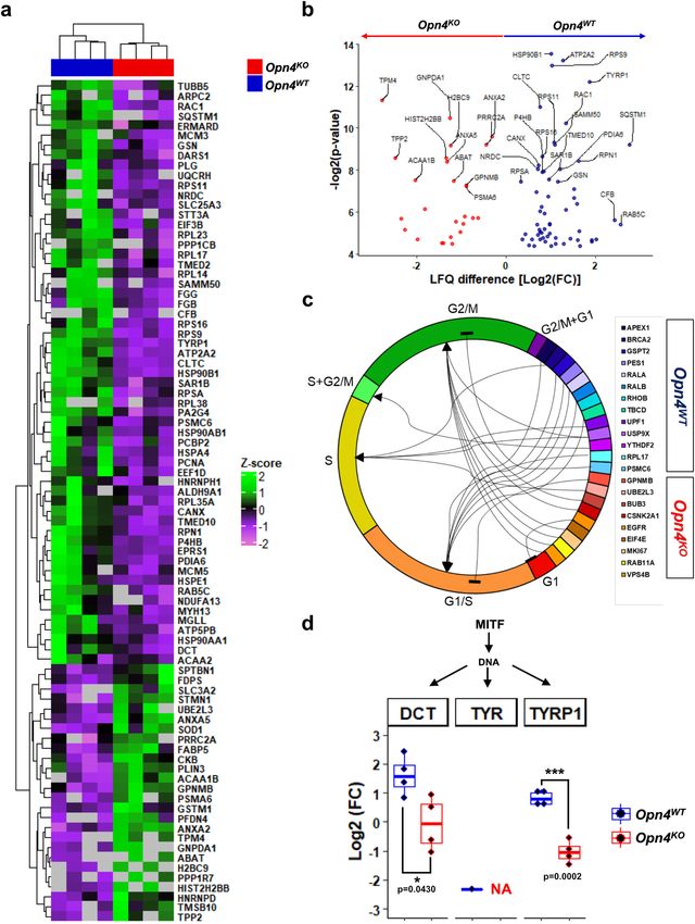
ARTICLE COMMUNICATIONS BIOLOGY | https://doi.org/10.1038/s42003-022-03425-6
C
utaneous melanoma (CM) cancer represents about 5% of increased energy requirements to sustain core body
all skin-related cancer cases, but it accounts for approxi- temperature28. Therefore, to avoid confounding factors caused
mately 80% of cancer-related deaths. The incidence of CM by cold stress, mice were kept in their thermal-neutral
has steadily increased over the years1,2, despite increasing temperature.
awareness of the deleterious effects of the sun on the skin3. The It has been previously shown that tumor growth is significantly
etiology of CM is multifactorial and includes risk factors such as faster in mice kept at temperatures below thermoneutrality than
UV radiation exposure, genetic susceptibility, high nevus density, at thermoneutrality29. Indeed, tumor growth was only measurable
reduced pigmentation, and immunosuppression4,5. from the 13th day onwards unlike previously shown in mice kept
The molecular biology of cutaneous melanoma is well at 22 °C and inoculated with Opn4WT cells20. On the 22nd and
understood due to an effort made by several pioneering 25th days after inoculation, tumor volume was significantly
endeavors that resulted in solid comprehension of the tumor smaller in Opn4KO inoculated animals compared to Opn4WT
biology landscape6. Currently, CM can be classified into four tumors (Fig. 1a). Tumor weight and melanin content on the 25th
subtypes based on the most prevalent mutations: mutant B-Raf day were also lower in Opn4KO tumors compared to Opn4WT
proto-oncogene, serine/threonine kinase (BRAF), mutant counterparts (Fig. 1b, c).
Kirsten rat sarcoma viral proto-oncogene (RAS), mutant neu- Hemogram analysis of tumor-bearing mice revealed no
rofibromin 1 (NF1), and triple wild-type6. Intriguingly, CM is difference in circulating white blood cells and the absolute
known to display the highest mutational load of all cancers7. number of lymphocytes, but a decreased frequency of lympho-
The temporal control of physiological processes is crucial for cytes was found in Opn4WT tumor-bearing mice compared to the
homeostasis and such regulation is dependent on the circadian remaining groups (Fig. S1a–c). The absolute number of
clock8. The molecular clock system is comprised of several genes monocytes did not differ between the groups, but increased
that engage in transcriptional feedback loops, whose transcripts frequency of monocytes was found in Opn4WT tumor-bearing
oscillate throughout the day8–10. In recent years, the antitumoral mice compared to sham controls (Fig. S1 d–e). The absolute
role of the molecular clock has been extensively investigated in number of granulocytes was higher in Opn4WT tumor-bearing
several types of cancer11–15. In CM, expression of clock genes and mice compared to sham animals. Granulocyte frequency was also
proteins are mostly downregulated when compared to healthy higher in Opn4WT inoculated mice compared to the remaining
skin or tumor-adjacent tissues in murine in vitro and in vivo, groups (sham control and Opn4KO inoculated mice, Fig. S1 f, g).
respectively, as well as in human tissues16–21. Remarkably, the levels of red blood cells, hemoglobin, and
Melanocytes are known to be light-responsive cells mainly platelets were severely reduced in Opn4WT inoculated mice in
because they express a complex photosensitive system comprised comparison to the remaining groups (Fig. S1 h–k).
of chromophores and light-sensitive molecules, known as Flow cytometry was used to assess the population and
opsins22,23. We have previously investigated the role of mela- subpopulations of tumor-associated immune cells in the tumor
nopsin (OPN4) in murine melanocytes and demonstrated that microenvironment (TME) of both genotypes (Fig. S2). Macro-
this protein acts as UVA-sensor for pigmentary and apoptosis- phages are known to be highly specialized in the removal of
dependent processes24,25 as well as a thermal sensor26. More debris and cells and to present antigens, thus playing an
recently, we demonstrated that OPN4 may also display light- and important role in the initial immune response30. No difference
thermo-independent roles as Opn4 knockout murine melanocytes in total tumor-associated macrophages, M1, and M2, frequencies
show faster proliferation and cell cycle progression27 between the tumors were found (Fig. 1d–f). Tumor-infiltrating
In this study, we evaluated the putative role of OPN4 in the lymphocytes play an important role in tumor development, as in
carcinogenic process of melanoma. To this end, we investigated the first stages of tumor growth the immune system can combat
how the absence of OPN4 in tumor cells would affect the and kill tumor cells, thus halting tumor growth. However, as the
development and progression of melanoma in a murine model. tumor progresses, tumor-infiltrating lymphocytes are often
Taken altogether, we provide evidence that OPN4 can act as an inactivated by the TME, thus leading to the exhaustion of
oncogene in melanoma. tumor-infiltrating lymphocytes, and consequently accelerated
tumor growth31,32.
Increased frequency of CD4+ and CD8+ in Opn4KO tumors
Results compared to Opn4WT was found, which is suggestive of increased
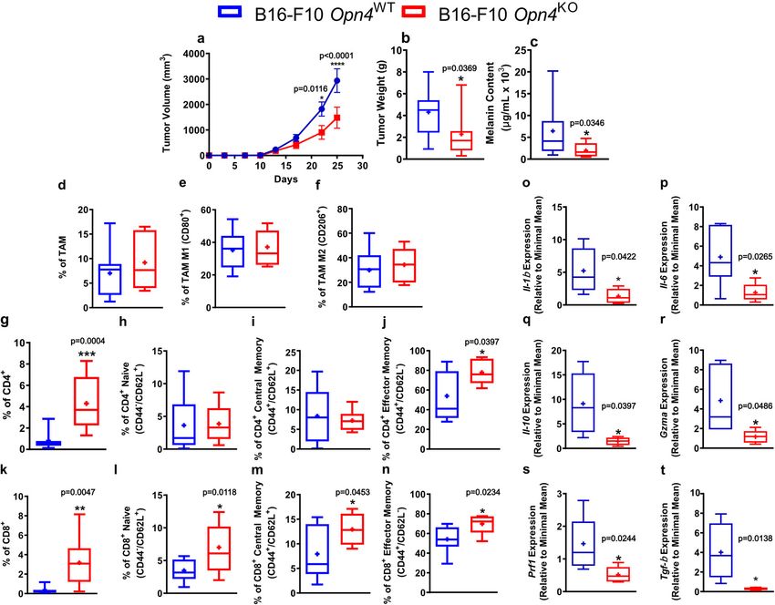
Absence of OPN4 results in slower tumor growth and migration of lymphocytes to the tumor site (Fig. 1g–n).
increased immune cell infiltration in the tumor micro- Compared to the naive population, memory cells are known to
environment (TME). We previously established and validated an be more persistent. While central and effector memory cells often
Opn4KO model using Clustered Regularly Interspaced Short display several phenotypic similarities, central memory cells
Palindromic Repeats (CRISPR) of B16-F10 cells25. In this process, persist longer than effector cells, which undergo a significant
three clones that exhibited no ultraviolet A radiation-induced decrease in terms of population but display immediate cytotoxic
pigmentation and apoptosis responses were identified. B16-F10 effector function. On the other hand, central memory cells retain
Opn4KO clone 16 was chosen and Sanger sequencing of the proliferative ability with little effector capacity, upon a reencoun-
CRISPR edited region showed alteration in the coding sequencing ter with an antigen33,34. No difference between CD4+ naive and
that led to the loss of function. Immunocytochemistry of OPN4 central memory lymphocytes was detected whereas an increase in
revealed increased protein presence in a region capping the the frequency of CD4+ effector memory cells was found in
nucleus, thus suggestive of protein retention likely due to altered Opn4KO tumors (Fig. 1h–j). We also found an increased
protein structure25. In this study, B16-F10 Opn4KO clone 16 was frequency of naive, central, and effector memory CD8+ T
chosen and used in the next steps. lymphocytes in Opn4KO tumors compared to Opn4WT ones
To evaluate whether OPN4 would impact tumor development, (Fig. 1 k–n). Intriguingly, decreased gene expression of both pro-
we inoculated C57Bl/6 J mice, kept in thermoneutrality (Il-1β and Il-6) and anti-inflammatory (Il-10 and Tgf-β) players,
(30 ± 1 °C), with B16-F10 Opn4WT or B16-F10 Opn4KO cells. At as well as T CD8+ dependent effector function genes such as
temperatures between 29 and 31 °C, mice do not activate granzyme (Gzma) and perforin (Prf1) in TME of Opn4KO was
thermogenesis to sustain core body temperature. In fact, mice found when compared to Opn4WT tumor-bearing mice (Fig. 1
kept below thermoneutrality are considered cold-stressed due to o–t). These data suggest that as tumor growth is less marked in
2 COMMUNICATIONS BIOLOGY | (2022)5:461 | https://doi.org/10.1038/s42003-022-03425-6 | www.nature.com/commsbio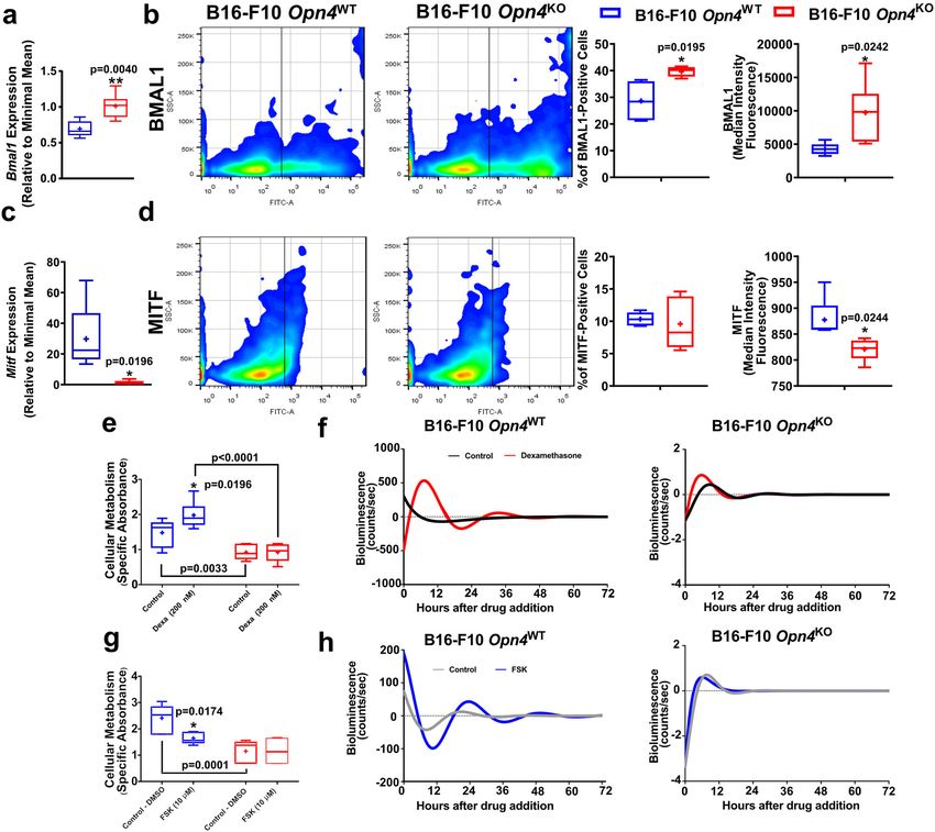
COMMUNICATIONS BIOLOGY | https://doi.org/10.1038/s42003-022-03425-6                                                                              ARTICLE

Fig. 1 In vivo tumor growth is reduced in mice inoculated with Opn4KO cells and correlates with higher immune system response. a–c Tumor volume,
weight, and melanin content in Opn4WT and Opn4KO tumor-bearing mice. In a, n = 17 and 11 for Opn4WT and Opn4KO tumors, respectively. Error bars are
shown as SEM; in b, n = 13 and 7, respectively; in c, n = 11 and 7, respectively. d–n Assessment of tumor microenvironment (TME) population represented
by tumor-associated macrophages (TAM), CD4+ T lymphocytes, and CD8+ T lymphocytes. Subtypes of each cell population are indicated in the Y axis.
Representative gate strategy is shown in Figure S2. In d–f, n = 13 and 7 for Opn4WT and Opn4KO groups, respectively; (g, h), n = 12 and 6, respectively; in
i–j, n = 13 and 6, respectively; (k–l), n = 12 and 7, respectively; (m), n = 11 and 6, respectively; (n), n = 13 and 5, respectively. o–t Gene expression of pro-
and anti-inflammatory markers as well as T CD8+ dependent effector function in TME of Opn4KO versus Opn4WT tumors. In o, n = 5 for Opn4WT and
Opn4KO tumors; in p, n = 6 and 5, respectively; in q, n = 6 and 4, respectively; in (r), n = 5; in s, n = 7 and 5, respectively; in t, n = 8 and 5, respectively. In
every analyzes, the n number is derived from independent samples. Asterisks represent significant differences between Opn4KO and Opn4WT tumors.

Opn4KO cells, one might speculate a better resolution of the                       central and effector memory cell frequency, was seen in spleens of
inflammatory process, thus leading to a less inflamed TME.                           both tumor-bearing mice compared to control sham animals
   Immune system evaluation was also performed in spleens of                       (Fig. S3 h–k).
tumor-bearing and sham control mice. No difference in the                             Taken altogether, we found marked differences in the immune
frequency of macrophages between Opn4WT and Opn4KO spleens                         profile between the tumor genotypes. Opn4KO bearing mice
as well as compared to sham control mice was found. However,                       showed increased CD4+ and CD8+ infiltration to the TME.
an increase of M1, but not M2, macrophage frequency in spleens                     Moreover, increased CD4+ effector memory presence was found
of both genotypes compared to sham control mice was found                          in spleens of Opn4KO bearing mice. Collectively, these data suggest
(Fig. S3 a–c). With regards to CD4+ lymphocytes, a frequency                       an increased immune system activity in the TME that may
reduction in Opn4WT tumor-bearing mice compared to Opn4KO                          contribute to the reduced tumor growth found in Opn4KO tumors.
and sham control mice was found. A reduction in the frequency
of naive CD4+ was accompanied by increased CD4+ central
memory population in spleens of both tumor-bearing mice                            Removal of OPN4 reduces metabolic activity, delays pro-
compared to sham control mice; however, spleens of Opn4KO                          liferation and cell cycle progression, and impairs the molecular
tumor-bearing mice showed a higher frequency of CD4+ effector                      clock in vitro. Interested in comprehending the reduced
memory compared to the remaining groups (Fig. S3 d–g). A                           growth of Opn4KO tumors, we focused on in vitro experiments.
similar profile of T CD8+ cells was also found as a reduction of                    Metabolic activity and proliferation of Opn4KO melanocytes were
CD8+ total frequency and naive cells, followed by increased                        significantly lower compared to Opn4WT counterparts (Fig. 2a).

COMMUNICATIONS BIOLOGY | (2022)5:461 | https://doi.org/10.1038/s42003-022-03425-6 | www.nature.com/commsbio                                                       3
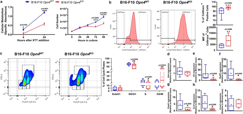
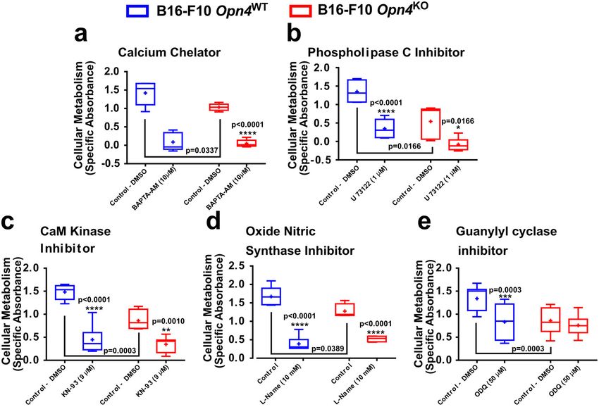
ARTICLE                                                                     COMMUNICATIONS BIOLOGY | https://doi.org/10.1038/s42003-022-03425-6

Fig. 2 In vitro growth of Opn4KO malignant melanocytes is reduced and is associated with delayed cell cycle progression. a Metabolic evaluation using
daily XTT assay for 4 and 24 h (n = 12 for each group) and proliferation assay by cell counting for 96 h (n = 6 for each group). b Proliferation assay by
Celltrace™ staining using flow cytometry. Celltrace™ positive cells are represented. In a and b, error bars are shown as SEM. Quantitative analysis of
Celltrace™ positive cells and median intensity fluorescence (MIF) of Opn4WT and Opn4KO malignant melanocytes (n = 15 for each group). c Evaluation of
cell cycle populations and expression of cell cycle-related genes in Opn4WT and Opn4KO malignant melanocytes. Negative control is shown in Fig. S2.
Arrows show the respective cell cycle phase. Boxplots show the quantitative evaluation of cell cycle phases by flow cytometry (n = 18 and 15 for Opn4WT
and Opn4KO cells, respectively). d–i Expression of cell cycle-related genes in Opn4WT and Opn4KO malignant melanocytes. In d, e, n = 11 and 6 for Opn4WT
and Opn4KO, respectively; in f, n = 11 and 5, respectively; in g, n = 11 and 6, respectively; in h, n = 9 and 5, respectively; in (i), n = 10 and 6, respectively. In
every analyzes, the n number is derived from independent samples.

To quantify the proliferative capacity, cells were loaded with                         Clock genes have been previously implicated in the
CellTrace™, a proliferative cell marker. As cells proliferate and                   melanoma carcinogenic process with exciting results16,18,20,21.
divide, a reduction of the median intensity of fluorescence (MIF)                    Increased Bmal1 gene expression, frequency, and fluorescence
is expected while slower proliferative cells show increased MIF.                    (MIF) of BMAL1 positive cells were found in Opn4KO
Corroborating our previous data, Opn4KO malignant melanocytes                       malignant cells compared to Opn4WT cells (Fig. 3a, b).
displayed an increased percentage of CellTrace™ positive cells and                  Interestingly, the microphthalmia-associated transcription fac-
fluorescence compared to Opn4WT cells (Fig. 2b). Cell cycle                          tor (MITF) – the master regulator of several biological processes
evaluation was also performed using the 7-AAD (DNA marker)                          of melanocytes42 – gene and protein expression, but not the
and BrdU (S Phase marker) dual staining procedure. Opn4KO                           frequency of positive cells, were severely less expressed in
malignant melanocytes displayed reduced G0-G1 and S phase cell                      Opn4KO melanocytes (Fig. 3c, d).
populations and increased G2/M population compared to                                  As important clock genes showed decreased expression, the
Opn4WT counterparts (Fig. 2c).                                                      investigation of the functionality of this system was performed.
   Based on our previous study27, 6 genes were selected for qPCR                    Since B16-F10 cells show a reduced synchronization capacity to
validation. The genes ataxia-telangiectasia-mutated (Atm) and                       different methods18,19,43, we focused on the evaluation of the
ataxia telangiectasia and Rad3-related (Atr) encode proteins that                   acute clock response in this assay. Opn4WT and Opn4KO cells
act on DNA damage response and are responsible for maintain-                        were challenged with dexamethasone and forskolin, both known
ing genome integrity35. We found no difference in Atm                               modulators of the molecular clock44, using a Per1:Luc biolumi-
expression while Atr transcripts were lower in Opn4KO malignant                     nescence reporter assay, as previously described25,27. As expected,
melanocytes compared to Opn4WT melanoma cells (Fig. 2d, e).                         dexamethasone increased cellular metabolism and Per1 biolumi-
Rad51 is a protein that plays a major role in homologous DNA                        nescence in Opn4WT melanocytes compared to control Opn4WT
recombination during a double-strand break36. We found a                            cells. Forskolin, on the other hand, led to a reduction in cellular
reduction of Rad51 in melanoma Opn4KO cells (Fig. 2f).                              metabolism and bioluminescence in Opn4WT melanocytes
Checkpoint kinase 1 (Chek1) gene encodes a serine/threonine-                        compared to control Opn4WT cells (Fig. 3 e–h). One would
specific protein kinase that is involved in DNA damage response                      expect an increase in Per1 bioluminescence in response to
and may elicit cell cycle arrest, DNA repair, and death37. Cyclin F,                forskolin, as previously shown for murine melanocytes27.
encoded by Ccnf, has an important role in the cell cycle as this                    However, the lack of Per1 bioluminescence induction may be
cyclin binds and activates cyclin-dependent kinase, thus leading                    related to an already upregulated cAMP signaling, which is a
to cell cycle progression38,39. Ccna1 (Cyclin A1) encodes a                         common feature known in melanoma cells45. Interestingly,
protein that is responsible for activating cyclin-dependent                         Opn4KO malignant cells were less responsive to both treatments
kinases, thus playing a positive role in the cell cycle40,41. In our                (Fig. 3 e–h). These data suggest that the acute molecular clock
model, Chek1 and Ccnf expression was reduced while Ccna1                            response is impaired in the absence of Opn4. Further circadian-
expression was not affected in Opn4KO cells compared to                             related experiments are needed to clarify the impact of OPN4 in
Opn4WT malignant melanocytes (Fig. 2g–i).                                           the regulation of the circadian clock function.

4                             COMMUNICATIONS BIOLOGY | (2022)5:461 | https://doi.org/10.1038/s42003-022-03425-6 | www.nature.com/commsbio
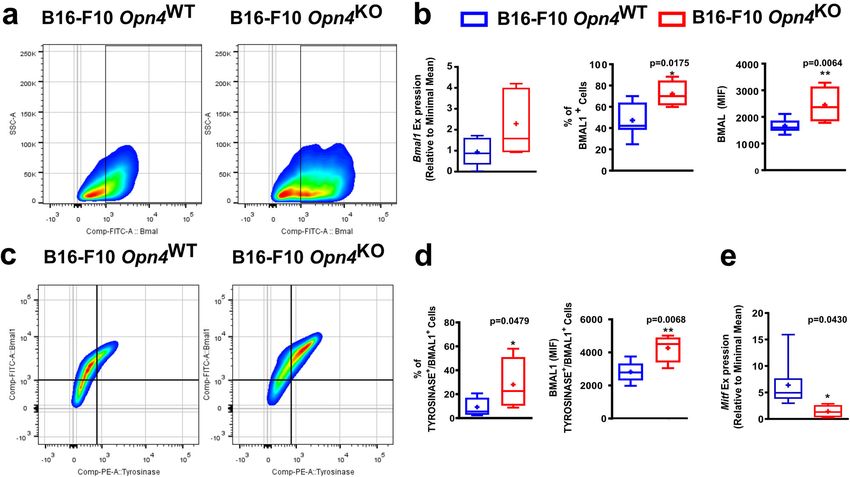
COMMUNICATIONS BIOLOGY | https://doi.org/10.1038/s42003-022-03425-6                                                                        ARTICLE

Fig. 3 Reduced growth of Opn4KO cells in vitro is associated with reduced MITF expression and reduced clock gene activation by classical activators.
a–d Bmal1 and Mitf gene and protein evaluation in Opn4WT and Opn4KO malignant melanocytes in vitro. Gene expression, BMAL1 and MITF positive cells,
median intensity fluorescence (MIF), and quantitative analyses are depicted. In a, n = 5 and 6 for Opn4WT and Opn4KO cells, respectively. In b, n = 4 and 5,
respectively. In c, n = 5 and 6, respectively. In d, n = 5 for each group. e–h Cellular metabolism assessment by XTT assay in response to classical clock
activators and molecular clock evaluation by bioluminescence assay (Per1:Luc) of Opn4WT and Opn4KO malignant melanocytes. In e, n = 6 and 11,
respectively; in f, n = 4 and 11, respectively. In g, n = 6 for both groups. In h, n = 6 and 9, respectively. In every analyzes, the n number is derived from
independent samples.

   We hypothesized that the inhibition of the OPN4 signaling                       Collectively, these data suggest an impairment in the cell cycle
cascade would differentially affect cellular metabolism in both                 progression and the molecular clock response associated with
cell genotypes. Thus, to evaluate a putative signaling pathway                  reduced cellular proliferation with important cell cycle regulators
that is triggered by OPN4 in a basal condition, classic players of              being affected at the mRNA level in vitro. The pharmacological
OPN4 signaling pathways46 were inhibited. Calcium and                           approach showed that the basal cellular metabolism of malignant
phospholipase C participation were ruled out as no difference                   melanocytes is dependent on calcium, CaM kinase, NOS, and
between genotypes was found (Fig. 4 a, b). The role of cGMP,                    cGMP pathways. However, in the absence of OPN4, guanylyl
which has been previously implicated in UVA-induced                             cyclase activity seems to be impaired as no inhibitory effect was
pigmentary response in an OPN4-dependent manner in normal                       seen when the enzyme was pharmacologically inhibited. One could
and malignant melanocytes, was evaluated24. CaM kinase and                      argue that in Opn4KO cells the lack of guanylyl cyclase inhibition
oxide nitric synthase (NOS) were evaluated and no difference                    may be due to the already downregulated enzyme activity.
between the genotypes was found (Fig. 4 c – d). Remarkably,
upon guanylyl cyclase inhibition a reduction of cellular                        BMAL1 and MITF gene and protein expression is affected in
metabolism in Opn4WT malignant melanocytes (Fig. 4e) was                        Opn4KO tumors and favors reduced tumor growth in vivo. As
found while Opn4KO malignant cells were insensitive to the                      Bmal1 has been recently shown to participate as a positive
enzyme inhibitor.                                                               prognostic marker and a putative biomarker for immunotherapy

COMMUNICATIONS BIOLOGY | (2022)5:461 | https://doi.org/10.1038/s42003-022-03425-6 | www.nature.com/commsbio                                                5
ARTICLE                                                                     COMMUNICATIONS BIOLOGY | https://doi.org/10.1038/s42003-022-03425-6

Fig. 4 Evaluation of the OPN4 classical signaling cascade in Opn4WT and Opn4KO malignant melanocytes in vitro. a–e Respective drugs’ actions are
shown in bold letters. Controls received either PBS or DMSO, depending on the drug vehicle. The highest DMSO concentration was 2%. *p < 0.05;
**p < 0.01; ***p < 0.001; ****p < 0.0001. Asterisks represent differences between control and treated groups within the same genotype. Brackets represent
differences between Opn4WT and Opn4KO malignant melanocytes (p values are shown). In a, n = 6 and 9, for Opn4WT and Opn4KO cells, respectively; In
b, n = 6 and 5, respectively; in c, n = 5 and 7, respectively; in d, n = 5 and 4, respectively; in e, n = 10 and 12, respectively. In every analyzes, the n number is
derived from independent samples.

success in metastatic melanoma21, BMAL1 expression in tumor                         expected42. Using cell cycle-specific gene expression signature,
bulk and in tyrosinase-positive cells, i.e., melanoma cells, was                    we inferred the abundance of G1-S and G2-M related genes in
investigated. In line with previous data, Opn4KO tumors displayed a                 each sample and showed that tumors with low MITF (low OPN4)
higher frequency and fluorescence of BMAL1-positive cells, despite                   expression display a reduced G1-S / G2-M ratio, indicating a
no change in Bmal1 gene expression was detected (Fig. 5 a, b). By                   higher proportion of cells in G2-M (Fig. S5c). We used the
probing tyrosinase expression in the tumor bulk, melanoma cells                     CIBERSORT deconvolution algorithm to estimate the abundance
(TYROSINASE-positive cells) were selected, which demonstrated                       of different immune cell types in each sample using the RNA-seq
higher levels of BMAL1 expression and increased frequency of                        data47. Low MITF expressing tumors displayed increased
BMAL1-positive cells (Fig. 5 c, d). Opn4KO tumors also displayed                    infiltration of CD4+ T memory cells, and M2 macrophages
reduced Mitf gene expression compared to Opn4WT tumors                              while high MITF tumors display higher infiltration of T gamma
(Fig. 5e), which agrees with previous experimental data (Fig. 3 a–d).               cells, natural killer, and mast cells frequency (Fig. S5d).
Furthermore, a significant downregulation of cell cycle-related                      Conversely, a negative correlation between OPN4 levels and the
genes, such as Atm, Atr, Ccna1, Chek1, Ccnf, and Rad51 was found                    abundance of several immune cells, such as B and CD4+
in Opn4KO tumors (Fig. S4 a–f).                                                     lymphocytes, and M1 macrophages was found (Fig. S5e); a
   Despite the limitations imposed by the comparison between                        positive correlation was found for mast cells and neutrophils in
in vitro and in vivo data due to a myriad of different experimental                 human melanoma (Fig. S5e).
conditions, conserved responses such as increased Bmal1 gene and                       Taken altogether, our previous studies21,25 associated with our
protein expression, and decreased Mitf expression in the absence of                 current data may provide a rationality why patients with low OPN4
Opn4 were observed. Indeed, reduced Mitf expression (gene and                       tumors display increased survival. In fact, reduced OPN4
protein) associated with a reduction at the gene level of important                 expression is associated with reduced MITF and increased BMAL1
cell cycle regulators suggest that Opn4KO malignant melanocytes                     expression. In our study, decreased MITF and OPN4 (also higher
continue to exhibit impaired proliferation in vivo.                                 BMAL1) gene expression is associated with a slower proliferation,
   We have previously shown that OPN4 expression decreases as                       higher antitumorigenic TME, and therefore, reduced tumor
tumor aggressiveness increases in humans. Moreover, low OPN4                        growth.
expressing tumors also have higher levels of BMAL125, which is a
biomarker for longer survival. Upon analyzing TCGA RNA-seq
data6, an association between OPN4 and MITF was found. Low                          Protein set enrichment analyses of Opn4WT and Opn4KO
expressing MITF melanomas, which are mostly metastatic, had a                       proteomes reveal specific alterations that contribute to reduced
significantly decreased expression of OPN4. A difference in MITF                     growth in Opn4KO tumors. To elucidate the dynamic molecular
expression between human tumor types, i.e., primary and                             changes and screen for molecular signatures in the in vivo
metastatic melanomas, was found (Fig. S5 a, b), which is                            Opn4WT and Opn4KO tumors, the proteomics approach was

6                             COMMUNICATIONS BIOLOGY | (2022)5:461 | https://doi.org/10.1038/s42003-022-03425-6 | www.nature.com/commsbio
COMMUNICATIONS BIOLOGY | https://doi.org/10.1038/s42003-022-03425-6                                                                     ARTICLE

Fig. 5 Evaluation of BMAL1 in tumor bulk and in TYROSINASE-positive cells in Opn4WT and Opn4KO tumors. BMAL1 and MITF gene and protein
expression. a and c Representative gates of flow cytometry assay. b Gene expression of Bmal1 and frequency and fluorescence of BMAL1 positive cells.
d Percentage and quantification of BMAL1 fluorescence in TYROSINASE and BMAL1 positive cells in Opn4WT and Opn4KO tumors. e Gene expression of
Mitf in tumor bulk. In b (qPCR), n = 8 and 5, for Opn4WT and Opn4KO tumors, respectively. In b and d (FACS), n = 8 and 4, respectively; (e), n = 8 and 4,
respectively. In every analyzes, the n number is derived from independent samples.

performed using differentially encoded protein analysis within                poly-ADP ribosylation of target proteins that participate in DNA
label-free quantification (Fig. 6a). In total, 1480 proteins were              repair and chromatin remodeling52,53. Indeed, the role of PARP1
identified in both tumors, of which 275 and 213 were unique to                 in melanoma is dependent on MITF signaling54. PARP1 and
Opn4WT and Opn4KO tumors, respectively. We also identified 992                 OTU domain-containing protein 5 (OTUD5, called also DUBA)
proteins that were shared by Opn4WT and Opn4KO tumors.                        were shown to suppress IL-17 synthesis in CD4+ T cells
Protein set enrichment analyses showed that these shared pro-                 (Th17)55. These proteins were grouped as “negative regulation of
teins belong to several biological processes such as RNA binding,             interleukin-17 secretion” in Opn4WT tumors. On the other hand,
GTP binding, tricarboxylic acid cycle, translation initiation, and            increased levels of Tgf-ß and Il-6 in Opn4WT tumors (Fig. 1) could
ATP metabolic processes (Fig. 6b; Supplementary Data 1 and 2).                result in CD4+ naive differentiation into Th17 cells56,57. One
   Protein set enrichment analyses of the exclusive proteins in               might consider that increased Th17 cells would lead to enhanced
Opn4WT tumors suggest processes associated with higher                        tumor growth as supported by experimental evidence. However,
translation, proliferation, and aggressiveness (Fig. 6 c). The most           in fact, anti-tumoral effects of Th17 in melanoma have also been
enriched GO-term in Opn4WT was “formation of translation                      described58. But since the CD4+ Th17 population was not
preinitiation complex” represented by eukaryotic translation                  evaluated in this study, further investigation is required.
initiation factor 3 subunits (Fig. 6 d). Indeed, the overexpression              On the other hand, the proteome signature of exclusive
of eukaryotic translation initiation factor has been associated with          proteins from Opn4KO tumor points to anti-tumoral effects. In
hyperactivation of the translation initiation machinery48, and                Opn4KO tumors “positive regulation of axon extension” and
therefore it may be associated with the rapid proliferation of                “myelin assembly” processes were identified (Fig. 6d). One can
Opn4WT tumors. Enrichment of GTPase-mediated signal trans-                    suggest that such processes may result in an attempt of tumor
duction process in Opn4WT tumors indicates faster melanoma                    cells to improve tumor innervation. Axon outgrowth in neurons
progression as overexpression of small GTPases has been                       is mediated by TRPV2 channel59, a protein identified in Opn4KO
associated with melanoma growth, aggressiveness, and dimin-                   tumors (Fig. 6c). By contrast, in human melanoma there is a
ished response to cancer drugs49. This protein class acts as                  significant reduction of intratumoral nerve fibers60. TRPV2
molecular switches that cycle between active GTP-bound and                    activation may also inhibit, in a Ca2+-dependent manner, small
inactive GDP-bound forms, interacting with downstream effec-                  GTPase activation (e.g., RAC1)61. Of note, enrichment of
tors to trigger signaling pathways50. For instance, overexpression            “cellular response calcium ion” suggests an upregulation of the
of the small GTPase RHOC has been reported to accelerate                      Ca2+ signaling pathway in the absence of Opn4.
melanoma progression via mechanisms that regulate PI3K/AKT                       Intriguingly, enrichment of the “activation of GTPase activity”
and ROCK signaling pathways51.                                                process (TIAM1, AKT2, NDEL1, and CORO1C) was also
   In addition, biological processes related to cell survival such            identified in Opn4KO tumors. Such proteins are known to
as “telomere maintenance”, “DNA replication”, and “double-                    increase activation of small GTPases (GTP-bound form), like
strand break repair” were also identified in Opn4WT tumors.                    RAC1, CDC42, and RALA62–65. We suggest that the proteins
For instance, poly-ADP-ribosyltransferase (PARP1) promotes                    involved in the “activation of GTPase activity” may represent a

COMMUNICATIONS BIOLOGY | (2022)5:461 | https://doi.org/10.1038/s42003-022-03425-6 | www.nature.com/commsbio                                            7
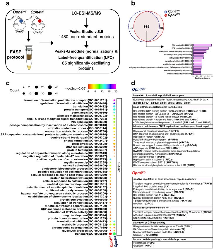
ARTICLE                                                               COMMUNICATIONS BIOLOGY | https://doi.org/10.1038/s42003-022-03425-6

Fig. 6 Overview of proteomic experimental design and selected results. a Experimental protocol. b Venn diagram representing the overlap of results
revealed by proteomes of Opn4WT and Opn4KO tumors and top 10 functional annotation clustering enriched (Classification Stringency: low) of commonly
identified proteins in the tumor tissues performed in DAVID (p < 0.05). c Enrichment analyses of differentially identified proteins in the tumor proteomes
using Gene Ontology (biological process, GO-BP) s performed in DAVID (p < 0.05). GO-BP terms were filtered for redundancy using REViGO. d Detailed
view of proteins related to some enriched terms (GO-BP) showing distinct molecular features between Opn4WT and Opn4KO tumor are shown. n = 4 for
each group comprised of independent samples.

compensatory mechanism of reduced small GTPase levels in                        Our next step was to apply the label-free quantification method
Opn4KO tumors, as will be seen below. However, this event may                to relatively quantify the commonly identified proteins between
contribute to the antitumoral effects seen in Opn4KO tumors. In              tumors. A total of 85 proteins out of 992 common proteins were
fact, it has been shown that TIAM1 and CORO1C are negative                   significantly different, being 59 and 26 proteins classified as up-
regulators of metastatic melanoma growth66,67.                               and downregulated in Opn4WT and Opn4KO tumors, respectively

8                          COMMUNICATIONS BIOLOGY | (2022)5:461 | https://doi.org/10.1038/s42003-022-03425-6 | www.nature.com/commsbio
COMMUNICATIONS BIOLOGY | https://doi.org/10.1038/s42003-022-03425-6                                                                ARTICLE

(Fig. 7 a; Supplementary Data 3). For instance, among the                     apoptotic processes of human melanocytes77. Contributing to the
upregulated proteins in Opn4WT tumors, GTPases (e.g. RAC1,                    new role of opsins, our group has recently demonstrated that
RAB5C, and SAR1B) 68,69and proteins involved in DNA                           OPN4 removal in malignant melanocytes resulted in insensitivity
replication and cell proliferation (MCM3 and MCM5) were                       to UVA-induced effects such as melanin content increase and
identified70. On the other hand, SOD1 protein, known to inhibit                apoptosis25. In normal melanocytes, OPN4 removal resulted in
RAC171, was upregulated in Opn4KO compared to Opn4WT                          the loss of UVA-induced reduced cellular growth and
tumors (Fig. 7 b).                                                            pigmentation 25and a higher proliferation and faster cell cycle
   Label-free quantification data show differences between the                 progression, which was associated with higher Mitf expression27.
proteome of both tumors and suggest a remarkable effect on                       Our results show that the removal of OPN4 in mouse mela-
GTPase activity. Furthermore, increased levels of Complement                  noma results in reduced proliferation and cell cycle progression
factor B in Opn4WT tumors compared to Opn4KO along with                       impairment, which differs from the previous results in normal
exclusively C5 and C4b proteins in Opn4WT proteome were                       melanocytes27. In human melanoma, OPN4 expression decreases
identified. It is known that complement system activation in                   with disease progression, and tumors expressing low levels of
cancer is linked with a higher inflammatory TME and melanoma                   OPN4 also display increased levels of BMAL125. Patients with
progression72,73. Therefore, these data together suggest a higher             high BMAL1 expressing tumors showed increased survival com-
inflammatory TME, which could explain the higher tumor                         pared to BMAL1 low expressing ones, a phenotype associated
growth of Opn4WT tumors.                                                      with increased mutational load21. Consequently, higher BMAL1
   We also evaluated the functionality of proteins unique to each             expressing tumors are more immunogenic, which affects patient
tumor genotype and those present in both proteomes but                        survival21. Remarkably, the BMAL1 gene was also shown to be a
differentially regulated, using some GO-categories keywords                   biomarker of immunotherapy success in metastatic melanoma21.
(e.g., “cell cycle”, “rhythm”, “circadian”, and “melanin”) to                    Furthermore, previous results from our group showed that the
identify associated proteins, following manual curation (Supple-              removal of Trpa1 channel results in reduced tumor growth via
mentary Data 4). Most proteins of the upregulated or exclusively              increased CD8+ cytotoxicity78. Collectively, we suggest that
identified in Opn4WT tumor were associated with positive                       pharmacological modulation of either Opn4 or Trpa1, as well as
regulation of G1/S progression (APEX1, GSPT2, PES1, RALA,                     others opsin-associated signaling pathway players79, may become
RALB, RHOB, TBCD, UPF1, USP9X, and RPL17) and negative                        interesting pharmacological targets in melanoma treatment.
regulation of G2/M transition (PSMC6). On the other hand,                        In vivo tumor growth corroborated our in vitro findings.
differentially expressed proteins found in Opn4KO tumors were                 Reduced tumor growth was followed by decreased melanin con-
associated with positive regulation of G2/M transition (EGFR,                 tent, which is also in line with the literature80,81. Despite the
MKI67, RAB11A, and VPS4B) and negative regulation of G1/S                     increased frequency of tumor-infiltrating lymphocytes in TME of
progression (GPNMB and EIF4E) (Fig. 7c; Supplementary                         Opn4KO tumors, gene expression of tumor bulk suggests a less
Data 4). These data, therefore, corroborate the slower cell cycle             inflammatory TME, a fact that can be associated with a higher
progression of Opn4KO cells.                                                  activity of the immune system and success of the immune system
   Although main clock proteins were not identified, several                   against tumor cells. However, we did not investigate the
players (DBP, NDUFA9, PTGDS, TOP1, TOP2a, USP9x, and                          mechanisms underlying the slower growth of Opn4KO tumors.
PP1CB) directly and indirectly involved in the positive loop                  We also found evidence that the molecular clock acute response is
regulation of the molecular clock (BMAL/CLOCK) were                           impaired in in vivo tumors, which is in line with the in vitro data.
upregulated in Opn4WT (Supplementary Data 4). In the absence                  In accordance with our previous data, we found that Opn4KO
of OPN4, increased protein expression of HNRNPD, a negative                   tumor bulk cells display a higher frequency of BMAL1 positive
regulator of the molecular clock, was identified (Supplementary                cells with increased protein expression; moreover, in TME we also
Data 4). Moreover, well-known target proteins of MITF in                      found that tyrosinase and BMAL1 positive cells (malignant
melanogenesis such as TYRP1, DCT, and TYR1 proteins were                      melanocytes) are also enriched in Opn4KO tumors compared to
positivity regulated in Opn4WT tumor (Fig. 7d; Supplementary                  Opn4WT ones. Importantly, Opn4KO tumors also show impaired
Data 4), and therefore, strongly suggest a reduction of MITF                  cell cycle-related gene expression, which argues for a slower cell
signaling in Opn4KO tumor.                                                    cycle progression in vivo as found for the malignant melanocytes
   Taken altogether, proteomic data brought to light possible                 in vitro.
mechanisms of slower proliferation and cell cycle progression in                 The in vitro data demonstrated that reduced cell proliferation
Opn4KO tumors. Moreover, proteomic data suggest a highly                      is associated with reduced cell metabolism and proliferative
immune suppressor TME in Opn4WT. This event associated with                   capacity. Of note, cell cycle progression was also impaired in the
a faster proliferation explains the faster growth of Opn4WT                   absence of Opn4, as these cells exhibited a decrease in G1 and S as
tumors. All these findings further provided robust evidence of                 well as an increase in the G2/M phases. These increased levels of
an intriguing role of OPN4 in a light- and thermo-independent                 G2/M could in fact represent a point of cell cycle arrest, related to
fashion that can be appreciated as an oncogene in melanoma.                   the absence of Opn4KO. Therefore, our findings suggest that
                                                                              Opn4KO malignant melanocytes display an impaired capacity to
                                                                              fully start a new cell cycle. Important cell cycle regulators were
Discussion                                                                    also affected at the mRNA level, which collectively confirm a
We provided evidence that a light- and thermo-sensing protein,                slower proliferation phenotype.
OPN4, whose role as an important light sensor responsible for                    Gene and protein levels of molecular clock components were
circadian entrainment has been well established74,75, plays a pro-            also differentially expressed in the absence of Opn4. Moreover,
tumoral role in melanoma. Extra-retinal OPN4 has been descri-                 the molecular clock of Opn4KO malignant cells was less sensitive
bed in skin cells, blood vessels, and other peripheral tissues                to dexamethasone and forskolin. The experimental design chosen
(reviewed in22). Some studies have shown that opsins, mainly                  in this study focused on the evaluation of the acute response of
encephalopsin (OPN3), also exert regulatory functions in cellular             the molecular clock and not on the rhythmicity aspect in the
biology other than light or thermal sensors. For instance, OPN3               absence of Opn4 since the B16-F10 circadian clock has previously
acts as a negative regulator of melanogenesis through melano-                 shown weak responses to synchronizing agents18,19. Therefore,
cortin 1 receptor (MC1R) interaction76 and participates in                    our experimental data are insufficient to evaluate the alterations

COMMUNICATIONS BIOLOGY | (2022)5:461 | https://doi.org/10.1038/s42003-022-03425-6 | www.nature.com/commsbio                                      9
ARTICLE COMMUNICATIONS BIOLOGY | https://doi.org/10.1038/s42003-022-03425-6 Fig. 7 Identification of differentially expressed proteins and their participation in cell cycle regulation and MITF signaling. a Heatmap of 85 differentially regulated proteins in Opn4WT and Opn4KO tumors. Significance was determined by two-sided t-test (p < 0.05) and FC ≥ 1.2 [log2(FC) ≥ 0.26 or ≤−0.26]. b Scatter plot showing the distribution of significantly regulated proteins in LFQ analysis. Proteins with higher fold change and statically significant values are shown. c Differentially encoded proteins among melanoma types showed GO-term related to different cell cycle phases. This shows the specific relationship between proteins and cell cycle progression. Arrows and blunt arrows represent positive and negative regulation, respectively. d Boxplots of MITF target proteins (DCT, TYR, and TYRP1) differentially encoded by Opn4 absence, which were associated with melanogenesis (tyrosine metabolism) pathway and melanoma. n = 4 for each group comprised of independent samples. 10 COMMUNICATIONS BIOLOGY | (2022)5:461 | https://doi.org/10.1038/s42003-022-03425-6 | www.nature.com/commsbio
COMMUNICATIONS BIOLOGY | https://doi.org/10.1038/s42003-022-03425-6                                                                         ARTICLE

that the absence of Opn4 may cause to the circadian clock                     exclusive presence of DNA repair proteins (PARP1, RPA2,
function. Noteworthy, the Bmal1 gene and protein expression was               APEX1, and XRCC6) and DNA replication (RCF2 and TOP1).
increased in Opn4KO compared to Opn4WT counterparts. One                      Increased DNA repair capacity has been related with more
can further suggest that increased acute clock gene response may              aggressive cancer development and treatment resistance in
represent an advantage for tumor growth. Therefore, our data                  melanoma88–91. We previously identified a link between higher
show that the proliferative capacity of malignant melanocytes                 expression of BMAL1 and lower base excision repair score.
seems to be dependent on Opn4 as in the absence of this protein,              Indeed, such association was found in our experimental model as
proliferation and cell metabolism were significantly lower.                    the absence of OPN4 resulted in elevated BMAL1 protein
   We further suggest that the impairment of guanylyl cyclase                 expression as well as reduction/absence of base excision repair-
activity demonstrated in vitro is associated with the decreased               related proteins (PARP1, RPA2, APEX192–94; uniprot database).
proliferation of Opn4KO cells. Proteomics analyses further cor-               Since these proteins were only identified in Opn4WT tumors, we
roborated these findings. In Opn4KO tumors, the RAC1 level was                 suggest a possible reduction of DNA repair via decreased base
reduced compared to Opn4WT tumors. RAC1 is known to acti-                     excision repair activity. It may be possible that these findings are
vate guanylyl cyclase and increase cGMP via RAC/PAK/GC/                       linked with reduced MITF signaling in the absence of OPN4 via
cGMP pathway82. Furthermore, enrichment of calcium-                           an elusive mechanism. Although MITF was not detected in the
dependent processes suggests a higher concentration of this ion               proteomics, we found that classical MITF targets such as DCT,
in Opn4KO tumors, and consequently, increased inhibition of                   TYR, and TYRP1 were less expressed in Opn4KO tumors.
guanylyl cyclase activity83. Experimental data strongly suggest                  Our findings are suggestive that OPN4 acts as an oncogene, but
that decreased guanylyl cyclase-cGMP activity is associated with              additional experiments using human melanoma cell lines and
reduced tumor growth and proliferation84,85. Within this line, our            different approaches, i.e., knockdown, knockout, and/or rescue
in vitro pharmacological guanylyl cyclase inhibition was effective            strategies are required to fully establish the OPN4 role as an
only in the presence of OPN4, thus suggesting that this enzyme                oncogene. One must consider that our study used mice kept at
activity in Opn4KO is at an already low level. Therefore, different           thermoneutrality, and thus, comparisons with other studies that
layers of evidence suggest impaired guanylyl cyclase activity in the          used temperature lower than the thermoneutral, should be made
absence of OPN4, which we suggest being one of the mechanisms                 with caution. With regards to the circadian aspects, our in vitro
by which removal of OPN4 reduces tumor growth and                             and in vivo sampling collection took place within a narrow time
proliferation.                                                                interval, thus, ruling out the influence of time in our analyses.
   Our data provided an interaction between OPN4 and MITF,                    However, additional circadian experiments are necessary to
which was first shown in normal melanocytes27. Since Mitf                      fully establish the impact of OPN4 in the regulation of the
expression is severely reduced in Opn4KO malignant melanocytes,               circadian clock.
and based on the literature42, a reduction in cellular proliferation             Taken altogether, our data add a novel layer of complexity to
would be expected. However, the novelty of our data lies in the               the opsin realm as we provide evidence that OPN4 can be a
fact that the lack of Opn4 resulted in reduced Mitf expression. In            tumor oncogene in melanoma. The traditional concept that
line with our results, Gaddameedhi’s lab recently reported that               opsins are light sensors has been challenged as opsins can also
MITF shows a rhythmic expression via BMAL1 interaction with                   detect thermal energy. Emerging evidence shows that opsins
the MITF promoter in human melanoma cells86. We suggest that                  also display light- and thermo-independent functions likely due
OPN4 could participate in the circadian regulation of MITF                    to protein-protein interaction, and therefore, opens a new field
either by interacting with BMAL1 at the DNA level and/or via                  of investigation. Understanding the function of opsins as light-
downstream pathways that lead to degradation of mRNA and/or                   and thermo-independent proteins in both physiological and
protein. Further research is needed to clarify this possible                  pathological contexts can yield promising therapeutic strategies
interaction.                                                                  and tools in a near future. Therefore, we suggest that OPN4 can
   Our experimental data well correlate with human melanoma                   be seen as a tumor oncogene in melanoma and could be
from the TCGA database. We found that OPN4 correlates with                    pharmacologically targeted.
MITF. Low MITF tumors display a lower ratio of G1-S/G2-M
gene expression, which shows higher gene expression of G2-M                   Methods
related targets. This is suggestive of a slower cell cycle progres-           Cell culture. Murine malignant B16-F10 Opn4KO melanocytes were generated
sion. Moreover, by estimating the frequency of immune cells in                using Clustered Regularly Interspaced Short Palindromic Repeats (CRISPR) tech-
the tumor bulk, we found an interesting correlation between                   nique. Opn4WT and Opn4KO cells were subject to Per1:Luc gene transfection as
                                                                              described previously25 and were used in this study. Wild type B16-F10 cells were
MITF and OPN4 with different immune cell types that is sug-                   initially donated by Prof. Roger Chammas (School of Medicine, University of Sāo
gestive of a role of OPN4 in TME and consequently tumor                       Paulo). Cells were cultured in phenol red-free RPMI 1640 medium (Atena, Brazil),
progression.                                                                  supplemented with 25 mM NaHCO3 (Sigma-Aldrich, USA), 20 mM HEPES (Santa
   Proteomics analyses confirmed our experimental data at all                  Cruz, USA), 10% fetal bovine serum (FBS, Atena, Brazil), 1% antibiotic/anti-
                                                                              mycotic solution (10,000 U/mL penicillin, 10,000 μg/mL streptomycin, and 25 μg/
levels and further shed light on additional pathways that collec-             mL amphotericin B, Thermo Fisher, USA), pH 7.2. Geneticin (200 µg/mL, Ther-
tively provide the rationality of why Opn4KO tumors show                      moFisher, USA) was used to guarantee selection during maintenance and removed
reduced proliferation. We uncovered a downregulation and                      during the experiments. Cells were kept at 37 °C with 5% CO2. In all experiments,
upregulation of positive and negative loops of the main clock                 unless otherwise mentioned, FBS was reduced to 2% and all-trans retinal (100 nM,
                                                                              Sigma-Aldrich, USA) was added as a supplement. For experiments in the absence
machinery, respectively, in Opn4KO compared to Opn4WT                         of CO2, HEPES concentration was increased to 50 mM. All in vitro experiments
tumors. With regards to the cell cycle, proteins related to G1/S              were carried out in the dark under red dim light (7 W Konex bulb and Safe-Light
and G2/M were down- and upregulated, respectively, in the                     filter GBX-2, Kodak, USA). Unless otherwise mentioned, cells were collected on the
absence of Opn4. Evidence of a less inflammatory TME in                        4th day after seeding.
Opn4KO tumors was found as reduced complement expression
and gene expression of inflammatory cytokines were identified in                Animal handling and care. Animal experimentation was performed according to
Opn4KO tumors. Reduced expression of GTPases has been linked                  Brazilian animal welfare regulations and approved by the Committee of Animal
                                                                              Ethical Experimentation of the Institute of Biosciences, University of Sāo Paulo
with slower cell cycle progression87, which is in line with                   (Approval 322/2018). Three- to eight-months old male C57BL/6 J mice were kept
the slower Opn4KO proliferation. Moreover, the higher pro-                    under a 12:12 light/dark cycle (800–1000 lux white LED light, ranging from 420 to
liferative capacity of Opn4WT tumors can be associated with the               750 nm) at controlled temperature (30 ± 1oC). Lights were on at 7 a.m. and off at

COMMUNICATIONS BIOLOGY | (2022)5:461 | https://doi.org/10.1038/s42003-022-03425-6 | www.nature.com/commsbio                                                 11
ARTICLE COMMUNICATIONS BIOLOGY | https://doi.org/10.1038/s42003-022-03425-6
7 p.m. Mice were subcutaneously inoculated in the right flank with 2 × 10⁶ Opn4WT thermocycler (BioRad Laboratories, USA) in the following conditions: 3 min at
or Opn4KO B16-F10 cells in 100 µL of phosphate-buffered saline (PBS). Sham 95 °C followed by 45 cycles of 15 s at 95 °C and 60 s at 60 °C. For SYBR Green
control animals were injected with the same volume of PBS. Then, mice were single assay, independent solutions were prepared with cDNA, specific primers, and Kapa
housed for the entire experiment. Tumor measurement, food intake, and weight Sybr Fast mix (Kapa Biosystems, USA), and run in duplicates in an iQ5
were assessed every three to four days at the same time of the day (from 2 to 3 pm). thermocycler in the following conditions: 10 min at 95 °C, followed by 45 cycles of
Length, width, and height of the tumors were measured with a caliper rule from the 15 s at 95 °C, 60 s at 60 °C, and 80 cycles of 10 s at 55 °C with a gradual increase
13th day onwards, and the volume (mm3) was calculated according to the formula: of 0.5 °C.
π/6 x length x width x height95. Animals were CO2-euthanized 25 days after
inoculation, and death was assured by cervical dislocation, between 9 and 10 am
Per1:Luc bioluminescence assay. One hundred thousand Opn4WT or Opn4KO
(Zeitgeber time 2–3). After euthanasia, every animal was visually inspected, and no
malignant melanocytes were seeded in 35 mm dishes in experimental media
visible metastasis nodules were found. The organs and blood were harvested and
(containing 50 mM HEPES) and kept in a CO2 incubator for 24 h. On the next day,
immediately processed or stored at −80 oC. The tumor was resected, weighed and
cells were treated with dexamethasone (200 nM) or forskolin (10 µM). Drugs and
melanin levels were quantified as previously described20.
vehicles remained in dishes until the end of the experiment. Luciferin (Promega,
USA, 100 µM) was also added, dishes were sealed with 35 mm round coverslips
XTT assay––metabolic evaluation. For XTT experiments, 104 Opn4WTor Opn4KO (VWR, England) and parafilm, and placed into the Lumicycle equipment (Acti-
malignant melanocytes were seeded in 96-well plates in 100 µL of experimental metris, USA) in an incubator without CO2 positive pressure, at 37 ± 0.5 °C. Bio-
medium and kept for 24 h in the incubator. On the following day, 50 µL of luminescence was recorded every 10 min. The temperature of the incubator was
XTT and electron coupling reagent (ECR) solution (7:1) was added. For phar- monitored every 10 min (iLog, Escort Data Loggers, USA). Baseline subtracted data
macological manipulation, drugs or respective vehicles were also added (Supple- were plotted using a dampened sine wave function in Graphpad Prism (7.0).
mentary Table S1). Cells were kept at 37 °C with 5% CO2 for 4 and 24 h,
and then the solutions’ absorbance was read in a spectrophotometer
Hematological analyses. After euthanasia, blood was collected by cardiac punc-
(SpectraMax 250, Molecular Devices, USA). Specific absorbance was calculated as
ture in EDTA (10.25 mg/mL) collection tubes and immediately processed. Analyses
(A450 sample − A450 blank) − A660 according to the manufacturer’s instructions
were performed on an automated hematology analyzer (BC-2800Vet, Mindray,
(CyQUANT™ XTT Cell Viability Assay, Thermofisher, USA).
USA) using mouse-specific algorithms and parameters.
Cellular growth evaluation. Fifty thousand Opn4WT or Opn4KO malignant mel-
Flow cytometry for tumor-associated macrophages, tumor-infiltrating lym-
anocytes were seeded in 12-well plates in the experimental medium and kept for
phocytes, and BMAL positive cells. The tumor and spleen were dissected and
24 h in the incubator. Twenty-four, 48, 72, and 96 h later, the cells were harvested
dissociated through a cell strainer (100 µm, Corning, USA) in PBS. Red blood cells
with Tyrode/EDTA solution and counted in a hemocytometer.
(RBC) were lysed using ACK (ammonium-chloride-potassium) and RBC lysing
buffer (0.15 M NH4Cl, 10.0 mM KHCO3, 0.1 mM Na2EDTA), followed by 1000 x g
CellTrace™ proliferative assay. One thousand Opn4WT or Opn4KO malignant centrifugation. The supernatant was removed and the remaining cells were
melanocytes were loaded with Celltrace dye (1 µL for every 106 cells), seeded in resuspended in PBS. One million cells per well were stained in a round bottom 96
6-well plates in the maintenance media (10% SFB), and kept in the incubator for well plate using a two-step staining protocol. Flow cytometry procedures were
four days. Then, cells were harvested with Tyrode/EDTA solution, stained with performed as described previously78. In short, first, cells were stained with a Live/
Live/ Dead violet fluorescent dye™ (405 nm, 1:500, ThermoFisher, USA) in PBS and Dead dye (Fixable aqua 405 nm, Invitrogen, USA) at 4 °C for 20 min, and after
kept at 37 °C. The cells were fixed in 4% paraformaldehyde (Electron Microscopy washing 100 μL of a solution containing surface antibodies diluted in staining
Science, USA) on ice for 30 min, and stored at 4 °C until processing in Canto II buffer (1% FBS, 1 mM EDTA, and 0.02% NaN3 in PBS) were added into each well.
flow cytometer (BD Biosciences, USA) using DiVA 8 acquisition software. At least After 30 min at 4 °C, the samples were washed (2X) and resuspended in staining
105 events were captured. Cells were gated using FSC and SCC, duplets were buffer until acquisition. The following antibodies from Biolegend, USA (1:200
excluded using FSC-H vs FSC-A. Negative Live/Dead stained cells (viable cells) dilution) were used unless otherwise mentioned: APC-Cy7 anti-Mouse F4/80 (Cat
were gated and from this population, CellTrace-positive cells were calculated in no. 123118), PerCP-Cy5.5 anti-Mouse CD80 (Cat no. 194722), FITC anti-mouse
terms of percentage and median intensity of fluorescence (MIF). Data were pro- CD206 (Cat no.141704), FITC anti-mouse CD4 (Cat no. 100509), PE-Cy7 anti-
cessed in FlowJo™ software (BD Biosciences, USA). Negative controls are shown in mouse CD44 (Cat no. 560569), PE anti-mouse CD62L, (Cat no. RM4304), and
Fig. S2 K. APC anti-mouse CD8 (Cat no. MCD0805, Invitrogen, USA). For tyrosinase and
BMAL1 dual staining, tumor cells were labeled with anti-tyrosinase (1:25, goat
polyclonal, Santa Cruz, USA SC 18182) and anti-BMAL1 (1:100, rabbit polyclonal,
Cell cycle analysis by flow cytometry. Cell staining followed the manufacturer’s
ABCAM, ab93806) in staining buffer as described above. Secondary antibodies
instructions (BD Biosciences, USA). In brief, cells were loaded with BrdU solution
anti-goat (555 nm) and anti-rabbit (488 nm) (both Alexa Fluor ThermoFisher,
(10 mM) for 2 h, and then harvested and fixed with Cytoperm Cytofix solution (BD
USA) were used (1:100). Samples were assessed in a FACSCanto II cell analyzer
Biosciences, USA) on ice for 30 min. Approximately 105 to 5 × 10⁵ cells/well were
(Becton Dickinson, USA) using DiVA 8 acquisition software and FlowJo 5 V10
placed onto a 96-well round bottom plate in Cytoperm Permeabilization Buffer
(Becton Dickinson, USA) data analysis software. Representative negative controls
Plus, kept on ice for 10 min, followed by another incubation with Cytofix/Cyto-
of all flow cytometry experiments can be found in Fig. S1A–I.
perm buffer on ice for 5 min. DNAse (300 µg/mL) was added and cells were placed
at 37 °C for 1 h. Cells were resuspended in anti-BrdU antibody in Perm/Wash
buffer (FITC, 1:50) and kept at room temperature for 20 min. Cells were resus- Sample preparation for proteomics. Tumor tissues were mechanically homo-
pended in 7-AAD solution and kept in staining buffer until the acquisition in genized in lysis buffer (2% SDS, 100 mM Tris-HCl, pH 7.8) supplemented with
Canto II Flow Cytometry (BD Biosciences, USA). In every step, cells were washed protease inhibitors (Complete™ ULTRA Tablets, Mini, EASYpack Protease Inhi-
with Perm/Wash buffer, followed by centrifugation (200 x g for 3 min). Negative bitor Cocktail, Roche, USA), using a POLYTRON® PT 1200 and sonicated for
controls are shown in Fig. S2L. three cycles at 30% amplitude (20 s bursts with 20 s pauses). Samples were cen-
trifuged at 16,000 x g for 10 min at 4 °C to remove the tissue debris. The super-
natants were collected and the protein concentration was measured by using a BCA
RNA extraction, cDNA synthesis, and quantitative PCR (qPCR). Total RNA
protein assay (Pierce™ BCA Protein Assay Kit, Thermo Scientific, USA). Aliquots of
was extracted from cells or tumors with Trizol (ThermoFisher, USA) according to
SDS-lysates containing 200 µg of total protein from each sample were processed
the manufacturer’s instruction, using 1-bromo-3-chloropropane (Sigma, USA),
according to the filter-aided sample preparation (FASP) method, using Microcon
isopropanol (Sigma, USA), and washed with 75% molecular grade ethanol (Sigma,
10 kDa centrifugal filter units (Merck, USA) operated at 10,000 x g for 50 min at
USA). DEPC water was used to resuspend the RNA pellets. Genomic con-
20 °C96–98. Next, Trypsin/LysC Mix (Promega, USA) was added to the filters at an
tamination was removed using TURBO DNAse (ThermoFisher, USA) and RNA
enzyme-to-protein ratio of 1:100 (w/w), and incubated for 12 h at 37 °C; a second
concentration and quality (OD260/OD280) were assessed in a spectrophotometer
digestion was carried out with trypsin (Promega, USA) at an enzyme-to-protein
(NanoDrop, USA). One μg of total RNA was subject to reverse transcriptase
ratio of 1:100 (w/w) at 37 °C for 4 h. Following protein digestion, peptides were
reaction using random hexamer primers and Superscript III, in addition to the
filtered through the membrane and purified with reversed-phase chromatography
reagents recommended by the enzyme manufacturer (Life Technologies, USA).
using C18 micro-pipette tips (TopTipTM, PolyLC inc, USA), according to the
Twenty-five ng of cDNA was subject to quantitative PCR reactions using
manufacturer’s instructions. Peptides were dried in a vacuum concentrator and
species-specific primers (Supplementary Table S2) spanning introns, based on
stored at −20 °C.
sequences obtained from the GenBank (http://www.ncbi.nlm.nih.gov/genbank),
designed by Primer Blast (http://www.ncbi.nlm.nih.gov/genbank) or Primer Quest
(IDT, USA), and synthesized by Integrated DNA Technologies (IDT, USA). Rpl37a Mass spectrometry data acquisition. Dried peptides were recovered in 20 µL of
was used to normalize the expression values of the genes of interest in the in vitro 0.1% formic acid in water and analyzed on a nano-ACQUITY UPLC system
and in vivo assays. Rpl37a showed a robust and stable expression across samples (Waters, USA) coupled online to a maXis 3 G quadrupole time-of-flight (Q-TOF)
(SD < 1). For TaqMan assay, reactions containing cDNA, primers, fluorescent mass spectrometer (Bruker Daltonics, Germany), equipped with a Captive Spray
probes, and Kapa Probe Fast Mix (Kapa Biosystems, USA) were used and run in nanoelectrospray source. One microliter of the sample was injected and peptide
triplicates for each cDNA sample. Reactions were carried out in an iQ5 mixture was loaded onto a trap column (nanoAcquity UPLC® 2G-V/M Trap 5 µm
12 COMMUNICATIONS BIOLOGY | (2022)5:461 | https://doi.org/10.1038/s42003-022-03425-6 | www.nature.com/commsbioYou can also read