In vivo partial cellular reprogramming enhances liver plasticity and regeneration
←
→
Page content transcription
If your browser does not render page correctly, please read the page content below
Article
In vivo partial cellular reprogramming enhances liver
plasticity and regeneration
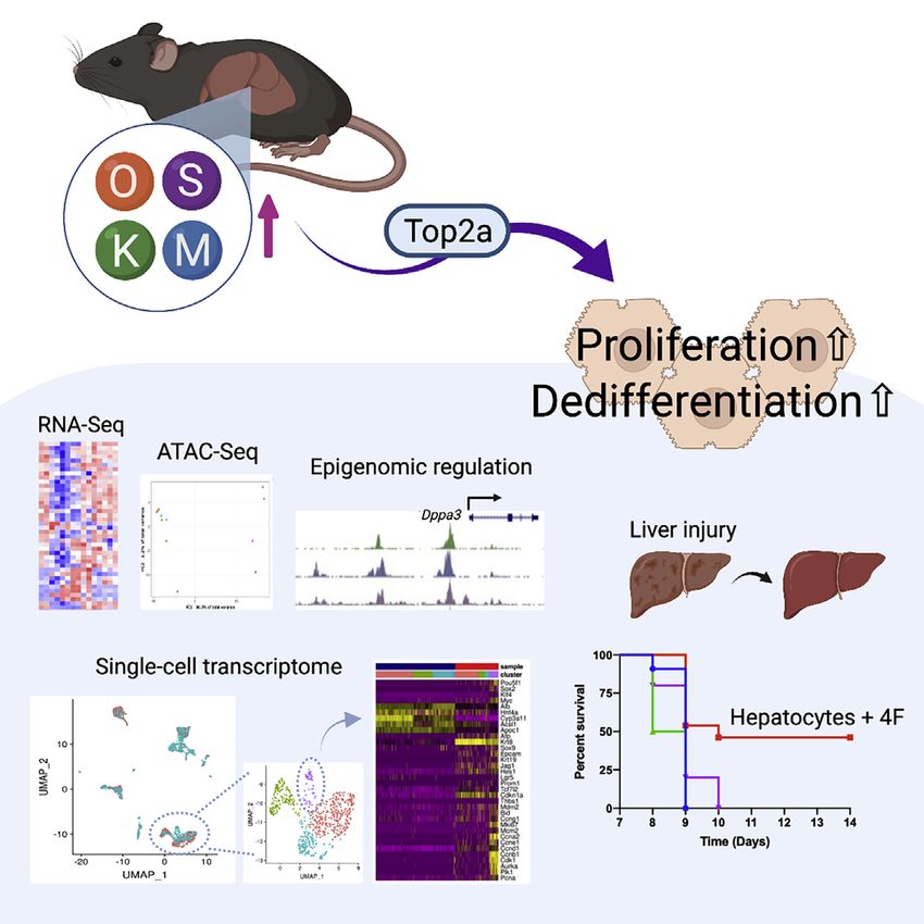
Graphical abstract Authors
Tomoaki Hishida, Mako Yamamoto,
Yuriko Hishida-Nozaki, ...,
Pradeep Reddy, Guang-Hui Liu,
Juan Carlos Izpisua Belmonte
Correspondence
jcbelmonte@altoslabs.com
In brief
In regenerating animals, such as fish and
salamanders, dedifferentiation followed
by proliferation contributes to tissue
regeneration. Hishida et al. show that
hepatocyte-specific cellular
reprogramming induces cell proliferation
and dedifferentiation in the liver and
enhances liver regenerative capacity
through topoisomerase2-mediated
partial reprogramming.
Highlights
d Hepatocyte-specific 4F expression induces cell proliferation
and dedifferentiation
d Hepatocyte-specific 4F expression induces a global change
in DNA accessibility
d Top2a is required for cellular reprogramming in vitro and
in vivo
d In vivo reprogramming has beneficial effects on regenerative
capacity
Hishida et al., 2022, Cell Reports 39, 110730
April 26, 2022 ª 2022 The Authors.
https://doi.org/10.1016/j.celrep.2022.110730 ll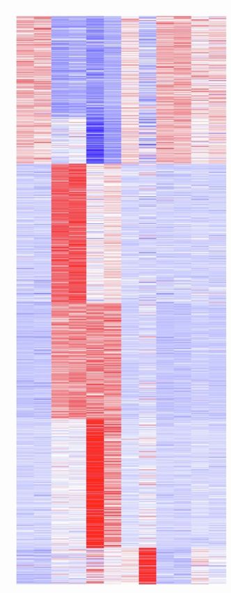
ll
                                                                                                                      OPEN ACCESS

Article
In vivo partial cellular reprogramming
enhances liver plasticity and regeneration
Tomoaki Hishida,1,14,15 Mako Yamamoto,1,15 Yuriko Hishida-Nozaki,1 Changwei Shao,1 Ling Huang,2 Chao Wang,1,3
Kensaku Shojima,1 Yuan Xue,1 Yuqing Hang,2 Maxim Shokhirev,2 Sebastian Memczak,1,3 Sanjeeb Kumar Sahu,1,3
Fumiyuki Hatanaka,1,3 Ruben Rabadan Ros,1,9 Matthew B. Maxwell,4,5 Jasmine Chavez,1 Yanjiao Shao,1,3 Hsin-Kai Liao,1
Paloma Martinez-Redondo,1 Isabel Guillen-Guillen,1,3 Reyna Hernandez-Benitez,1,3 Concepcion Rodriguez Esteban,1,3
Jing Qu,6 Michael C. Holmes,7 Fei Yi,7 Raymond D. Hickey,7 Pedro Guillen Garcia,8 Estrella Nuñez Delicado,9
Antoni Castells,10 Josep M. Campistol,10 Yang Yu,1 Diana C. Hargreaves,4 Akihiro Asai,11,12 Pradeep Reddy,1
Guang-Hui Liu,13 and Juan Carlos Izpisua Belmonte1,3,16,*
1Gene   Expression Laboratory, Salk Institute for Biological Studies, 10010 North Torrey Pines Road, La Jolla, CA 92037, USA
2Razavi  Newman Integrative Genomics and Bioinformatics Core, Salk Institute for Biological Studies, 10010 North Torrey Pines Road, La
Jolla, CA 92037, USA
3Altos Labs, 5510 Morehouse Drive, San Diego, CA 92121, USA
4Molecular and Cell Biology Laboratory, Salk Institute for Biological Studies, 10010 North Torrey Pines Road, La Jolla, CA 92037, USA
5Division of Biological Sciences, UCSD, La Jolla, CA 92037, USA
6State Key Laboratory of Stem Cell and Reproductive Biology, Institute of Zoology, Chinese Academy of Sciences, Beijing 100101, China
7Ambys Medicines, 131 Oyster Point Boulevard, Suite 200, South San Francisco, CA 94080, USA
8Clinica CEMTRO, 28035 Madrid, Spain
9Universidad Católica San Antonio de Murcia (UCAM), Campus de los Jerónimos, Nº 135 12, 30107 Guadalupe, Spain
10Hospital Clinic of Barcelona, Carrer Villarroel, 170, 08036 Barcelona, Spain
11Division of Gastroenterology, Hepatology and Nutrition, Cincinnati Children’s Hospital Medical Center, Cincinnati, OH 45229, USA
12Department of Pediatrics, College of Medicine, University of Cincinnati, Cincinnati, OH 45229, USA
13State Key Laboratory of Membrane Biology, Institute of Zoology, Chinese Academy of Sciences, Beijing 100101, China
14Laboratory of Biological Chemistry, School of Pharmaceutical Sciences, Wakayama Medical University, 25-1 Shitibancho, Wakayama,

Wakayama 640-8156, Japan
15These authors contributed equally
16Lead contact

*Correspondence: jcbelmonte@altoslabs.com
 https://doi.org/10.1016/j.celrep.2022.110730

SUMMARY

Mammals have limited regenerative capacity, whereas some vertebrates, like fish and salamanders, are able
to regenerate their organs efficiently. The regeneration in these species depends on cell dedifferentiation
followed by proliferation. We generate a mouse model that enables the inducible expression of the four Ya-
manaka factors (Oct-3/4, Sox2, Klf4, and c-Myc, or 4F) specifically in hepatocytes. Transient in vivo 4F
expression induces partial reprogramming of adult hepatocytes to a progenitor state and concomitantly in-
creases cell proliferation. This is indicated by reduced expression of differentiated hepatic-lineage markers,
an increase in markers of proliferation and chromatin modifiers, global changes in DNA accessibility, and an
acquisition of liver stem and progenitor cell markers. Functionally, short-term expression of 4F enhances liver
regenerative capacity through topoisomerase2-mediated partial reprogramming. Our results reveal that
liver-specific 4F expression in vivo induces cellular plasticity and counteracts liver failure, suggesting that
partial reprogramming may represent an avenue for enhancing tissue regeneration.

INTRODUCTION                                                            Somatic cells can be reprogrammed to a pluripotent state by
                                                                     overexpressing the four Yamanaka factors (Oct-3/4, Sox2,
Mammals lack the regenerative capacity exhibited by some ver-        KLF4, and c-Myc, hereafter referred to as 4F; Takahashi et al.,
tebrates, such as fish and salamanders (Simon, 2012). In these       2007; Takahashi and Yamanaka, 2006) for several weeks.
regenerating animals, it has been shown that dedifferentiation       Although systemic 4F overexpression can induce dedifferentia-
followed by proliferation contributes to tissue regeneration (Jo-    tion even in vivo, the end result in most instances is cancer
pling et al., 2010; Wang and Simon, 2016). The scarcity of dedif-    (Abad et al., 2013; Ohnishi et al., 2014; Shibata et al., 2018).
ferentiation in mammal tissues (Rinkevich et al., 2011) may be the   We therefore switched to a short-term 4F induction protocol
reason they cannot regenerate.                                       and demonstrated that 4F ameliorates aging processes in a

                                                                          Cell Reports 39, 110730, April 26, 2022 ª 2022 The Authors. 1
                                   This is an open access article under the CC BY license (http://creativecommons.org/licenses/by/4.0/).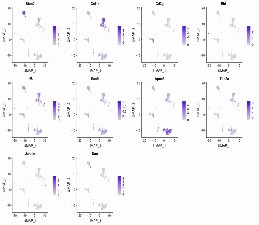
ll
     OPEN ACCESS                                                                                                                                                                                  Article

 A                                          B                                                                                        C

                                                                          8
                                                                                 *P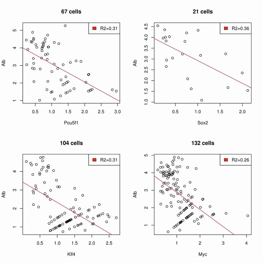
ll
Article                                                                                                                                        OPEN ACCESS

mouse model of premature aging (Ocampo et al., 2016). Recent                       scription factors (Hnf1a, Hnf4a, and Hnf6) were downregulated,
studies showed that in vivo reprogramming can improve regen-                       whereas Foxa2, Gata4, and Gata6, which play more important
eration of muscle, optic nerve, and cardiomyocytes (Chen et al.,                   roles during liver ontogeny, were upregulated. Interestingly, the
2021; Lu et al., 2020; Wang et al., 2021). However, it remains an                  gene Afp, which encodes a serum protein that is normally
open question whether short-term 4F induction can transiently                      silenced after birth, was also upregulated, both at the transcrip-
and partially reprogram mature cells to a plastic state in vivo to                 tion and protein levels (Figures 1D and S1C). This may also
promote the regeneration of mammalian tissues without also                         indicate cellular proliferation, as Afp is elevated during liver
driving tumor formation. This question is difficult to answer                      regeneration (Nakano et al., 2017).
without a stringent lineage-tracing system but is important to                        Two days of Dox treatment caused the mice to die within
address since an increase in regenerative capacity could, in prin-                 5 days. A failure of liver function was identified as one possible
ciple, be harnessed to treat many human diseases.                                  reason for mouse death, because mature hepatocyte markers,
   With this in mind, here, we developed a mouse model that                        such as Alb and Cyp3a11, were downregulated. To investigate
enables both the inducible expression of 4F in specific tissues,                   that possibility, comprehensive metabolic panel analyses were
as well as the ability to track 4F-expressing cells. Although                      performed (Figure S1D). These metabolic analyses indeed
tissue regeneration in mammals generally is poor, the liver,                       showed poor liver function, and therefore, liver failure caused
if not severely injured, has some regenerative abilities. Thus,                    by 4F may be one reason for mouse death.
we decided to focus our question on the liver and therefore                           To optimize the experimental condition where lethal effects
used hepatocyte-specific 4F (Hep-4F) mice for all subsequent                       can be minimized, we systematically varied the amount of Dox
experiments.                                                                       that was administered, as well as the duration of treatment.
                                                                                   Limiting Dox treatment to 1 day at 0.1 mg/mL allowed the
RESULTS                                                                            Hep-4F mice to survive (Figures 1E–1G). We then used this pro-
                                                                                   tocol to perform time course experiments, collecting liver
Hepatocyte-specific 4F expression induces cell                                     samples at the indicated time points (Figure 1H). Dox treatment
proliferation and the loss of hepatic characteristics                              downregulated differentiated hepatocyte markers and upregu-
The Hep-4F mouse model includes the albumin (Alb)-Cre trans-                       lated Gata4, Gata6, Foxa2, and Sox9 (Figure 1I). The dedifferen-
gene, allowing for liver-specific Cre recombinase expression,                      tiation effect of 4F appeared to be transient, as the expression of
tetO-4F, and LoxP-STOP-LoxP-rtTA-IRES-GFP (Figure 1A). In                          adult hepatocyte markers returned to normal levels after Dox
this mouse model, the LoxP-STOP-LoxP cassette is excised                           withdrawal. In contrast to previous studies where the dose and
by Alb-Cre, resulting in the expression of rtTA and GFP in hepa-                   timing was higher (Abad et al., 2013; Ohnishi et al., 2014; Shibata
tocytes, which enables lineage tracing. Thus, administration of                    et al., 2018), this transient liver-specific 4F induction never re-
doxycycline (Dox) allows for liver-specific 4F expression. We first                sulted in tumor formation (data not shown). This analysis was
treated Hep-4F mice with Dox for 2 days. Quantitative RT-PCR                       performed up to 9 months after Dox withdrawal, although any
(qRT-PCR) analysis of collected tissues confirmed that 4F induc-                   tumorigenic activity mediated by 4F will need to be further in-
tion was specific to the liver (Figure S1A). We also observed that                 vestigated. Similarly, the effect of short-term 4F on cellular
4F expression resulted in a large, pale liver (Figure 1B). Any                     proliferation was also relatively transient (Figure 1J), showing a
noticeable histological change was not observed in the lung                        correlation between the loss of differentiated hepatocyte
and kidney, in which negligible gene expression of 4F was                          markers and proliferation. To see whether hepatic zonation is
observed after Dox treatment (Figure S1B). Immunostaining for                      altered by 4F induction, immunostaining was performed for
Ki67, a proliferation marker, showed that the number of Ki67-                      E-cadherin (E-Cad) and glutamine synthetase (GS) as zonally ex-
positive proliferating cells was increased 2 days after Dox treat-                 pressed markers for zone 1 and zone 3, respectively (He et al.,
ment (Figure 1C). Thus, 4F quickly promoted the proliferation of                   2021; Wei et al., 2021; Figure S1E). It seems that the impact of
liver cells. We next examined the expression of liver-specific                     4F on zonation was minimal. We also found that Sox9+ cells
marker genes via qRT-PCR (Figure 1D). Markers of mature hepa-                      diffusely emerged in the liver after Dox treatment in a transient
tocytes (Alb and Cyp3a11) and differentiated liver-enriched tran-                  manner (Figures 1J and S1F). Importantly, these Sox9+ cells

Figure 1. Induction of transient, partial reprogramming by liver-specific 4F expression
(A) Schematic representation of the genetic makeup for lineage-traceable, liver-specific 4F inducible mouse models. In this model, rtTA can be activated by Alb-
Cre, allowing for specific 4F induction in the liver in a Tet-ON manner.
(B) Livers collected from Dox-treated and untreated Hep-4F mice 2 days after Dox administration. Left: representative images are shown. Right: relative liver
weight (% body weight) is shown. Data represent the mean with SE (n = 5). *p < 0.01 (unpaired t test).
(C) Immunostaining for Klf4 and Ki67. Livers were collected 2 days after Dox administration. Scale bar, 200 mm.
(D) qPCR analysis for liver-related genes in the liver of Hep-4F mice treated with different concentrations of Dox (0.1 or 0.2 mg/mL) for 2 days. Data represent the
mean with SD (n = 3; technical replicates).
(E) Schematic representation for Dox treatment protocol.
(F and G) Body weight (F) and survival (G) of Hep-4F mice after Dox treatment (0.1 mg/mL; 1d-on, n = 9; 2d-on, n = 12).
(H) Schematic representation of time course for Dox treatment protocol.
(I and J) Time course experiment of qPCR (I) and IHC for Sox9 and Ki67 (J). Data represent the mean (n = 2; biological replicates). Scale bar, 200 mm.
(K) GFP-based lineage-tracing experiments for hepatocytes after 4F induction. Livers were collected 1 day after Dox withdrawal. White arrow and white
arrowhead indicate atypical Sox9+ cells and Sox9+ cholangiocytes, respectively. Scale bar, 100 mm.

                                                                                                                 Cell Reports 39, 110730, April 26, 2022 3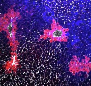
ll
      OPEN ACCESS                                                                                                                              Article

           A                                                                                         D

           B                                              C

                                                                                                     E

           F

           G

Figure 2. Global analysis of transcriptome and chromatin accessibility of Hep-4F mice
(A) Venn diagrams summarizing overlapping upregulated or downregulated genes responsive to 4F expression (day 2 versus day 0) and CCl4 treatment. GO an-
alyses were performed for overlapping DEGs.
(B and C) PCA analysis for RNA-seq (B) and ATAC-seq (C). Livers were collected at the indicated time points: 0d, 1d, 2d, 1d-on_1d-off, and 1d-on_2d-off. To
compare reprogramming with regeneration, B6 mice were treated with or without CCl4 for acute injury, and the livers were collected 3 days after the treatment:
control and CCl4. Each sample was prepared in duplicates except CCl4 samples, which are in triplicates.

                                                                                                                           (legend continued on next page)

4 Cell Reports 39, 110730, April 26, 2022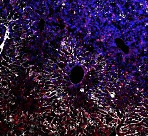
ll
Article                                                                                                                                  OPEN ACCESS

were GFP positive and distinct from GFPdim cholangiocytes,                      changes in DNA accessibility, and subsequent changes to the
which are epithelial cells of the bile duct that also express                   hepatocyte transcriptional signature.
Sox9 (Athwal et al., 2017; Figure 1K). This suggests that 4F                       We next analyzed RNA-seq data in detail. A previous report
expression partially reprogrammed hepatocytes to Sox9+ cells.                   showed that the embryonic stem cell (ESC) transcription signa-
Levels of Sox9 expression remained elevated following Dox                       ture can be dissected into three modules: core, Myc, and PRC
withdrawal, suggesting that 4F may induce dedifferentiation to                  modules (Kim et al., 2010). Focusing on these modules, we found
an undetermined plastic state and that these cells are then redir-              that core and Myc modules were transiently activated, indicating
ected back to the hepatocyte lineage (following Dox withdrawal)                 that 4F was able to activate the ESC program in liver cells (Fig-
by the surrounding niche. This will require further investigation.              ure 2D). However, core pluripotency factors, such as Nanog,
  We next tested whether Myc alone, which is known to induce                    Utf1, and Rex1, were not expressed (data not shown). Of note,
hepatocyte proliferation (Shachaf et al., 2004), is sufficient to               CCl4 did not induce core modules but was able to affect Myc
induce loss of mature hepatocyte markers. For this analysis,                    modules to some degree, consistent with previous reports that
we used a Hep-Myc mouse model carrying Alb-Cre, tetO-MYC                        c-Myc is upregulated during liver regeneration (White et al.,
(human), and LSL-rtTA-IRES-GFP. qRT-PCR results showed                          2005). We next assessed hepatocyte-related genes. Markers
that, unlike 4F, MYC did not induce the loss of mature hepato-                  of mature hepatocytes were globally downregulated soon after
cyte markers (Figure S1G), suggesting that partial reprogram-                   Dox administration (Figures 2E and S3). At the same time, cell-
ming does not solely depend on cell proliferation.                              cycle-related genes were being activated (Figure 2E). Expres-
                                                                                sion of liver progenitor and stem cell markers, such as Sox9
Global analysis of chromatin accessibility and gene                             and Epcam, were upregulated (Figure 2E) together with in-
transcription revealed that 4F induces partial                                  creases in chromatin accessibility within putative enhancer re-
reprogramming in vivo                                                           gions (Figure S2C). Of note, such changes induced by 4F were
We next performed global transcriptomic and DNA accessibility                   almost reverted 20 days after Dox withdrawal as analyzed by
analyses using RNA sequencing (RNA-seq) and an assay for                        RNA-seq (Figure 2F). This is consistent with 4F inducing partial
transposase-accessible chromatin using sequencing (ATAC-                        reprogramming in the liver in a transient manner.
seq). As a reference, we included liver samples collected                          Alb, Afp, and Afm are tandemly arranged in the same tran-
from mice injected with carbon tetrachloride (CCl4), which                      scriptional orientation, although little is known about how
causes acute liver injury followed by liver regeneration (Bezerra               these genes are regulated (Jin et al., 2009). Interestingly, our
et al., 1999; Nakano et al., 2017). Consistent with previous re-                RNA-seq data revealed that Afp and Afm are reciprocally regu-
ports, CCl4 administration activated genes related to cell                      lated (Figure 2G). Since Afp expression is correlated with can-
proliferation. These genes partially overlapped with the genes                  cers (Chen et al., 2020; Mizejewski, 2002), we asked whether
activated by 4F expression (Figure 2A). We performed prin-                      this reciprocal regulation is observed in hepatocellular carci-
cipal-component analysis (PCA) of the RNA-seq and ATAC-                         noma (HCC) using public data. We analyzed 313 cases and
seq data (Figures 2B and 2C). CCl4 treatment clearly affected                   found that there is a reverse correlation between AFP and AFM
gene expression but had little if any effect on chromatin acces-                (p = 0.036; Figure S4A). More importantly, the AFPlowAFMhigh
sibility. In sharp contrast, 4F dramatically changed chromatin                  group had a higher survival rate than the AFPhighAFMlow group
accessibility, presumably leading to massive alterations in                     (p = 0.011; Figure S4B), indicating the importance of the recip-
gene expression. We next performed unbiased clustering of                       rocal regulation between AFP and AFM in the context of cancer.
ATAC-seq peaks, resulting in six clusters (Figure S2A). Two                        We next performed Gene Ontology (GO) analysis for differen-
of these six clusters were associated with a downregulation                     tially expressed genes (DEGs) (Figure 3A). Cell-cycle-related GO
of DNA accessibility after Dox treatment, suggesting that these                 terms, such as DNA replication and cytokinesis, were upregu-
regions switched from an ‘‘open to closed’’ chromatin state                     lated, whereas metabolic pathway GO terms were downregu-
(OC). The other clusters switched from ‘‘closed to open’’                       lated. Interestingly, epigenetic-modification-related terms,
(CO), with variation in the timing of this switch. We performed                 such as DNA conformation change and covalent chromatin
motif analysis, revealing that motifs associated with hepato-                   modification, were also upregulated, driving us to investigate
cyte-enriched transcription factors were largely enriched in                    epigenetic modifiers. Indeed, 4F induced lots of epigenetic mod-
the OC1 cluster and that 4F motifs were enriched in CO1/2                       ifiers (Figure 3B), which would explain 4F-mediated global
groups (Figure S2B). Of note, motifs for Jun/Fos (AP1 complex),                 changes in DNA accessibility. With the RNA- and ATAC-seq
Gata4/6, and Foxa2 were generally activated by Dox treatment.                   data, we found that 4F activated several silenced genes, such
These pioneer factors (Biddie et al., 2011; Iwafuchi-Doi et al.,                as Dppa3 and L1td1, as well as the distal enhancer of Oct-3/4
2016; Zaret et al., 2016) may have contributed to the 4F-medi-                  (Figures 3C and 3D). During induced pluripotent stem cell
ated global changes in chromatin accessibility. Taken together,                 (iPSC) reprogramming in vitro, these genes are not activated until
these data indicate that 4F induces reprogramming, global                       the pluripotent state is established (Xu et al., 2015), indicating

(D) Bar chart for false discovery rate (FDR)-q value for ESC modules (core, Myc, and PRC modules) as calculated from RNA-seq. WT, wild type.
(E) Heatmap for liver gene-expression signature in Hep-4F mice.
(F) Long-term effect of 4F after Dox withdrawal. Boxplots show gene expression of each gene set (related to Figures 2E and 3B), normalized to that of control
samples (0 days) based on RNA-seq.
(G) Genome browser tracks of RNA-seq for Afp and Afm loci.

                                                                                                            Cell Reports 39, 110730, April 26, 2022 5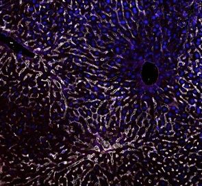
ll
     OPEN ACCESS                                      Article

 A                                          B
                                                              Kat2B
                                                              Jmjd8
                                                              Hdac11
                                                              Sirt3
                                                              Sirt1
                                                              Kdm7A
                                                              Mina
                                                              Smarcc1
                                                              Kdm3A
                                                              Hdac4
                                                              Tet2
                                                              Cdyl
                                                              Hdac1
                                                              Kdm5B
                                                              Kat7
                                                              Arid1A
                                                              Kdm6A
                                                              Ino80D
                                                              Kat6B
                                                              Ino80
                                                              Kdm1A
                                                              Kdm1B
                                                              Ezh2
                                                              Smarca5
                                                              Hdac2
                                                              Prmt5
                                                              Ruvbl1
                                                              Smyd5
                                                              Kat2A
                                                              Jarid2
                                                              Tet1
                                                              Mta2
                                                              Elp3
                                                              Smarca4
                                                              Kdm3B
                                                              Chd4
                                                              Ino80C
                                                              Ncoa3
                                                              Hat1
                                                              Jmjd1C
                                                              Setd8
                                                              Dnmt1

 C

 D     50                                   E
                               Dppa3
       40                      L1td1

       30

       20

       10

         0

                                                (legend on next page)

6 Cell Reports 39, 110730, April 26, 2022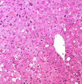
ll
Article                                                                                                                                 OPEN ACCESS

that in vivo reprogramming is unique (e.g., in vivo reprogramming              Top2a is required for reprogramming in vitro and in vivo
induces totipotency; Abad et al., 2013). Consistent with the acti-             We found that in vivo reprogramming rapidly induces the expres-
vation of these genes, DNA methylation analysis with bisulfite                 sion of epigenetic genes. We therefore hypothesized that epige-
conversion revealed that Dox treatment reduced levels of DNA                   netic regulators might be involved in partial reprogramming
methylation within the Dppa3 and Oct-3/4 loci (Figure 3E). Taken               (Figures 3B and 4F). Since the mechanisms of partial reprogram-
together, 4F transiently induced very notable changes in chro-                 ming are largely unknown and this is an important knowledge
matin, supporting the notion that 4F mediates epigenetic                       gap that must be addressed before therapeutic application, we
reprogramming.                                                                 investigated these downstream factors in more detail. Among
   We next performed single-cell RNA-seq (scRNA-seq), collect-                 them, topoisomerase2a (Top2a) was a good candidate for medi-
ing liver samples 1 day after Dox withdrawal (1d-on_1d-off).                   ating 4F’s function because its expression is largely restricted to
We obtained 2,284 single-cell transcriptomes (Dox: 1,258                      development, playing crucial roles in regulating the epigenome
cells; +Dox: 1,026 cells). Uniform manifold approximation and                  (Miller et al., 2017; Thakurela et al., 2013). Moreover, Top2a,
projection (UMAP) plots revealed seven clusters, which could                   not Top2b, was highly induced by 4F at mRNA and protein levels
be identified based on marker gene expression (Figures 4A                      and scRNA-seq showed Top2a induction was more specific to
and S5A). Dox treatment induced 4F expression specifically in                  the reprogrammed population (H3 cluster; Figures 5A–5D).
hepatocyte populations and changed their gene expression sig-                  Moreover, Top2 activity increased at day 2 after 4F induction
natures (Figures 4B and S5B). The gene Alb was downregulated,                  as assessed by decatenation assay (Figure 5E), which prompted
inversely correlating with 4F expression (Figure S5C). DEGs for                us to investigate the role of Top2a on cellular reprogramming. To
each cell type were next analyzed (Figure S5D). These analyses                 do so, we first treated reprogrammable mouse embryonic fibro-
show that hepatocytes have the highest number of DE genes,                     blasts (MEFs) with Dox in the presence or absence of ICRF-193,
indicating that these cell types are the most affected by 4F induc-            a Top2 inhibitor, which inhibits both TOP2A and TOP2B (Tanabe
tion. Upregulated genes in Dox-treated condition are specific to               et al., 1991). ICRF-193 treatment blocked iPSC reprogramming
hepatocytes, while some common signatures (liver metabolism                    in all conditions tested (Figure 5F). Since Top2a expression is
related) were observed in downregulated genes across cell                      induced by 4F (unlike Top2b), we performed RNAi experiment
types. Macrophages and endothelial cells were most affected                    for Top2a (Figure 5G). RNAi-mediated knockdown led to a
among non-hepatocyte cell types in transcriptomes. Consistent                  40-fold reduction in reprogramming efficiency, highlighting the
with these data, Dox-treated livers had fewer endothelial cells                crucial role of Top2a in reprogramming (Figure 5H). We next
and more macrophages, which may be a secondary effect of he-                   asked whether Top2 inhibition affects in vivo reprogramming.
patocyte proliferation. The hepatocyte population could be                     To do so, we treated Hep-4F mice with Dox in the presence of
further subdivided into four clusters (H1, H2, H3, and H4; Fig-                PBS or dexrazoxane hydrochloride (DRZ), another Top2 inhibi-
ure 4B). H4 cells were more prevalent in the Dox-treated con-                  tor, which inhibits both isoforms TOP2A and TOP2B. We used
dition (Figure 4B) and expressed high levels of 4F, prolifera-                 DRZ because it can be dissolved in PBS and therefore is more
tion-related genes, and epigenetic modifiers, suggesting that                  suitable for in vivo purposes (Hasinoff et al., 1997; Figure 5I).
the H4 cluster exhibits the strongest signature of reprogramming               DRZ inhibited the change in the color of the liver (Figure 5J),
(Figures 4C–4F). We also found that Dox-treated hepatocytes                    although it did not affect relative liver weight (Figure 5K). DRZ
upregulated p53 target genes, such as Cdkn1a and Mdm2.                         blocked the induction of progenitor markers and epigenetic reg-
This is consistent with a previously described compensatory                    ulators, while the loss of mature hepatic markers was not
mechanism, as 4F has been shown to activate the p53 pathway                    affected (Figure 5L). DRZ treatment itself did not affect gene
(Hong et al., 2009; Kawamura et al., 2009). Finally, Dox-treated               expression (data not shown). Collectively, our data show that
H3 cells exhibited partial restoration of gene signatures associ-              in vitro and in vivo reprogramming depends on Top2a.
ated with mature hepatocytes (Alb and Cyp3a11) and had lower
levels of cdkn1a expression (Figures 4E and 4G). Thus, short-                  In vivo reprogramming has beneficial effects on
term 4F may induce partial reprogramming and cellular prolifer-                regenerative capacity
ation, but after Dox withdrawal, the cells begin to restore normal             We next asked whether 4F induction could have beneficial ef-
hepatic characteristics, such as quiescence. Our results hint at a             fects on the liver. To do so, we treated 1-day-Dox-treated
possible cell trajectory after partial reprogramming in the liver,             Hep4F mice with CCl4 to monitor alanine transaminase (ALT),
although we cannot distinguish between cells that are on their                 a marker of liver injury (Figure 6A). We observed an earlier
way back to normal versus those that were never strongly re-                   clearance of ALT in Dox-treated Hep-4F mice and an earlier in-
programmed to begin with.                                                      crease in relative liver weight (Figures 6B and 6C). We next

Figure 3. Epigenetic reprogramming by liver-specific 4F expression
(A) GO analysis for differentiation-expressed genes in RNA-seq.
(B) Heatmap for epigenetic modifiers in Hep-4F mice.
(C) Genome browser view of the ATAC-seq data at the indicated gene loci.
(D) Expression levels of Dppa3 and L1td1 genes in Dox-treated Hep-4F mice. Data were extracted from RNA-seq (Figure 2). Data represent the mean (n = 2;
biological replicates). FPKM, fragments per kilobase of exon per million mapped fragments.
(E) Bisulfite sequencing of the Dppa3 promoter and distal enhancer of Oct-3/4 with the liver samples collected from the mice treated with or without Dox for
2 days.

                                                                                                            Cell Reports 39, 110730, April 26, 2022 7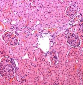
ll
OPEN ACCESS Article
A B
E
sample sample
cluster minusDox
Pou5f1 plusDox
Sox2
Klf4 cluster
Myc H1
Alb H2
Hnf4a
Cyp3a11 H3
Acsl1 H4
Apoc1
Afp
Krt8
Sox9
Epcam
Krt19
Jag1
Hes1
C D Lgr5
Prom1
Tcf7l2
100 15 Cdkn1a
Pou5f1 Thbs1 2.5
Mdm2
80 Sox2 Bid 2
Klf4 Ccng1
1.5
Myc
10 Mki67
60 Mcm2 1
MKi67 Ccna2
Ccne1 0.5
Ccnd1
40 0
Ccnb1
5 Cdk1
−0.5
Aurka
20 Plk1 −1
Pcna
0 0
H1 H2 H3 H4
0 1 2 3 4 5
G
F
sample
sample
cluster minusDox
Tet1 plusDox
Tet2 cluster
Smarcc1 H1
Cdyl H2
Hdac1 H3
Smyd5 H4
Mina
Hat1
Prmt5
Ino80
Setd8
Hdac4
Elp3
Jarid2
Dnmt1
Smarca4
Ezh2
Kat7 2.5
Hdac2 2
Chd4
Ncoa3 1.5
Smarca5 1
Sirt1
0.5
Mta2
Ruvbl1 0
Jmjd8
−0.5
Sirt3
Hdac11 −1
Figure 4. Single-cell transcriptome analysis of Hep-4F mice
(A) UMAP visualization of liver cell clusters. Hep-4F mice were treated with or without Dox for 1 day and single-cell suspensions were prepared from the livers
isolated at 1 day after Dox withdrawal. Each cell type was characterized based on gene expression (Figure S5A).
(B) Left: UMAP plot of untreated (Dox) or Dox-treated (+Dox) liver cell clusters: Dox (blue dots) and +Dox (red dots). Right: enlarged hepatocyte clusters are
shown.
(C) Relative cell number for the indicated genes in each hepatocyte cluster.
(D) Volcano plots of GO analyses for DEGs between H4 cells and the other hepatocyte cells.
(E and F) Heatmap of gene expression of the liver-related and cell-cycle-related genes (E) and of the epigenetic modifiers (F) in the cells of hepatocyte sub-
clusters.
(G) Violin plots for gene expression of the reprogramming-related genes in each hepatic cluster.
administered a lethal dose of acetaminophen (APAP) to Hep-4F ficient liver regeneration (Bhushan and Apte, 2019). Hep-4F
mice. APAP is commonly used to study acute liver injury mice were treated with Dox for 1 day and then APAP was
(Jaeschke et al., 2014). In this model, liver regeneration is administered 6 days later (day 7) following Dox withdrawal (Fig-
tightly associated with survival, as mice die rapidly without suf- ure 6D). Two days after APAP administration, all control mice
8 Cell Reports 39, 110730, April 26, 2022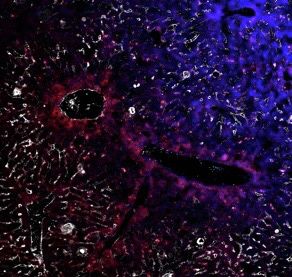
ll
Article OPEN ACCESS
A 6 Top2a B E
Top2b
4
log2 (FPKM)
2.0
2
1.5
0
1.0
-2
0.5
E
0.0
0d 1d 2d
C
F
800
600
400
200
10
8
6
4
2
0
D
R 0
IC F 50
R 50 0
50 > 0
0
IC F 50
IC F
>
IC F
R
0
IC
R
F
R
L
Luc KD 1500 15 1.5 1.5
Top2a KD
G H
1.5 1.5 1000 10 1.0 1.0
200
500 5 0.5 0.5
1.0 1.0
0 0 0.0 0.0
100
0.5 0.5
1.5 20000 6×10⁶ 2000
0
0.0 0.0
D
D
15000 1500
K
K
4×10⁶
c
a
1.0
Lu
p2
0d
2d
4d
0d
2d
4d
To
10000 1000
0.5 2×10⁶
I 5000 500
K 0.0 0 0 0
8
-Dox+PBS 10
Relative liver weight (%)
100 4
-Dox+DRZ 8
6 80
3 -Dox+PBS
+Dox+PBS
J 60 6 -Dox+DRZ
4 +Dox+DRZ 2
+Dox+PBS
40 4
+Dox+DRZ
2 1
20 2
0 0 0
0
Figure 5. Role of Top2a on reprogramming in vitro and in vivo
(A) Time course for gene expression of Top2a and Top2b in reprogramming livers. Data were extracted from RNA-seq (Figure 2). Data represent the mean (n = 2;
biological replicates).
(B) Western blotting for Top2a and Top2b in liver samples collected at indicated time points after Dox administration.
(legend continued on next page)
Cell Reports 39, 110730, April 26, 2022 9ll
OPEN ACCESS Article
died, but half of the Dox-treated mice survived (Figure 6E). DISCUSSION
Associated with longer survival of Dox-treated Hep-4F mice
(Figure 6E), Dox-treated livers were more proliferative In summary, here, we have developed a mouse model that en-
compared with control livers before and after APAP treatment ables hepatocyte-specific 4F induction and subsequent lineage
(Figure 6F). Importantly, this beneficial effect of 4F was not tracing of 4F-expressing cells. We demonstrate that liver-spe-
observed when mice were treated with DRZ (Figure 6E), high- cific 4F expression rapidly and transiently induced partial reprog-
lighting the importance of Top2a for liver regeneration. DRZ ramming and that this enhanced liver regeneration. This study,
treatment did not affect cell proliferation before APAP treat- the first to perform lineage tracing and single-cell transcriptome
ment; however, it resulted in a 2-fold reduction in Ki67+ cells analyses for 4F-expressing cells in vivo, shows that 4F-mediated
along with the decrease in Sox9+ cells (Figures 6F and 6G). cellular partial reprogramming is a potential avenue for inducing
Top2a may be involved in 4F enhancing regeneration compe- a proliferative, plastic progenitor state.
tency in a cell-proliferation-independent mechanism, which It was originally reported that systemic 4F expression in vivo
might be attributed to epigenetic reprogramming. Further ana- leads to cancers with totipotency (Abad et al., 2013). Subsequent
lyses are needed to clarify this mechanism. reports demonstrated the beneficial effects of 4F in several
We next asked whether the partially reprogrammed, prolifer- different contexts (Doeser et al., 2018; Lu et al., 2020; Ocampo
ative hepatocytes that express 4F can contribute to liver regen- et al., 2016; Browder and Reddy, 2022). Our previous report
eration. To measure this, we added a bromodeoxyuridine showed that cyclic 4F expression ameliorates age-associated
(BrdU) injection on day 2 of our APAP protocol and assessed hallmarks in a mouse model of premature aging (Ocampo et al.,
whether BrdU+ cells expressed Alb (Figure S6A). Alb and 2016). Another recent paper showed that expression of Oct-3/4,
BrdU signals overlapped, suggesting that BrdU-labeled cells Sox2, and KLF4 in the retina restores youthful DNA methylation
contributed to liver regeneration (Figure S6B). Ki67+ cells patterns and transcriptomes and promotes axon regeneration af-
were largely labeled with BrdU before APAP treatment; howev- ter injury (Lu et al., 2020). However, it remains unknown whether
er, Ki67+ cells that emerged post-APAP-mediated injury were short-term 4F induction leads to dedifferentiation in vivo because
not always positive for BrdU (Figures 6H and 6I), suggesting of a lack of a stringent system for lineage tracing. Here, we demon-
that enhancing effect of 4F on proliferation may not simply strate that 4F-expressing cells partially reprogram to progenitor
explain the increase in cell number prior to liver injury and state, associated with cell proliferation in the liver consistent with
may be mediated by other mechanisms, where, for instance, the report that dedifferentiation is associated with cell proliferation.
the cells that did not express 4F but proliferate in a compensa- Our findings raise several questions, namely whether the effect
tory mechanism might contribute to the regeneration. RNA-seq of 4F is tissue or context specific and what is the molecular basis
was performed for the liver samples collected before and after of quick reprogramming induced by 4F? Further study will be
APAP treatment. DEG analyses show that GO terms associated needed to thoroughly answer these questions, but here, we report
with cell cycle and proliferation were enriched in Hep-4F mice that Top2a is a critical component of the mechanism underlying
consistent with better regeneration (Figure S6C), although this in vivo reprogramming. Inhibition of Top2 activity dramatically
may not be the cause but rather a consequence of better reduced the beneficial effect of 4F and inhibited the increase in
regeneration. gene expression of epigenetic regulators and progenitor markers
Lastly, to test whether 4F can have a beneficial effect even while not affecting the loss of mature hepatocyte markers. This
when 4F is induced on or after liver injury, we treated mice with suggests that Top2a-mediated partial cellular reprogramming is
APAP and Dox simultaneously (Figure 6J). The expression of required for 4F-mediated benefits. Notably, Top2a inhibition
4F and epigenetic regulators, such as Top2a, was still induced dramatically suppressed the expression of Tet1, which was
in this context (Figure 6K). Importantly, Dox-treated mice recently reported to have a crucial role in reprogramming-medi-
showed lower levels of ALT (Figure 6L). Taken together, in vivo ated regeneration induced by 4F (Lu et al., 2020), implying that
reprogramming shows beneficial effects by promoting the Top2a may act upstream of epigenetic modifiers, such as Tet1.
regeneration of injured liver. Our study is therefore an important We observed a strong correlation between the loss of mature
step toward developing partial reprogramming therapies for hepatic markers and proliferation in reprogramming hepatocytes.
treating human diseases. This highlights a causal link between dedifferentiation and
(C) UMAP visualization for Top2a and Top2b.
(D) Violin plots for gene expression of Top2a and Top2b in each hepatic cluster.
(E) Decatenation assay for topoisomerase activity in liver samples. Data represent the mean with SD (n = 4). ns, not significant.
(F) Effect of ICRF-193 (Top2 inhibitor) on iPSC reprogramming. Left: schematic representation for ICRF-193 (ICRF) treatment protocol is shown. Right: re-
programming efficiency is shown. Data represent the mean with SD (n = 3).
(G) qPCR for Top2a and Top2b in the cells expressing short hairpin RNA (shRNA) for Top2a (Top2a knockdown [KD]) or for Luciferase (Luc KD). Data represent the
mean with SD (n = 3; technical replicates).
(H) Reprogramming efficiency of Top2a or Luc KD cells. Data represent the mean with SD (n = 3).
(I) Schematic representation of dexrazoxane (DRZ) treatment protocol.
(J) Livers collected from PBS-treated and DRZ-treated Hep-4F mice 1 day after Dox withdrawal.
(K) Relative liver weight (% body weight). Data represent the mean with SE (n = 5).
(L) qPCR analysis for PBS- or DRZ-treated Hep-4F mice with or without Dox for 2 days. Data represent the mean with SD (n = 3; technical replicates).
Statistical analyses were conducted by unpaired t test or one-way ANOVA with Tukey’s post hoc analysis. *p < 0.05; **p < 0.01; ***p < 0.001; ****p < 0.0001.
10 Cell Reports 39, 110730, April 26, 2022ll
Article OPEN ACCESS
A D
Alb-Cre +Dox (n=11)
B C ALT E Hep-4F +Dox (n=7)
Hep-4F-Dox (n =2)
8 15000 100
Relative liver weight (%)
Control Hep-4F+Dox+DRZ (n=5)
6 Hep-4F Hep-4F-Dox-DRZ (n =3)
Percent survival
75
arbitrary unit
10000
4
50
Control
5000
2 Hep-4F 25
0 0
7 9 10 8 9 10
0
7 8 9 10 11 12 13 14
Time (Days)
F H
****
100 ns
****
Ki67+ cells per field
80
60 ns
*
40 **
20
I
0
Day7 Day8
G ns
****
80 ****
Sox9+ cells per field
60 ns
ns
***
40
20
0
Day7 Day8
J K 15 L
-Dox
6000
Relative expression
+Dox
ALT (Arbitrary unit)
10
4000
5
2000
0
0
-Dox +Dox
4
x2
c- f4
yc
yp lb
L1 1
St 1
To la
a
t1
B 0
1
1
ct
td
p2
o8
al
el
l
A
Te
3a
So
m
K
m
O
In
C
Figure 6. Improvement of liver regeneration capacity induced by 4F-mediated partial reprogramming
(A) Schematic representation for CCl4 treatment protocol. Alb-Cre mice (no 4F cassette) or Hep-4F mice were treated with Dox for 1 day. Six days following Dox
withdrawal, the mice were injected with CCl4.
(B) Relative liver weight (% body weight). Data represent the mean with SE (n = 5).
(legend continued on next page)
Cell Reports 39, 110730, April 26, 2022 11ll
OPEN ACCESS Article
proliferation, which is consistent with previous reports where B Tissue preparation and IHC
dedifferentiation followed by proliferation contributes to tissue B RNA isolation and quantitative-PCR (qPCR)
regeneration in vertebrate species with high regenerative capac- B Bulk RNA-Sequencing
ities (Jopling et al., 2010; Tanaka et al., 2016). Therefore, in vivo B ATAC-sequencing
4F-mediated partial reprogramming may share some of the mech- B Principle component analysis (PCA)
anisms that underlie the tissue and organ regeneration observed in B Pathway analysis
nature. Further analyses of dedifferentiation mechanisms should B ScRNA-sequencing
lead to new strategies for expanding the regenerative capacity B Bisulfite sequencing
of mammalian tissues. B Comprehensive metabolic panel analyses
B Western blotting
B Decatenation assay
Limitations of the study
B Top2a knockdown
While we demonstrated that hepatocyte-specific 4F induction en-
B iPSC reprogramming
hances liver regenerative capacity by partial reprogramming medi-
ated through topoisomerase2, this study still has several limita- d QUANTIFICATION AND STATISTICAL ANALYSIS
tions. First, prevalent chronic liver injuries, such as fibrosis and
non-alcoholic fatty liver disease, were not tested here but should SUPPLEMENTAL INFORMATION
be addressed in future projects. Second, longevity and tumori-
Supplemental information can be found online at https://doi.org/10.1016/j.
genic risks were not fully assessed with longer term monitoring celrep.2022.110730.
of Dox-treated Hep-4F mice, and evaluating maximum lifespan
and tumor incidence after Dox treatment will be required for com- ACKNOWLEDGMENTS
plete safety assessment. Third, mechanistically, it remains to be
elucidated how Top2a can participate in hepatocyte cellular re- We thank May Schwarz and Peter Schwarz for administrative help and Yuta
programming and whether Top2a activation alone is sufficient to Takahashi, Mariana Morales Valencia, and Josephine Ho for experimental
enhance liver regeneration. Lastly, cell trajectories of reprogram- help. We also thank David O’Keefe for help with manuscript preparation.
T.H. was supported by a Uehara Memorial Foundation research fellowship.
ming hepatocytes were not fully investigated, and for now, it re-
Work in the laboratory of J.C.I.B. was supported by UCAM and Fundacion
mains unclear how these reprogrammed cells contribute to the Dr. Pedro Guillen.
enhancement of liver regeneration in detail. Deeper single-cell an-
alyses with additional in vivo tracing systems for the partially re- AUTHOR CONTRIBUTIONS
programmed cells will help understand their behaviors during liver
regeneration. T.H. and M.Y. designed the research, performed most experiments, and
analyzed and interpreted data. T.H., M.Y., S.M., P.R., and J.C.I.B. wrote the
manuscript. Y.H.-N., C.S., C.W., K.S., Y.X., R.R.R., M.B.M., and J.C. helped
STAR+METHODS perform experiments. S.K.S., F.H., Y.S., H.-K.L., P.M.-R., I.G.-G., R.H.-B.,
C.R.E., J.Q., R.D.H., P.G.G., E.N.D., A.C., J.M.C., Y.Y., and G.-H.L. analyzed
Detailed methods are provided in the online version of this paper and interpreted data. L.H., Y.H., and M.S. analyzed sequencing data. M.H.,
and include the following: F.Y., D.C.H., and A.A. provided critical advice, interpreted data, and aided in
manuscript preparation. J.C.I.B. designed and supervised the experiments
d KEY RESOURCES TABLE and provided financial and administrative support.
d RESOURCE AVAILABILITY
B Lead contact DECLARATION OF INTERESTS
B Materials availability
The authors declare no competing interests.
B Data and code availability
d EXPERIMENTAL MODEL AND SUBJECT DETAILS
Received: May 17, 2021
B Mice Revised: December 28, 2021
B Cell lines and cell culture Accepted: April 1, 2022
d METHODS DETAILS Published: April 26, 2022
(C) ALT levels of Alb-Cre or Hep-4F mice. Data represent the mean with SE (control: n = 5 [8 days], n = 8 [9 days], n = 4 [10 days]; Hep-4F, n = 4 [8 days], n = 7
[9 days], n = 5 [10 days]).
(D) Schematic representation for APAP treatment protocol. Hep-4F mice were treated with or without Dox for 1 day. Six days following Dox withdrawal, the mice
were injected with APAP.
(E) Survival curve of Hep-4F mice.
(F) Quantification of Ki67+ cells. Data represent the mean with SE (n = 12).
(G) Quantification of Sox9+ cells. Data represent the mean with SE (n = 12).
(H) Schematic representation for APAP treatment protocol for cell tracking of BrdU-positive cells.
(I) IHC for Ki67 and BrdU. Scale bar, 200 mm.
(J) Schematic representation for the protocol of simultaneous injection of Dox and APAP.
(K) qPCR analysis for Hep-4F mice with or without Dox for 2 days. Data represent the mean with SD (n = 3; technical replicates).
(L) ALT levels of Dox-treated or untreated Hep-4F mice 2 days after APAP treatment. Data represent the mean with SE (n = 6).
Statistical analyses were conducted by unpaired t test or one-way ANOVA with Tukey’s post hoc analysis.*p < 0.05; **p < 0.01; ***p < 0.001; ****p < 0.0001.
12 Cell Reports 39, 110730, April 26, 2022ll
Article OPEN ACCESS
REFERENCES Hong, H., Takahashi, K., Ichisaka, T., Aoi, T., Kanagawa, O., Nakagawa, M.,
Okita, K., and Yamanaka, S. (2009). Suppression of induced pluripotent
Abad, M., Mosteiro, L., Pantoja, C., Canamero, M., Rayon, T., Ors, I., Grana, stem cell generation by the p53-p21 pathway. Nature 460, 1132–1135.
O., Megias, D., Dominguez, O., Martinez, D., et al. (2013). Reprogramming Iwafuchi-Doi, M., Donahue, G., Kakumanu, A., Watts, J.A., Mahony, S., Pugh,
in vivo produces teratomas and iPS cells with totipotency features. Nature B.F., Lee, D., Kaestner, K.H., and Zaret, K.S. (2016). The pioneer transcription
502, 340–345. factor FoxA maintains an accessible nucleosome configuration at enhancers
Athwal, V.S., Pritchett, J., Llewellyn, J., Martin, K., Camacho, E., Raza, S.M., for tissue-specific gene activation. Mol. Cell 62, 79–91.
Phythian-Adams, A., Birchall, L.J., Mullan, A.F., Su, K., et al. (2017). SOX9 pre- Jaeschke, H., Xie, Y., and McGill, M.R. (2014). Acetaminophen-induced liver
dicts progression toward cirrhosis in patients while its loss protects against injury: from animal models to humans. J. Clin. Transl. Hepatol. 2, 153–161.
liver fibrosis. EMBO Mol. Med. 9, 1696–1710.
Jin, L., Long, L., Green, M.A., and Spear, B.T. (2009). The alpha-fetoprotein
Belteki, G., Haigh, J., Kabacs, N., Haigh, K., Sison, K., Costantini, F., Whitsett, enhancer region activates the albumin and alpha-fetoprotein promoters during
J., Quaggin, S.E., and Nagy, A. (2005). Conditional and inducible transgene liver development. Develop. Biol. 336, 294–300.
expression in mice through the combinatorial use of Cre-mediated recombina-
Jopling, C., Sleep, E., Raya, M., Marti, M., Raya, A., and Izpisua Belmonte, J.C.
tion and tetracycline induction. Nucleic Acids Res. 33, e51.
(2010). Zebrafish heart regeneration occurs by cardiomyocyte dedifferentia-
Bezerra, J.A., Bugge, T.H., Melin-Aldana, H., Sabla, G., Kombrinck, K.W., tion and proliferation. Nature 464, 606–609.
Witte, D.P., and Degen, J.L. (1999). Plasminogen deficiency leads to impaired Kawamura, T., Suzuki, J., Wang, Y.V., Menendez, S., Morera, L.B., Raya, A.,
remodeling after a toxic injury to the liver. Proc. Natl. Acad. Sci. U S A 96, Wahl, G.M., and Izpisua Belmonte, J.C. (2009). Linking the p53 tumour sup-
15143–15148. pressor pathway to somatic cell reprogramming. Nature 460, 1140–1144.
Bhushan, B., and Apte, U. (2019). Liver regeneration after acetaminophen hep- Kim, J., Woo, A.J., Chu, J., Snow, J.W., Fujiwara, Y., Kim, C.G., Cantor, A.B.,
atotoxicity: mechanisms and therapeutic opportunities. Am. J. Pathol. 189, and Orkin, S.H. (2010). A Myc network accounts for similarities between em-
719–729. bryonic stem and cancer cell transcription programs. Cell 143, 313–324.
Biddie, S.C., John, S., Sabo, P.J., Thurman, R.E., Johnson, T.A., Schiltz, R.L., Lu, Y., Brommer, B., Tian, X., Krishnan, A., Meer, M., Wang, C., Vera, D.L.,
Miranda, T.B., Sung, M.H., Trump, S., Lightman, S.L., et al. (2011). Transcrip- Zeng, Q., Yu, D., Bonkowski, M.S., et al. (2020). Reprogramming to recover
tion factor AP1 potentiates chromatin accessibility and glucocorticoid recep- youthful epigenetic information and restore vision. Nature 588, 124–129.
tor binding. Mol. Cell 43, 145–155. Miller, E.L., Hargreaves, D.C., Kadoch, C., Chang, C.Y., Calarco, J.P., Hodges,
Browder, K.C., Reddy, P., et al. (2022). In vivo partial reprogramming alters C., Buenrostro, J.D., Cui, K., Greenleaf, W.J., Zhao, K., et al. (2017). TOP2 syn-
age-associated molecular changes during physiological aging in mice. Nature ergizes with BAF chromatin remodeling for both resolution and formation of
Aging 2, 243–253. facultative heterochromatin. Nat. Struct. Mol. Biol. 24, 344–352.
Buenrostro, J.D., Giresi, P.G., Zaba, L.C., Chang, H.Y., and Greenleaf, W.J. Mizejewski, G.J. (2002). Biological role of alpha-fetoprotein in cancer: pros-
(2013). Transposition of native chromatin for fast and sensitive epigenomic pects for anticancer therapy. Expert Rev. Anticancer Ther. 2, 709–735.
profiling of open chromatin, DNA-binding proteins and nucleosome position. Nakano, Y., Nakao, S., Sumiyoshi, H., Mikami, K., Tanno, Y., Sueoka, M., Ka-
Nat. Methods. 10, 1213–1218. sahara, D., Kimura, H., Moro, T., Kamiya, A., et al. (2017). Identification of a
Cabral, F., Miller, C.M., Kudrna, K.M., Hass, B.E., Daubendiek, J.G., Kellar, novel alpha-fetoprotein-expressing cell population induced by the Jagged1/
B.M., and Harris, E.N. (2018). Purification of hepatocytes and sinusoidal endo- Notch2 signal in murine fibrotic liver. Hepatol. Commun. 1, 215–229.
thelial cells from mouse liver perfusion. J. Vis. Exp. 12, 56993. Ocampo, A., Reddy, P., Martinez-Redondo, P., Platero-Luengo, A., Hatanaka,
Carey, B.W., Markoulaki, S., Beard, C., Hanna, J., and Jaenisch, R. (2010). Sin- F., Hishida, T., Li, M., Lam, D., Kurita, M., Beyret, E., et al. (2016). In Vivo
gle-gene transgenic mouse strains for reprogramming adult somatic cells. Nat. amelioration of age-associated hallmarks by partial reprogramming. Cell
Methods. 7, 56–59. 167, 1719–1733.e1712.
Ohnishi, K., Semi, K., Yamamoto, T., Shimizu, M., Tanaka, A., Mitsunaga, K.,
Chen, T., Dai, X., Dai, J., Ding, C., Zhang, Z., Lin, Z., Hu, J., Lu, M., Wang, Z.,
Okita, K., Osafune, K., Arioka, Y., Maeda, T., et al. (2014). Premature termina-
Qi, Y., et al. (2020). AFP promotes HCC progression by suppressing the HuR-
tion of reprogramming in vivo leads to cancer development through altered
mediated Fas/FADD apoptotic pathway. Cell Death Dis. 11, 822.
epigenetic regulation. Cell 156, 663–677.
Chen, Y., Luttmann, F.F., Schoger, E., Scholer, H.R., Zelarayan, L.C., Kim,
Postic, C., Shiota, M., Niswender, K.D., Jetton, T.L., Chen, Y., Moates, J.M.,
K.P., Haigh, J.J., Kim, J., and Braun, T. (2021). Reversible reprogramming of
Shelton, K.D., Lindner, J., Cherrington, A.D., and Magnuson, M.A. (1999).
cardiomyocytes to a fetal state drives heart regeneration in mice. Science
Dual roles for glucokinase in glucose homeostasis as determined by liver
373, 1537–1540.
and pancreatic beta cell-specific gene knock-outs using Cre recombinase.
Corces, M.R., Trevino, A.E., Hamilton, E.G., Greenside, P.G., Sinnott-Arm- J. Biol. Chem. 274, 305–315.
strong, N.A., Vesuna, S., Satpathy, A.T., Rubin, A.J., Montine, K.S., Wu, B.,
Rinkevich, Y., Lindau, P., Ueno, H., Longaker, M.T., and Weissman, I.L. (2011).
et al. (2017). An improved ATAC-seq protocol reduces background and en-
Germ-layer and lineage-restricted stem/progenitors regenerate the mouse
ables interrogation of frozen tissues. Nat. Methods. 14, 959–962.
digit tip. Nature 476, 409–413.
Doeser, M.C., Scholer, H.R., and Wu, G. (2018). Reduction of fibrosis and scar Shachaf, C.M., Kopelman, A.M., Arvanitis, C., Karlsson, A., Beer, S., Mandl, S.,
formation by partial reprogramming in vivo. Stem Cell 36, 1216–1225. Bachmann, M.H., Borowsky, A.D., Ruebner, B., Cardiff, R.D., et al. (2004).
Felsher, D.W., and Bishop, J.M. (1999). Reversible tumorigenesis by MYC in MYC inactivation uncovers pluripotent differentiation and tumour dormancy
hematopoietic lineages. Mol. Cell 4, 199–207. in hepatocellular cancer. Nature 431, 1112–1117.
Hasinoff, B.B., Kuschak, T.I., Creighton, A.M., Fattman, C.L., Allan, W.P., Shibata, H., Komura, S., Yamada, Y., Sankoda, N., Tanaka, A., Ukai, T., Ka-
Thampatty, P., and Yalowich, J.C. (1997). Characterization of a Chinese ham- bata, M., Sakurai, S., Kuze, B., Woltjen, K., et al. (2018). In vivo reprogramming
ster ovary cell line with acquired resistance to the bisdioxopiperazine dexra- drives Kras-induced cancer development. Nat. Commun. 9, 2081.
zoxane (ICRF-187) catalytic inhibitor of topoisomerase II. Biochem. Pharma- Simon, H.G. (2012). Salamanders and fish can regenerate lost structures–why
col. 53, 1843–1853. can’t we? BMC Biol. 10, 15.
He, L., Pu, W., Liu, X., Zhang, Z., Han, M., Li, Y., Huang, X., Han, X., Li, Y., Liu, Takahashi, K., Tanabe, K., Ohnuki, M., Narita, M., Ichisaka, T., Tomoda, K.,
K., et al. (2021). Proliferation tracing reveals regional hepatocyte generation in and Yamanaka, S. (2007). Induction of pluripotent stem cells from adult human
liver homeostasis and repair. Science 371, eabc4346. fibroblasts by defined factors. Cell 131, 861–872.
Cell Reports 39, 110730, April 26, 2022 13ll
OPEN ACCESS Article
Takahashi, K., and Yamanaka, S. (2006). Induction of pluripotent stem cells Wang, C., Rabadan Ros, R., Martinez-Redondo, P., Ma, Z., Shi, L., Xue, Y.,
from mouse embryonic and adult fibroblast cultures by defined factors. Cell Guillen-Guillen, I., Huang, L., Hishida, T., Liao, H.K., et al. (2021). In vivo partial
126, 663–676. reprogramming of myofibers promotes muscle regeneration by remodeling the
stem cell niche. Nat. Commun. 12, 3094.
Takahashi, Y., Wu, J., Suzuki, K., Martinez-Redondo, P., Li, M., Liao, H.K., Wu,
M.Z., Hernandez-Benitez, R., Hishida, T., Shokhirev, M.N., et al. (2017). Inte- Wang, H., and Simon, A. (2016). Skeletal muscle dedifferentiation during sala-
gration of CpG-free DNA induces de novo methylation of CpG islands in plurip- mander limb regeneration. Curr. Opin. Genet. Dev. 40, 108–112.
otent stem cells. Science 356, 503–508. Wei, Y., Wang, Y.G., Jia, Y., Li, L., Yoon, J., Zhang, S., Wang, Z., Zhang, Y.,
Zhu, M., Sharma, T., et al. (2021). Liver homeostasis is maintained by midlob-
Tanabe, K., Ikegami, Y., Ishida, R., and Andoh, T. (1991). Inhibition of topo-
ular zone 2 hepatocytes. Science 371, eabb1625.
isomerase II by antitumor agents bis(2,6-dioxopiperazine) derivatives. Cancer
White, P., Brestelli, J.E., Kaestner, K.H., and Greenbaum, L.E. (2005). Identifi-
Res. 51, 4903–4908.
cation of transcriptional networks during liver regeneration. J. Biol. Chem. 280,
Tanaka, H.V., Ng, N.C.Y., Yang Yu, Z., Casco-Robles, M.M., Maruo, F., Tsonis, 3715–3722.
P.A., and Chiba, C. (2016). A developmentally regulated switch from stem cells Xu, X., Smorag, L., Nakamura, T., Kimura, T., Dressel, R., Fitzner, A., Tan, X.,
to dedifferentiation for limb muscle regeneration in newts. Nat. Commun. 7, Linke, M., Zechner, U., Engel, W., et al. (2015). Dppa3 expression is critical for
11069. generation of fully reprogrammed iPS cells and maintenance of Dlk1-Dio3
Thakurela, S., Garding, A., Jung, J., Schubeler, D., Burger, L., and Tiwari, V.K. imprinting. Nat. Commun. 6, 6008.
(2013). Gene regulation and priming by topoisomerase IIalpha in embryonic Zaret, K.S., Lerner, J., and Iwafuchi-Doi, M. (2016). Chromatin scanning by dy-
stem cells. Nat. Commun. 4, 2478. namic binding of pioneer factors. Mol. Cell 62, 665–667.
14 Cell Reports 39, 110730, April 26, 2022ll
Article OPEN ACCESS
STAR+METHODS
KEY RESOURCES TABLE
REAGENT or RESOURCE SOURCE IDENTIFIER
Antibodies
Goat polyclonal anti-GFP Abcam Cat# 6673; RRID: AB_305643
Mouse monoclonal anti-GFP Clontech Cat# JL-8; RRID: AB_10013427
Rabbit monoclonal anti-Ki67 Cell signaling Cat# 12202; RRID: AB_2620142
Goat polyclonal anti-Klf4 R&D Cat# AF3158; RRID: AB_2130245
Rat monoclonal anti-Sox9 Abcam Cat# 185230; RRID: AB_2715497
Rat monoclonal anti-BrdU Abcam Cat# 6326; RRID: AB_305426
Rabbit monoclonal anti-Top2a Abcam Cat# 52934; RRID: AB_883143
Rabbit monoclonal anti-Top2b Abcam Cat# 220385; RRID: N/A
(No longer available)
Mouse monoclonal anti-b-actin Santa Cruz Cat# 47778; RRID: AB_2714189
Mouse monoclonal anti-Afp Santa Cruz Cat# sc-8399; RRID: AB_626665
Rabbit monoclonal anti-GS Abcam Cat# 49873; RRID: AB_880241
Mouse monoclonal anti-E-cadherin MBL Cat# NCH-38; RRID: AB_10982676
Chemicals, peptides, and recombinant proteins
Doxycycline Sigma-Aldrich Cat# D9891
DAPI Fluoromount-G SouthernBiotech Cat# 100-20
OCT compound Fisher HealthCare Cat# 4585
TRIzol Invitrogen Cat# 15596026
RNeasy Mini kit QIAGEN Cat# 74134
RNase-Free DNase Set QIAGEN Cat# 79254
Maxima H Minus Mastermix Thermo Scientific Cat# M1662
Platinum SYBR Green quantitative PCR supermix Bio Rad Cat# 1725274
Carbon tetrachloride Sigma-Aldrich Cat# 289116
Zymo EZ DNA Methylation-Gold kit Zymo Research Cat# D5005
Epi-Taq TaKaRa Cat# R110A
ICRF-193 Enzo Life Sciences Cat# BML-GR332
Dexrazoxane hydrochloride MedChemExpress Cat# HY-76201
4-Acetamidophenol, 98% Thermo Scientific Cat# 102332500
NE-PERTM Nuclear and Cytoplasmic Thermo Fisher Scientific Cat# 78833
Extraction Reagents
NuPAGETM 4 to 12%, Bis-Tris, 1.0 mm, Thermo Fisher Scientific Cat# NP0321BOX
Mini Protein Gel, 10-well
Immobilon-P Membrane, PVDF Millipore Cat# IPVH00010
SuperSignalTM West Femto Thermo Fisher Scientific Cat# 34094
Maximum Sensitivity Substrate
DMEM Thermo Fisher Scientific Cat# 11995
Penicillin-Streptomycin Thermo Fisher Scientific Cat# 15140122
FBS Gemini Bio-products Cat# 25300054
Trypsin-EDTA (0.05%) ThermoFisher Scientific Cat# 15140122
Lipofectamine3000 Invitrogen Cat# L3000015
Hygromycin B Invitrogen Cat# 10687010
Polybrene Sigma Cat# H9268
Critical commercial assays
Human Topoisomerase II Assay Kit TopoGEN Cat# TG1001-2
InfinityTM ALT (GPT) Liquid Stable Reagent ThermoFisher Cat# TR71121
(Continued on next page)
Cell Reports 39, 110730, April 26, 2022 e1ll
OPEN ACCESS Article
Continued
REAGENT or RESOURCE SOURCE IDENTIFIER
Deposited data
RNA-Seq, ATAC-Seq, sc-RNA-Seq This paper GSE144600
Experimental models: Cell lines
Reprogrammable MEFs This paper N/A
293FT cells Salk Stem cell core N/A
LIF-expressing 293A cells This paper N/A
Experimental models: Organisms/strains
Mouse: B6N.Cg-Speer6-ps1Tg(Alb-cre)21Mgn/J The Jackson Laboratory RRID: IMSR_JAX:018961
Mouse: Gt(ROSA)26Sortm1(rtTA*M2)Jae The Jackson Laboratory RRID: IMSR_JAX:011004
Col1a1tm3(tetO-Pou5f1,-Sox2,-Klf4,-Myc)Jae/J
Mouse: FVB/N-Tg(tetO-MYC)36aBop/J The Jackson Laboratory RRID: IMSR_JAX:019376
Mouse: B6.Cg-Gt(ROSA)26Sortm1(rtTA, EGFP)Nagy/J The Jackson Laboratory RRID: IMSR_JAX:005,670
Mouse: Gt(ROSA)26Sortm1(rtTA*M2)Jae The Jackson Laboratory RRID: IMSR_JAX:011,011
Col1a1tm4(tetO-Pou5f1,-Sox2,-Klf4,-Myc)Jae/J
Oligonucleotides
See Table S1 for qPCR primers N/A N/A
See Table S2 for bisulfite primers N/A N/A
See Table S3 for Top2a target N/A N/A
Recombinant DNA
pCAG-LIF-Ires-Puro This paper N/A
pCS-tetO-shTop2a-EThygro This paper N/A
pCS-tetO-shLuc-EThygro This paper N/A
pCAG-HIVgp Miyoshi. H et al., 1998 Cat# RDB04394
pCMV-VSVG-RSV-Rev Miyoshi. H et al., 1998 Cat# RDB04393
Software and algorithms
Prism 8.0 GraphPad Software http://www.graphpad.com
ImageJ Software https://imagej.nih.gov/ij/
Excel Microsoft N/A
RESOURCE AVAILABILITY
Lead contact
Further information and requests for resources and reagents should be directed to and will be fulfilled by the lead contact, Juan Car-
los Izpisua Belmonte (belmonte@salk.edu).
Materials availability
All unique/stable reagents/cells generated in this study are available from the lead contact with a completed Materials Transfer
Agreement.
Data and code availability
dRNA-Seq, ATAC-Seq and sc-RNAseq data have been deposited to Gene Expression Omnibus and are available to the public as
of data of publication. The accession number is listed in the key resources table.
dThis paper does not report any original code.
dAny additional information required to reanalyze the data reported in this paper is available from the lead contact upon request.
EXPERIMENTAL MODEL AND SUBJECT DETAILS
Mice
Alb-Cre (Postic et al., 1999), Col1a1tetO4F/tetO4F;ROSA26rtTA/rtTA, Col1a12loxP-tetO4F/2loxP-tetO4F;ROSA26rtTA/rtTA (Carey et al.,
2010), tetO-MYC (Felsher and Bishop, 1999), and ROSALSL-rtTA-IRES-GFP/LSL-rtTA-IRES-GFP (Belteki et al., 2005) have been previously
e2 Cell Reports 39, 110730, April 26, 2022You can also read