Mid-luteal angiogenesis and function in the primate is dependent on vascular endothelial growth factor
←
→
Page content transcription
If your browser does not render page correctly, please read the page content below
409

Mid-luteal angiogenesis and function in the primate is dependent
on vascular endothelial growth factor
S E Dickson, R Bicknell1 and H M Fraser
MRC Human Reproductive Sciences Unit, Centre for Reproductive Biology, 37 Chalmers Street, Edinburgh EH3 9ET, UK
2
Molecular Angiogenesis Laboratories, Imperial Cancer Research Fund, Institute of Molecular Medicine, John Radcliffe Hospital, Oxford OX3 9DS, UK
(Requests for offprints should be addressed to H M Fraser, MRC Human Reproductive Sciences Unit, Centre for Reproductive Biology, 37 Chalmers Street,
   Edinburgh EH3 9ET, UK; E-mail: h.fraser@hrsu.mrc.ac.uk)

Abstract
Vascular endothelial growth factor (VEGF) is essential for                     ian sections were stained using antibodies to BrdU, the
the angiogenesis required for the formation of the corpus                      endothelial cell marker, CD31, the pericyte marker,
luteum; however, its role in ongoing luteal angiogenesis                       alpha-smooth muscle actin, and 3 end DNA fragments as
and in the maintenance of the established vascular network                     a marker for apoptosis. VEGF immunoneutralisation sig-
is unknown. The aim of this study was to determine                             nificantly suppressed endothelial cell proliferation and the
whether VEGF inhibition could intervene in ongoing                             area occupied by endothelial cells while increasing peri-
luteal angiogenesis using immunoneutralisation of VEGF                         cyte coverage and the incidence of endothelial cell apop-
starting in the mid-luteal phase. In addition, the effects on                   tosis. Luteal function was markedly compromised by
endothelial cell survival and the recruitment of peri-                         anti-VEGF treatment as judged by a 50% reduction in
endothelial support cells were examined. Treatment with                        plasma progesterone concentration. It is concluded that
a monoclonal antibody to VEGF, or mouse gamma globu-                           ongoing angiogenesis in the mid-luteal phase is primarily
lin for control animals, commenced on day 7 after ovu-                         driven by VEGF, and that a proportion of endothelial cells
lation and continued for 3 days. Bromodeoxyuridine                             of the mid-luteal phase vasculature are dependent on
(BrdU), used to label proliferating cells to obtain a                          VEGF support.
proliferation index, was administered one hour before                          Journal of Endocrinology (2001) 168, 409–416
collecting ovaries from control and treated animals. Ovar-

Introduction                                                                   utralisation of VEGF for the first 3 days of the luteal
                                                                               phase markedly suppressed angiogenesis, extending treat-
Formation of the primate corpus luteum (CL) is ac-                             ment to 10 days post ovulation failed to affect mid-luteal
companied by prolific angiogenesis in the early luteal                         cell proliferation (Fraser et al. 2000). It is possible that
phase, and throughout the life span of the CL angiogenesis                     endothelial cell proliferation was unaffected at this stage
continues at a lower rate until the late luteal phase when                     as a result of (1) induction of a compensatory effect
endothelial cell proliferation decreases further (Jablonka-                    from other growth factors after chronic inhibition of
Shariff et al. 1993, Rodger et al. 1997, Dickson & Fraser                       VEGF, (2) an immune response against the mouse mono-
2000). The angiogenic process is regulated by a number of                      clonal antibody or (3) the possibility that VEGF was
growth factors, degradation of the extracellular matrix                        not involved in stimulating endothelial cell prolifer-
and cell–cell interactions. One growth factor primarily                        ation during this period. In the light of this observation
involved is vascular endothelial growth factor (VEGF).                         the current study investigated the role of VEGF specifi-
   The importance of VEGF in the onset of luteal angio-                        cally during the mid-luteal phase to determine whether
genesis has been demonstrated by specific VEGF neutral-                        VEGF inhibition would intervene in ongoing luteal angio-
isation in the rat (Ferrara et al. 1998) and the marmoset                      genesis.
monkey (Fraser et al. 2000) which resulted in suppression                         First, using quantitative immunocytochemistry we es-
of endothelial cell proliferation, restricted development of                   tablished that VEGF was present in high amounts during
the microvascular tree and decreased progesterone produc-                      the mid-luteal phase. We then investigated whether it was
tion. These studies have solely addressed prevention of                        possible to intervene in the already established angiogenic
angiogenesis by targeting the early luteal phase just before                   process by administering anti-VEGF treatment for 3 days
the onset of angiogenesis (see Fig. 1) (Dickson & Fraser                       in the mid-luteal phase and examining the effects on
2000, Fraser et al. 2000). Surprisingly, while immunone-                       angiogenesis and luteal function.

Journal of Endocrinology (2001) 168, 409–416                                                             Online version via http://www.endocrinology.org
0022–0795/01/0168–409  2001 Society for Endocrinology Printed in Great Britain

                                                                                                          Downloaded from Bioscientifica.com at 02/07/2022 11:22:12PM
                                                                                                                                                        via free access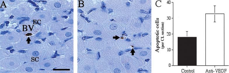
410   S E DICKSON   and others ·       Intervention in luteal angiogenesis

         Previous studies on the effects of withdrawal of VEGF
      in other angiogenic-dependent systems report the require-
      ment of VEGF for the survival of immature endothelial
      cells. For example, in human prostate cancer the loss of
      VEGF as a result of androgen ablation therapy leads to
      selective apoptosis of endothelial cells in vessels devoid of
      periendothelial support cells (Benjamin et al. 1999). To
      investigate whether endothelial cells of the primate CL
      may be susceptible to withdrawal of VEGF, we deter-
      mined the incidence of apoptosis after anti-VEGF treat-
      ment. Finally, to assess the relationship of periendothelial
      support cells to endothelial cell survival, the area of
      pericyte coverage in control CL and after anti-VEGF
      treatment was quantified.
                                                                             Figure 1 Schematic diagram showing the different treatment
      Materials and Methods                                                  regimes (1, 2 and 3) with VEGF monoclonal antibody designed to
                                                                             determine whether inhibition of angiogenic factors can prevent
      Animals and treatments                                                 angiogenesis (regimes 1 and 2) or intervene once the process is
                                                                             underway (regime 3). The approach used in the current study was
      Marmoset monkeys (Calithrix jacchus) were housed as                    to intervene in the already established angiogenic process by
      described previously (Fraser et al. 1999a) and procedures              administering a 3-day treatment with VEGF monoclonal antibody
                                                                             in the mid-luteal phase according to regime 3. PG, prostaglandin
      were carried out in accordance with the Animals (Scien-                F2 analogue.
      tific Procedures) Act 1986. Blood samples were collected
      by femoral venepuncture three times per week without
      anaesthesia. Ovulatory cycles were monitored by radio-                 after administration of 20 mg bromodeoxyuridine (BrdU;
      immunoassay of plasma progesterone as previously de-                   Boehringer Mannheim, Lewes, East Sussex, UK) dissolved
      scribed (Smith et al. 1990). The day of ovulation (day 0 of            in 500 µl physiological saline, to label proliferating cells
      the luteal phase) was taken as the day on which proges-                in the S phase of the cell cycle. Ovaries were fixed
      terone concentration rose above 30 nmol/l when followed                immediately in 4% paraformaldehyde in 0·01 M PBS
      by a sustained increase, characteristic of the luteal phase.           (phosphate-buffered saline, pH 7·4, containing 2·7 mM
          Ovaries collected from animals in the early (luteal days           KCl, 0·137 M NaCl) for paraffin-embedding.
      2–4), mid- (days 8–10) and late (days 16–20) luteal phase
      of the ovulatory cycle (n=4–5 animals per group) for
                                                                             Immunocytochemistry
      previous studies (Fraser et al. 1999a,b, Dickson & Fraser
      2000) were used for immunocytochemical localisation and                Paraffin-embedded ovarian sections (5 µm) were mounted
      quantification of VEGF protein.                                        onto TESPA- (Sigma, Poole, Dorset, UK) coated glass
          The mid-luteal phase was targeted for anti-VEGF                    slides and dried at 50 C overnight. To carry out immu-
      treatment to determine whether luteal angiogenesis could               nocytochemistry for VEGF, sections were dewaxed in
      be prevented at the stage when high levels of VEGF are                 Histoclear (National Diagnostics, Aylesbury, Bucks, UK),
      present, as seen by VEGF immunolocalisation throughout                 rehydrated in descending concentrations of industrial
      the luteal phase of the marmoset ovulatory cycle.                      methylated spirits and washed in distilled water. VEGF
          Animals were given 1 µg prostaglandin F2 analogue                 antigen was retrieved by pressure cooking slides on full
      (Planate; Coopers Animal Health Ltd, Crewe, Cheshire,                  power in 3 M glycine, 0·1% EDTA buffer, pH 3·5, for
      UK) i.m. in the mid- to late luteal phase of the pre-                  5 min. The slides remained in hot buffer for a further
      treatment cycle to induce luteolysis and to synchronise                20 min and were washed in TBS (0·05 M Tris–buffered
      subsequent ovulation which was presumed to occur 10                    saline, pH 7·4, containing 50 mM Tris–HCl, 150 mM
      days after prostaglandin treatment (luteal day 0) (Fig. 1)             NaCl). Endogenous peroxidase activity was quenched
      (Summers et al. 1985). Blood samples were collected every              with a 30-min incubation in 3% hydrogen peroxide in
      second day, then daily from the first day of treatment. The            methanol at room temperature. The following procedures,
      properties of the VEGF monoclonal antibody have been                   with the exception of immunostaining development, were
      described previously (Fraser et al. 2000). Four animals                carried out in Sequenza racks (Shandon Scientific Ltd,
      were subsequently treated with 2 mg VEGF monoclonal                    Runcorn, Cheshire, UK). To block endogenous biotin,
      antibody on luteal day 7 and 1 mg on luteal days 8 and 9,              sections were incubated in avidin (Vector Laboratories,
      and four control animals were given equivalent doses of                Burlingame, CA, USA) at a concentration of 8 drops/ml
      mouse gamma globulin (Fig. 1). Ovaries were collected on               normal swine serum (NSS, 1:5 dilution in TBS+0·25 g
      day 10 as previously described by Fraser et al. (1998), 1 h            bovine serum albumin), followed by a 20-min incubation
      Journal of Endocrinology (2001) 168, 409–416                                                                             www.endocrinology.org
                                                                                                   Downloaded from Bioscientifica.com at 02/07/2022 11:22:12PM
                                                                                                                                                 via free access
Intervention in luteal angiogenesis · S E DICKSON and others 411
in biotin (Vector Laboratories) in TBS (8 drops/ml). modifications according to Sharpe et al. (1998). An ad-
Rabbit polyclonal VEGF antibody (2 µg/ml pre-diluted in ditional proteinase K digestion step was performed after
NSS, Santa Cruz Biotechnology, Santa Cruz, CA, USA) dewaxing and rehydrating the sections. Slides were incu-
was added and sections were incubated at 4 C overnight. bated for 6 min at room temperature in 20 µg/ml proteinase
Negative controls were incubated in primary antibody K in buffer containing 0·05 M Tris, pH 8, 0·05 M EDTA,
pre-absorbed with VEGF peptide in 1:5 ratio of VEGF pH 8, in distilled H2O. After blocking of endogenous
antibody:blocking peptide (Santa Cruz). Immunolocalisa- peroxidase activity, it was necessary to block endogenous
tion was undertaken using the rabbit EnVision kit (Dako biotin using the avidin-biotin block in PBS as above.
Ltd, Cambridge, Cambs, UK) according to the guidelines
of the manufacturer.
BrdU immunocytochemistry was carried out using a Analysis
monoclonal antibody (Boehringer Mannheim) as pre- A BrdU proliferation index (PI) was obtained as previously
viously described (Fraser et al. 2000). Immunocytochemis- described (Dickson & Fraser 2000). Endothelial cell and
try for the endothelial cell marker, CD31 (Dako Ltd), and pericyte areas were quantified in at least 6 randomly
the pericyte marker, alpha-smooth muscle actin (SMA; chosen areas of 5·3104 µm2 (approximately two thirds
Dako Ltd), were performed using monoclonal antibodies of each CL) as described by Fraser et al. (2000) for the
(20·5 µg/ml in TBS and 4·3 µg/ml in TBS respectively) endothelial cell marker, Factor VIII. The area of VEGF
and the same method as for BrdU immunostaining. immunostaining was measured using Photoshop version
Apoptosis was detected by 3 end labelling as previously 5·0 according to the method of Otani et al. (1999).
described in the marmoset CL (Young et al. 1997), with Statistical analysis of VEGF immunostaining throughout
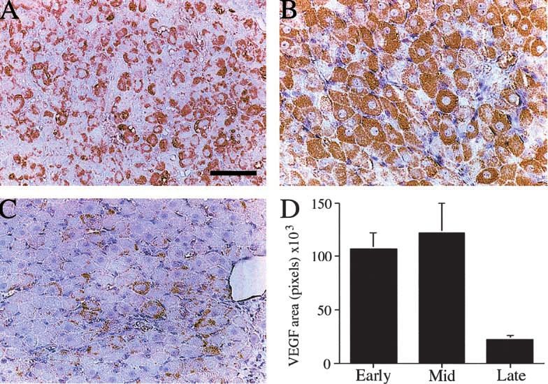
Figure 2 Photomicrographs of marmoset corpora lutea showing VEGF localisation (brown staining cytoplasm) in (A) an early luteal phase
corpus luteum, (B) a mid-luteal phase corpus luteum and (C) a late luteal phase corpus luteum. Bar represents 50 m. (D) Quantification
of the area of VEGF immunostaining in the marmoset corpus luteum throughout the luteal phase of the ovulatory cycle. Early and
mid-luteal values are significantly higher (P=0·04 and P=0·02 respectively) than the late luteal value. Data are means S.E.M.
www.endocrinology.org Journal of Endocrinology (2001) 168, 409–416
Downloaded from Bioscientifica.com at 02/07/2022 11:22:12PM
via free access412 S E DICKSON and others · Intervention in luteal angiogenesis
the ovulatory cycle was carried out using a factorial thelial cell associated with a blood vessel can be seen in Fig.
one-way analysis of variance (ANOVA) with Fisher’s 4A. The incidence of apoptotic nuclei increased with
PLSD (protected least significant difference) post-hoc test anti-VEGF treatment as seen in Fig. 4B which shows two
at 5% significance. The effects of anti-VEGF treatment on positive nuclei probably of endothelial cell origin. Positive
PI, endothelial cell and pericyte areas, apoptotic index and apoptotic nuclei remained sparsely distributed, even
VEGF immunostaining, as compared with controls, were though anti-VEGF treatment significantly (P=0·03) in-
determined using separate two-tailed, unpaired t-tests, creased the occurrence of apoptosis (Fig. 4C). The inci-
with 95% confidence intervals. Serum progesterone con- dence of apoptotic cells was measured per CL section as
centrations were analysed using a repeated measures the size of the control and treated CL did not differ
ANOVA with Fisher’s PLSD post-hoc test at 5% signifi- significantly. The regular shape of the steroidogenic lutein
cance. All tests were performed using Statview version 4·0. cells in control CL was retained in the treated CL.
Results Plasma progesterone concentration
VEGF immunocytochemistry throughout the cycle A sustained elevation of plasma progesterone was observed
Figure 2 illustrates the localisation and quantification of prior to treatment in all animals. After administration of
VEGF immunostaining in the CL during the early, mid- anti-VEGF treatment starting on luteal day 7 there was a
and late luteal phase. Early luteal sections show intense, marked reduction in plasma progesterone concentrations
punctate VEGF staining in the cytoplasm of lutein cells which had fallen by over 50% by day 10 (Fig. 5) (P=0·01).
(Fig. 2A) which becomes more uniform and widespread in
the mid-luteal phase CL (Fig. 2B). Staining was absent
from recognisable endothelial cells. In the late luteal phase Discussion
CL, VEGF staining was markedly reduced (Fig. 2C).
Quantification confirmed a high area of staining in the This study has demonstrated for the first time that VEGF
early and mid-luteal phase which declined markedly in the is essential for luteal angiogenesis even when the process is
late luteal phase CL, as compared with early and mid- already established. Furthermore, inhibition of VEGF at
luteal levels (P=0·04 and P=0·02 respectively) (Fig. 2D). this time suppresses the function of the CL. We have also
demonstrated that VEGF appears to have a role in
BrdU, CD31 and SMA immunocytochemistry maintenance of the vascular network in the mid-luteal CL
as shown by an increase in 3 end labelling after VEGF
Figure 3 shows comparisons of BrdU incorporation, CD31 withdrawal.
and SMA immunostaining, and their quantification in Our findings also show that the expression of VEGF is
mid-luteal control and anti-VEGF-treated CL. Moderate high not only in the early luteal period of intense angio-
BrdU incorporation into endothelial cells was observed in genesis, but also in the less prolific mid-luteal phase CL,
control CL (Fig. 3A) but this was significantly lowered by and is only down regulated after initiation of luteolysis.
anti-VEGF treatment (Fig. 3B). This was confirmed This agrees with VEGF measurements throughout the life
by comparing the PI from control and treated groups span of the CL in the bovine (Goede et al. 1998) and the
(PIntervention in luteal angiogenesis · S E DICKSON and others 413 Figure 3 Low power photomicrographs of marmoset corpora lutea showing the general distribution of endothelial cell BrdU incorporation (dark-staining nuclei) in a control section (A) and its reduced incorporation after anti-VEGF treatment (B). (C) The proliferation index in corpora lutea from mid-luteal phase controls (solid bar) and anti-VEGF-treated (open bar) marmoset corpora lutea. Values from treated animals were significantly lower (P
414   S E DICKSON   and others ·       Intervention in luteal angiogenesis

      Figure 4 High power photomicrographs of 3 end labelled apoptotic cells (brown staining) in (A) a mid-luteal phase control
      marmoset corpus luteum and (B) after anti-VEGF treatment. Note the presence of endothelial cells (EC) surrounding a blood vessel
      (BV) in the control section, and smaller dark haematoxylin-stained nuclei representing endothelial cells and periendothelial support cells
      in both control and treated CL. Arrows point to positive apoptotic nuclei assumed to be endothelial cells by their small, elongated
      form. The morphology of steroidogenic cells (SC) appears unaffected by treatment. (C) Quantification of positive cells in control (solid
      bar) and treated sections (open bar) shows increased presence of positive nuclei in anti-VEGF-treated sections. Note that values are
      per corpus luteum section so the incidence of apoptosis even after treatment is very low. Data are means S.E.M. Bar represents
      20 m.

                                                                             at least during the first 1–2 days of treatment. The rapid
                                                                             decline in plasma progesterone concentration seen on the
                                                                             first day of this acute mid-luteal phase anti-VEGF treat-
                                                                             ment, indicates that in addition to the regulation of
                                                                             ongoing mid-luteal angiogenesis, VEGF also modulates
                                                                             vascular permeability in the CL. An effect on CL vascular
                                                                             permeability could deprive the lutein cells of both the
                                                                             necessary precursors for progesterone production and the
                                                                             efficient release of their products into the bloodstream,
                                                                             which would result in marked reductions in plasma
                                                                             progesterone values.
                                                                                After anti-VEGF treatment, morphological changes in
                                                                             lutein steroidogenic cells were not apparent. This contrasts
                                                                             with observations on the characteristic effects of with-
                                                                             drawal of the trophic factor, luteinising hormone (LH),
                                                                             which have been described recently (Fraser et al. 1999b,
                                                                             Dickson & Fraser 2000), suggesting that anti-VEGF treat-
                                                                             ment does not markedly interfere with pituitary LH
                                                                             secretion.
                                                                                In this study, we have also observed an increase in
                                                                             pericyte coverage as a result of VEGF withdrawal. Peri-
                                                                             cytes are believed to be involved in the remodelling
                                                                             process which occurs during blood vessel maturation
                                                                             (reviewed by Darland & D’Amore 1999), a process also
                                                                             thought to take place in the CL (Tsukada et al. 1996,
                                                                             Goede et al. 1998). Benjamin et al. (1999) demonstrated
                                                                             that in both xenografted tumour and primary human
                                                                             tumours, VEGF withdrawal resulted in specific obliter-
                                                                             ation of immature vessels and that in human prostate
      Figure 5 Plasma progesterone concentrations in control (•) and
      anti-VEGF-treated (●) marmosets. Treatment started on day 7 after      cancer loss of VEGF led to selective apoptosis of endo-
      ovulation and is associated with a significant suppression             thelial cells devoid of periendothelial cells. Mature vessels
      (P=0·01). Data are means S.E.M.                                       with associated pericytes are believed to be VEGF
      Journal of Endocrinology (2001) 168, 409–416                                                                              www.endocrinology.org
                                                                                                    Downloaded from Bioscientifica.com at 02/07/2022 11:22:12PM
                                                                                                                                                  via free access
Intervention in luteal angiogenesis · S E DICKSON and others 415
independent (Benjamin et al. 1998). It is possible that, in References
the current study, the increase in apparent apoptosis in
endothelial cells after anti-VEGF treatment is a conse- Benjamin LE, Hemo I & Keshet E 1998 A plasticity window for
quence of a lack of VEGF support to susceptible, imma- blood vessels remodelling is defined by pericyte coverage of the
preformed endothelial network and is regulated by PDGF-B and
ture vessels with no associated pericytes. It could follow, VEGF. Development 125 1591–1598.
therefore, that endothelial cells of more mature vessels Benjamin LE, Golojanin D, Itin A, Pode D & Keshet E 1999
with associated pericytes would be VEGF independent Selective ablation of immature blood vessels in established human
and not susceptible to anti-VEGF treatment. Our finding tumors follows vascular endothelial growth factor withdrawal.
Journal of Clinical Investigation 103 159–165.
that mid-luteal VEGF withdrawal led to an increase in Darland DC & D’Amore PA 1999 Blood vessel maturation: vascular
endothelial cell apoptosis, the occurrence of which was development comes of age. Journal of Clinical Investigation 103
rare, in the presence of extended pericyte coverage sug- 157–158.
gests that it is the endothelial cells associated with the few Davis S, Aldrich TH, Jones PF, Acheson A, Compton DL, Jain V,
remaining immature capillaries which were susceptible to Ryan TE, Bruno J, Radziejewski C, Maisonpierre PC &
Yancopoulos GD 1996 Isolation of angiopoietin-1, a ligand for the
loss of VEGF support. This, in turn, indicates that the TIE2 receptor, by secretion-trap expression cloning. Cell 87
reduction in endothelial cell area after anti-VEGF treat- 1161–1169.
ment may be a consequence not only of a decreased Dickson SE & Fraser HM 2000 Inhibition of early luteal angiogenesis by
angiogenic rate, but also of increased endothelial cell gonadotropin-releasing hormone antagonist treatment in the primate.
Journal of Clinical Endocrinology and Metabolism 85 2339–2344.
death. Ferrara N, Chen H, Davis-Smyth T, Hans-Peter G, Nguyen T-N,
Factors involved in the recruitment of pericytes Peers D, Chisholm V, Hillan K & Schwall R 1998 Vascular
include platelet-derived growth factor-B (reviewed in endothelial growth factor is essential for corpus luteum angiogenesis.
Darland & D’Amore 1999) and angiopoietin-1 (Davis Nature Medicine 4 336–340.
et al. 1996, Maisonpierre et al. 1997, Koblizek et al. 1998), Fraser HM, Lunn SF, Kim H & Erickson GF 1998 Insulin-like
growth factor binding protein-3 (IGFBP-3) mRNA in the
while transforming growth factor is thought to regulate endothelial cells of the primate corpus luteum. Human Reproduction
pericyte differentiation (Hirschi et al. 1999). It remains 13 2180–2185.
to be determined whether factors such as angiopoietin-1 Fraser HM, Dickson SE, Morris KD, Erickson GF & Lunn SF 1999a
may be activated after VEGF inhibition to act as a sur- The effects of the angiogenesis inhibitor TNP-470 on luteal
establishment and function in the primate. Human Reproduction 14
vival mechanism to ‘rescue’ existing vessels from the 2054–2060.
absence of the immature endothelial cell survival factor, Fraser HM, Lunn SF, Harrison DJ & Kerr JB 1999b Luteal regression
VEGF. in the primate: different forms of cell death during natural and
In conclusion, this study demonstrates that VEGF is a gonadotropin-releasing hormone antagonist or prostaglandin
primary factor controlling angiogenesis and luteal function analogue-induced luteolysis. Biology of Reproduction 61 1468–1479.
Fraser HM, Dickson SE, Lunn SF, Wulff C, Morris KD, Carroll V &
in the mid-luteal phase. The rapid decline in plasma Bicknell R 2000 Suppression of luteal angiogenesis in the primate
progesterone after VEGF inhibition suggests that another by neutralization of vascular endothelial growth factor. Endocrinology
function of VEGF at this stage may be in the maintenance 141 995–1000.
of vascular permeability. In addition, the increase in Goede V, Schmidt T, Kimmina S, Kozian D & Augustin HG 1998
Analysis of blood vessel maturation processes during cyclic ovarian
endothelial cell apoptosis indicates a role for VEGF in the angiogenesis. Laboratory Investigations 78 1385–1394.
survival of a proportion of endothelial cells, perhaps those Hirschi K, Rohovsky S, Beck L, Smith S & D’Amore P 1999
without associated periendothelial support cells. However, Endothelial cells modulate the proliferation of mural-cell precursors
further investigation into the role of VEGF in pericyte via platelet-derived growth factor-BB and heterotypic cell contact.
recruitment is required to further elucidate the processes Circulation Research 84 298–305.
Jablonka-Shariff A, Grazul-Bilska AT, Redmer DA & Reynolds LP
of blood vessel maturation in the CL. Since the mid-luteal 1993 Growth and cellular proliferation of ovine corpora lutea
phase is a crucial period in the establishment of early throughout the estrous cycle. Endocrinology 133 1871–1879.
pregnancy and a time at which survival of the luteal Koblizek TI, Weiss C, Yancopoulos GD, Deutsch U & Risau W
vasculature may be important in the ‘rescue’ of the CL, 1998 Angiopoietin-1 induces sprouting angiogenesis in vitro. Current
Biology 8 529–532.
manipulation of VEGF may have clinical importance Maisonpierre PC, Suri C, Jones PF, Bartunkova S, Wiegand SJ,
with respect to, on the one hand, treatment of early Radziejewski C, Compton D, McLain J, Aldrich TH, Papadopoulos
pregnancy loss, and, on the other hand, interruption N, Daly TJ, Davis S, Sato TN & Yancopoulos GD 1997 Angiopoietin-
of pregnancy. 2, a natural antagonist for Tie2 that disrupts in vivo angiogenesis. Science
277 55–60.
Otani N, Sawako M, Yamoto M, Shikone T, Otani H, Nishiyama R,
Otani T & Nakano R 1999 The vascular endothelial growth
Acknowledgements factor/fms-like tyrosine kinase system in human ovary during the
menstrual cycle and early pregnancy. Journal of Clinical Endocrinology
We thank the staff of our primate unit for animal care, and Metabolism 84 3845–3851.
Rodger FE, Young FM, Fraser HM & Illingworth PJ 1997
M Millar and S MacPherson for expert support with Endothelial cell proliferation follows the mid-cycle luteinizing
histology, and P Hartley and I Swanston for progesterone hormone surge, but not human chorionic gonadotrophin rescue, in
RIAs. the human corpus luteum. Human Reproduction 12 1723–1729.
www.endocrinology.org Journal of Endocrinology (2001) 168, 409–416
Downloaded from Bioscientifica.com at 02/07/2022 11:22:12PM
via free access416 S E DICKSON and others · Intervention in luteal angiogenesis
Sharpe RM, Atanassova N, McKinnell C, Parte P, Turner KJ, Fisher JS, Tsukada K, Matsushima T & Yamanaka N 1996 Neovascularization of
Kerr JB, Groome NP, MacPherson S, Millar MR & Saunders PTK the corpus luteum of rats during the estrous cycle. Pathology
1998 Abnormalities in functional development of the Sertoli cells in International 46 408–416.
rats treated neonatally with diethylstilbestrol: a role for estrogens in Young FM, Illingworth PJ, Lunn SF, Harrison DJ & Fraser HM 1997
Sertoli cell development. Biology of Reproduction 59 1084–1094. Cell death during luteal regression in the marmoset monkey
Smith KB, Lunn SF & Fraser HM 1990 Inhibin secretion during the (Callithrix jacchus). Journal of Reproduction and Fertility 111
ovulatory cycle and pregnancy in the common marmoset monkey. 109–119.
Journal of Endocrinology 126 489–495.
Summers PM, Wennink J & Hodges JK 1985 Cloprostenol-induced
luteolysis in the marmoset monkey (Callithrix jacchus). Journal of Received 16 October 2000
Reproduction and Fertility 73 133–138. Accepted 10 November 2000
Journal of Endocrinology (2001) 168, 409–416 www.endocrinology.org
Downloaded from Bioscientifica.com at 02/07/2022 11:22:12PM
via free accessYou can also read