Mechanisms of fibrinolysis resistance and potential targets for thrombolysis in acute ischaemic stroke: lessons from retrieved stroke emboli ...
←
→
Page content transcription
If your browser does not render page correctly, please read the page content below
Open access Review
Mechanisms of fibrinolysis resistance
Stroke Vasc Neurol: first published as 10.1136/svn-2021-001032 on 26 July 2021. Downloaded from http://svn.bmj.com/ on October 24, 2021 by guest. Protected by copyright.
and potential targets for thrombolysis in
acute ischaemic stroke: lessons from
retrieved stroke emboli
Waleed Brinjikji,1,2 Oana Madalina Mereuta ,1 Daying Dai,1 David F Kallmes,1
Luis Savastano,2 Yang Liu ,1 Shahid M Nimjee,3 Raul G Nogueira,4
Mehdi Abbasi ,1 Ramanathan Kadirvel1
To cite: Brinjikji W, ABSTRACT fibrinolysis resistance. Among acute patients
Madalina Mereuta O, Dai D, There has been growing interest and insight into the with AIS with large vessel occlusion (LVO),
et al. Mechanisms of fibrinolysis histological composition of retrieved stroke emboli. One of
resistance and potential targets
alteplase (tissue-type plasminogen activator)
the main focuses of the stroke clot analysis literature has recanalised only 10% of occlusions while a
for thrombolysis in acute
ischaemic stroke: lessons from been the implications of clot composition on mechanical newer fibrinolytic agent, tenecteplase (TNK),
retrieved stroke emboli. Stroke thrombectomy procedures. However, the holy grail of clot recanalised 22%.13 The mechanisms under-
& Vascular Neurology 2021;0. analysis may not be in the field of clot–device interaction,
lying the low response to fibrinolytic therapy,
doi:10.1136/svn-2021-001032 but rather, in understanding mechanisms of fibrinolysis
even with the newer, more powerful agents,
resistance. The mechanisms underlying the low response
Received 31 March 2021 remain poorly understood. While factors such
to fibrinolytic therapy, even with the newer, more powerful
Accepted 30 June 2021
agents, remain poorly understood. While factors such as embolus size, location and collateral status
as embolus size, location and collateral status influence influence alteplase delivery and recanalisa-
alteplase delivery and recanalisation rates; compositional tion rates, compositional analyses focused
analyses focused on histological and ultrastructural on histological and ultrastructural character-
characteristics offer unique insights into mechanisms of istics offer unique insights into mechanisms
alteplase resistance. In this review, we strive to provide of alteplase resistance. In this article, we
comprehensive review of current knowledge on clot will provide a comprehensive review of clot
composition and ultrastructural analyses that help explain composition and ultrastructural analyses that
resistance to fibrinolysis. help explain resistance to fibrinolysis and
discuss potential future directions in the field
INTRODUCTION of thrombolysis.
Over the past two decades, there has been
growing interest and insight into the histolog- WHAT ARE EMBOLI FROM PATIENTS WITH ACUTE
ical composition of retrieved stroke emboli. ISCHAEMIC STROKE COMPOSED OF?
Numerous groups have published instructive To date, several thousand of clots have
analyses of emboli from patients with acute been collected and analysed with emphasis
ischaemic stroke (AIS) providing additional primarily on histological composition. The
© Author(s) (or their
employer(s)) 2021. Re-use information regarding clot organisation, dominant components studied in these anal-
permitted under CC BY-NC. No composition and ultrastructural analyses.1–9 yses include RBCs, platelets, fibrin and white
commercial re-use. See rights One of the main focuses of the stroke clot blood cells (WBCs). Histological analyses have
and permissions. Published by
analysis literature has been the implications demonstrated that emboli are highly heter-
BMJ.
1 of clot composition on mechanical throm- ogeneous. A review of our multicenter data-
Radiology, Mayo Clinic,
Rochester, Minnesota, USA bectomy procedures. There is a growing base of 1430 retrieved emboli from patients
2
Neurosurgery, Mayo Clinic, consensus in the literature that stiff, fibrin with acute ischaemic stroke found that 56% of
Rochester, Minnesota, USA and platelet-rich clots are harder to retrieve emboli had RBCs as their dominant compo-
3
Neurosurgery, Ohio State with current devices than softer, red blood nent while 21% had fibrin as their dominant
University Medical Center,
Columbus, Ohio, USA
cell (RBC)-rich clots.1 10 11 In fact, the science component and 23% had platelets as their
4
Neurology, Emory University has been so compelling that some companies dominant component. Thus, the spectrum of
School of Medicine, Atlanta, have developed devices solely for the retrieval clots can be classified as RBC-rich, fibrin-rich
Georgia, USA of these tough clots.12 or platelet-rich clots.5 6 14
However, the holy grail of clot analysis may Review of the literature and large data-
Correspondence to
Dr Waleed Brinjikji; not be in the field of clot–device interaction, bases including those of the STRIP (Stroke
brinjikji.waleed@mayo.edu but rather, in understanding mechanisms of Thromboembolism Registry of Imaging and
Brinjikji W, et al. Stroke & Vascular Neurology 2021;0. doi:10.1136/svn-2021-001032 1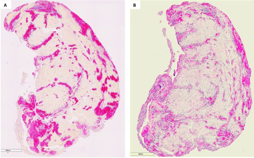
Open access
Pathology) and RESTORE (REgiStry of cloT histOlogy from the STRIP registry having a median RBC composi-
Stroke Vasc Neurol: first published as 10.1136/svn-2021-001032 on 26 July 2021. Downloaded from http://svn.bmj.com/ on October 24, 2021 by guest. Protected by copyright.
and stRokE pathology) registries have found that there tion of 43%. Fibrin networks in RBC-rich clots tend to be
are two distinct structural components: (1) RBC- rich loose, which renders these clots somewhat more porous,
and fibrin-poor regions that have dense packing of RBCs thus allowing for improved delivery of fibrinolytic agents
within a network of thin fibrin strands with very few plate- to their therapeutic target. Meanwhile, preclinical studies
lets and von Willebrand Factor (vWF) in between and have shown that platelet and fibrin-rich clots are gener-
(2) well-structured regions that are rich in platelets and ally more resistant to fibrinolysis than RBC-rich clots.
fibrin and poor in RBCs and the platelet-rich areas have
very dense fibrin structures and are rich in vWF with few Fibrin heterogeneity
RBCs.9 15 In general, these RBC- rich and platelet- rich Fibrin structure and properties have been shown to be
regions are interspersed throughout the clot, while some affected by numerous internal and external factors. Several
clots are very homogeneous in their composition. Inter- studies have shown that genetic variants of fibrinogen
estingly, a sizeable proportion of clots consist of core, and post-translational modifications could contribute to
which is RBC- rich and an outer crust of platelet- rich the heterogeneity of fibrin present in the clot. Mutations
material.16 to fibrin gene can result in defective fibrinogen mole-
WBCs make up a minority of the cellular composition cule, which is generally observed in dysfibrinogenemia.21
of emboli from patients with acute ischaemic stroke and Genetic polymorphism of fibrinogen gene, cross-links of
comprise, on average, 2%–4% of clot area. The most fibrin chains and oxidation, phosphorylation, methyla-
common WBCs within emboli are neutrophils. These are tion, glycosylation of amino acid moieties in fibrin may
predominantly found at the interface between the RBC alter structure and function of fibrin.22–24
and platelet-rich areas as well as within the platelet-rich
zones. However, they are not commonly found in RBC- Therapeutic target
rich areas.15 Since its approval by the FDA in 1996, alteplase has been
One factor that is largely ignored in the ischaemic stroke the standard pharmacological agent for lysing clots in
literature is the concept of clot contraction. It is inter- the setting of AIS. alteplase binds to fibrin within the clot
esting that clots are often composed of distinct regions of and activates plasminogen within the clot. By cleaving
RBC and platelet-rich areas, but why do different parts of plasminogen at its Arg561-Val562 peptide bond, alteplase
the clots have such distinct architecture? Clot contraction creates plasmin, which is a fibrinolytic enzyme that cleaves
likely plays a role in this process. It is thought that the the cross-links between the fibrin polymers. By cleaving
contractile forces of platelets on fibrin can separate the the fibrin bonds within the clot, the clot will typically
RBC and platelet aggregates. This contractile process can dissolve. Alteplase is inhibited by plasminogen activator
compress RBCs as well to the form of polyhedrocytes.17 18 inhibitor 1 (PAI-1), which binds to the drug and forms
Clot contraction likely results in increased organisation, an inactive complex. Fibrinolysis by plasmin is short lived
density and stiffness.19 Further work is needed to charac- due to the presence of plasmin inhibitors, which inacti-
terise this process in patients with ischaemic stroke. vate plasmin.
More recently, there has been growing interest in
TNK as an alternative to alteplase. TNK is a recombinant
FIBRIN fibrin-specific plasminogen activator, which also binds to
Description and function the fibrin component of the clot and converts clot-bound
Fibrin, which is also known as Factor Ia, is a fibrous plasminogen to plasmin. One major difference between
protein involved in haemostasis and is formed following TNK and alteplase, however, is that TNK has higher fibrin
cleavage of fibrinogen by thrombin resulting in polymer- specificity and is more resistant to inactivation by PAI-1.
isation. Platelets are rich in thrombin receptors that bind As explained previously, the RBC- rich areas tend to
serum thrombin molecules. Thus, when platelets start be composed of loose fibrin networks while the platelet-
aggregating, they can turn local-soluble fibrinogen into rich areas are composed of dense fibrin networks rich
fibrin.20 As the fibrin aggregates, it forms long strands in WBCs, vWF and, as we will see later, neutrophil extra-
of this tough insoluble protein that bind to the plate- cellular traps (NETs). A number of previously reported
lets. Factor XIII helps in completing the cross-linking of studies have found that RBC-rich clots respond better
fibrin, so that it hardens and contracts.20 to alteplase than platelet-rich clots.25–28 We hypothesise
that this increased efficacy is due to improved delivery of
Identification in emboli alteplase into the loose meshwork of fibrin in the RBC-
Histological studies of retrieved stroke emboli have rich areas when compared with the denser fibrin struc-
demonstrated marked heterogeneity in the spatial distri- tures copresented with platelet-rich regions.
bution of fibrin and the proportion of fibrin in these clots
(figure 1). Data from the STRIP clot registry show fibrin Other factors affecting fibrinolysis
density ranges from 0.2% to 88% with the median fibrin Several studies have acknowledged the importance of
content being around 27%. RBCs are generally the most biophysical determinants of fibrinolysis.29 A key player
well-
represented component within emboli with clots in the inhibition of fibrinolysis is the activated factor
2 Brinjikji W, et al. Stroke & Vascular Neurology 2021;0. doi:10.1136/svn-2021-001032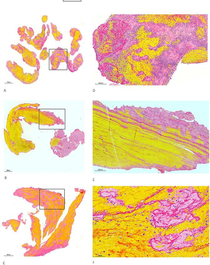
Open access
Stroke Vasc Neurol: first published as 10.1136/svn-2021-001032 on 26 July 2021. Downloaded from http://svn.bmj.com/ on October 24, 2021 by guest. Protected by copyright.
Figure 1 Heterogeneous feature of stroke emboli. At least two serial sections of thrombi retrieved from ischaemic
stroke patients were stained with Martius scarlet blue to identify different component of thrombus. (A–C) Representative
microphotographs taken with lower magnification from three different patients’ thrombi, showing the thrombus tissue consists
of different component which is stained with different colour (yellow, pinkish, red and blue). (D–F) Microphotographs taken
with higher magnification from the rectangular area in (A–C), showing red blood cells (yellow), fibrin strands (red/ intense pink),
platelets (pinkish) and white blood cells (blue) all are present.
XIIIa (FXIIIa), which catalyses the cross-linking of fibrin plasmin.30 Moreover, FXIIIa promotes RBC retention in
between residues in the γ-chains and α-chains of fibrin clots by α-chain cross-linking. This adds to the contribu-
monomers within individual fibres. Although cross- tion of platelet contractile force and RBC deformability
linking has minor effects on the overall fibrin network to clot structure and may determine thrombus composi-
morphology, it significantly decreases the extensibility tion.31
and elasticity of individual fibres and, therefore, stabilises Inhibition of the fibrinolytic system may occur at the
the clot and makes it more resistant to degradation by level of plasminogen activation, mainly by PAI-1 and PAI-2.
Brinjikji W, et al. Stroke & Vascular Neurology 2021;0. doi:10.1136/svn-2021-001032 3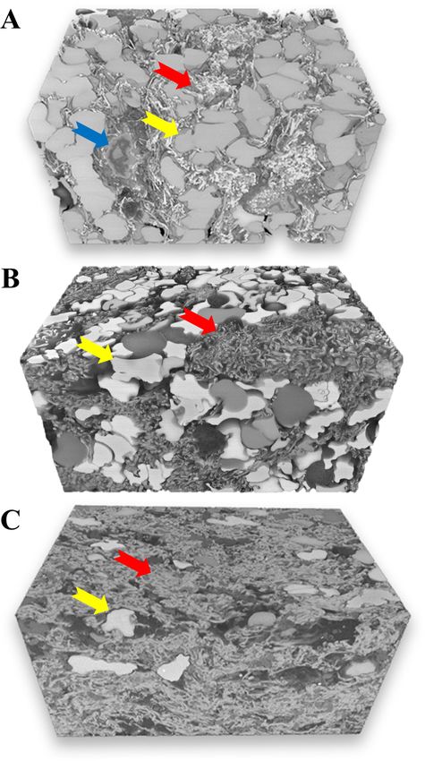
Open access
Fibrin degradation is additionally inhibited by thrombin- Identification in emboli from patients with acute ischaemic
Stroke Vasc Neurol: first published as 10.1136/svn-2021-001032 on 26 July 2021. Downloaded from http://svn.bmj.com/ on October 24, 2021 by guest. Protected by copyright.
activatable fibrinolysis inhibitor (TAFI). FXIIIa does not stroke
only catalyse intramolecular cross-links within fibrin but Until recently, platelets, surprisingly, were ignored in the
also catalyses intermolecular cross-links between fibrin ischaemic stroke literature as major contributors to clot
and other proteins including fibrinolytic inhibitors such composition. Our group and others have been working on
as 2-antiplasmin (2AP), PAI-2 and TAFI and complement identifying platelets using CD42b immunohistochemical
C3. In particular, the cross-linking of 2AP to fibrin has the staining as well as Martius scarlet blue staining. Data from
strongest effect on fibrinolysis. The inhibition of clot lysis
large clot registries have now confirmed, as suspected,
becomes stronger when clot retraction occurs and FXIIIa
that platelets represent a major structural component of
prevents α2AP being expelled from the clot.30
stroke emboli. In the STRIP registry, platelets comprise,
on average, 25%–30% of clot area.3 In general, it seems
PLATELETS that platelets colocalise with vWF (figure 2A,B), which
Description and function binds to platelets through GPIb (glycoprotein Ib) recep-
Platelets are nucleus-free cell fragments produced by bone tors. It is thought that the vWF GPIb binding results in
marrow megakaryocytes. They function to preserve vessel
platelets slowing down, thus, allowing for binding of GPVI
wall health in healthy blood vessels and are a primary
and integrin a2B1 to arrest platelet movement.32 33 This
mediator of haemostasis in the setting of endothelial
injury. When a vessel wall is injured or an atherosclerotic leads to an intracellular signalling cascade, which allows
plaque ruptures, subendothelial collagen is exposed and the release of platelet agonists such as thromboxane A2,
platelets adhere to the collagen GPVI (glycoprotein VI) thrombin and ADP, which then triggers activation of GpII-
and integrin a2B1 receptors. This stimulates intracellular bIIIa.34 Activated GpIIb/IIIa then binds to fibrinogen
signalling mechanisms, which trigger platelet activation and vWF, which results in platelet–platelet cross-linking.34
and promote clot formation. In the setting of athero- One interesting facet of platelet activation in clots is the
sclerosis, however, platelet adhesion is more dependent process of contraction. Activated platelets can remodel
on platelet GP1b receptor binding to vWF, which will be clot fibrin networks and actually increase the fibrin
described further below. density and increase clot stiffness.15 In fact, this process is
thought to be responsible for the poorer revascularisation
outcomes seen in platelet-rich clots. Platelets also play a
role in activation of WBCs.3 Activated platelets release
cytokines which then go on to activate WBCs and localise
Figure 2 Colocalisation of platelets and Von Willebrand Factor (vWF). Serial sections of thrombi retrieved from ischaemic
stroke patients were stained with antibodies against CD42b and vWF to visualise the copresence of platelets and vWF. (A and
B) Representative macrophotographs of serial sections from one thrombus stained for CD42b (A) and vWF (B), showing the
distribution (red/pink) of platelets and vWF are same (Immunohistochemistry, original magnification ×4.0).
4 Brinjikji W, et al. Stroke & Vascular Neurology 2021;0. doi:10.1136/svn-2021-001032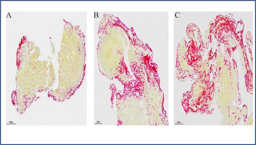
Open access
them to the clot. Studies have shown a strong spatial link was present in all emboli with amounts ranging from 5%
Stroke Vasc Neurol: first published as 10.1136/svn-2021-001032 on 26 July 2021. Downloaded from http://svn.bmj.com/ on October 24, 2021 by guest. Protected by copyright.
between platelets and WBCs in emboli.3 to 50%. Furthermore, data from the RESTORE registry
suggest a strong correlation between vWF and WBC
Therapeutic target density, suggesting a possible relationship between vWF
Platelets represent a prime therapeutic target for throm- and inflammation, something which will be discussed
bolysis in AIS. As mentioned above, much of the platelet– later.3 One interesting finding from STRIP is the fact that
platelet cross-linking is primarily mediated via GpIIb/IIIa vWF seemed to be spatially distributed along the surface
receptors. GpIIb/IIIa antagonists have been a mainstay of of the clot. One additional study on vWF in retrieved
intravenous and intra-arterial antiplatelet therapy for well emboli found that relative vWF content was significantly
over a decade in helping to manage and prevent throm- higher in retrieved emboli treated with alteplase than
botic complications during endovascular procedures.2 those that were not treated with alteplase. This supports
In fact, simple intravenous therapy with these agents is the idea that emboli resistant to fibrinolysis is rich in vWF.
extremely effective in the management of complications
such as in- stent thrombosis, while alteplase therapy is Therapeutic target
not, largely due to the fact that these thrombotic compli- vWF is cleaved by the metalloprotease ADAMTS13,
cations are primarily platelet mediated.2 Thus, it seems which cleaves the vWF A2 domain. ADAMTS13 has been
logical that if such therapy could be integrated with fibro- studied as a thrombolytic agent in both animal and
lytic therapy, superior revascularisation outcomes could benchtop models. In a mouse study in which the middle
be achieved. There is currently a trial ongoing titled the cerebral artery (MCA) was occluded by vWF-rich emboli,
Multi-arm Optimization of Stroke Thrombolysis trial alteplase did not lyse the occlusions, but dose-dependent
comparing the efficacy of adjunctive thrombolysis with administration of ADAMTS13 dissolved the emboli with
argatroban (a direct thrombin inhibitor), eptifibatide no systemic bleeding side effects.25 Another potential
(a GpIIbIIIa inhibitor) and placebo is being compared therapeutic strategy for targeting vWF is by blocking
(NCT03735979). vWF–platelet interactions and reducing vWF monomer–
monomer disulfide bonds using N-acetyle cysteine. More
VON WILLEBRAND FACTOR recently, preclinical studies of thromboembolic stroke
Description and function demonstrated that an inhibitor targeting the A1 domain
vWF is increasingly recognised as an important factor in of vWF recanalised carotid artery occlusions better than
stroke clot pathology and pathophysiology. vWF is the alteplase.36Results such as these suggest that vWF could
largest circulating plasma glycoprotein that mediates be a novel target for thrombolysis.
thrombosis by recruiting platelets to sites of vascular
injury. Along with fibrinogen and fibrin, vWF links plate-
lets together and helps in the stabilisation of clots. vWF NEUTROPHIL EXTRACELLULAR TRAPS AND WBC
is produced in the endothelium as an ultralarge protein Description and function
within Weibel-Palade bodies, megakaryocytes (a-granules NETs have recently been implicated in thrombosis. NETs
of platelets) and in the subendothelial connective tissues. consistent of decondensed chromatic lined with granular
vWF’s primary function is to bind other proteins and fibrous structures, which are released by neutrophils
involved in haemostasis. Factor VIII is bound to vWF in order to kill foreign pathogens. One of the key compo-
while inactive in the circulation and is released from nents of NETs formation is citrullination of histones,
vWF by the action of thrombin. Without vWF, factor VIII which allows for decondensation of nuclear chromatin.
rapidly degrades. vWF also binds to collagen when it is Recently, NETs have been implicated in thrombosis as well
exposed beneath endothelial cells. vWF binds to platelet as they have been found to provide a scaffold for platelets
GpIb receptors, mostly under high shear stress conditions and RBCs and can influence the coagulation cascade.37 38
as well as to multiple additional platelet receptors when NETs have been shown to play a very important role
activated by thrombin. Finally, vWF also self-associates, in thrombosis where they can promote both arterial and
extending from the luminal surface, serving as a scaffold venous clots. NETs assist in the localisation of procoag-
for thrombus formation.35 ulant factors including tissue factor and factor XII. The
decondensed DNA fibres within the extracellular space
Identification in emboli from patients with acute ischaemic act as a scaffold for RBCs, platelets and prothrombotic
stroke molecules and play a putative role in thrombus stability.
To date, there have only been a few studies examining the Cell-free DNA from NETs mediates thrombin generation
proportion and distribution of vWF in retrieved emboli. in the Factor XII and Factor XI- dependent pathways.
If vWF was to be found to be present in high quantities Histones induce thrombin generation by activating plate-
within these emboli, it could represent a potential ther- lets through inducing fibrinogen-mediated platelet aggre-
apeutic target. In a study of 91 emboli samples from the gation. NETs can also accelerate the extrinsic coagulation
RESTORE registry, the mean vWF content was 30% with cascade by their interaction with tissue factor. In vitro
a high correlation between platelet and vWF levels and experiments have shown that NET-induced tissue factor
an inverse correlation between vWF and RBC levels. vWF expression in endothelial cells accelerates clotting.37 38
Brinjikji W, et al. Stroke & Vascular Neurology 2021;0. doi:10.1136/svn-2021-001032 5Open access
Stroke Vasc Neurol: first published as 10.1136/svn-2021-001032 on 26 July 2021. Downloaded from http://svn.bmj.com/ on October 24, 2021 by guest. Protected by copyright.
Figure 3 Neutrophil extracellular traps (NETs) in stroke emboli. Serial sections of thrombi retrieved from ischaemic stroke
patients were stained with antibody against citrullinated histone H3 (H3Cit) to reveal the presence of NETs. (A–C) Representative
microphotographs of three different patients’ thrombi stained for H3Cit (red), showing NETs is expressed along the surface of
thrombus (A), at junctions between RBC-rich and platelet rich-regions (B and C). (Immunohistochemistry, original magnification
×3.7 (A), ×4.9 (B and C)).
Identification in emboli from patients with acute ischaemic thrombectomy more difficult with increased number of
stroke passes and longer procedure times.38–40 However, DNA,
Quantification of NETs in AIS emboli is lacking and the main component of NETs, is degraded by DNases, in
most studies have quantified the WBC level. Interest- particular, DNase 1 and DNase1-like 3.
ingly, WBCs make up a minority of the cellular compo- In a seminal article by Jimenez-Alcazar et al, the authors
nents of AIS emboli. The median proportion of WBCs found that DNase knockout mice produced vascular occlu-
in AIS emboli is about 3% with an IQR of just 2%–5%.38 sions with NET-rich clots, resulting in significantly higher
However, WBCs, particularly neutrophils (CD66b posi- rates of end-organ damage than normal controls.41 In a
tive), play an outsized role in thrombosis in the form of study from De Meyer et al, addition of DNase 1 to alteplase
NETs, which can cover up to 60% of the surface area of resulted in significantly higher rates of ex vivo dissolution
AIS emboli. of emboli freshly retrieved from stroke patients. DNase 1
NETs are revealed by the presence of prominent extra- has also been shown to improve thrombolytic activity for
cellular nucleic acid- rich areas located in neutrophil- coronary thrombi as well.
rich zones. These areas stain highly positive for H3Cit
(figure 3), the defining marker of NETs as well as neutro-
phil elastase. As mentioned above, there are growing data NETS–VWF–PLATELET AXIS
suggesting that NETs for a scaffold for platelets and RBCs. There is growing evidence linking NETs, platelets and
This is supported by work from a number of groups, vWF in a prothrombotic and proinflammatory axis. This
which show that NETs are abundant at junctions between axis is going to be very important to understand both for
RBC-rich and platelet-rich regions within AIS emboli as acute treatment of ischaemic stroke with thrombolysis
well as along the surface of emboli. and in primary and secondary prevention strategies; espe-
cially given the fact that NETs, platelets and vWF are the
NETs as a therapeutic target primary agents responsible for alteplase resistance of AIS
Recent work has focused on the role of NETs as a poten- emboli. vWF binds to pure DNA via electrostatic inter-
tial therapeutic target in thrombolysis. NETs serve as a actions, which, interestingly, can be blocked by heparin.
thrombotic scaffold contributing the clot stability and are The DNA binds to the same domain that the Gp1a recep-
thought to confer some degree of alteplase resistance. tors of platelets bind to as well. Neutrophils also adhere
In fact, the addition of extracellular DNA and histones to vWF and can be recruited to the vessel wall via the
to fibrin has been shown to increase the thickness, stiff- vWF–DNA interaction. Neutrophil elastase, an abundant
ness and stability of the fibrin network; possibly making component of NETs, also binds to vWF via electrostatic
it harder to deliver alteplase to the interstices of the clot. interactions due to its positive charge. Thus, it is probable
High NET density has also been shown to make mechanical that vWF released from endothelial cells and platelets
6 Brinjikji W, et al. Stroke & Vascular Neurology 2021;0. doi:10.1136/svn-2021-001032Open access
Stroke Vasc Neurol: first published as 10.1136/svn-2021-001032 on 26 July 2021. Downloaded from http://svn.bmj.com/ on October 24, 2021 by guest. Protected by copyright.
Table 1 Summary table
Clot component Structure Interacts with Potential therapeutic agent(s)
Fibrin (factor Ia) Fibrous protein Platelets, factor XIII, RBCs Alteplase, tenecteplase
Platelets Nucleus-free cell fragments Collagen, vWF, WBCs GpIIb/IIIa blockers
vWF Plasma glycoprotein Fibrin, platelets, collagen ADAMTS13, N-acetyle cysteine
(NAC)
NET Decondensed DNA/chromatin Thrombin, platelets, vWF DNase1
NET, neutrophil extracellular trap; RBC, red blood cell; vWF, von Willebrand Factor; WBC, white blood cell.
interact with NETs to promote progression of thrombosis consisted of polyhedrocytes, a morphological marker of
and inflammation. clot contraction, which, as described above, is a common
The vWF–NETs–platelet axis is further supported by process, resulting from contractile platelets pulling on
histological studies of emboli. In a study by de Meyer et fibrin. Furthermore, the 3DEM data showed that WBCs
al, WBCs and extracellular DNA were present mainly in were concentrated mainly in the fibrin-rich and platelet-
platelet-rich regions and at the boundary between platelet- rich areas and occasionally at the interface between RBC-
rich and RBC-rich regions. It is thought that vWF serves as rich and platelet-rich areas, lending further support to
a mediator to bring NETs and fibrin into close proximity, the potential role of NETs as a mechanism of fibrinolysis
thus facilitating the ability of NETs to modify the fibrin resistance. 3DEM was also able to show how alteplase
structure and increase local coagulation.38 Platelets play resulted in thinning of fibrin fibres and disruption of
a key role in this axis as well and are thought to promote fibrin fibre organisation (figure 5).
uncontrolled NETosis. Endothelial cell bound vWF Recently, many groups have explored the role of trans-
promotes initial platelet adhesion by binding to platelet mission electron microscopy in evaluating the efficacy of
GPIba (glycoprotein Iba) receptors and activating plate- thrombolysis. For example, in a study evaluating alteplase
lets. These activated platelets promote neutrophil migra- versus TNK for in vitro clots, Fruhwald et al found that
tion via toll-like receptors, which can promote the growth clots treated with TNK had significant reductions in fibrin
of clots.42 In fact, in one recently published study, platelet fibre density than those treated with alteplase.43
TLR4 was found to mediate NET formation in a mouse
model of acute ischaemic stroke and ex vivo fresh platelet-
rich emboli from patients were lysed by DNase-I.42
FUTURE DIRECTIONS
In summary, NETs and vWF recruit platelets and WBCs
Over the past several decades, the main focus for stroke
and, thus, promote coagulation. We believe that the
thrombolysis has been on fibrinolysis. However, if there is
NET–vWF interaction provides an extremely strong scaf-
any chance for the field of pharmacological-based revas-
fold within emboli and facilitates interactions between
cularisation to progress, the knowledge of clot compo-
platelets–WBCs–NETs and fibrin–vWF and NETs. There-
sition, organisation and physical characteristics that we
fore, devising attempts to target the NET–vWFvplatelet
have gained from the multitudes of retrieved AIS emboli
axis could represent a viable therapeutic strategy for isch-
needs to be applied. First and foremost, more in vitro and
aemic stroke. A summary table is provided in table 1.
animal studies studying the basic biologic mechanism of
resistance to thrombolysis needs to be conducted, specif-
ULTRASTRUCTURAL ANALYSIS OF CLOT ically targeting platelets, NETs and vWF. This requires
Electron microscopy has opened the door to new insights the production of physiologically representative AIS
into the ultrastructural characteristics of stroke emboli clot analogues that are rich in these components and
and their implications for fibrinolysis resistance. In one have similar structural organisation (ie, shell of alteplase
recently published study, Di Meglio et al analysed nearly resistant material) and physical characteristics and the
200 emboli using immunohistochemistry and scanning application of thrombolytic agents in in vitro models,
electron microscopy and found that stroke emboli shared which replicate the haemodynamics of a vascular occlu-
a common structural feature comprising of an outer shell sion. This last point is especially true as one of the main
composed of densely packed fibrin, vWF and aggregated challenges in thrombolysis is how to deliver drug to a
platelets.16 In vitro experiments confirmed that platelets blood vessel that has no flow.
were essential to the formation of this outer shell and this Studies examining the effects of various novel throm-
outer shell was found to decrease the efficacy of alteplase bolytic agents on the ultrastructure of clots would also be
(figure 4). useful, simply to understand what the exact mechanisms
Another recently published study of 3D electron micros- are for success and failure of these agents are. In addi-
copy (3DEM) in AIS emboli was also able to provide some tion, studies on mechanically altering the clot ultrastruc-
novel insights into the ultrastructure of AIS emboli.8 In ture (such as sonothrombolysis) to enhance diffusion of
this particular analysis, 3DEM showed that RBC-rich areas thrombolytic agents into the clot can also be useful.
Brinjikji W, et al. Stroke & Vascular Neurology 2021;0. doi:10.1136/svn-2021-001032 7Open access
Stroke Vasc Neurol: first published as 10.1136/svn-2021-001032 on 26 July 2021. Downloaded from http://svn.bmj.com/ on October 24, 2021 by guest. Protected by copyright.
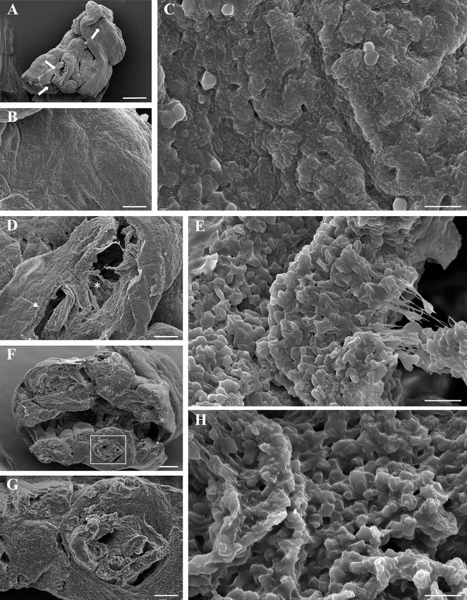
Figure 4 Representative scanning electron microscopy (SEM) images of an acute ischaemic stroke thrombus. (A) Arrows
correspond to the locations analysed by SEM. The surface of the clot shows multiple folds and ridges. (B) Distinct thrombotic
components are not discernible on the surface that appears smooth. (C) High magnification of the surface suggestive of dense
integration of components and advanced organisation. Only a few red blood cells are evident. (D) The rift on the surface shows
the presence of a dense outer shell (arrow) and a different structure of the interior of the clot (asterisk). (E) Magnified area of
the interior of the clot displays numerous individual polyhedrocytes and some distinct fibrin strands suggesting an immature
structure. (F) The cross-section exposes the thrombus core which is detailed in (G). (H) Identifiable thrombus components
indicate a limited maturity and incomplete integration. Scale bar=10 µm (C, E, H), 100 µm (B, D, G), 200 µm (F) and 500 µm (A).
CONCLUSIONS our experience of retrieved emboli, these clots are
We are only starting to understand the complex biolog- composed of a complex mixture of RBCs, fibrin, WBCs,
ical and structural characteristics of thromboembolisms vWF, NETs and platelets with the fibrinolytic-resistant
causing LVO stroke. Based on what we now know from components (ie, vWF, NETs and platelets) located
8 Brinjikji W, et al. Stroke & Vascular Neurology 2021;0. doi:10.1136/svn-2021-001032Open access
Contributors WB, OMM, DD, DFK, LS, YL, SMN, RGN, MA and RK made substantial
Stroke Vasc Neurol: first published as 10.1136/svn-2021-001032 on 26 July 2021. Downloaded from http://svn.bmj.com/ on October 24, 2021 by guest. Protected by copyright.
contributions to the conception or design of the work or the acquisition, analysis or
interpretation of data for the work; and drafting of the work or revising it critically
for important intellectual content. All authors provided final approval of the version
to be published. All authors agree to be accountable for all aspects of the work in
ensuring that questions related to the accuracy or integrity of any part of the work
are appropriately investigated and resolved.
Funding This work was supported by the National Institutes of Health grant
number (R01 NS105853).
Competing interests None declared.
Patient consent for publication Not required.
Provenance and peer review Not commissioned; externally peer reviewed.
Open access This is an open access article distributed in accordance with the
Creative Commons Attribution Non Commercial (CC BY-NC 4.0) license, which
permits others to distribute, remix, adapt, build upon this work non-commercially,
and license their derivative works on different terms, provided the original work is
properly cited, appropriate credit is given, any changes made indicated, and the use
is non-commercial. See: http://creativecommons.org/licenses/by-nc/4.0/.
ORCID iDs
Oana Madalina Mereuta http://orcid.org/0000-0003-3741-3784
Yang Liu http://orcid.org/0000-0002-9132-0258
Mehdi Abbasi http://orcid.org/0000-0001-6978-2563
REFERENCES
1 Brinjikji W, Duffy S, Burrows A, et al. Correlation of imaging and
histopathology of thrombi in acute ischemic stroke with etiology and
outcome: a systematic review. J Neurointerv Surg 2017;9:529–34.
2 Brinjikji W, Morales-Valero SF, Murad MH, et al. Rescue treatment
of thromboembolic complications during endovascular treatment
of cerebral aneurysms: a meta-analysis. AJNR Am J Neuroradiol
2015;36:121–5.
3 Douglas A, Fitzgerald S, Mereuta OM, et al. Platelet-rich emboli
are associated with von Willebrand factor levels and have poorer
revascularization outcomes. J Neurointerv Surg 2020;12:557–62.
4 Fitzgerald S, Dai D, Wang S, et al. Platelet-Rich emboli in
cerebral large vessel occlusion are associated with a large artery
atherosclerosis source. Stroke 2019;50:1907–10.
5 Fitzgerald S, Rossi R, Mereuta OM, et al. Per-pass analysis of
acute ischemic stroke clots: impact of stroke etiology on extracted
clot area and histological composition. J Neurointerv Surg 2020.
doi:10.1136/neurintsurg-2020-016966. [Epub ahead of print: 09 Dec
2020].
6 Fitzgerald S, Rossi R, Mereuta OM, et al. Large artery atherosclerotic
Figure 5 Three-dimensional scanning electron microscopy clots are larger than clots of other stroke etiologies and have poorer
analysis in three representative cases. Volume renderings recanalization rates. J Stroke Cerebrovasc Dis 2021;30:105463.
7 Fitzgerald ST, Wang S, Dai D, et al. Platelet-rich clots as identified by
assembled from serial block-face imaging highlight detailed Martius scarlet blue staining are isodense on NCCT. J Neurointerv
ultrastructural organisation and characteristics of individual Surg 2019;11:1145–9.
components for each clot (red blood cells (RBC)=yellow 8 Mereuta OM, Fitzgerald S, Christensen TA, et al. High-resolution
arrows, fibrin=red arrows, white blood cells=blue arrow). scanning electron microscopy for the analysis of three-dimensional
ultrastructure of clots in acute ischemic stroke. J Neurointerv Surg
(A) Clot area with tightly packed RBC as polyhedrocytes 2020. doi:10.1136/neurintsurg-2020-016709. [Epub ahead of print:
intermixed with a limited volume of thin fibrin fibres. (B) Clot 23 Dec 2020].
area with mixed composition consisting of both packed 9 Staessens S, Fitzgerald S, Andersson T, et al. Histological
polyhedrocytes and dense network of thick fibrin fibres. (C) stroke clot analysis after thrombectomy: technical aspects and
recommendations. Int J Stroke 2020;15:467–76.
Fibrin-rich area containing dense fibrin masses with sparse 10 Johnson S, Chueh J, Gounis MJ, et al. Mechanical behavior of in
polyhedrocytes. vitro blood clots and the implications for acute ischemic stroke
treatment. J Neurointerv Surg 2020;12:853–7.
11 Weafer FM, Duffy S, Machado I, et al. Characterization of strut
indentation during mechanical thrombectomy in acute ischemic
predominantly at the surface of the clot. Further experi- stroke clot analogs. J Neurointerv Surg 2019;11:891–7.
12 Fennell VS, Setlur Nagesh SV, Meess KM, et al. What to do about
ments are needed to design and test novel thrombolytics fibrin rich 'tough clots'? Comparing the Solitaire stent retriever
that target not just fibrin, but these other components. with a novel geometric clot extractor in an in vitro stroke model. J
Neurointerv Surg 2018;10:907–10.
Also, more research is needed to better understand the 13 Campbell BCV, Mitchell PJ, Churilov L, et al. Tenecteplase versus
ultrastructural characteristics of stroke emboli in order alteplase before thrombectomy for ischemic stroke. N Engl J Med
2018;378:1573–82.
for us to better understand mechanisms of thrombolysis 14 Brouwer PA, Brinjikji W, De Meyer SF. Clot pathophysiology: why is it
resistance. clinically important? Neuroimaging Clin N Am 2018;28:611–23.
Brinjikji W, et al. Stroke & Vascular Neurology 2021;0. doi:10.1136/svn-2021-001032 9Open access
15 Staessens S, Denorme F, Francois O, et al. Structural analysis 29 Hudson NE. Biophysical mechanisms mediating fibrin fiber lysis.
Stroke Vasc Neurol: first published as 10.1136/svn-2021-001032 on 26 July 2021. Downloaded from http://svn.bmj.com/ on October 24, 2021 by guest. Protected by copyright.
of ischemic stroke thrombi: histological indications for therapy Biomed Res Int 2017;2017:1–17.
resistance. Haematologica 2020;105:498–507. 30 Rijken DC, Uitte de Willige S. Inhibition of fibrinolysis by coagulation
16 Di Meglio L, Desilles J-P, Ollivier V, et al. Acute ischemic stroke factor XIII. Biomed Res Int 2017;2017:1–6.
thrombi have an outer shell that impairs fibrinolysis. Neurology 31 Byrnes JR, Duval C, Wang Y, et al. Factor XIIIa-dependent retention
2019;93:e1686–98. of red blood cells in clots is mediated by fibrin α-chain crosslinking.
17 Cines DB, Lebedeva T, Nagaswami C, et al. Clot contraction: Blood 2015;126:1940–8.
compression of erythrocytes into tightly packed polyhedra and 32 Denorme F, De Meyer SF. The VWF-GPIb axis in ischaemic stroke:
redistribution of platelets and fibrin. Blood 2014;123:1596–603. lessons from animal models. Thromb Haemost 2016;116:597–604.
18 Tutwiler V, Mukhitov AR, Peshkova AD, et al. Shape changes of 33 Denorme F, Vanhoorelbeke K, De Meyer SF. Von Willebrand factor
erythrocytes during blood clot contraction and the structure of and platelet glycoprotein Ib: a Thromboinflammatory axis in stroke.
polyhedrocytes. Sci Rep 2018;8:17907. Front Immunol 2019;10:2884.
19 Tutwiler V, Peshkova AD, Andrianova IA, et al. Contraction of blood 34 Offermanns S. Activation of platelet function through G protein-
clots is impaired in acute ischemic stroke. Arterioscler Thromb Vasc coupled receptors. Circ Res 2006;99:1293–304.
Biol 2017;37:271–9. 35 Savage B, Sixma JJ, Ruggeri ZM. Functional self-association of von
20 Khismatullin RR, Nagaswami C, Shakirova AZ, et al. Quantitative Willebrand factor during platelet adhesion under flow. Proc Natl Acad
morphology of cerebral thrombi related to intravital contraction and Sci U S A 2002;99:425–30.
clinical features of ischemic stroke. Stroke 2020;51:3640–50. 36 Nimjee SM, Dornbos D, Pitoc GA, et al. Preclinical development of a
21 Casini A, Duval C, Pan X, et al. Fibrin clot structure in patients with vWF aptamer to limit thrombosis and engender arterial recanalization
of occluded vessels. Mol Ther 2019;27:1228–41.
congenital dysfibrinogenaemia. Thromb Res 2016;137:189–95.
37 Bonaventura A, Liberale L, Carbone F, et al. The pathophysiological
22 Kotzé RC, Nienaber-Rousseau C, De Lange Z, et al. Genetic
role of neutrophil extracellular traps in inflammatory diseases.
polymorphisms influencing total and γ' fibrinogen levels and fibrin
Thromb Haemost 2018;118:006–27.
clot properties in Africans. Br J Haematol 2015;168:102–12.
38 Ducroux C, Di Meglio L, Loyau S, et al. Thrombus neutrophil
23 Weisel JW, Litvinov RI. Fibrin formation, structure and properties.
extracellular traps content impair tPA-induced thrombolysis in acute
Subcell Biochem 2017;82:405–56. ischemic stroke. Stroke 2018;49:754–7.
24 Zubairova LD, Nabiullina RM, Nagaswami C, et al. Circulating 39 Ducroux C, Desilles J-P, Ho-Tin-Noe B. Response by ducroux et al
microparticles alter formation, structure, and properties of fibrin to letter regarding article, "thrombus neutrophil extracellular traps
clots. Sci Rep 2015;5:17611. content impair tPA-induced thrombolysis in acute ischemic stroke".
25 Denorme F, Langhauser F, Desender L, et al. ADAMTS13-mediated Stroke 2018;49:e266.
thrombolysis of t-PA-resistant occlusions in ischemic stroke in mice. 40 Longstaff C, Varjú I, Sótonyi P, et al. Mechanical stability and
Blood 2016;127:2337–45. fibrinolytic resistance of clots containing fibrin, DNA, and histones. J
26 Niesten JM, van der Schaaf IC, van der Graaf Y, et al. Predictive Biol Chem 2013;288:6946–56.
value of thrombus attenuation on thin-slice non-contrast CT for 41 Jiménez-Alcázar M, Rangaswamy C, Panda R, et al. Host DNases
persistent occlusion after intravenous thrombolysis. Cerebrovasc Dis prevent vascular occlusion by neutrophil extracellular traps. Science
2014;37:116–22. 2017;358:1202–6.
27 Puig J, Pedraza S, Demchuk A, et al. Quantification of thrombus 42 Peña-Martínez C, Durán-Laforet V, García-Culebras A, et al.
hounsfield units on noncontrast CT predicts stroke subtype and early Pharmacological modulation of neutrophil extracellular traps reverses
recanalization after intravenous recombinant tissue plasminogen thrombotic stroke tPA (tissue-type plasminogen activator) resistance.
activator. AJNR Am J Neuroradiol 2012;33:90–6. Stroke 2019;50:3228–37.
28 Choi MH, Park GH, Lee JS, et al. Erythrocyte fraction within retrieved 43 Frühwald T, Gärtner U, Stöckmann N, et al. In vitro examination of
thrombi contributes to thrombolytic response in acute ischemic the thrombolytic efficacy of tenecteplase and therapeutic ultrasound
stroke. Stroke 2018;49:652–9. compared to rt-PA. BMC Neurol 2019;19:181.
10 Brinjikji W, et al. Stroke & Vascular Neurology 2021;0. doi:10.1136/svn-2021-001032You can also read


