Lung microbiome of stable and exacerbated COPD patients in Tshwane, South Africa
←
→
Page content transcription
If your browser does not render page correctly, please read the page content below
www.nature.com/scientificreports

                OPEN              Lung microbiome of stable
                                  and exacerbated COPD patients
                                  in Tshwane, South Africa
                                  T. Goolam Mahomed1, R. P. H. Peters1,2, M. Allam3, A. Ismail3, S. Mtshali3,
                                  A. Goolam Mahomed4, V. Ueckermann5, M. M. Kock1,6 & M. M. Ehlers1,6*

                                  Chronic obstructive pulmonary disease (COPD) is characterised by the occurrence of exacerbations
                                  triggered by infections. The aim of this study was to determine the composition of the lung
                                  microbiome and lung virome in patients with COPD in an African setting and to compare their
                                  composition between the stable and exacerbated states. Twenty-four adult COPD patients were
                                  recruited from three hospitals. Sputum was collected and bacterial DNA was extracted. Targeted
                                  metagenomics was performed to determine the microbiome composition. Viral DNA and RNA were
                                  extracted from selected samples followed by cDNA conversion. Shotgun metagenomics sequencing
                                  was performed on pooled DNA and RNA. The most abundant phyla across all samples were Firmicutes
                                  and Proteobacteria. The following genera were most prevalent: Haemophilus and Streptococcus. There
                                  were no considerable differences for alpha and beta diversity measures between the disease states.
                                  However, a difference in the abundances between disease states was observed for: (i) Serratia (3%
                                  lower abundance in exacerbated state), (ii) Granulicatella (2.2% higher abundance in exacerbated
                                  state), (iii) Haemophilus (5.7% higher abundance in exacerbated state) and (iv) Veillonella (2.5%
                                  higher abundance in exacerbated state). Virome analysis showed a high abundance of the BeAn 58058
                                  virus, a member of the Poxviridae family, in all six samples (90% to 94%). This study is among the first
                                  to report lung microbiome composition in COPD patients from Africa. In this small sample set, no
                                  differences in alpha or beta diversity between stable and exacerbated disease state was observed, but
                                  an unexpectedly high frequency of BeAn 58058 virus was observed. These observations highlight the
                                  need for further research of the lung microbiome of COPD patients in African settings.

                                  Abbreviations
                                  16S rRNA	16S ribosomal ribonucleic acid
                                  cDNA	Complementary deoxyribonucleic acid
                                  COPD	Chronic obstructive pulmonary disease
                                  DNA	Deoxyribonucleic acid
                                  DNase	Deoxyribonuclease
                                  dsDNA	Double stranded deoxyribonucleic acid
                                  dsRNA	Double stranded ribonucleic acid
                                  DTT	Dithiothreitol
                                  FEV1%	Percentage of the forced vital capacity
                                  HIV	Human immunodeficiency virus
                                  IQR	Interquartile range
                                  N/A	Not available
                                  NGS	Next generation sequencing
                                  NICD	National Institute for Communicable Diseases of South Africa
                                  OTUs	Operational taxonomic units
                                  PCoA	Principal component analysis

                                  1
                                   Department of Medical Microbiology, University of Pretoria, Pretoria, South Africa. 2Foundation for Professional
                                  Development, Research Unit, East London, South Africa. 3National Institute for Communicable Diseases,
                                  National Health Laboratory Service, Johannesburg, South Africa. 4Louis Pasteur Private Hospital, Pretoria,
                                  South Africa. 5Department of Internal Medicine, University of Pretoria, Pretoria, South Africa. 6Department of
                                  Medical Microbiology, Tshwane Academic Division, National Health Laboratory Service, Johannesburg, South
                                  Africa. *email: marthie.ehlers@up.ac.za

Scientific Reports |   (2021) 11:19758               | https://doi.org/10.1038/s41598-021-99127-w                                                1

                                                                                                                                            Vol.:(0123456789)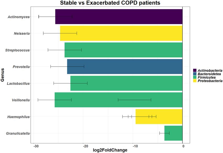
www.nature.com/scientificreports/
QIIME2 Quantitative insights into microbial ecology 2
REC Research ethics committee
RNA Ribonucleic acid
rRNA Ribosomal ribonucleic acid
RT-PCR Reverse transcriptase polymerase chain reaction
ssDNA Single stranded deoxyribonucleic acid
ssRNA Single stranded ribonucleic acid
UK United Kingdom
USA United States of America
Chronic obstructive pulmonary disease (COPD) is a progressive lung disease that results in progressive airflow
limitation (i.e. obstruction)1,2. COPD is one of the world’s leading causes of death and was projected to be the
third leading cause of death in 2 0203. Symptoms of COPD include a chronic cough, dyspnoea and sputum
production4,5. These symptoms affect the quality of life of the individual suffering from this disease6. There is
limited data about the prevalence of COPD in the African continent; the last reported prevalence data on COPD
in South Africa was in 2005 (19% in men and women over 40 years of age)7–10. This disease has been linked to
smoking, exposure to occupational dust (e.g. working in a mine), burning of biomass and fossil fuels, previous
tuberculosis infection and to HIV; all of these risk factors are highly prevalent in South Africa10.
Exacerbation of airway inflammation and associated symptoms is another factor that affects the quality of
life for these individuals10. Patients suffering from COPD often move between a stable state of disease (where
symptoms are absent to mild) to an exacerbated state of disease (defined as worsening of symptoms, respiratory
and/or non-respiratory and over the course of the disease, as the lung damage due to COPD progresses, the
frequency of these exacerbations increases11–13. Exacerbations are triggered by environmental pollutants, may
have an unknown cause or by infection with bacteria and/or v iruses14. Bacterial and viral infections account for
between 30 to 50% of all e xacerbations15. However, bacteria have been detected in the stable state of disease as
well and the association between these microorganisms and disease is unclear16,17.
To better understand the role of microorganisms in COPD disease, the use of next-generation sequencing
(NGS) can be employed to study the microbiome (defined as the genetic material of the microorganism in
the community)18. NGS is high-throughput, parallel sequencing technology which has been used to sequence
whole genomes of bacteria and viruses, perform transcriptomics (studying the complete set of RNA transcripts
produced by the genomes) and to study the microbiome/metagenome19,20. The advantage of NGS over culturing
and other molecular methods is that it can detect unculturable bacteria and provide information regarding the
diversity, composition and functional roles of members of the m icrobiome21,22. An important drawback is that
the cost of sequencing is still relatively high, especially in the African c ontinent23. The NGS technology can be
employed in one of two ways: (i) using a targeted approach or (ii) using a metagenomic approach24,25.
The targeted approach is commonly used to study the microbiome and is employed by targeting the 16S
rRNA gene26,27. This gene is useful for studying the bacterial microbiome as it is universally present and con-
served within all b acteria28–30. Studying the virome, i.e. viral component of the microbiome is more challenging
as (i) most viruses are difficult to culture, (ii) there is no consensus sequence to study viruses and (iii) viruses
are diverse and may be ssDNA, ssRNA, dsDNA or d sRNA31–33. By using shotgun metagenomics (i.e. random
sequencing of the DNA from the microbial community) along with cDNA synthesis to study the virome, these
challenges can be overcome34–36.
In South Africa, there is no data on the composition of the lung microbiome in COPD patients.
Previous studies on the lung microbiome of COPD patients were conducted in Europe and the USA37–39.
Furthermore, there have been limited studies on the lung virome in COPD40,41. It is important to study not only
the microbiome in the African continent, in countries such as South Africa but also the virome as local environ-
mental conditions e.g. climate and clinical co-morbidities, e.g. HIV and tuberculosis infection (both of which are
highly prevalent in sub-Saharan Africa) have the potential to affect the microbiome. Therefore, the aim of this
study was to determine the composition of the lung microbiome and the lung virome in the sputum of COPD
patients from South Africa and to compare their composition between stable and exacerbated states of disease.
Methods
Study setting and patient recruitment criteria. COPD patients admitted to or attending clinics (for
scheduled check-ups) at one of three hospitals (one academic, one district and one private) in the Tshwane
Health district, South Africa were invited to participate in the study. Written informed consent was obtained
from all participants if the inclusion and exclusion criteria were met (Supplementary materials Table S1). Par-
ticipants were classified as either in the stable or in the exacerbated state based on the definition by Vogelmeier
et al. (2017). Ethical approval was granted from the Research Ethics Committee, Faculty of Health Sciences,
University of Pretoria (REC no: 237/2017).
Extraction of DNA and RNA and cDNA synthesis. Spontaneously expectorated sputum specimens
were collected from participants at a single time point, transported on ice and stored at − 80 °C (Innova U535
Upright, Eppendorf, Germany) until batch processing could occur (no preservation medium was used). The
sputum specimens were treated with an equal volume of 0.1% dithiothreitol (DTT) (Roche Diagnostics, Switzer-
land) to reduce sputum viscosity and homogenised for 30 s (Vortex-Genie® 2; Scientific Industries Inc., USA)42–44.
The samples were split into three aliquots for: (i) bacterial DNA extraction (aliquot 1), (ii) viral DNA and RNA
extraction (aliquot 2) and (iii) storage at − 80 °C (aliquot 3, for future processing and/or studies) (Innova U535
Upright, Eppendorf, Germany).
Scientific Reports | (2021) 11:19758 | https://doi.org/10.1038/s41598-021-99127-w 2
Vol:.(1234567890)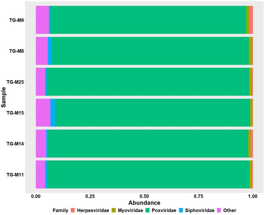
www.nature.com/scientificreports/
The bacterial extraction aliquot was centrifuged (Spectrafuge™ 24D, Labnet International Inc., USA) at 4000×g
for 30 min before extraction. Bacterial DNA was extracted using the Isolate II Genomic DNA Kit (Bioline, UK).
The manufacturer’s instructions (protocol 9.2) were followed with the addition of 10 mg/mL lysozyme (Sigma-
Aldrich, USA), 3 U/µL lysostaphin (Sigma-Aldrich, USA) and 6.75 µL of 10 U/µL mutanolysin (Sigma-Aldrich,
USA) to the hard-to-lyse buffer [20 mM Tris (Sigma-Aldrich, USA) pH 8.0; 1% Triton X-100 (Amresco, USA);
2 mM EDTA (Sigma-Aldrich, USA)].
The viral DNA and RNA aliquot was treated with DNase I to remove host (human) DNA [10 U/mL TURBO™
DNase (Ambion, USA)] at 37 °C for 30 min (AccuBlock™ Digital Dry Bath, Labnet International Inc., USA),
followed by inactivation with 15 mM ethylenediaminetetraacetic acid (EDTA) (Sigma-Aldrich, USA) at 75 °C
for 10 min (AccuBlock™ Digital Dry Bath, Labnet International Inc., USA) according to the manufacturer’s
instructions45. The viral DNA aliquot was centrifuged (Spectrafuge™ 24D, Labnet International Inc., USA) at
4000×g for 30 min before extraction. The viral DNA was extracted using the Isolate II Genomic DNA Kit (Bioline,
UK) according to the manufacturer’s instructions (protocol 9.13). The RNA extraction was performed according
to the manufacturer’s instructions using the QIAmp Viral RNA kit (Qiagen, Germany). The RNA was converted
to cDNA using the SuperScript First Strand Synthesis System for RT-PCR (Invitrogen, USA) using the random
hexamer primers supplied according to the manufacturer’s instructions (Bio-rad T100™ Thermal cycle, Bio-rad
Laboratories Inc., USA). The second synthesis (to convert cDNA and ssDNA) was performed using Klenow Frag-
ment (New England Biolabs, USA) (Bio-rad T100™ Thermal cycle, Bio-rad Laboratories Inc., USA). The converted
cDNA and ssDNA (along with dsDNA) were amplified with KAPA HiFi polymerase (Roche, Switzerland) and
the FR20RV primer as described previously (Bio-rad T100™ Thermal cycle, Bio-rad Laboratories Inc., USA)46.
All converted cDNA, ssDNA and double-stranded were pooled together.
Targeted and shotgun metagenomics approach. The targeted metagenomics was performed at
Inqaba Biotechnical Industries (Pretoria, South Africa), a commercial NGS service provider. Briefly, the
extracted bacterial DNA was amplified by targeting the V1–V3 region of the 16S rRNA gene (using 27F and
518R primers). Paired-end libraries (2 × 300 bp) were prepared using the NEBNext® Ultra™ II DNA library prep
kit for Illumina® (New England Biolabs, USA) and sequencing was performed on an Illumina MiSeq instru-
ment (Illumina, USA). After, the targeted approach, a subset of six samples were selected for virome sequencing
according to the following criteria: (i) samples should be from both states of disease and (ii) samples should
be representative of the diversity in the samples (one for low diversity, one for intermediate diversity and one
for high diversity). For shotgun metagenomics of the amplified and pooled virome samples, paired-end librar-
ies (2 × 300 bp) were prepared with the Nextera DNA Flex library preparation kit (Illumina, San Diego, CA,
USA) and sequencing performed on an Illumina MiSeq instrument by the National Institute of Communica-
ble Diseases Sequencing Core Facility, South Africa. The fragments of the 16S rRNA sequences were analysed
using QIIME2 version 2019.1 (1548866877) and the Greengenes database version 13.847–49. Human DNA was
removed from the virome sequences using Bowtie2 Galaxy version 2.3.4.3 using Hg38 genome as a reference
genome50,51. Thee virome sequences were analysed using Kraken 2 Galaxy version 2.1.1in the Galaxy platform
with 2019 virome d atabase52,53. The viral sequencing results were compared to the virus-host database (https://
www.genome.jp/virushostdb/view/) to determine the host of the viruses identified54.
Statistical analysis and data visualisation. The data was analysed on R using the following packages:
(i) Qiime2R version 0.99.21 (to import QIIME2 data), (ii) phyloseq version 1.30.0 (alpha diversity, beta diver-
sity, statistical tests, principal component analysis (PCoA), hierarchical clustering and relative abundance of
the taxa), (iii) ggplot2 version 3.3.2 (for the plotting of all graphs), (iv) DESeq2 version 1.26.0 (to determine if
there was a log2fold difference) and (v) ALDex2 version 1.20.055–59. A p-value greater than 0.05 was considered
significant (for any of the statistical tests unless otherwise specified). The Wilcoxon sum rank test was used as
statistical test for the alpha diversity measures.
Ethics approval and consent to participate. Ethics approval was obtained from the Research Ethics
committee, Faculty of Health Sciences, University of Pretoria (REC no: 237/2017). Written informed consent
was received from all participants. All methods were performed in accordance with the guidelines and regula-
tions as stipulated by the REC.
Consent for publication. All authors consent to the publication.
Results
Patient demographics. A total of 24 participants were enrolled in the study; 18 males and six females the
aged from 50 years old to 82 years old (median age was 60 years old). Only one of the participants was HIV-
infected. Participants were distributed across the three hospitals as follows: (i) Hospital A (Tertiary Academic
Hospital): 16 participants, (ii) Hospital B (District Hospital): one participant and Hospital C (Private Hospital):
seven participants. Eighteen of the participants were in the stable state of disease at the time of sampling and six
of the participants were in the exacerbated state of disease at the time of sampling. The clinical characteristics
are shown in Table 1.
The sputum microbiome. A total of 631 operational taxonomic units (OTUs) were identified across the
24 samples for the microbiome. These OTUs were divided into 14 phyla, 27 classes, 37 orders, 70 families and 77
genera. Twenty-two percent (140/631) of all OTUs could be classified to a species level. The relative abundance
Scientific Reports | (2021) 11:19758 | https://doi.org/10.1038/s41598-021-99127-w 3
Vol.:(0123456789)www.nature.com/scientificreports/
Patients in stable state of disease Patients in exacerbated state of
Characteristics Total patients (n = 24) (n = 18) disease (n = 6)
Age (years) 62 .17 ± 7 .34 61 .22 ± 7 .45 65 ± 6 .19
Gender (M:F) 18:6 13:0 2:4
Current smoker 9 8 3
Ex-smoker 11 7 2
Never smoker 4 3 1
Years smoked 27 .8 ± 10 .34 27 .4 ± 10 .03 29 ± 11 .14
Worked in a mine (yes: no) 5:19 4:14 1:5
HIV status (positive: negative) 1:23 1:17 0:6
Previous TB diagnosis (yes: no) 3:21 2:16 1:5
Hospital recruited from
Hospital A 16 12 4
Hospital B 1 0 1
Hospital C 7 6 1
Table 1. Clinical characteristics of COPD participants.
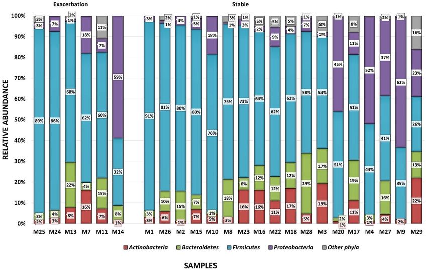
Figure 1. Bar plots showing the relative abundance of the differing phyla by disease state occurring in the
sputum microbiome of 24 COPD participants using targeted metagenomics across the different samples.
Firmicutes are shown in blue, Proteobacteria in purple, Bacteroidetes in green and Actinobacteria in red.
The graph is separated into the exacerbated state (n = 6) and stable state (n = 18). The specimens are ordered
according to the prevalence of Firmicutes.
of unclassified species ranged from 32 to 94% between samples. The most abundant phyla identified were Firmi-
cutes (ranging from 41 to 91%), Proteobacteria (ranging from 3 to 62%), Bacteroidetes (ranging from 3 to 22%)
and Actinobacteria (ranging from 1 to 22%) (Fig. 1).
The most abundant genera were Streptococcus (detected in all 24 samples, with abundances ranging from
19 to 82%), Haemophilus (detected in all 24 samples, with abundances ranging from 0.02% to 61%), Prevotella
(detected in all 24 samples, with abundances ranging from 0.1% to 22%), Veillonella (detected in all 24 samples,
Scientific Reports | (2021) 11:19758 | https://doi.org/10.1038/s41598-021-99127-w 4
Vol:.(1234567890)www.nature.com/scientificreports/
Figure 2. Graph of the DESeq2 analysis showing the log2fold differential abundance of the different genera
between the exacerbated state and stable state of disease (n = 24) in the sputum microbiome of COPD
participants. Differences were considered significant with the p-value (adjusted for false discovery rate using
Benjamini–Hochberg correction) cut-off of 0.2. Log2fold changes greater than zero indicated an increase in the
relevant genera, whereas log2fold changes less than zero indicated a decrease in the relevant genera. All genera
shown below the zero line had a decreased relative abundance with the stable state of disease i.e. these genera
were increased during the exacerbated state of disease. The error bars corresponding to the calculated lfcSE
(standard error).
with abundances ranging from 0.15% to 19%) and Granulicatella (detected in all 24 samples, with abundances
ranging from 0.12% to 11%).
Comparison of exacerbation and stable states of disease for the microbiome. The relative
abundance of the Actinobacteria, Bacteroidetes, Firmicutes, Fusobacteria and Proteobacteria phyla differed across
the disease states; with a higher abundance of Firmicutes (63% in the exacerbated state and 61% in the stable
state) and a lower abundance of Actinobacteria (5% in the exacerbated state and 5% in the stable state), Bacteroi-
detes (11% in the exacerbated state and 9% in the stable state) and Proteobacteria (19% in the exacerbated state
and 17% in the stable state), during the exacerbated state (Figure S1).
At a genus level (Figure S2), the exacerbated state showed changes in 75 genera; with 49 genera that had a
lower relative abundance and 26 genera that had a higher abundance. Key genera that showed lower relative
abundance during the exacerbated state included Porphyromonas (0.19% in the exacerbated state and 3.92% in the
stable state), Serratia (0.00% in the exacerbated state and 2.99% in the stable state), Staphylococcus (0.00% in the
exacerbated state and 1.02% in the stable state) and Streptococcus (47.88% in the exacerbated state and 49.61% in
the stable state). Genera that showed a higher relative abundance in the exacerbated state included Granulicatella
(5.30% in the exacerbated state and 3.06% in the stable state), Haemophilus (16.82% in the exacerbated state and
11.08% in the stable state), Prevotella (10.02% in the exacerbated state and 7.87% in the stable state) and Veil-
lonella (6.92% in the exacerbated state and 4.44% in the stable state). Although, the relative abundance differed
across the disease state, with DESeq2 analysis and ALDEx2 analysis no significant difference were observed
when a false discovery rate (FDR) of 0.05 was used. When an FDR of 0.2 was used, significant differences were
observed across the disease states (Fig. 2) for DESeq2 analysis but not for ALDEx2 analysis.
There was no significant difference in the alpha-diversity between disease states (Fig. 3) for the micro-
biome using the Wilcoxon sum rank test for both Chao1 (p-values = 0.58) and Simpson diversity measures
(p-value = 0.72). Beta-diversity measures showed no clustering for any of the variables using PCoA and weighted
UniFrac (for microbiome) measures (Fig. 4).
The sputum virome. A total of 3480 operational taxonomic units (OTUs) were identified across the six
samples for the virome. The taxonomic classification identified 16 phyla, 34 classes, 53 orders, 141 families and
826 genera. Most of the OTUs [95% (3306/3480)] could be classified up to a species level. The most abundant
Scientific Reports | (2021) 11:19758 | https://doi.org/10.1038/s41598-021-99127-w 5
Vol.:(0123456789)www.nature.com/scientificreports/
Figure 3. The alpha diversity box-plot of the sputum microbiome compared across the exacerbated state
(n = 6) and stable state (n = 18) of COPD using Chao1 and Simpson diversity measures. Each dot on the graph
represents a sample. The boxes represent the interquartile range (IQR) and the horizontal line represents
the median. The median values for Chao1 diversity measure were as follows: (i) stable state = 147.06 and (ii)
exacerbated state = 115.56. The median values for the Simpson diversity measures were as follows: (i) stable
state = 0.84 and (ii) exacerbated state = 0.86.
family across all samples was the Poxviridae family (detected in all six samples, with abundances ranging from 90
to 93%), followed by the bacteriophage families Myoviridae (detected in all six samples, with abundances 0.63%
to 2.11%) and Siphoviridae (detected in all six samples, with abundances 1.08% to 1.55%) (Fig. 5).
The most prevalent species was BeAn 58058, a member of the Poxviridae family that was detected in all
specimens sent for virome sequencing followed by bacteriophages (associated with both Gram-positive and
Gram-negative bacteria). Most of the viruses identified were dsDNA viruses (ranging from 97.23 to 98.15%).
Discussion
In this study, the composition of the sputum microbiome of COPD participants was investigated and was com-
pared between the different disease states i.e. stable state of disease and exacerbated state of disease. Two phyla
predominated, Firmicutes and Proteobacteria, with Streptococcus and Haemophilus being the most prevalent
genera. However, this study observed no significant differences between the exacerbated and stable states of
disease in COPD, in terms of, alpha diversity and beta diversity for the sputum microbiome in COPD. When an
FDR of 0.2 was used for DESeq2 analysis, significant differences were observed between the two disease states
for the relative abundance. With the virome, a high prevalence of the viruses, BeAn 58058 was observed. In this
study, there was difficulty in recruiting HIV-positive individuals with COPD and as a result, only a single HIV-
positive participant was recruited in this study.
In both disease states, four phyla dominated: Firmicutes (ranging from 41 to 91%), Proteobacteria (rang-
ing from 3 to 62%), Bacteroidetes (ranging from 3 to 22%) and Actinobacteria (ranging from 1 to 22%). This
is in agreement with other studies conducted on the lung microbiome in healthy individuals and other lung
diseases60,61. Even though some of these studies had different patient groups (e.g. asthmatics and smokers), used
different specimen types [e.g. bronchoalveolar lavage (BAL)] and used different sequencing technologies (e.g. 454
sequencing), these four phyla were always found to be dominant in the lung microbiome60–63. However, the most
prevalent phylum has been found to differ between different disease e.g. in severe COPD, Proteobacteria is more
prevalent whereas in the healthy lung Firmicutes is more prevalent60–65. This study showed a higher prevalence
Scientific Reports | (2021) 11:19758 | https://doi.org/10.1038/s41598-021-99127-w 6
Vol:.(1234567890)www.nature.com/scientificreports/
Figure 4. Principal component analysis (PCoA) plot derived using weighted UniFrac diversity measure
comparing the different disease states of COPD in the sputum microbiome The ellipses show the different states
of disease with the exacerbated state (n = 6) indicated in red and the stable state (n = 18) indicated in blue; with
the dots represent in each sample.
of Firmicutes; previous studies have shown that the microbiome in mild COPD is similar to that of the healthy
lung64,65. When the stable and exacerbated states of disease were compared in this study a higher abundance
of the Firmicutes phylum (2% higher in the exacerbated state) and lower abundances of the Proteobacteria (2%
higher in the exacerbated state), Actinobacteria (3% higher in the exacerbated state) and Bacteroidetes phyla (2%
higher in the exacerbated state) was observed. Previous studies (all conducted using sputum specimens) that
have compared the two diseases states in COPD have observed the same trend, where one of the phyla shows an
increased prevalence and the other phyla showed a decreased prevalence in the exacerbated state, however, none
of these studies reported the percentage increase66–71. In most of these studies, Proteobacteria increased, however,
two studies [Jubinville et al. (2018) and Wang et al. (2020)], showed an increased prevalence of Firmicutes (as
observed in this study). These studies had a variety of different sample sizes (ranging from nine participants to
281 participants), were conducted in USA, Europe and China and used different sequencing technologies (454
sequencing, MiSeq sequencing and PhyloChip)66–71.
The genera that showed the highest frequency in this study (in both disease states) were: Granulicatella
(Firmicutes), Haemophilus (Proteobacteria), Prevotella (Bacteroidetes), Streptococcus (Firmicutes) and Veillonella
(Firmicutes). Previous studies conducted on the lung microbiome of healthy individuals and COPD patients have
observed these genera in high abundances along with Pseudomonas and Porphyromonas.72,73. Most of these stud-
ies were conducted using 454 sequencing in the USA or Europe with a variety of different specimens. A study by
Wang et al. (2016) showed the most similarity to this study with one key difference; the changes in abundance of
genera during the exacerbated was different from this study. In this study, Haemophilus had a higher abundance
[5.7% higher in this study and 3% increase in Wang et al. (2016)] whereas Streptococcus had a lower abundance
[1.7% decrease in this study and 3% decrease in Wang et al. (2016)]. These genera i.e. Granulicatella, Haemophilus,
Prevotella and Veillonella showed significant increase in the relative abundance during the exacerbated state of
disease when DESeq2 analysis was used. The differences in abundances of the genera could be attributed to the
different study population size (87 individuals in the Wang et al. (2016) study vs 24 in this study), the type of
country (with United Kingdom (UK) being a developed country and South Africa a developing country) and the
difference in the sequencing methodology (MiSeq platform (Illumina, USA) and V1-V3 region for sequencing
was used in this study whereas Wang et al. (2016) used 454 sequencing (Roche Diagnostics, UK) and the V3-V5
region of 16S rRNA). Geographical location and local environmental conditions, such as air pollution have been
shown to affect the lung microbiome and could explain the difference in relative abundance between the two
Scientific Reports | (2021) 11:19758 | https://doi.org/10.1038/s41598-021-99127-w 7
Vol.:(0123456789)www.nature.com/scientificreports/
Figure 5. Bar plots showing the most abundant of viruses at a family level; the most prevalent families were as
follows: (i) Poxviridae (indicated in bright green), (ii) Siphoviridae (indicated in blue), (iii) Myoviridae (indicated
in olive green) and (iv) Herpesviridae (indicated in red). The rest of the viruses are grouped together as other
(indicated in pink).
studies13,74. Additionally, seasonal variation may play a role in bacteria identified75. Most of the exacerbation
samples in this study were collected in either autumn or winter. In Pretoria, the dry season is in winter which is
in contrast to the United Kingdom, where the dry season generally falls in summer. Additionally, the bacteria that
showed a higher prevalence (between 2 to 6% higher) during the exacerbated state of disease, i.e. Granulicatella,
Haemophilus, Prevotella and Veillonella have been associated with gastrointestinal reflux disease (GERD)76. As
a result of COPD patients having a common cough, GERD is associated with COPD and is considered a co-
morbidity77. In fact, GERD has been observed to be a predictor of exacerbations in COPD and implies that a
higher prevalence of these bacteria could be used as a potential indicator of COPD e xacerbations77,78.
In this study, bacterial alpha diversity and beta-diversity analysis showed no difference between disease states.
This observation is in agreement with previous COPD studies except for a study by Jubinville et al. (2018) who
observed a difference in alpha diversity when comparing paired samples i.e. the diversity in the paired samples
differed across the disease state with most exacerbated samples showing a higher diversity66–68,70. All these studies
were conducted in Europe (the UK and Spain) or Northern America (Canada and USA) using sputum specimens,
with most studies having less than 30 participants and having used 454 sequencing. The only difference between
these studies and the study by Jubinville et al. (2018) was the diversity measure used; most of the other studies
used the Shannon index (often combined with Chao1 and Faith PD diversity measure), whereas Jubinville et al.
(2018) used the Simpson index. Unlike, the Shannon index, the Simpson index is more affected by the relative
abundances (i.e. evenness) of the species in a sample; this suggests that during the exacerbated state of disease,
the abundances of species/OTUs changes but not the number of species/OTUs (richness)79.
In this study, the most prevalent viral family was Poxviridae followed by Siphoviridae and Myoviridae. When
compared to the only two other studies that have focused on the COPD lung virome, this study differed in the
relative abundance of the key families40,41,87. The study by Garcia-Nunez et al. (2018) used sputum specimens
(n = 10) from paired stable and exacerbated patients (n = 5) in Spain. The study by van Rijn et al. (2019) used
nasopharyngeal swabs (n = 88) collected from exacerbated patients between 2006 and 2010 and was conducted in
Norway. The most prevalent viral families in these studies were Anelloviridae (negative sense DNA virus with no
known pathogenicity in humans) and Siphoviridae (double-stranded DNA bacteriophages that have been found
in the lung virome of CF patients as well as in the gastrointestinal tract virome and the oral v irome40,41,80–85. These
bacteriophages i.e. Siphoviridae and Myoviridae may act as reservoirs for antibiotic resistance genes (contain
antibiotic resistance genes in their genomes), mobile genetic elements and may contain virulence genes and other
genes that affect bacterial metabolic pathways35,86.
Scientific Reports | (2021) 11:19758 | https://doi.org/10.1038/s41598-021-99127-w 8
Vol:.(1234567890)www.nature.com/scientificreports/
A high abundance of Poxviridae was observed in this study, particularly the BeAn 58,085 virus (BAV). Pox-
viridae is a family of complex, double-stranded DNA (dsDNA) viruses that are often zoonotic and are known to
cause skin lesion, with the most well-known virus being variola virus, the causative agent for smallpox (has been
eradicated)87. Only two other virome studies, one that studied fluid in the human body (conducted in Spain) and
one that studied ocular adnexa (conducted in Denmark on samples collected between 2005 and 2014) detected
the BeAn 58058 virus in h umans88,89. This virus (BeAn 58058) was originally isolated from rodents (Oryzomys
90
sp.) in Brazil in 1 963 . According to the viral-host database, the only known host for the BeAn 58058 virus is the
Oryzomys sp., however, other Poxviridae have been known to infect a wide variety of hosts including h umans54.
The BeAn 58085 virus is considered a variant of the Vaccinia virus, a close relative of the smallpox virus that was
used as a vaccine vector for smallpox until 197091,92. There are three possible explanations for the high abundance
of BeAn 58058 virus detected in this study. The first theory is that the BeAn 58058 virus is an ancient virus that
over time has incorporated as part of the human genome; the theory is supported by (i) A study by Mollerup
et al. (2019) conducted on the virome of the ocular adnexa, which showed that viral reads (i.e. the BeAn 58058
virus) identified had high sequence homology to sequences of human origin, (ii) A study that was conducted
on the human genome (studying structural variants) identified the BeAn 58058 virus as part of the genome and
iii) Poxviridae re dsDNA viruses and can easily integrate into the double-stranded human genome93. The second
theory is that BeAn 5808 is a DNA artefact of the smallpox vaccine (which was a live attenuated vaccine) received
years earlier; evidence supporting this theory includes the following: (i) the study population in this study were
all over the age of 50 years and would have received the smallpox vaccine before the vaccination programme
for the smallpox virus was terminated in South Africa (in 1970) and (ii) the Vaccinia virus, which was used
for the smallpox vaccine showed high homology with the BeAn 58058 v irus91,92,94. The third theory is that the
participants in this study encountered an environmental exposure from which the virus was contracted, e.g. rats
and its similarity to the cotia virus, which can infect human cells95. The fourth theory is that the BeAn 5808 is a
contaminant (i.e. a sequence not truly in the sample) from the extraction kit, from animal cells, reagents used
or even from a previous sequencing run96,97. Further analysis of the lung virome, as well as the human genome
of healthy individuals (i.e. not suffering from any lung disease) across different geographical regions and age
groups, should provide insight into this in the future.
This study had several limitations. First, this study had a small population size and did not have paired
samples for the different disease states. Second, a sputum specimen was chosen for this study (instead of BAL,
which has been used by most studies on the COPD microbiome) as it is the most patient-friendly method i.e. is
non-invasive98. The sputum microbiome has a mixture of the microbiomes from both the upper respiratory tract
and the lower respiratory t ract98–101. Additionally, sputum specimens have higher bacterial loads and are better
for longitudinal studies (as these specimens are non-invasive)99. Third, as only a single HIV participant could be
recruited into this study, no comparison between HIV positive individuals and HIV negative individuals could be
performed for the sputum microbiome in COPD patients; this aspect therefore requires further research. Lastly,
no controls were included in the study; the lack of negative controls for the extraction procedure (conducted in
a Biosafety level 2 cabinet with DNase away and RNase away) means that the laboratory contamination from
extraction reagents, from a previous sequencing run, etc. cannot be ruled out96,97,101. However, a strength of this
study was that it provided a good pilot overview of the sputum microbiome and the sputum virome of the COPD
lung in a South African setting. A diverse microbiome was observed in this study in both the stable and the
exacerbated states of disease; with Proteobacteria predominating in the exacerbated state of disease. Conversely,
the virome (studied both DNA and RNA viruses) was dominated by a single virus, the BeAn 58058 virus (a
dsDNA virus). Most viruses found previously found in respiratory tract were shown to be RNA viruses, such as
Influenza viruses, however, most shotgun metagenomics approaches favour DNA viruses, such as members of the
Siphoviridae. As result members of the Siphoviridae family and other DNA viruses, such as BeAn 58058 dominate
the lung virome. However, the origins of the BeAn 58085 virus and its possible clinical relevance is unknown.
Future studies into the virome would require further investigation into this virus by studying the lung virome in
healthy individuals and other lung diseases in the South African and international context. Future studies into
the COPD lung microbiome should include longitudinal studies that compared the stable and exacerbated states
of disease over several time points in the same individuals.
Conclusions
This study is among the first to report lung microbiome composition in COPD patients from Africa. No sta-
tistically significant differences in the microbiome of COPD patients during the different states of disease were
observed in this study. However, this study did note differences in the frequencies of key phyla and genera when
compared to other studies from Europe and the USA. However, the reason for this differing microbial profile is
unknown and warrants further research. In the virome, a high frequency of the BeAn 58058 virus was observed
in the six samples; the explanation for this observation is unclear. To conclude, the sputum microbiome in South
African COPD patients is diverse, regardless of the disease state, while the sputum virome warrants further
research.
Data availability
The sequencing data from this study is available in the NCBI Sequence Read Archive (SRA) database (https://
www.ncbi.nlm.nih.gov/sra) Bioproject PRJNA683885 (Accession numbers SAMN17041381 to SAMN17041404
and SAMN17065738 to SAMN17065743). The scripts used in R and in QIIME2 were added to a Github respira-
tory at https://github.com/tgmahomed/COPDMicrobiome.
Scientific Reports | (2021) 11:19758 | https://doi.org/10.1038/s41598-021-99127-w 9
Vol.:(0123456789)www.nature.com/scientificreports/
Received: 27 January 2021; Accepted: 14 September 2021
References
1. Terzikhan, N. et al. Prevalence and incidence of COPD in smokers and non-smokers: The Rotterdam Study. Eur. J. Epidemiol.
31(8), 785–792 (2016).
2. Owuor, N., Nalamala, N., Gimenes, J. A. Jr. & Sajjan, U. S. Rhinovirus and COPD airway epithelium. Pulm. Crit. Care Med. 2(3),
3 (2017).
3. Lopez-Campos, J. L., Tan, W. & Soriano, J. B. Global burden of COPD. Respirology 21(1), 14–23 (2016).
4. Vogelmeier, C. F. et al. Global strategy for the diagnosis, management, and prevention of chronic obstructive lung disease 2017
report: GOLD executive summary. Am. J. Respir. Crit. Care Med. 195(5), 557–582 (2017).
5. Lee, S. W., Kuan, C. S., Wu, L. S. & Weng, J. T. Metagenome and metatranscriptome profiling of moderate and severe COPD
sputum in Taiwanese Han males. PLoS ONE 11(7), e0159066 (2016).
6. Deslee, G. et al. Impact of current cough on health-related quality of life in patients with COPD. Int. J. Chron. Obstruct. Pulmon.
Dis. 11, 2091–2097 (2016).
7. Buist, A. S. et al. International variation in the prevalence of COPD (the BOLD study): A population-based prevalence study.
Lancet 370(9589), 741–750 (2007).
8. Salvi, S. The silent epidemic of COPD in Africa. Lancet Glob. Health 3(1), e6–e7 (2015).
9. Viviers, P. J. & Van Zyl-Smit, R. N. Chronic obstructive pulmonary disease – Diagnosis and classification of severity. S. Afr. Med.
J. 105(9), 9 (2015).
10. Abdool-Gaffar, M. S. et al. Management of chronic obstructive pulmonary disease-A position statement of the South African
Thoracic Society: 2019 update. J. Thorac. Dis. 11(11), 4408–4427 (2019).
11. Miravitlles, M. & Anzueto, A. Antibiotic prophylaxis in COPD: Why, when, and for whom?. Pulm. Pharmacol. Ther. 32, 119–123
(2015).
12. Pavord, I. D., Jones, P. W., Burgel, P. R. & Rabe, K. F. Exacerbations of COPD. Int. J. Chron. Obstruct. Pulmon. Dis. 11, 21–30
(2016).
13. Bouquet, J. et al. Microbial burden and viral exacerbations in a longitudinal multicenter COPD cohort. Respir. Res. 21(1), 77
(2020).
14. Global Initiative for Chronic Obstructive Lung Disease: Global Strategy for the Diagnosis, Management, and Prevention of Chronic
Obstructive Pulmonary Disease (2020 Report). (2020).
15. Aaron, S. D. Management and prevention of exacerbations of COPD. BMJ 349, g5237 (2014).
16. Doring, G., Parameswaran, I. G. & Murphy, T. F. Differential adaptation of microbial pathogens to airways of patients with cystic
fibrosis and chronic obstructive pulmonary disease. FEMS Microbiol. Rev. 35(1), 124–146 (2011).
17. D’Anna, S. E., Balbi, B., Cappello, F., Carone, M. & Di Stefano, A. Bacterial-viral load and the immune response in stable and
exacerbated COPD: Significance and therapeutic prospects. Int. J. Chron. Obstruct. Pulmon. Dis. 11, 445–453 (2016).
18. Clooney, A. G. et al. Comparing apples and oranges?: Next generation sequencing and its impact on microbiome analysis. PLoS
ONE 11(2), e0148028 (2016).
19. Kulski, J.K. Next-generation sequencing—An overview of the history, tools, and “omic” applications. in Next Generation Sequenc-
ing—Advances, Applications and Challenges (2016).
20. Park, S. T. & Kim, J. Trends in next-generation sequencing and a new era for whole genome sequencing. Int. Neurourol. J.
20(Suppl 2), S76-83 (2016).
21. Ji, B. & Nielsen, J. From next-generation sequencing to systematic modeling of the gut microbiome. Front. Genet. 6, 219 (2015).
22. Ito, T., Sekizuka, T., Kishi, N., Yamashita, A. & Kuroda, M. Conventional culture methods with commercially available media
unveil the presence of novel culturable bacteria. Gut Microbes 10(1), 77–91 (2019).
23. Helmy, M., Awad, M. & Mosa, K. A. Limited resources of genome sequencing in developing countries: Challenges and solutions.
Appl. Transl. Genom. 9, 15–19 (2016).
24. Thurber, R. V., Haynes, M., Breitbart, M., Wegley, L. & Rohwer, F. Laboratory procedures to generate viral metagenomes. Nat.
Protoc. 4(4), 470–483 (2009).
25. Wommack, K. E. et al. VIROME: A standard operating procedure for analysis of viral metagenome sequences. Stand. Genomic
Sci. 6(3), 427–439 (2012).
26. Kembel, S. W., Wu, M., Eisen, J. A. & Green, J. L. Incorporating 16S gene copy number information improves estimates of
microbial diversity and abundance. PLoS Comput. Biol. 8(10), e1002743 (2012).
27. Martin, C. et al. Host-microbe interactions in distal airways: Relevance to chronic airway diseases. Eur. Respir. Rev. 24(135),
78–91 (2015).
28. Hiergeist, A., Glasner, J., Reischl, U. & Gessner, A. Analyses of intestinal microbiota: Culture versus sequencing. ILAR J. 56(2),
228–240 (2015).
29. Woese, C. R. & Fox, G. E. Phylogenetic structure of the prokaryotic domain: The primary kingdoms. Proc. Natl. Acad. Sci. U S
A 74(11), 5088–5090 (1977).
30. Gürtler, V., Subrahmanyam, G., Shekar, M., Maiti, B., & Karunasagar, I. Chapter 12- Bacterial Typing and Identification By
Genomic Analysis of 16S–23S rRNA Intergenic Transcribed Spacer (ITS) Sequences. In Methods in Microbiology. (Eds. Goodfel-
low, M., Sutcliffe, I., Chun, J.). Vol. 41. 253–274. (Academic Press, 2014).
31. King, A.M., Adams, M.J., Carstens, E.B., & Lefkowitz, E.J. Virus taxonomy. in Ninth report of the International Committee on
Taxonomy of Viruses. 486–487. (2012).
32. Williams, S. C. The other microbiome. Proc. Natl. Acad. Sci. U S A 110(8), 2682–2684 (2013).
33. Wylie, K. M. The virome of the human respiratory tract. Clin. Chest Med. 38(1), 11–19 (2017).
34. Bragg, L. & Tyson, G. W. Metagenomics using next-generation sequencing. Methods Mol. Biol. 1096, 183–201 (2014).
35. Wylie, K. M., Weinstock, G. M. & Storch, G. A. Emerging view of the human virome. Transl. Res. 160(4), 283–290 (2012).
36. Amato, K. R. An introduction to microbiome analysis for human biology applications. Am. J. Hum. Biol. 29(1), 1 (2017).
37. Cabrera-Rubio, R. et al. Microbiome diversity in the bronchial tracts of patients with chronic obstructive pulmonary disease. J.
Clin. Microbiol. 50(11), 3562–3568 (2012).
38. Dickson, R. P., Martinez, F. J. & Huffnagle, G. B. The role of the microbiome in exacerbations of chronic lung diseases. Lancet
384(9944), 691–702 (2014).
39. Sze, M. A. et al. Host response to the lung microbiome in chronic obstructive pulmonary disease. Am. J. Respir. Crit. Care Med.
192(4), 438–445 (2015).
40. Garcia-Nunez, M. et al. The respiratory virome in chronic obstructive pulmonary disease. Future Virol. 13(7), 457–466 (2018).
41. van Rijn, A. L. et al. The respiratory virome and exacerbations in patients with chronic obstructive pulmonary disease. PLoS
ONE 14(10), e0223952 (2019).
42. Hamid, Q. et al. Methods of sputum processing for cell counts, immunocytochemistry and in situ hybridisation. Eur. Respir. J.
20(Supplement 37), 19S-23S (2002).
Scientific Reports | (2021) 11:19758 | https://doi.org/10.1038/s41598-021-99127-w 10
Vol:.(1234567890)www.nature.com/scientificreports/
43. Terranova, L. et al. How to process sputum samples and extract bacterial DNA for microbiota analysis. Int. J. Mol. Sci. 19(10),
3256–3568 (2018).
44. Stokell, J. R., Khan, A. & Steck, T. R. Mechanical homogenization increases bacterial homogeneity in sputum. J. Clin. Microbiol.
52(7), 2340–2345 (2014).
45. de la Cruz Pena, M. J. et al. Deciphering the human virome with single-virus genomics and metagenomics. Viruses 10(3), 3
(2018).
46. Allander, T. et al. Cloning of a human parvovirus by molecular screening of respiratory tract samples. Proc. Natl. Acad. Sci. U
S A 102(36), 12891–12896 (2005).
47. Bolyen, E. et al. Reproducible, interactive, scalable and extensible microbiome data science using QIIME 2. Nat. Biotechnol.
37(8), 852–857 (2019).
48. McDonald, D. et al. An improved Greengenes taxonomy with explicit ranks for ecological and evolutionary analyses of bacteria
and archaea. ISME J. 6(3), 610–618 (2012).
49. DeSantis, T. Z. et al. Greengenes, a chimera-checked 16S rRNA gene database and workbench compatible with ARB. Appl.
Environ. Microbiol. 72(7), 5069–5072 (2006).
50. Langmead, B. & Salzberg, S. L. Fast gapped-read alignment with Bowtie 2. Nat. Methods 9(4), 357–359 (2012).
51. Langmead, B., Trapnell, C., Pop, M. & Salzberg, S. L. Ultrafast and memory-efficient alignment of short DNA sequences to the
human genome. Genome Biol. 10(3), R25 (2009).
52. Wood, D. E., Lu, J. & Langmead, B. Improved metagenomic analysis with Kraken 2. Genome Biol. 20(1), 257 (2019).
53. Afgan, E. et al. The Galaxy platform for accessible, reproducible and collaborative biomedical analyses: 2018 update. Nucleic
Acids Res. 46(W1), W537–W544 (2018).
54. Mihara, T. et al. Linking virus genomes with host taxonomy. Viruses 8(3), 66 (2016).
55. Love, M. I., Huber, W. & Anders, S. Moderated estimation of fold change and dispersion for RNA-seq data with DESeq2. Genome
Biol. 15(12), 550 (2014).
56. Bisanz, J.E. qiime2R: Importing QIIME2 Artifacts and Associated Data into R Sessions. (2018).
57. McMurdie, P. J. & Holmes, S. phyloseq: An R package for reproducible interactive analysis and graphics of microbiome census
data. PLoS ONE 8(4), e61217 (2013).
58. Wickham, H. ggplot2: Elegant Graphics for Data Analysis. (Springer, 2016).
59. Gloor, G. B., Macklaim, J. M., Pawlowsky-Glahn, V. & Egozcue, J. J. Microbiome datasets are compositional: And this is not
optional. Front. Microbiol. 8, 2224 (2017).
60. Invernizzi, R., Lloyd, C. M. & Molyneaux, P. L. Respiratory microbiome and epithelial interactions shape immunity in the lungs.
Immunology 160(2), 171–182 (2020).
61. Fabbrizzi, A., Amedei, A., Lavorini, F., Renda, T. & Fontana, G. The lung microbiome: Clinical and therapeutic implications.
Intern. Emerg. Med. 14(8), 1241–1250 (2019).
62. Segal, L. N. et al. Enrichment of lung microbiome with supraglottic taxa is associated with increased pulmonary inflammation.
Microbiome 1(1), 19 (2013).
63. Hilty, M. et al. Disordered microbial communities in asthmatic airways. PLoS ONE 5(1), e8578 (2010).
64. Huffnagle, G. B., Dickson, R. P. & Lukacs, N. W. The respiratory tract microbiome and lung inflammation: A two-way street.
Mucosal Immunol. 10(2), 299–306 (2017).
65. Haldar, K. et al. The sputum microbiome is distinct between COPD and health, independent of smoking history. Respir. Res.
21(1), 183 (2020).
66. Jubinville, E. et al. Exacerbation induces a microbiota shift in sputa of COPD patients. PLoS ONE 13(3), e0194355 (2018).
67. Millares, L. et al. Functional metagenomics of the bronchial microbiome in COPD. PLoS ONE 10(12), e0144448 (2015).
68. Wang, Z. et al. Sputum microbiome temporal variability and dysbiosis in chronic obstructive pulmonary disease exacerbations:
An analysis of the COPDMAP study. Thorax 73(4), 331–338 (2018).
69. Wang, Z. et al. Lung microbiome dynamics in COPD exacerbations. Eur. Respir. J. 47(4), 1082–1092 (2016).
70. Huang, Y. J. et al. Airway microbiome dynamics in exacerbations of chronic obstructive pulmonary disease. J. Clin. Microbiol.
52(8), 2813–2823 (2014).
71. Wang, J., Chai, J., Sun, L., Zhao, J. & Chang, C. The sputum microbiome associated with different sub-types of AECOPD in a
Chinese cohort. BMC Infect. Dis. 20(1), 610 (2020).
72. Ubags, N. D. J. & Marsland, B. J. Mechanistic insight into the function of the microbiome in lung diseases. Eur. Respir. J. 50(3),
1602467–1602489 (2017).
73. Faner, R. et al. The microbiome in respiratory medicine: Current challenges and future perspectives. Eur. Respir. J. 49(4), 4 (2017).
74. Rylance, J. et al. Household air pollution and the lung microbiome of healthy adults in Malawi: A cross-sectional study. BMC
Microbiol. 16(1), 182 (2016).
75. Kumpitsch, C., Koskinen, K., Schopf, V. & Moissl-Eichinger, C. The microbiome of the upper respiratory tract in health and
disease. BMC Biol. 17(1), 87 (2019).
76. Park, C. H. et al. Treatment of non-erosive reflux disease and dynamics of the esophageal microbiome: A prospective multicenter
study. Sci. Rep. 10(1), 15154 (2020).
77. Lee, A. L. & Goldstein, R. S. Gastroesophageal reflux disease in COPD: Links and risks. Int. J. Chron. Obstruct. Pulmon. Dis. 10,
1935–1949 (2015).
78. Sanchez, J. et al. Laryngopharyngeal reflux in chronic obstructive pulmonary disease - A multi-centre study. Respir. Res. 21(1),
220 (2020).
79. Johnson, K. V. & Burnet, P. W. Microbiome: Should we diversify from diversity?. Gut Microbes 7(6), 455–458 (2016).
80. Mitchell, A. B., Oliver, B. G. & Glanville, A. R. Translational aspects of the human respiratory virome. Am. J. Respir. Crit. Care
Med. 194(12), 1458–1464 (2016).
81. Zarate, S., Taboada, B., Yocupicio-Monroy, M. & Arias, C. F. Human virome. Arch. Med. Res. 48(8), 701–716 (2017).
82. Freer, G. et al. The virome and its major component, anellovirus, a convoluted system molding human immune defenses and
possibly affecting the development of asthma and respiratory diseases in childhood. Front. Microbiol. 9, 686 (2018).
83. Simmonds, P. & Sharp, C. P. Anelloviridae. Clin. Virol. 2016, 701–711 (2016).
84. Fermin, G. Virion structure, genome organization, and taxonomy of viruses. Viruses 2018, 17–54 (2018).
85. Malathi, V. G. & Renuka Devi, P. ssDNA viruses: Key players in global virome. Virusdisease 30(1), 3–12 (2019).
86. Keen, E. C. & Dantas, G. Close encounters of three kinds: Bacteriophages, commensal bacteria, and host immunity. Trends
Microbiol. 26(11), 943–954 (2018).
87. Isaacs, S. N. & Buller, R. M. Poxviruses. Clin. Virol. 2016, 385–413 (2016).
88. Mollerup, S., Mikkelsen, L. H., Hansen, A. J. & Heegaard, S. High-throughput sequencing reveals no viral pathogens in eight
cases of ocular adnexal extranodal marginal zone B-cell lymphoma. Exp. Eye Res. 185, 107677 (2019).
89. Blanco-Picazo, P. et al. Unravelling the consequences of the bacteriophages in human samples. Sci. Rep. 10(1), 6737 (2020).
90. Wanzeller, A. L. et al. Complete genome sequence of the BeAn 58058 virus isolated from Oryzomys sp. rodents in the Amazon
Region of Brazil. Genome Announc 5(9), 9 (2017).
91. Marques, J. T. et al. Characterization of ATI, TK and IFN-alpha/betaR genes in the genome of the BeAn 58058 virus, a naturally
attenuated wild Orthopoxvirus. Virus Genes 23(3), 291–301 (2001).
Scientific Reports | (2021) 11:19758 | https://doi.org/10.1038/s41598-021-99127-w 11
Vol.:(0123456789)www.nature.com/scientificreports/
92. Silva, D. C. M., Moreira-Silva, EAd. S., Gomes, Jd. A. S., Fonseca, FGd. & Correa-Oliveira, R. Clinical signs, diagnosis, and case
reports of Vaccinia virus infections. Braz. J. Infect. Dis. 14, 129–134 (2010).
93. Oliveira, G. P., Rodrigues, R. A. L., Lima, M. T., Drumond, B. P. & Abrahao, J. S. Poxvirus host range genes and virus-HOST
SPECTRum: A critical review. Viruses 9(11), 11 (2017).
94. Abrahao, J. S. et al. Long-lasting stability of Vaccinia virus strains in murine feces: Implications for virus circulation and envi-
ronmental maintenance. Arch. Virol. 154(9), 1551–1553 (2009).
95. Haller, S. L., Peng, C., McFadden, G. & Rothenburg, S. Poxviruses and the evolution of host range and virulence. Infect. Genet.
Evol. 21, 15–40 (2014).
96. Davis, N. M., Proctor, D. M., Holmes, S. P., Relman, D. A. & Callahan, B. J. Simple statistical identification and removal of
contaminant sequences in marker-gene and metagenomics data. Microbiome 6(1), 226 (2018).
97. Marti, J. M. Recentrifuge: Robust comparative analysis and contamination removal for metagenomics. PLoS Comput. Biol. 15(4),
e1006967 (2019).
98. Ditz, B. et al. Sputum microbiome profiling in COPD: Beyond singular pathogen detection. Thorax 75(4), 338–344 (2020).
99. Carney, S. M. et al. Methods in lung microbiome research. Am. J. Respir. Cell Mol. Biol. 62(3), 283–299 (2020).
100. Sulaiman, I., Schuster, S. & Segal, L. N. Perspectives in lung microbiome research. Curr. Opin. Microbiol. 56, 24–29 (2020).
101. Salter, S. J. et al. Reagent and laboratory contamination can critically impact sequence-based microbiome analyses. BMC Biol.
12, 87 (2014).
Acknowledgements
The authors would like to thank the late Prof Anton Stoltz for his assistance with patient identification and with
the study design. The authors would also like to thank all staff at the hospitals and the participants.
Author contributions
T.G.M. was involved in all aspects of the study including study design, patient recruitment, data analysis and
writing of the manuscript; R.P.H.P. and M.M.E. were involved in the study design and outline and edited the
manuscript; M.M.K. edited the manuscript. M.A., A.I. and S.M. were involved in the analysis of the virome data
and edited the manuscript. A.G.M. and V.U. helped with the study design, patient recruitment and edited the
manuscript.
Funding
National Health Laboratory Service of South Africa (NHLS) Research Trust (Grant number: GRANT004 94626).
Competing interests
The authors declare no competing interests.
Additional information
Supplementary Information The online version contains supplementary material available at https://doi.org/
10.1038/s41598-021-99127-w.
Correspondence and requests for materials should be addressed to M.M.E.
Reprints and permissions information is available at www.nature.com/reprints.
Publisher’s note Springer Nature remains neutral with regard to jurisdictional claims in published maps and
institutional affiliations.
Open Access This article is licensed under a Creative Commons Attribution 4.0 International
License, which permits use, sharing, adaptation, distribution and reproduction in any medium or
format, as long as you give appropriate credit to the original author(s) and the source, provide a link to the
Creative Commons licence, and indicate if changes were made. The images or other third party material in this
article are included in the article’s Creative Commons licence, unless indicated otherwise in a credit line to the
material. If material is not included in the article’s Creative Commons licence and your intended use is not
permitted by statutory regulation or exceeds the permitted use, you will need to obtain permission directly from
the copyright holder. To view a copy of this licence, visit http://creativecommons.org/licenses/by/4.0/.
© The Author(s) 2021
Scientific Reports | (2021) 11:19758 | https://doi.org/10.1038/s41598-021-99127-w 12
Vol:.(1234567890)You can also read