Long term follow-up of pediatric-onset Evans syndrome: broad immunopathological manifestations and high treatment burden
←
→
Page content transcription
If your browser does not render page correctly, please read the page content below
Complications in Hematology                                                           SUPPLEMENTARY APPENDIX
Long term follow-up of pediatric-onset Evans syndrome: broad
immunopathological manifestations and high treatment burden
Thomas Pincez,1,2 Helder Fernandes,1,3 Thierry Leblanc,4 Gérard Michel,5 Vincent Barlogis,5 Yves Bertrand,6 Bénédicte
Neven,7,8,9 Wadih Abou Chahla,10 Marlène Pasquet,11 Corinne Guitton,12 Aude Marie-Cardine,13 Isabelle Pellier,14 Corinne
Armari-Alla,15 Joy Benadiba,16 Pascale Blouin,17 Eric Jeziorski,18 Frédéric Millot,19 Catherine Paillard,20 Caroline
Thomas,21 Nathalie Cheikh,22 Sophie Bayart,23 Fanny Fouyssac,24 Christophe Piguet,25 Marianna Deparis,26 Claire Brian-
det,27 Eric Doré,28 Capucine Picard,9,29 Frédéric Rieux-Laucat,8,9 Judith Landman-Parker,30 Guy Leverger30 and Nathalie
Aladjidi1,3 on the behalf of members of the French Reference Center for Pediatric Autoimmune Cytopenia (CEREVANCE)
and of collaborators from the French Reference Center for Adult Autoimmune Cytopenia (CERECAI).
1
 Centre de Référence National des Cytopénies Auto-immunes de l’Enfant (CEREVANCE), Bordeaux, France; 2Division of Pediatric Hema-
tology-Oncology, Charles-Bruneau Cancer Center, Department of Pediatrics, Sainte-Justine University Hospital, Université de Montréal,
Montréal, Québec, Canada; 3Pediatric Oncology Hematology Unit, University Hospital, Plurithématique CIC (CICP), Centre d’Investiga-
tion Clinique (CIC) 1401, INSERM, Bordeaux, France; 4Pediatric Hematology Unit, Robert Debré University Hospital, AP-HP, Paris,
France; 5Department of Pediatric Hematology, La Timone Hospital, Marseille University Hospital, Marseille, France; 6Institute of Pedi-
atric Hematology and Oncology, Lyon University Hospital, Lyon, France; 7Pediatric Immuno-Hematology and Rheumatology Department,
Necker-Enfants Malades University Hospital, AP-HP, Paris, France; 8Laboratory of Immunogenetics of Pediatric Autoimmune Diseases,
Paris, France; 9Imagine Institute, UMR 1163 INSERM, University of Paris, Paris, France; 10Department of Pediatric Hematology, Jeanne
de Flandre Hospital, Lille University Hospital, Lille, France; 11Pediatric Oncology Immunology Hematology Unit, Children’s University
Hospital, Toulouse, France; 12Department of Pediatrics, Bicêtre University Hospital, AP-HP, Le Kremlin-Bicêtre, France; 13Department of
Pediatric Hematology and Oncology, Rouen University Hospital, Rouen, France; 14Pediatric Unit, Angers University Hospital, Angers,
France; 15Pediatric Oncology Hematology Unit, Grenoble University Hospital, Grenoble, France; 16Department of Hemato-Oncology Pedi-
atric, Nice University Hospital, Nice, France; 17Department of Pediatric Hematology-Oncology, Clocheville Hospital, Tours University Hos-
pital, Tours, France; 18Pediatric Oncology Hematology Unit, Arnaud de Villeneuve University Hospital, Montpellier, France; 19Department
of Pediatric Hematology, Poitiers University Hospital, Poitiers, France; 20Department of Pediatric Hematology and Oncology, Hautepierre
University Hospital, Strasbourg, France; 21Pediatric Hematology Unit, Nantes University Hospital, Nantes, France; 22Department of Pedi-
atric Hematology-Oncology, Besançon University Hospital, Besançon, France; 23Pediatric Hematology Unit, Rennes University Hospital,
Rennes, France; 24Pediatric Hematology Unit, Nancy University Hospital, Nancy, France; 25Pediatric Oncology Hematology Unit, Limoges
University Hospital, Limoges, France; 26Pediatric Oncology-Hematology Unit, Caen University Hospital, Caen, France; 27Department of
Pediatrics, Dijon University Hospital, Dijon, France; 28Pediatric Unit, Clermont-Ferrand University Hospital, Clermont-Ferrand, France;
29
   Study Center for Primary Immunodeficiencies, Necker-Enfants Malades University Hospital, AP-HP, Paris, France and 30Pediatric On-
cology Immunology Hematology Unit, Armand-Trousseau University Hospital, AP-HP, Paris, France

©2022 Ferrata Storti Foundation. This is an open-access paper. doi:10.3324/haematol.2020.271106

Received: August 31, 2020.
Accepted: December 22, 2020.
Pre-published: January 14, 2021.
Correspondence: NATHALIE ALADJIDI - nathalie.aladjidi@chu-bordeaux.fr
Long term follow-up of pediatric-onset Evans syndrome: broad immunopathological
                               manifestations and high treatment burden

Supplemental data                                                                          Page
Supplemental Table 1. Patient selection and definitions                                        2
Supplemental Table 2. Immunopathological and other manifestations                              3
Supplemental Table 3. Second-line treatments received                                          6
Supplemental Table 4. Severe or recurrent infections                                           7
Supplemental Table 5. Characteristics of patients who died                                     8
Supplemental Figure 1. Selection of patients within the OBS’CEREVANCE database                 9
Supplemental Figure 2. Clinical immunopathological manifestations                              10
Supplemental Figure 3. Second-line treatments                                                  11
Appendix                                                                                       12

                                                                                           1
Supplemental Table 1. Patient selection and definitions
Inclusion criteria
Evans syndrome (ES) is defined as the simultaneous (less than 1 month) or sequential
association of immune thrombocytopenic purpura (ITP) and autoimmune hemolytic
anemia (AIHA).
Patients are included in the cohort if they are less than 18 years old at first cytopenia
diagnosis, regardless of the age at inclusion. Follow-up is calculated from first cytopenia
diagnosis.
ITP, AIHA and ES definitions
ITP is defined according to the international working group criteria (Rodeghiero et al., Blood
2009).
AIHA is defined as Hb < 110 g/L with a positive direct antiglobulin test (DAT) and at least
one of the following hemolysis criteria: reticulocyte count > 120 G/L, free bilirubin > 17
mmol/L, or haptoglobin < 10 mg/dL.
Autoimmune neutropenia (AIN) is defined as peripheral neutropenia < 1 G/L persisting for
more than 6 months that is neither infection nor drug driven, regardless of the presence or
absence of neutrophil autoantibodies.
Patients with AIHA and autoimmune neutropenia (AIN) or with ITP and AIN are not
classified as ES in the present analysis; patients with isolated DAT or anti-platelet
antibodies, or compensated hemolysis without AIHA were only considered as having ES
when cytopenia was present.
Exclusion criteria
Inherited red cell or platelet disease.
Autoimmune cytopenia secondary to human immunodeficiency virus infection.
Chemotherapy before ES.
Bone marrow or organ transplantation before ES.
Known PIDs before ES onset.
Systemic lupus erythematosus known before ES onset.
Patients living outside metropolitan France.
Immune thrombocytopenic purpura and autoimmune hemolytic anemia status
ITP and AIHA status were separately classified as follows: no remission (NR) = platelet
count < 30 G/L or Hb < 70 g/L; partial remission (PR) = platelet count 30–100 G/L or Hb
70–110 g/L, with reticulocytes > 120 G/L; complete remission (CR) = platelet count > 100
G/L or Hb ≥ 110 g/L, with reticulocytes ≤ 120 G/L.
Cytopenia flare was defined as change of status from CR or PR to NR, whether the CR or
PR has been reached spontaneously or after a first-line treatment.
Immunopathological manifestations
Lymphoproliferation: significant splenomegaly without hemolysis or significant persistent
lymphadenopathy (> 1 cm), eventually requiring a biopsy.
Clinical IMs: lymphoproliferation or autoimmune / autoinflammatory organ disease :
pulmonary (granulomatous-lymphocytic interstitial lung disease), liver and digestive tract
2problems (lymphoïd enteropathy, coeliac-like disease, inflammatory bowel disease,
chronic gastritis, autoimmune or giant cell hepatitis, cirrhosis), neurologic (autoimmune or
autoinflammatory or granulomatous or vascular manifestations), endocrinologic
(autoimmune thyroiditis, Grave’s disease or isolated specific autoantibodies), dermatologic
(psoriasis, vitiligo, alopecia, vasculitis) and other autoimmune or autoinflammatory
manifestations (ophthalmologic, cardiac, renal or hematological).
Biological IMs*: hypogammaglobulinemia, systemic lupus erythematosus (SLE)
biomarkers, or autoimmune lymphoproliferative syndrome (ALPS) first line biomarkers.
Hypogammaglobulinemia: Immunoglobulin (Ig)G levels below the mean for the subject’s
age (at least 2 SD) on two separate samples, associated or not with IgA levels below the
mean for the subject’s age (at least 2 SD) or with defects in vaccine immunizations.
SLE biomarkers*: antinuclear antibodies titer > 1/160; anti double-stranded DNA or
Smith, hypocomplementemia, and lupus anticoagulant (all in two separate samples).
ALPS biomarkers*: persistent hypergammaglobulinemia (at least 2 SD over the mean for
age), or high counts of circulating TCRαβ CD4- CD8- double negative T lymphocytes.
Severe or recurrent infections
Bacteremia, pneumonia, bronchiectasis, recurrent pyogenes infections, severe herpes virus
infections, or opportunistic infections.
Second-line treatments
All immunomodulatory or immunosuppressive treatments (including splenectomy), except
steroids or intravenous immunoglobulins (considered as first-line treatments).
Loss to follow-up
A patient is classified “lost to follow-up” if no data are available for more than 2 years and
no future appointments are scheduled.
* The biological workup is made at the clinician’s discretion, which is annually in most cases.
Supplemental Table 2. Immunopathological and other manifestations
Median age (years)
Number (%)
at onset (min–max)
Clinical IMs
Lymphoproliferation 71 (47) 7.8 (0–41)
Superficial (palpable) adenopathies 61 (40.4) 8 (0–41)
Deep (abdominal or thoracic) adenopathies 16 (10.6) 10.9 (2.7–19.1)
Splenomegaly 49 (32.5) 7.6 (0–19)
Associated hepatomegaly 17 (11.3) 5.0 (0.3–19)
Pneumological 16 (10.6) 14.3 (4.6–27.7)
Granulomatous–lymphocytic interstitial lung disease 16 (10.6) 14.3 (4.6–27.7)
Gastrointestinal/hepatic 23 (15.2) 13 (0.7–25.9)
Coeliac disease 3 (2) 1 (0.7–14.9)
3Autoimmune hepatitis 7 (4.6) 11.3 (1.6–14.7)
Lymphoid enteropathy 5 (3.3) 16.4 (6–25.9)
Chronic gastritis 5 (3.3) 16.5 (6.5–19.4)
Inflammatory bowel disease 2 (1.3) 14.4 (9.8–18.9)
Rosai–Dorfman 1 (0.7) 7.3
Primary sclerosing cholangitis 1 (0.7) 13 (13–13)
Neurological 13 (8.6) 16.3 (7.8–27.9)
Vasculitis 3 (2) 13.1 (12.1–17.8)
Lymphoproliferation 1 (0.7) 7.8
Myelitis 2 (1.3) 26.7 (25.4–27.9)
Infratentorial inflammatory lesions 2 (1.3) 18.2 (11.9–24.6)
Subtentorial inflammatory lesions 7 (4.6) 14.2 (7.8–27.9)
Sensitive ganglionopathy 1 (0.7) 16.3
Endocrinological 5 (3.3) 12.2 (3–41.1)
Type 1 diabetes 2 (1.3) 3.2 (3–3.3)
Grave's hyperthyroidism 2 (1.3) 29.1 (17.1–41.1)
Hashimoto thyroiditis 1 (0.7) 12.2
Dermatological 26 (17.2) 15.5 (0–27.3)
Vitiligo 4 (2.6) 3 (0–15.5)
Chronic urticaria 2 (1.3) 8.2 (0.8–15.6)
Psoriasis 3 (2) 21.1 (15.6–27.3)
Bullous pemphigoid 1 (0.7) 15.5
Eczema 4 (2.6) 10.9 (0–17.2)
Cutaneous lupus erythematosus involvement 8 (5.3) 17.2 (12.1–21.5)
Granuloma 2 (1.3) 7.8 (0–15.6)
Immunoglobulin A vasculitis 2 (1.3) 11.8 (5.7–17.9)
Sjögren's syndrome 1 (0.7) 15.5
Livedo reticularis 1 (0.7) 17.6
Rheumatological 12 (7.9) 15.1 (2.6–41.1)
Monoarthritis 3 (2) 13.7 (2.6–15.4)
Oligoarthritis 2 (1.3) 16.6 (16.3–16.9)
Polyarthritis 5 (3.3) 14.4 (5.9–41.1)
Polyarticular pain without arthritis 3 (2) 15 (13.7–19.3)
Cardiological 4 (2.6) 15.9 (12.7–20.9)
Pericarditis 3 (2) 15.2 (12.7–20.9)
Granulomatous aortitis 1 (0.7) 16.6
Ophthalmological 8 (5.3) 9.6 (6.1–15.6)
Keratitis 4 (2.6) 8.7 (6.1–9.7)
Uveitis 4 (2.6) 12 (7.9–15.6)
Ischemic optic neuropathy 1 (0.7) 12.1
4Nephrological 3 (2) 14.2 (10.6–16.4)
Tubulopathy 1 (0.7) 10.6
Granulomatous infiltration 2 (1.3) 15.3 (14.2–16.4)
Hematological, other 2 (1.3) 6.9 (2.9–10.9)
Myelofibrosis 1 (0.7) 10.9
Acquired thrombotic thrombocytopenic purpura 1 (0.7) 2.9
Biological IMs
Hypogammaglobulinemia 54 (35.8) 10 (2.3–23)
Low IgG before anti-CD20 44 (29.1) 10.5 (2.3–22.4)
Low IgG persisting after anti-CD20 8 (5.3) 9.5 (5–23)
Low IgA 5 (3.3) 11 (6.5–15.8)
Immunoglobulin replacement therapy 24 (15.9) 9.4 (2.3–22.4)
SLE biomarkers 42 (27.8) 10.2 (0.3–19.5)
Antinuclear antibodies 38 (25.2) 9.8 (0.3–19.5)
Anti double-stranded DNA or anti Smith 6 (4) 14.7 (6–19.5)
Hypocomplementemia 10 (6.6) 12.6 (0.3–19.5)
Lupus anticoagulant 6 (4) 10.6 (0.8–16)
ALPS biomarkers 24 (15.9) 8.4 (2.8–26.7)
Hypergammaglobulinemia 14 (9.3) 7.8 (2.8–26.7)
Elevated Fas ligand 5 (3.3) 8 (4–10.8)
Defective Fas-mediated apoptosis 3 (2) 9.6 (4–13.5)
Increase IL-10 and/or vitamin B12 2 (1.3) 13.8 (13.5–14)
Elevated alpha beta double-negative T cells 10 (6.6) 9.2 (5.2–15)
Other manifestations
Granuloma on pathology, any localization 9 (6) 16.57 (0–26.2)
Lymph node 3 (2) 14.6 (2.7–17.8)
Lung 3 (2) 10.1 (7.4–26.2)
Cutaneous 2 (1.3) 7.8 (0–15.6)
Another organ 4 (2.6) 15.4 (2.7–18)
Malignancies 4 (2.6) 20.5 (16.3–28.8)
Angioimmunoblastic T-cell lymphoma 1 (0.7) 28.8
Juvenile myelomonocytic leukemia 1 (0.7) 20
EBV-negative Hodgkin Lymphoma IVBb 1 (0.7) 16.3
Large granular lymphocytic leukemia 1 (0.7) 21
A given patient may have several manifestations within the same category.
Abbreviations: IMs, immunopathological manifestations; EBV, Epstein–Barr Virus; Ig, immunoglobulin;
SLE, systemic lupus erythematosus; ALPS, autoimmune lymphoproliferative syndrome; IL, interleukin.
5Supplemental Table 3. Second-line treatments received
At treatment start (years)
Number
Percentage Median time after
Second-line treatment of
of patients first cytopenia (min– Median age (min–max)
patients
max)
Rituximab 79 52.3 5.6 (0.0–35.5) 14.1 (0.8–47.4)
Azathioprine 55 36.4 3.8 (0.0–27.8) 12.4 (0.5–39.8)
Splenectomy 36 23.8 6.7 (0.1–16.0) 11.1 (1.5–19.8)
Mycophenolate 29 19.2 6.1 (0.1–29.1) 11.5 (2.4–14.7)
Ciclosporin 28 18.5 4.0 (0.2–15.0) 10.8 (0.8–19.8)
Hydroxychloroquine 27 17.9 5.9 (0.0–17.5) 15.8 (5.6–39.8)
Mercaptopurine 12 7.9 4.8 (0.2–11.9) 13.9 (2.8–18.3)
Romiplostim 12 7.9 14.7 (0.5–38.1) 19.4 (5.3–50.0)
Vinblastine 11 7.3 5.5 (0.3–24.3) 13.8 (1.8–36.5)
Cyclophosphamide 10 6.6 3.2 (0.2–18.5) 11.6 (1.8–23.8)
Colchicine 8 5.3 4.3 (0.9–20.9) 15.3 (9.3–30.6)
Sirolimus 8 5.3 6.7 (3.2–11.4) 11.4 (8.0–19.8)
Vincristine 7 4.6 7.9 (0.1–16.6) 12.3 (0.5–20.8)
Eltrombopag 6 4.0 14.7 (6.1–35.4) 22.6 (9.0–47.3)
Abatacept 5 3.3 12.5 (7.5–20.5) 20.0 (9.7–26.5)
Dapsone 4 2.6 6.7 (1.6–11.2) 11.9 (4.3–19.0)
Anti-D immunoglobulin 3 2.0 0.2 (0.0–7.1) 2.9 (2.4–14.7)
Allogenic hematopoietic
3 2.0 8.7 (1.2–14.2) 11.5 (2.5–18.2)
stem-cell transplantation
Danazol 2 1.3 6.7 (2.9–10.5) 12.7 (5.7–19.7)
Everolimus 1 0.7 11.7 14.3
For treatment start assessments only, each separate course has been included for a given patient.
6Supplemental Table 4. Severe or recurrent infections
Infection type Number (%) Infection type Number (%)
Oral or genital herpes
Herpes zoster 17 (11.3) 4 (2.6)
simplex infection
Sinusitis/otitis media 15 (9.9) Aphthous stomatitis 4 (2.6)
Pneumopathy 12 (8.0) Cellulitis 3 (2.0)
Nontuberculous
Bronchopathy 11 (7.3) 2 (1.3)
mycobacteria
Bacteremia* 8 (5.3) Meningitis*** 2 (1.3)
Impetigo/furuncle/cutaneous Chronic blood EBV
7 (4.6) 2 (1.3)
abscesses replication
Other 7 (4.6) Cutaneous warts 2 (1.3)
Gastrointestinal infections** 5 (3.1) Molluscum contagiosum 2 (1.3)
Other (once each): cerebral toxoplasmosis, pityriasis versicolor, oral abscess, anogenital warts, bacterial
arthritis, CMV disseminated infection, parotitis.
* Microorganisms identified: Staphylococcus aureus, Pasteurella, Streptococcus pneumococcus,
Pseudomonas aeruginosa
** Microorganisms identified: Campylobacter, Salmonella
*** Microorganisms identified: HSV, Streptococcus pneumococcus
The onset times of recurrent infections could not be determined. Therefore, age data were not collected.
Abbreviations: HSV, herpes simplex virus; EBV, Epstein–Barr virus; CMV, cytomegalovirus.
7Supplemental Table 5. Characteristics of patients who died
Second-line treatments
Delay
Year Age at
after first
Sex of death IM Nb Sequence Cause of death
cytopenia
death (years)
(years)
M 2006 0.1 1.7 No 1 VBL Cerebral hemorrhage
CsA, RTX, SPX, AZT, VCR, CPX,
F 2002 1.8 3.8 Yes 7 Sepsis
HSCT
F 2018 0.9 3.9 Yes 1 CD20 Fulminant hepatitis
M 2001 1.6 5.0 Yes 3 CsA, AZT, CD20 Sepsis
M 1996 3.0 8.3 Yes 4 AZA, VCR, DAP, SPX Cerebral hemorrhage
F 2017 5.2 10.7 Yes 1 AZT HLH and cerebral vasculitis
M 2001 1.3 11.6 Yes 2 CD20, CsA Pancreatitis
M 2010 10.3 11.9 Yes 3 CsA, AZT, CD20 Gastrointestinal hemorrhage
M 1995 8.1 12.7 Yes 3 CsA, SPX, VBL Cerebral hemorrhage
M 1995 8.8 15.5 Yes 2 SPX, CsA Sepsis
F 2007 12.9 17.8 Yes 1 CD20 Sepsis
M 2010 16.4 18.2 Yes 4 AZT, SPX, MMF, CD20 Sepsis
F 2014 8.9 18.4 Yes 4 AZT, CD20, CsA, 6MP EBV-related lymphoproliferation
SPX, CPX, HCQ, AZT, CD20,
F 2002 14.6 18.5 Yes 7 Post-HSCT CMV infection
CsA, HSCT
F 2016 12.4 19.3 Yes 4 CD20, AZT, CPX, MMF Fulminant pneumococcal infection
F 2017 6.0 19.4 Yes 0 Unknown
M 2013 15.1 20.0 Yes 3 CD20, SPX, 6MP JMML
F 2013 5.1 21.1 Yes 4 CD20, HCQ, SPX, CPX Vasculitis, SLE
F 2012 12.9 24.5 Yes 3 SPX, CD20, AZT Probably sepsis
M 2011 24.2 28.1 Yes 5 SPX, CsA, AZT, HCQ, MMF Sepsis
M 2017 24.3 28.9 Yes 1 DAP Probably sepsis
MMF, CsA, HCQ, CD20, SPX,
M 2015 18.3 31.5 Yes 6 Probably sepsis
TPO-RA
Patients are ordered by age at death. Among the 22 patients with available data, 16 were being followed by pediatric teams, and six
patients (aged 20–31.5 years) were being followed by adult teams.
Abbreviations: IM, immunopathological manifestation; Nb, number; DAP, dapsone; AZT, azathioprine; CD20, rituximab; CPX,
cyclophosphamide; MMF, mycophenolate; CsA, ciclosporine; HCQ, hydroxychloroquine; TPO-RA, thrombopoietin receptor agonist;
VBL, vinblastine; SLE, systemic erythematosus lupus; SPX, splenectomy; HSCT, hematopoietic stem-cell transplantation; HLH,
hemophagocytic lymphohistiocytosis; EBV, Epstein–Barr virus; CMV, cytomegalovirus. JMML: Juvenile myelomonocytic leukemia
8Total number of patients included in OBS’CEREVANCE cohort
n = 1,505
Evans syndrome Isolated AIHA Isolated cITP
n = 216 n = 444 n = 845
Known secondary Evans
syndrome at inclusion
n = 6*
Secondary cytopenia
Isolated AIHA n = 23
Isolated cITP n = 15
Less than 5 years of follow-up
from first cytopenia
n = 59
Patients included
n = 151
Patients included as comparators in the survival analysis
Patients included in the survival analysis Isolated AIHA n = 421
n = 210 Isolated cITP n = 830
*post allogeneic hematopoietic stem cell transplantation (n = 3), autoimmune lymphoproliferative syndrome (n = 2), 22q11 microdeletion (n = 1).
Supplemental Figure 1. Selection of patients within the OBS’CEREVANCE database
The numbers shown are those recorded on the day of data extraction (21 June 2019). Among the
151 patients with Evans syndrome with more than 5 years of follow-up, 115 were alive and
actively followed by a pediatric hematologist (n = 73), an adult internist (n = 37), an adult
hematologist (n = 4), or an adult gastroenterologist (n = 1).
Abbreviations: AIHA, autoimmune hemolytic anemia; cITP, chronic immune thrombocytopenic
purpura.
9A B 60
Percentage of patients
51 53 50
(34%) (35%)
40 40
Number of patients
25
30 (17%) 30
14
20 (9%) 20
4
10 (3%) 2 2 10
(1%) (1%)
0 0
0 1 2 3 4 5 6 0 5 10 15 20 25 30
Number of cIM Age (years)
Number at risk
Lymphoproliferation 131 100 66 37 16 7
Dermatological 147 133 101 52 19 8
Pneumological 151 134 103 58 23 8
Gastrointestinal/Hepatic 149 132 97 55 23 9
Neurological 151 137 106 60 25 9
Rheumatological 151 136 106 58 24 9
Ophthalmological 151 137 108 63 27 10
Cardiological 151 136 106 62 26 10
Endocrinological 150 137 110 62 26 10
Nephrological 151 139 110 64 27 10
Hematological, other 150 138 110 64 27 10
Supplemental Figure 2. Clinical immunopathological manifestations (cIMs)
(A) Number of cIMs across the cohort. (B) Cumulative incidence of each cIM and numbers at
risk.
10A B
40 34 35
(23%) (23%)
Number of patients
30 26
(17%)
18
16
20 (12%)
(11%)
9
10 (6%) 6 5
(4%) (3%) 2
(1%)
0
0 1 2 3 4 5 6 7 8 9
Number of treatments
C
100 All treatments
Second-line only
Percentage of patients
80
60
40
20
0
0 3 6 9 12
Time after complete remission
Number at risk
All treatments 19 8 5 3
Second-line only 28 18 11 6
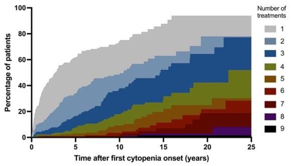
Supplemental Figure 3. Second-line treatments
(A) Number and percentage of second-line treatments in the 151 patients with Evans syndrome
(ES). Among these patients, 34 (23%) had none, 35 (23%) had one, and 82 (54%) had two or
more second-line treatments. The median number of treatments received (2 [0–9]) was higher for
ES than for cITP (1 [0–14]; p < 0.0001) but not AIHA (2 [0–7]; p = 0.7) alone. (B) Cumulative
number of second-line treatments after first cytopenia onset. Half of the patients had received at
least one, two, and three different treatments at 2.7, 10.5, and 14.7 years after first cytopenia
diagnosis, respectively. (C) Cumulative incidence of treatments received after complete
hematological remission, including the 61 patients with > 1 year of follow-up after complete
remission from cytopenia. Second-line treatments are shown in red. All treatments (i.e., second-
line, steroid, and therapeutic intravenous immunoglobulin treatments) are shown in blue.
11Appendix
Collaborators:
Louis Terriou,1 Jean-François Viallard,2 Pierre Duffau,3 Arnaud Hot,4 Isabelle Durieu,5 Lionel
Galicier,6 Claire Fieschi,6 Pierre Cougoul,7 Françoise Sarrot-Reynauld,8 Bertrand Godeau,9 Marc
Michel,9 Gaëtan Sauvetre,10 Mikael Ebbo,11 Felipe Suarez,12 Cyrille Hoarau,13 Mohamed
Hamidou,14 Agathe Masseau,14 Christian Lavigne,15 Frederic Bauduer,16 Dominique
Bordessoule,17 Robert Navarro,18 Alexis Mathian,19 Olivier Fain,20 Frédérique Roy-Peaud,21
Guillaume Denis,22 and Anne-Sophie Korganow.22
1 Department of Internal Medicine and Immunology, Claude-Huriez University Hospital, Lille, France
2 Department of Internal Medicine, Haut-Lévêque Hospital, Bordeaux University Hospital, Pessac, France
3 Department of Internal Medicine, Saint-André Hospital, Bordeaux University Hospital, Bordeaux,
France
4 Department of Internal Medicine, Edouard Herriot University Hospital, Lyon, France
5 Department of Internal Medicine, Lyon University Hospital, and Équipe d’Accueil Health Services and
Performance Research (HESPER) 7425, Lyon University, Lyon, France
6 Department of Clinical Immunology, Saint Louis University Hospital, AP-HP, and EA3518, Paris
University, Paris, France
7 Department of Internal Medicine, Cancer University Institute of Toulouse-Oncopole, Toulouse, France
8 Department of Internal Medicine, University Hospital of Grenoble, Grenoble, France
9 Department of Internal Medicine, National Referral Center for Adult's Immune Cytopenias (CERECAI),
Henri Mondor University Hospital, AP-HP, Créteil, France
10 Department of Internal Medicine, Charles Nicolle University Hospital, Rouen, France
11 Department of Internal Medicine, La Timone Hospital, Marseille University Hospital, Marseille,
France
12 Department of Hematology, French National Reference Center for Primary Immune Deficiencies,
Necker Hospital for Sick Children University Hospital, Imagine Institute, Paris, France
13 Department of Allergology and Immunology, Clocheville Hospital, CHRU de Tours, Tours, France
14 Department of Internal Medicine, Hôtel Dieu University Hospital, Nantes, France
15 Internal Medicine and Vascular Diseases Department, Angers University Hospital, Angers, France
16 Department of Hematology, Côte Basque Hospital, Bayonne, France
17 Department of Hematology, Limoges University Hospital, Limoges, France
18 Department of Hematology, Montpellier University Hospital, Montpellier, France
19 Department of Internal Medicine 2, Pitié-Salpêtrière University Hospital, AP-HP, Paris, France
20 Department of Internal Medicine, Saint Antoine University Hospital, AP-HP, and Sorbonne University,
Paris, France
21 Department of Internal Medicine and Infectious Diseases, Poitiers University Hospital, Poitiers, France
22 Department of Internal Medicine and Hematology, Rochefort Hospital, Rochefort, France
23 Department of internal Medicine and Clinical immunology, Strasbourg University Hospital,
Strasbourg, France
We thank the following pediatricians and their teams in charge of the patients at the pediatric age:
G. Leverger (AP-HP A.Trousseau, n = 22); N. Aladjidi (Bordeaux, n = 15); V. Barlogis (AP-HM,
12Marseille, n = 12); Y. Bertrand (IHOP Lyon, n = 12); B. Neven (AP-HP Necker, n = 12); W.
Abou Chahla (Lille, n = 11); M. Pasquet (Toulouse, n = 9); T. Leblanc (AP-HP R.Debré, n = 7);
C. Guitton (AP-HP Bicêtre, n = 6); A. Marie-Cardine (Rouen, n = 5); I. Pellier (Angers, n = 5);
C. Armari-Alla (Grenoble, n = 4); J. Benadiba (Nice, n = 4); P. Blouin (Tours, n = 4); E. Jeziorski
(Montpellier, n = 3); F. Millot (Poitiers, n = 3); C. Paillard (Strasbourg, n = 3); C. Thomas
(Nantes, n = 3); N. Cheikh (Besançon, n = 2); S. Bayart (Rennes, n = 2); F. Fouyssac (Nancy, n =
2); C. Piguet (Limoges, n = 2); M. Deparis (n = 1, Caen); C. Briandet (Dijon, n = 1); and E. Dore
(Clermont-Ferrand, n = 1).
In total, 51 of the 151 patients with Evans syndrome were actively followed by an adult team. We
thank the following adult clinicians and their teams: L.Terriou (Lille, n = 7); J.F Viallard, P.
Duffau (Bordeaux, n = 5); A. Hot, I. Durieu (Lyon, n = 4); L. Galicier, C. Fieschi (AP-HP, Saint
Louis, n = 4); P. Cougoul (Toulouse, n = 4); F. Sarrot-Reynauld (Grenoble, n = 3); B. Godeau,
M. Michel (AP-HP, H. Mondor, n = 3); G. Sauvetre (Rouen, n = 3); M. Ebbo (Marseille, n = 3);
F. Suarez (AP-HP, Necker, n = 2); C. Hoarau (Tours , n = 2); M. Hamidou, A Masseau (Nantes ,
n = 2); C. Lavigne (Angers, n = 1); F. Bauduer (Bayonne, n = 1); D. Bordessoule (Limoges, n =
1); R. Navarro (Montpellier, n = 1); A. Mathian (APHP, Pitié Salpêtrière, n = 1); O. Fain (APHP,
St Antoine, n = 1); F. Roy-Peaud (Poitiers, n = 1); G. Denis (Rochefort, n = 1); and A.S
Korganow (Strasbourg, n = 1).
13You can also read