Leaf morphological and anatomical structure of pregenerative individuals of Ferula tadshikorum in ex situ conditions
←
→
Page content transcription
If your browser does not render page correctly, please read the page content below
Acta Biologica Sibirica 7: 193–210 (2021)
                                                                                                             doi: 10.3897//abs.7.e 63714
                                                                                                                  https://abs.pensoft.net
                                                            RESEARCH ARTICLE

       Leaf morphological and anatomical structure
     of pregenerative individuals of Ferula tadshikorum
                    in ex situ conditions

                               Dilovar T. Khamraeva1, 2, Nina V. Grabovec2,
                               Rainer W. Bussmann3, Olim K. Khojimatov1

1 Institute of Botany, Academy of Science of the Republic of Uzbekistan, Durmon Yoli str., 32, Tashkent,
100125, Uzbekistan
2 Tashkent State Pedagogical University named after Nizami, 100043, Chilanzar district, Bunyodkor
str., 27, Tashkent, 100043, Uzbekistan
3 Ilia State University Institute of Botany and Bakuriani Alpine Botanical Garden Department of Eth-
nobotany, Tbilisi, Georgia

Corresponding author: Dilovar T. Khamraeva (hamraeva.dilovar@mail.ru)

Academic editor: R. Yakovlev | Received 28 January 2021 | Accepted 1 June 2021 | Published 8 July 2021

                                http://zoobank.org/A46704E1-F9EC-4A6B-9B4D-E2C7055377A4

Citation: Khamraeva DT, Grabovec NV, Bussmann RW, Khojimatov OK (2021) Features of morphological and
anatomical structure of the leaf of pregenerative individuals of Ferula tadshikorum in ex situ conditions. Acta
Biologica Sibirica 7: 193–210. https://doi.org/10.3897//abs.7.e63714

Abstract
For the first time, the morphological and anatomical structures of the leaves of different age indi-
viduals of the medicinal and rare species F. tadshikorum were studied in Tashkent Botanical Garden.
Currently, natural populations of the species are in decline, and for the preservation and production
of medicinal raw materials, research on the ex situ development conditions is needed. According to
the results, adaptive responses to new habitat conditions in the anatomical structures of the vegetative
organs were found. Plants of juvenile and immature development stages retained the the mesophyll's
isolateral-palisade type, but differed in other quantitative and qualitative indicators of leaf tissues.
Immature plants of the second year of life with more developed leaves showed modifications in the
anatomical structure of the leaves, with powerfully developed vascular bundles, mechanical tissue,
secretory ducts, as well as the presence of deep fibers both above the phloem of peripheral bundles,
and also above the phloem of the central bundles of the petiole.

Copyright Dilovar T. Khamraeva et al. This is an open access article distributed under the terms of the Creative Commons Attribution License
(CC BY 4.0), which permits unrestricted use, distribution, and reproduction in any medium, provided the original author and source are
credited.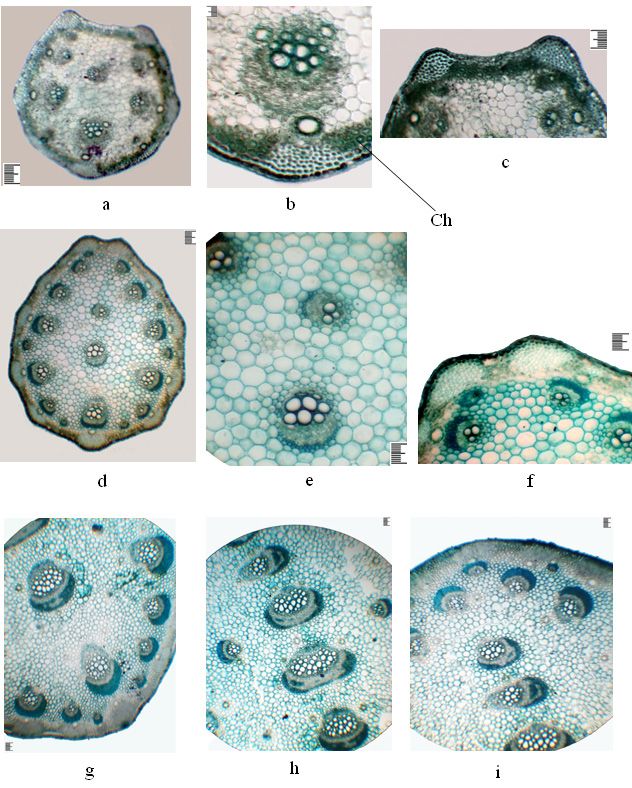
194 Dilovar T. Khamraeva et al. / Acta Biologica Sibirica 7: 193–210 (2021)

Keywords
Hypoderma, immature plant, mesophyll, palisade parenchyma, secretory duct, juvenile plant

Introduction
The genus Ferula L. belongs to the tribe Scandiceae Spreng. (subtribe Ferulinae
Engl.) The genus includes 180-185 species of perennial monocarpic and polycarpic
herbaceous plants, usually with a tall and thick stem and a strong root system. Most
of them are used in traditional eastern and folk medicine (Korovin, 1947; Panahi et
al., 2018; Plunkett et al., 2018). Ferula species are a rich source of biologically active
compounds, including coumarin derivatives, sesquiterpene compounds, esters or
resinous substances, flavonoids, less often saponins (Barnaulov et al., 1974; Golovi-
na et al., 1978; Rakhmankulov et al., 1981; Zhou et al., 2017; Mohammadhosseinia
et al., 2019). The use of these biologically active components with antimicrobial and
insecticidal effects can provide new strategies for the development of drugs and
"green" pesticides, and protect endangered plant resources (Zhou et al., 2017). The
literature contains ample information on the study of the pharmacological activity
of isolated biologically active compounds from various organs of Ferula species.
Many new secondary metabolites found in Ferula species belong to classes of natu-
ral products with interesting biological activities from antiproliferative to anti-in-
flammatory and neuroprotective (Mohammadhosseinia et al., 2019). The soluble li-
pid fraction from the roots of Ferula ferulaeoides (Steud.) Korovin revealed antiviral
activity against the Hepatitis B virus (Zhai et al., 2011). Volatile components of the
leaves of Ferula vesceritensis Coss. & Durieu ex Trab. showed antibacterial activity
of essential oils against gram-positive and gram-negative bacteria (Zellagui et al.,
2012). Compounds of other Ferula species (F. vesceritensis Coss. & Durieu ex Trab.,
F. assa-foetida (L.) Falc., F. gummosa Boiss., F. communis L., F. sinkiangensis K.M.
Shen.) showed cytotoxic effects, and the main mechanisms of action are associated
with slowing cell growth, making them an interesting choice as an adjuvant therapy
for certain diseases (Iranshahi et al., 2018). Ferula tadshikorum Pimenov has been
used as a medicinal species in folk medicine from ancient times in treating cough,
meteorism, seizures, arteriosclerosis, cataracts, nervous and mental disorders. In
addition, the plant has strong anti-helminthic properties, and it is used as a spice in
cooking. Both underground (milky root juice solidified in the air) and the plant's
aboveground parts are used as medicinal raw materials (Sadykov, 2003; Small, 2012;
Sharopov, 2018). F. tadshikorum is distributed in the southwestern Pamiro-Alai, and
is endemic to southern Uzbekistan and southern Tajikistan. F. tadshikorum is not
included in the Umbelliferae of the multivolume monograph "Flora of Uzbekistan"
(Korovin, 1959). For the first time in 2017, when conducting resource studies of
medicinal plants in southern Uzbekistan, O. Khojimatov et al. (2018) collected the
herbarium material Ferula tadshikorum, which was identified as a new species for
the flora of Uzbekistan, and its species identity was also confirmed by the mono-
Ferula tadshikorum in ex-situ conditions 195

graph of the Umbelliferae family of Central Asia, Prof. M.G. Pimenov. Unsustain-
able human economic activity often leads to a deterioration of the vegetation cover,
and as a result leads to a reduction in the number of plant populations, and possibly
complete extinction. During the last two decades, due to the strong exploitation
of natural populations of the species for the sake of obtaining gum resin from the
roots, the condition has worsened, and the species was included in the latest edition
of the Red Book of the Republic of Uzbekistan with status 3 (endangered) (Ma-
khmudov, 2019). To preserve natural populations and prevent a further decrease in
the number of Ferula tadshikorum, it is necessary to study the biological features of
this valuable medicinal plant, by propagating seed material in ex situ conditions and
conducting further monitoring to determine the viability of sprouted individuals.
An important mechanism for the conservation of plant species is primarily the need
to organize protected natural areas in natural ecosystems or create the artificial re-
serves, among which botanical gardens have a leading role. Botanical gardens need
to pay special attention to creating plant expositions including Red-listed species
and conduct educational work with the public. For the first time, the Tashkent Bo-
tanical Garden at the Institute of Botany of the Academy of Sciences of the Republic
of Uzbekistan, established experimental sites to study the adaptability of F. tadshiko-
rum to ex situ conditions. According to our data, in ex situ conditions, the length
of the juvenile phase of F. tadshikorum development is much reduced. According
to literary data, in natural conditions the maximum age of the species can reach
6–7 years (Rakhmonov, 2017), and in introduced conditions, some individuals in
the first year of life go into the immature phase of development (Khamraeva et al.,
2019). Shortening the pre-generative period of promising medicinal plants during
cultivation is considered an important biological feature from an economic per-
spective, for the rapid production of medicinal raw materials from the underground
or aboveground part. To identify the biological stability of the species in new envi-
ronmental conditions, it is necessary to study the rates of ontogenesis, phenology,
growth processes from which assimilating organs predefine the normal vital state
of the plant. The main function of leaves is the production of organic substances,
which eventually accumulate in underground organs, and a study of the structure
of this organ will determine the range of its ecological adaptability to completely
different environmental conditions.
     The present study aimed to conduct a comparative study to detect the diagnostic
signs in the morphological and anatomical structure of leaves of the individuals in
the pre-generative age state F. tadshikorum in ex situ conditions.

Material and methods
F. tadshikorum grows in the middle mountain zone in the territories of the Kashka-
darya and Surkhandarya regions of Uzbekistan on loess and finely ground soils and
limestone, along dry river valleys and on river terraces, at altitudes of 1400–1800 m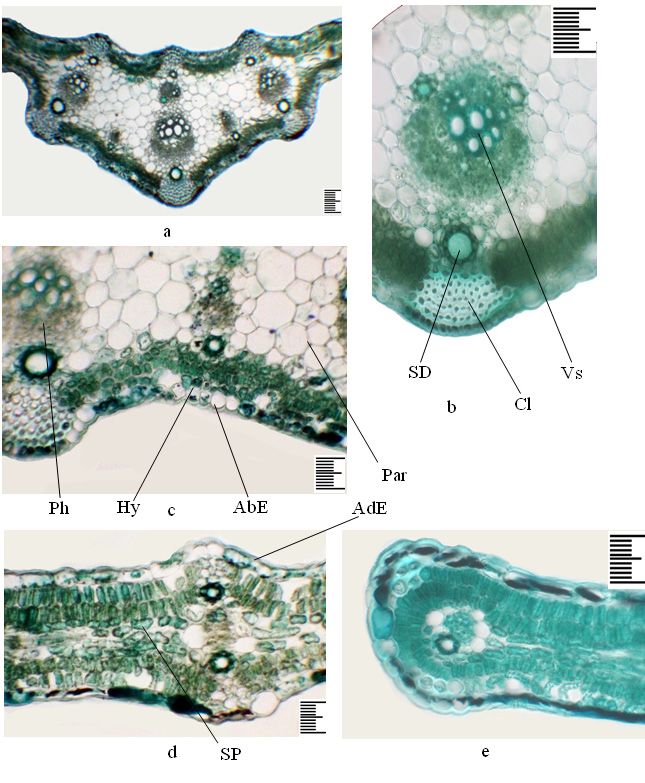
196 Dilovar T. Khamraeva et al. / Acta Biologica Sibirica 7: 193–210 (2021)

above sea level. The places of collection of seeds for subsequent cultivation under
introduced conditions and a detailed description of the studied age states of Ferula
tadshikorum were given by us in a previously published work (Khamraeva et al.,
2019). The material for studying the morpho-anatomical structure of leaves of dif-
ferent ages was collected from experimental sites of the Tashkent Botanical Garden
in 2019–2020 and preserved in 70% ethyl alcohol. The transverse sections of the
leaf plate and petiole from the middle part of the leaves were stained with meth-
ylene blue and enclosed in glycerol gelatin according to the conventional method
(Barykina, Chubatova, 2005). A Canon A 2300 was used to photograph the cross-
sections. Statistical processing of quantitative data was carried out by G.N. Zaitsev
(Zaitsev, 1990) using MS Excel. For each anatomical parameter of the leaf blade and
petiole, the arithmetic mean was calculated from 30 values.
     The soil and climatic conditions of the Botanical Garden are described in detail
in I.V. Belolipov (1989). The garden is located in the northeastern part of Tashkent
at an altitude of 480 m above sea level. The soil is an ancient rusty typical gray soil.
The climate of Tashkent is sharply continental, characterized by dryness, significant
daily temperature fluctuations, hot summers, dry warm autumn and moderately
cold winters. The absolute minimum temperature is... – 25.8°С, the absolute maxi-
mum is... + 44.6°С. According to long-term data, the main rainfall is 380–440 mm,
which falls during the autumn-winter-spring period. At the beginning of the vegeta-
tion period, watering and weeding were carried out twice in the experimental areas.
     Abbreviations for figures: AbE – abaxial epidermis; AdE – adaxial epidermis;
SP – spongy parenchyma; Hy – hypodermis; Cl – collenchyma; BF – bast fibers; Par
– parenchyma; SD – secretory dust; Vs – vessels; Ph – phloem; Ch – chlorenchyme.

Results and discussion

Morphological structure of the leaf
We have previously studied the initial stages of the ontogenesis of Ferula tadshiko-
rum in the introduction and given a detailed description of the morphological
structure of the aboveground and underground parts (Khamraeva et al., 2019). Ac-
cording to this data, the leaf of juvenile plants of the first year of life is simple,
diamond-shaped, the plate is elongated, finely serrated along the edge, 4–5 cm long
and 2–2.5 cm wide, the petiole 3.0–4.0 cm long. As it grows 1–3 (5) rosette leaves
form on the rosette shoot (Fig. 1a). Rosette leaves are of various lengths, from 14 to
24 cm, with the leaf plate 9–12 cm long, 4–5 cm wide, and petioles 5–12 cm long.
Immature individuals of the first year of life have 4–6 simple leaves, with one triple
leaf 17–25 (28) cm long, lobes obovate or wide-oval, petioles 8–16 cm long (Fig.
1b). Immature plants of the second year of life develop 1–2 scaly leaves, 7–11 cm
long, which quickly wilt after the appearance of rosette leaves. Rosette leaves range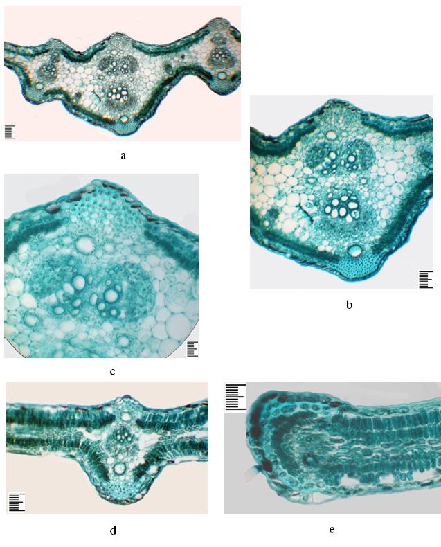
Ferula tadshikorum in ex-situ conditions 197

from 3 to 6, of which 1–2 are simple, and 2–3 triple-dissected. In some plants more
complex 5–6-lobed leaves develop (Fig. 1c, d). Triple-dissected leaves show obo-
vate or wide oval lobes, leaf plates 15–26 cm long, petioles 12–16 cm long, while
5–6-lobed leaves have elongated oval, ovoid or broad ovate segments, leaf plates are
wide-triangular in outline, up to 27 cm long, the segments 10–16 cm long, 4–6 cm
wide, the primary segments with short petioles, the others sessile, with petioles up
to 14 cm long.

Anatomical structure of the leaves of the juvenile plant of the first year
On the cross-section, the sheet along the entire plate is unevenly thickened, plate-
like, bare (Fig. 2 a, e). Collenchyma in the central vein area is located in the ribs, on
the abaxial side of 9–15 rows, adaxial – 5–6 rows, and in the lateral part - above and
under the bundles, only on the abaxial side above large bundles are more multirow.
The central vein area on the abaxial part shows three prominent widely spaced pro-
trusions, and the adaxial part is closely spaced, three more or less prominent protru-
sions and is more thickened due to multilayer aquiferous parenchyma. In the area of
large lateral veins, the leaf is biconvex (Fig. 2 d). The mesophyll consists of isolateral
palisade. Under the epidermis on the abaxial side is a hypoderm single layer formed
by rounded-oval cells. The palisade parenchyma on the abaxial side is 2-layered,
with tabloid cells. On the adaxial side it is also 2-layered, the cells are more or less
elongated (Table 1). The spongy parenchyma is 2–3-layered, with horizontally elon-
gated homogeneous cells (Table 1).
     The aquiferous parenchyma is only found in the central vein area, multilayered,
consisting of up to 10 layers of various sizes thin-walled rounded-oval cells. The
abaxial and adaxial epidermis are single-layered, their external walls are thickened,
and both epidermises are covered with a toothed cuticle (Table 1). The central vein
is represented by two vascular bundles, on the abaxial side an extensive bundle,
and the adaxial part a horizontally oriented more minor (Fig. 2 b, c). Two lateral
vascular bundles are located close by one above the other, and one vascular bundle
is located in the side parts of the leaf in the central plane. A large main vascular
bundle is represented by ten vessels, of which four are large. The vascular bundles
are collateral. One secretory duct is found throughout the leaf above and below the
vascular bundles, in the adaxial part with 6–7, in the area of the central vein with 10
epithelial cells, and in the abaxial part with 7–8, in the area of the central vein with
11 epithelial cells (Table 1). A large main bundle on the xylem side is accompanied
by two small secretory ducts with six epithelial cells. At the edge of the leaf, the
vascular bundle is surrounded by lining cells and there is one secretory duct on the
abaxial part with 7–8 epithelial cells (Fig. 2 e).
198 Dilovar T. Khamraeva et al. / Acta Biologica Sibirica 7: 193–210 (2021)
Anatomical structure of the leaves of the immature plant of the first year
On the cross-section, the sheet along the entire plate is unevenly thickened, plate-
shaped, abaxially pubescent with simple hairs (Fig. 3 a, b). Collenchyma in the cen-
tral vein area is located in the ribs, on the abaxial side 7–18 rows, adaxial 7–10
rows, and in the lateral part - above and under the bundles, more multi-row only
on the abaxial side above large bundles (Fig. 3 b, c). In the central vein area on the
abaxial part there are three widely spaced, strongly prominent and two more or less
prominent protrusions, and on the adaxial part with three slightly spaced more or
less prominent protrusions. In the central part, the leaf is even thicker due to the
multilayering of the aquifer parenchyma than in juvenile plants, and is biconvex in
the area of the lateral veins. The mesophyll has the isolateral palisades. Under the
epidermis on the abaxial side is a single-layer hypoderm formed by rounded oval
cells. The palisade parenchyma on the abaxial side consists of 2-layered tabloid cells.
On the adaxial side, it is also 2-layered, consisting of elongated cells (Table 1). The
spongy parenchyma is 2–3-layered, with horizontally elongated small cells (Fig. 3 d,
e; Table 1). The aquifer parenchyma is found only in the central vein area, multilay-
ered, consisting of thin-walled round-oval cells of various sizes in up to 15 layers.
The abaxial and adaxial epidermis are single-layered, their external walls thickened,
and both epidermises are covered with a toothed cuticle (Table 1). The vascular
bundles are of the collateral type. The central vein is represented by two vascular
bundles, on the abaxial side by a large bundle, and the adaxial part is also a large
double, from oppositely oriented bundles (Fig. 3 b, c). The double bundle consists
of 13–15 vessels, of which seven are large; the second bundle of 8–10 vessels, of
which five are large. Near the phloem there are two small secretory ducts with 5-6
epithelial cells. On two sides of the central vein one above the other there are two
side bundles; on the adaxial side the bundle is inverted. One vascular bundle is ar-
ranged in the central plane of the side parts of the leaf. Large secretory ducts with up
to 12 epithelial cells, and smaller ones with up to 6 epithelial cells are located above
and below the lateral vascular bundles. In the central part of the leaf on the abaxial
side, the secretory ducts have up to 11–13 epithelial cells, with adaxial up to 9–10
epithelial cells (Table 1). At the edge of the leaf, secretory ducts in the abaxial part
with 8, and with adaxial – 6 epithelial cells (Fig. 3 e).
Anatomical structure of the leaf of an immature plant of the second year
On the cross-section, the sheet is plate-shaped, rounded-triangular in the cen-
tral vein area, pubescent on the abaxial side with simple hairs (Fig. 4 a). Collenchy-
ma in the area of central vein is located in the ribs on the abaxial side, consisting of
9–20 rows, adaxial - 7–18 rows, and in the lateral part above and under the bundles,
on the abaxial side above the large bundles they are more multi-rowed (Fig. 4 b). In
the central vein area, the leaf is thicker due to aquifer parenchyma than in immature
individuals of the first year of vegetation, which is biconvex in the area of the lateralFerula tadshikorum in ex-situ conditions 199 Figure 1. Different-aged plants of Ferula tadshikorum in ex situ conditions. a – juvenile plants of the first year (26.03.2019); b – immature plants of the first year (27.05.2019); c – immature plant of the second year (13.03.2020); d – immature plant of the second year (18.05.2020). Scale ruler 1 mm.
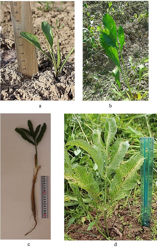
200 Dilovar T. Khamraeva et al. / Acta Biologica Sibirica 7: 193–210 (2021)

Figure 2. The anatomical structure of the leaves of a juvenile plant of the first year, Ferula
tadshikorum. a – detail of the central part of the leaf; b – the main vascular bundle; c – part
in the main bundle; d – lateral part; e – the edge of the leaf. Scale ruler 100 µm.
Ferula tadshikorum in ex-situ conditions 201

Figure 3. The anatomical structure of the leaves of an immature plant of the first year,
Ferula tadshikorum. a – detail of the central part of the leaf; b – the main vascular bundle;
c – part in the main bundle; d – lateral part; e – the edge of the leaf. Scale ruler 100 µm.
202 Dilovar T. Khamraeva et al. / Acta Biologica Sibirica 7: 193–210 (2021)

veins. The mesophyll is isolateral palisade. Under the epidermis in the abaxial and
adaxial parts there is a single-layer hypoderm with rounded-oval cells. The palisade
parenchyma in the abaxial part is 2-layered, with shortened cells; in the adaxial part
it also consists of 2 layers of elongated cells (Table 1). The sponge parenchyma is 4-5
layered, with horizontally elongated small cells (Table 1). The aquifer parenchyma
is found only in the central vein area, with thin-walled rounded oval cells of various
sizes in up to 22 layers (Fig. 4 c, d). The abaxial and adaxial epidermis are single-
layered, their external walls thickened, and both epidermises are covered with a
toothed cuticle (Table 1). Vascular bundles of collateral type. The central vein is
multi-bundles, represented by several large and small peripheral and one central
vascular bundle. Peripheral bundles are located opposite the collenchyma strands
and carry one secretory duct on the phloem side. On the phloem side, bundles are
deep fibers, in large multilayer bundles (Fig. 4 b, d). There is one vascular bundle in
the side parts of the leaf in the central plane (Fig. 4 e). Secretory ducts are located
above and below the lateral vascular bundles, from the abaxial to 10 epithelial cells,
from adaxial to 9 (Table 1). Vascular bundles in the central part of the leaf on the
abaxial side have up to 10–11 epithelial cells, on adaxial up to 8–9 epithelial cells.
The edge of the leaf on the abaxial side is one secretory duct with seven epithelial
cells (Fig. 4 e).

Anatomical structure of the petiole of a juvenile plant of the first year
On the cross-section, the petiole is almost rounded, weakly ribbed, nominally on
the adaxial side with two tubercles, strengthened by a collinear up to 10–12 rows
(Fig. 5 a, c). Collenchyma strains from 7 to 12–13 rows, which alternate with 2 place
three-layered chlorenchyma (Fig. 5 b). The epidermis is single-layered, covered with
a cuticle, and the outer and inner walls are thickened. Under the epidermis lies a
single layer of sub-epidermis whose cells contain chlorophyll grains. Around the
periphery there are nine collateral-type vascular bundles, of which four are small,
one is large and four are mediums. In the central part there is one vascular bundle
of medium size. A large peripheral bundle carries ten large vessels and in the xylem
part two secretory ducts with 6–7 epithelial cells (Table 2). The peripheral vascular
bundles from the phloem part have one secretory duct with 6–10 epithelial cells,
which corresponds to the size of the bundles. The main part of the petiole is rep-
resented by thin-walled rounded-oval parenchymal cells. The part phloem of the
central bundle has one secretory duct with six epithelial cells.

Anatomical structure of the petiole of an immature plant of first year

On the cross-section, the petiole is rounded ovate, ribbed, nominally on the adaxial
part with a small recess, collenchyma in two tubercles up to 15–18 rows (Fig. 5 d,
f). The collenchyma in ribs has 8 to 20–22 rows, with large size above large bundles,
Ferula tadshikorum in ex-situ conditions 203

smaller ones above small ones. Collenchyma strains alternate with chlorenchyma,
which is a 3–4 layer. The epidermis is single-layered, covered with a cuticle, the
outer and inner walls are thickened, and under it is a single-layer sub-epidermis,
the cells of which contain chloroplasts. Peripheral tufts are 17, of which 11 are large,
6 smalls, all of the collateral type. There are two vascular bundles in the central part
(Fig. 5 e). Above the phloem are bast fibers, above them secretory ducts with ten
epithelial cells (Table 2). There are 1 or 2 secretory ducts with 6–8 epithelial cells
in some large peripheral bundles of the xylem part. Above each peripheral bundle
there is also one secretory duct with 10–11 epithelial cells, in small bundles up to 8.
The main part of the petiole is represented by thin-walled rounded oval parenchy-
mal cells.

Table 1. Quantitative characteristics of the leaf blade. µm

               Indicators                 Juvenile plants   Immature plant        Immature plant
                                                            of first year         of second year
 The thickness        abaxial epidermis   5.13±0.14         5.55±0.06             4.41±0.14
 of the cell outer
                      adaxial epidermis 5.69±0.17           5.73±0.12             4.63±0.16
 walls
 Abaxial              height              17.17±0.55        13.05±0.22            20.98±1.15
 epidermis
                      width               23.14±0.70        22.76±0.54            29.14±1.93
 Adaxial              height              16.99±0.39        14.57±0.55            20.31±0.72
 epidermis
                      width               24.31±0.83        24.52±0.48            26.45±1.08
 Palisade tissue on   height              17.28±0.38        26.4±0.58             32.8±1.19
 the abaxial part
                      width               10.36±0.22        9.62±0.26             12.16±0.45
 Palisade tissue on   height              23.48±0.46        25.9±0.88             41.86±1.76
 the adaxial part
                      width               11.9±0.19         9.56±0.23             13.46±0.46
 Spongy               thickness           63.52±1.61        42.68±0.78            64.28±2.12
 parenchyma
 Large secretory      height              10.44±0.28        6.42±0.11             7.63±0.27
 duct
                      width               15.76±0.31        14.39±0.34            16.67±0.18
                      number              10.96±0.18        10.5±0.13             9.66±0.23
 Small secretory      height              6.66±0.15         5.28±0.12             7.05±0.21
 duct
                      width               11.42±0.23        7.84±0.11             13.87±0.25
                      number              8.1±0.16          7.6±0.11              9.86±0.19
 Cavity diameter      large               49.82±0.91        42.93±0.75            64.7±1.22
 of ducts
                      small               27.98±0.84        25.94±0.36            41.42±0.83
204 Dilovar T. Khamraeva et al. / Acta Biologica Sibirica 7: 193–210 (2021)
Table 2. Quantitative characteristics of the petiole. µm
Indicators Juvenile plants Immature plant of Immature plant of
the first year the second year
The thickness of the outer 7.7±0.14 5.4±0.16 5.4±0.20
wall of the epidermis
Height of the epidermis 10.74±0.24 17.68±0.28 12.78±0.62
Cavity diameter of vessels 30.44±1.11 42.5±1.27 40±1.16
Cavity diameter of ducts 42.24±0.92 43.82±1.27 53.8±1.05
Number of ducts epithelial 10.16±0.18 9.23±0.17 10.23±0.34
cells
Height of ducts epithelial 9.82±0.19 10.56±0.36 13.8±0.53
cells
Width of ducts epithelial 16.16±0.39 16.14±0.47 18.22±0.66
cells
Anatomical structure of the petiole of an immature plant of the second
year
On the cross-section there is an ellipsoidal petiole. The epidermis is single-layered,
the surface is covered with simple hairs, and the outer and inner walls are thickened.
Under it is one layer of sub-epidermis with numerous chlorophyll grains. Collen-
chyma strains correspond to the sizes of peripheral bundles, which alternate with
4-5 layered chlorenchyma (Fig. 5 g, i). Vascular bundles are of the collateral type.
Peripheral vascular bundles are represented by alternating large, medium and small
numerous bundles. There are five parallel vascular bundles of various sizes in the
petiole's central part, and perpendicular to the middle bundle 2 small side bundles
(Fig. 5 h). Sometimes one of the central bundles is obliquely oriented concerning
the rest of the central bundles. The deep fibers form caps above the phloem of the
bundles; the central bundles are less multilayered. Above the peripheral bundles is
one secretory duct with 10–12 epithelial cells. Numerous secretory ducts with 6–8
epithelial cells are located in the parenchymal part of the petiole. In large peripheral
bundles on the xylem side there are two secretory ducts.
Growing conditions or diagnostic, morphological and anatomical features of
vegetative and generative organs of some species of the genus Ferula have been
studied (Akalın et al., 2020; Safina et al., 2014; Wang et al., 2016, Khamraeva et
al., 2018; Sagyndukova, Imanbaeva, 2020). As a result of studying the anatomical
structure of the above ground vegetative organs of the three ceno-populations of the
rare and endemic species Ferula iliensis Krasn. ex Korovin found that in the stems
developed mechanical and assimilation tissues located in alternating areas along
the periphery directly under the epidermis, differing in volume among individuals
from the different ceno-populations associated with different environmental con-Ferula tadshikorum in ex-situ conditions 205
ditions (Akhmetova, 2013). When studying the morphological features of leaves,
inflorescences, fruits, and the anatomical structure of mericarp in Ferula caspica
M. Bieb. and Ferula szowitsiana DC., diagnostic species-specific features of these
structures were identified (Tuncay et al., 2019). For Ferula foetida, the diagnostic
features of the leaf are the shape and structure of the epidermis cells, the presence
of simple one- and multicellular trichomes, the placement of vascular bundles and
the structure of secretory ducts in the leaf blade and petiole, which differ slightly
in the leaves of different age individuals (Imanbaeva et al., 2015). According to S.
Rakhimov and Kh. Rakhmonov (2015), in juvenile plants under natural conditions
of Southern Tajikistan, in the second and third years, two scale-shaped and one
diamond-shaped rosette leaves are formed annually on the rosette shoot. From the
fourth to the eighth-tenth year of life in immature plants, 3-4 scaly, and 3-6 assimi-
lating rosette leaves are formed on each annual rosette shoot, the edges of which are
wavy, sometimes cut, leaves are simple, pinnately-dissected.
However, as our study in ex situ conditions showed, the absolute majority of
juvenile individuals (95-97%) of F. tadshikorum go through the immature phase of
development in the second year of vegetation, when the individuals have 1-2 scaly
and 3-6 rosette leaves, of which 1-2 are simple and 2-3 triple-dissected, or some still
pinnately-dissected with 5-6-lobes. Perhaps, in ex situ conditions with good mois-
ture availability, less insolation and the absence of sharp daily fluctuations in air and
soil temperature typical for mountain conditions, there is a significant reduction in
the initial stages of development of the virginial period associated with new soil cli-
matic conditions of growth. S. Rakhimov, Kh. Rakhmonov (2015) noted in natural
individuals of Ferula tadshikorum different leaves according to the morphological
structure. Studies on the morphogenesis and anatomical structure of the leaves in
the closely related species Ferula foetida in Kyzylkum showed in juvenile and imma-
ture plants an isolateral palisade type of mesophyll, an increase in leaf pubescence
on the adaxial side and an increase in the number of layers of aquifer and palisade
parenchyma in the second year of vegetation (Butnik et al., 2009). According to our
results, Ferula tadshikorum also has such signs in the structure of the leaf blade, as
an isolateral palisade type of mesophyll in juvenile and immature individuals, the
absence of pubescence in juvenile plants and its amplification on the adaxial side
in immature individuals, and characteristic features, such as the multiplicity of the
central vein, the multilayering of the aquifer parenchyma and the presence of bast
fibers above the phloem of the bundles of the central vein in immature plants of the
second year, the structure and location of secretory ducts. The petiole of the leaf in
juvenile individuals of the first year is smaller, with a straightforward blade, and in
immature plants of the second year of life it has a large, dissected leaf blade, and is
characterized by the robust development of collenchyme weights and vascular bun-
dles. Xeromorphic features in the anatomical structure of the petiole are manifested
in the development of wide and narrow collenchyma strains in the ribs but differing
in different age individuals in the number of rows, the thickening of the outer walls
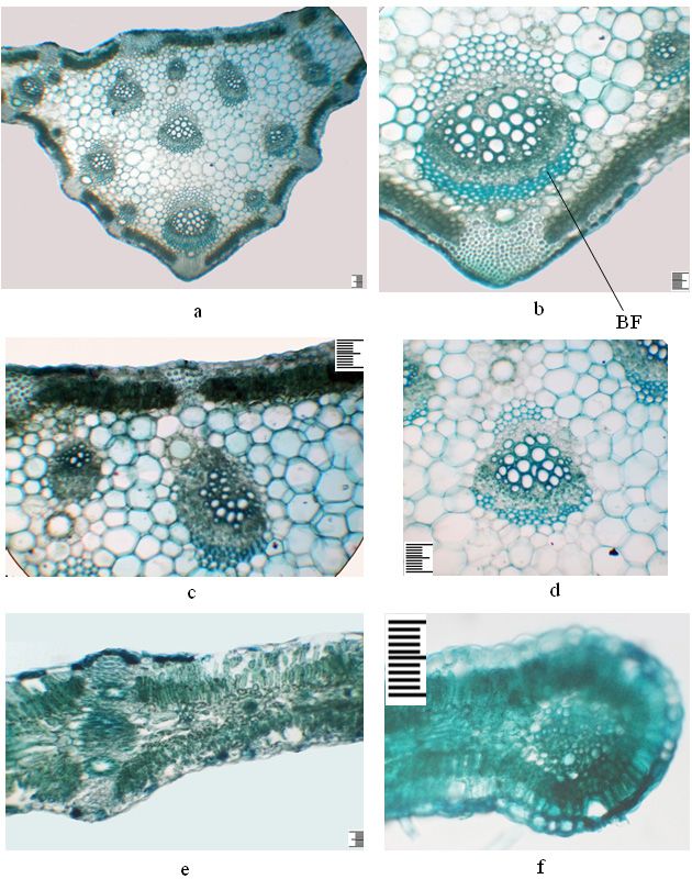
of the epidermal cells in juvenile individuals, which are most susceptible to stress-206 Dilovar T. Khamraeva et al. / Acta Biologica Sibirica 7: 193–210 (2021) Figure 4. The anatomical structure of the leaf of an immature plant of the second year, Ferula tadshikorum in cultural conditions. a – details of the central part of the leaf; b – the main vascular bundle; c – part of the main bundle; d – lateral part; e – the edge of the leaf. Scale ruler 100 µm.
Ferula tadshikorum in ex-situ conditions 207

Figure 5. The anatomical structure of the petiole of Ferula tadshikorum plants. Juvenile
plants of the first year (a, b, c): a – general view; b – peripheral vascular bundle; c – adaxial
part. Immature plants of the first year (d, e, f): d – general view; e – central vascular bundle;
f – adaxial part. Immature plant of the second year (g, h, i): g – detail of the abaxial part,
h – central part, i – detail of the adaxial part. Scale ruler 100 µm.
208 Dilovar T. Khamraeva et al. / Acta Biologica Sibirica 7: 193–210 (2021)

ful environmental conditions, as well as in the development of additional central
vascular bundles. The volume and number of central bundles increase with age. It
should be especially noted that deep fibers are formed over the phloem of periph-
eral bundles and the phloem of central bundles in immature plants of the second
year. Diagnostic features of the petiole are the shape on the cross-section of differ-
ent-age individuals, which in juvenile plants are almost rounded, with two tubercles
conventionally on the adaxial part, in immature plants of the first year are round
ovate, with two tubercles, and in immature plants of the second year ellipsoidal.
According to Kh. Rakhmonov (2017), the rosettes of shoots in species of the genus
Ferula were formed quite early, when they developed in open habitats. This feature
turned out to be adaptive in the xerophilic line of evolution of the genus Ferula. In
this regard, the internal structure of the sheet is being rebuilt by the transition to an
isolateral palisade structure, compaction of spongy tissue or reduction in the central
vein area due to high air temperature differences during the day intense insolation
in mountain conditions.
    The results of our study showed that when plants transition from one age state to
another, morphological changes in their structure, including the internal structure
of the assimilated organ occur. For example, in the juvenile and immature state, the
main elements of the leaf 's cover, mechanical and vascular tissues are laid, which are
more powerfully developed in immature plants.

References
Akalın E, Tuncay H, Olcay B, Miski M (2020) A New Ferula (Apiaceae) species from South-
    west Anatolia: Ferula pisidica Akalın & Miski. Plants 9 (6): 740. https://doi.org/10.3390/
    plants9060740
Akhmetova АB, Mukhitdinov NМ, Ydyrys А (2013) Anatomical research of vegetative
    organs of the Ferula iliensis Krasn. ex Korov., the rare and endemic species. Modern
    Phytomorphology 4: 223-227. [In Russian with an abstract in English] DOI: 10.5281/
    zenodo.161378
Barnaulov SD, Kiryalov NP, Bukreeva TV (1974). Pharmacological properties of some cou-
    marins of the genus Ferula L. Plant resources 10 (2): 259-262. [In Russian]
Barykina RP, Chubatova NV (2005) Great workshop on botany. Ecological anatomy of flow-
    ering plants. KMK Scientific press Ltd, Moscow, 77 pp. [In Russian]
Belolipov IV (1989) The introduction of herbaceous plants of the natural flora of Central
    Asia [Introduktsiya travyanistykh rasteniy prirodnoy flory Sredney Azii]. Fan, Tashkent.
    [In Russian]
Butnik АА, Ashurmetov ОА, Nigmanova RN, Begbaeva GF (2009) Ecological anatomy of
    desert plants of Central Asia 3. Herbs. Fan, Tashkent, 155 pp. [In Russian]
Golovina LА, Khasanov TX, Saidkhodjaev AI, Malikov VM, Rakhmankulov U (1978) Cou-
    marins and esters of Ferula microcarpa. Chemistry of natural compounds 5: 566-570.
    [In Russian]
Ferula tadshikorum in ex-situ conditions 209
Imanbaeva AA, Sarsenbayev KN, Sagyndukova MS (2015) Anatomical structure of above-
and underground organs of Ferula foetida (Bunge) Regel in Mangystau natural popula-
tions. Siberian ecological journal 6: 899-908. [In Russian with an abstract in English]
Iranshahi M, Rezaee R, Najaf Najafi M, Haghbin A, Kasaian J (2018). Cytotoxic activity of
the genus Ferula (Apiaceae) and its bioactive constituents. Avicenna Journal of Phyto-
medicine 8 (4): 296-312.
Khamraeva DT, Beshko NYu, Abdullayeva AT, Sharipova VK (2018) Structural investiga-
tion of the secretory system of some endemic and medicinal species of Apiaceae from
Uzbekistan. Iranian Journal of Botany 24 (1): 53-64.
Khamraeva DT, Khojimatov ОК, Uralov AI (2019) Growth and development of Ferula tad-
shikorum Pimenov in culture. Acta Biologica Sibirica 5 (3): 172 – 177. [In Russian with
an abstract in English] https://doi.org/10.14258/abs.v5.i3.6588
Khojimatov O, Maltsev I, Turginov O (2018) On the issue of stocks of the medicinal plant
Ferula tadshikorum in Uzbekistan. Ecological Bulletin of Uzbekistan 1 (201): 24-26. [In
Russian]
Korovin EP (1947) Genus Ferula (Tourn.) L. monographia illustrata. Academiae Scien-
tiarum UzRSS, 91 pp. [In Russian]
Korovin ЕP (1959) Flora of Uzbekistan 4. Fan, Tashkent, 257-470. [in Russian]
Makhmudov АV (2019) Ferula tadshikorum Pimenov. In: The Red Data Book of the Repub-
lic of Uzbekistan. Plants 2: 95-96. [In Russian]
Mohammadhosseinia M, Vendittib A, Sarker S, Naharc L, Akbarzadeh A (2019) The genus
Ferula: Ethnobotany, phytochemistry and bioactivities – A review. Industrial Crops &
Products 129: 350-394.
Panahi M, Banasiak Ł, Piwczyński M, Puchałka R, Kanani MR, Oskolski AA, Modnicki D,
Miłobędzka A, Spalik K (2018) Taxonomy of the traditional medicinal plant genus Feru-
la (Apiaceae) is confounded by incongruence between nuclear rDNA and plastid DNA.
Botanical Journal of the Linnean Society 188 (2): 173-189. https://doi.org/10.1093/bot-
linnean/boy055
Plunkett GM, Pimenov MG, Reduron JP, Kljuykov EV, Van Wyk BE, Ostroumova TA,
Henwood MJ, Tilney PM, Spalik K, Watson MF, Lee BY, Pu FD, Webb CJ, Hart JM,
Mitchell AD, Muckensturm B (2018) Apiaceae. In: Kadereit J, Bittrich V (eds) Flower-
ing Plants. Eudicots. The Families and Genera of Vascular Plants 15: 1-198. https://doi.
org/10.1007/978-3-319-93605-5_2
Rakhimov S, Rakhmonov Kh (2015) Ontogenesis of the monocarpic shoot Ferula tadshiko-
rum M. Pimen. Bulletin of the Academy of Sciences of the Republic of Tajikistan 1
(189): 7-11.
Rakhmankulov U, Melibaev S, Saidxodjaev AI (1981). Central Asian species of the genus
Ferula L. ‒ sources of sesquiterpene derivatives. Biological features and distribution of
promising medicinal plants. Fan, Tashkent, 138-153. [In Russian]
Rakhmonov KhS (2017) Biology and resources of Ferula tadshikorum M. Pimen. in the
South of Tajikistan. Dissertation for the degree of Candidate of Agricultural Sciences.
Dushanbe, 179 pp. [In Russian]210 Dilovar T. Khamraeva et al. / Acta Biologica Sibirica 7: 193–210 (2021)
Sadykov YuD (2003) Biologically active substances of wild medicinal plants in Tajikistan:
Content, biosynthesis and practical use. Dissertation for the degree of Doctor of Bio-
logical Sciences. Dushanbe, 325 pp. [In Russian]
Safina K, Ostroumova T, Pimenov M (2014) Carpology of the species of Ferula subgen.
Merwia (Umbelliferae–Apioideae) and some taxonomic implications. Nordic Journal of
Botany, 33: 140-150. https://doi.org/10.1111/j.1756-1051.2013.00315.x
Sagyndukova MS, Imanbaeva AA (2020) Study of the anatomical structure of Ferula foetida
of different age states and origin. Bulletin of the Karaganda University 1 (97), 73-78. [In
Russian with an abstract in English]
Sharopov FS, Khalifaev PD, Satyal P, Sun Y, Safomuddin A, Musozoda S, Wink M, Setze
WN (2019) The chemical composition and biological activity of the essential oil from
the underground parts of Ferula tadshikorum (Apiaceae). Records of Natural Products
13(1): 18-23. http://doi.org/10.25135/rnp.65.18.02.089
Small E (2012) Top 100 exotic food plants. CRC Press, New York, 708 pp.
Tuncay H, Olcay B, Uruşak E (2019) Comparative morphology and fruit anatomy of Ferula
szowitsiana DC. and Ferula caspica M.Bieb. Journal of Research Pharmacy 23(3): 577-
583. https://doi.org/10.12991/jrp.2019.165
Wang X, Liu M, Ru J, Wang J, Wang Yu (2016) Morphological and anatomical characteristics
of the fruit of Chinese Ferula and related taxa in Apiaceae. Acta Prataculturae Sinica 25
(6): 81-93. DOI: 10.11686/cyxb2015458
Zaitsev GN (1990) Mathematics in experimental botany. Nauka, Moscow, 296 pp. [In Rus-
sian]
Zellagui A, Gherraf N & Rhouati S (2012) Chemical composition and antibacterial activity
of the essential oils of Ferula vesceritensis Coss et Dur. leaves, endemic in Algeria. Or-
ganic and Medicinal Chemistry Letters 2: 31. https://doi.org/10.1186/2191-2858-2-31
Zhai LL, Liu T, Xie HQ, Xie YH, Mu Q (2012) Inhibition effects on HBV replication by hy-
drophobic extracts from Ferula ferulaeoides (Steud.) Korov. Journal of Medicinal Plants
Research 6 (8): 1486-1488. https://doi.org/10.5897/JMPR11.553
Zhou Yu, Xin F, Zhang G, Qu H, Yang D, Han X (2017) Recent Advances on Bioactive Con-
stituents in Ferula. Drug development research 78 (7): 321–331. https://doi.org/10.1002/
ddr.21402You can also read