Laparoscopic T-tube feeding jejunostomy as an adjunct to staging laparoscopy for upper gastrointestinal malignancies: the technique and review of ...
←
→
Page content transcription
If your browser does not render page correctly, please read the page content below
Siow et al. BMC Surgery (2017) 17:25
DOI 10.1186/s12893-017-0221-2

 RESEARCH ARTICLE                                                                                                                                Open Access

Laparoscopic T-tube feeding jejunostomy
as an adjunct to staging laparoscopy for
upper gastrointestinal malignancies: the
technique and review of outcomes
Sze Li Siow1,2, Hans Alexander Mahendran2, Chee Ming Wong1,2, Nirumal Kumar Milaksh2 and Myo Nyunt1*

  Abstract
  Background: In recent years, staging laparoscopy has gained acceptance as part of the assessment of resectability
  of upper gastrointestinal (UGI) malignancies. Not infrequently, we encounter tumours that are either locally
  advanced; requiring neoadjuvant therapy or occult peritoneal disease that requires palliation. In all these cases, the
  establishment of enteral feeding during staging laparoscopy is important for patients’ nutrition. This review
  describes our technique of performing laparoscopic feeding jejunostomy and the clinical outcomes.
  Methods: The medical records of all patients who underwent laparoscopic feeding jejunostomy following staging
  laparoscopy for UGI malignancies between January 2010 and July 2015 were retrospectively reviewed. The data
  included patient demographics, operative technique and clinical outcomes.
  Results: Fifteen patients (11 males) had feeding jejunostomy done when staging laparoscopy showed unresectable
  UGI maligancy. Eight (53.3%) had gastric carcinoma, four (26.7%) had oesophageal carcinoma and three (20%) had
  cardio-oesophageal junction carcinoma. The mean age was 63.3 ± 7.3 years. Mean operative time was 66.0 ± 7.
  4 min. Mean postoperative stay was 5.6 ± 2.2 days. Laparoscopic feeding jejunostomy was performed without
  intra-operative complications. There were no major complications requiring reoperation but four patients had
  excoriation at the T-tube site and three patients had tube dislodgement which required bedside replacement of
  the feeding tube. The mean duration of feeding tube was 127.3 ± 99.6 days.
  Conclusions: Laparoscopic feeding jejunostomy is an important adjunct to staging laparoscopy that can be
  performed safely with low morbidity. Meticulous attention to surgical techniques is the cornerstone of success.
  Keywords: Laparoscopic jejunostomy, Feeding jejunostomy, Tube jejunostomy, Staging laparoscopy,
  Oesophagogastric cancer

Background                                                                            laparoscopy is often necessary to establish enteral feed-
Staging laparoscopy has emerged as an important sta-                                  ing when oral intake is not possible or a gastrointestinal
ging modality for upper gastrointestinal (UGI) malignan-                              obstruction is expected to occur, such as in the presence
cies. It is most useful in detecting and confirming nodal                             of unresectable obstructed tumour or advanced meta-
involvement and small liver and peritoneal metastases                                 static cancer. Patients with severe sarcopenia will also
that can potentially alter the prognosis and treatment                                benefit especially if they are to undergo neoadjuvant
strategy from curative to palliative intent [1, 2]. The                               chemotherapy for down-staging or palliative chemother-
placement of a feeding jejunostomy tube during staging                                apy. The benefits of a feeding jejunostomy to enable
                                                                                      improvement of nutrition in those requiring chemother-
* Correspondence: drmyonyunt2010@gmail.com
                                                                                      apy and maintenance of enteral access during the period
1
 Department of Surgery, Faculty of Medicine and Health Sciences, Universiti           of profound gastrointestinal toxicity while on chemo-
Malaysia Sarawak, 94300Kota Samarahan, Kuching, Sarawak, Malaysia                     therapy cannot be underestimated.
Full list of author information is available at the end of the article

                                        © The Author(s). 2017 Open Access This article is distributed under the terms of the Creative Commons Attribution 4.0
                                        International License (http://creativecommons.org/licenses/by/4.0/), which permits unrestricted use, distribution, and
                                        reproduction in any medium, provided you give appropriate credit to the original author(s) and the source, provide a link to
                                        the Creative Commons license, and indicate if changes were made. The Creative Commons Public Domain Dedication waiver
                                        (http://creativecommons.org/publicdomain/zero/1.0/) applies to the data made available in this article, unless otherwise stated.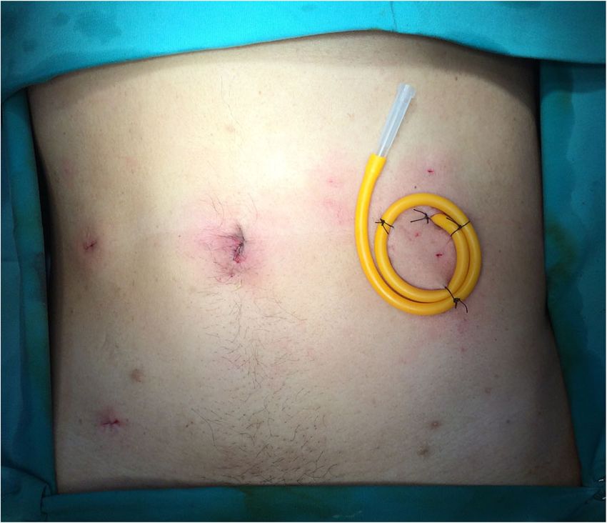
Siow et al. BMC Surgery (2017) 17:25                                                                               Page 2 of 10

  The first laparoscopic technique of feeding jejunostomy       loss of patients to follow-up during the study period. Pa-
was described by O’Regan et al. in 1990 [3]. The technique      tients or the next of kin were contacted in the event of a
underwent modifications with several descriptions and           missed clinic appointment.
commercially available products that facilitated the inser-        Complications were broadly classified into early (those oc-
tion of feeding tubes were introduced. However, the use of      curring within 30 days of jejunostomy placement) and late
commercially available product increases the cost of the        (those occurring ≥30 after the procedure). These complica-
surgery, making it unfavorable in developing countries          tions were either tube-placement related, or feed related
where health budget is a concern. Thus, we devised a total      (bloating, diarrhoea and abdominal colic). Complications
laparoscopic technique using a T-tube to overcome this          were further categorized as minor (catheter occlusion, cath-
limitation. This review describes our initial experience        eter dislodgement, pericatheter leakage, tube site infection,
with laparoscopic feeding jejunostomy with its technical        and feed intolerance) or major (bleeding requiring blood
details when used as an adjunct to staging laparoscopy.         transfusion, intestinal obstruction, peritonitis, volvulus, as-
                                                                piration and any potentially life threatening adverse event
Methods                                                         requiring the need of a surgical or radiologic intervention).
A retrospective review of all patients (15 patients) who
underwent laparoscopic feeding jejunostomy during sta-          Operative technique
ging laparoscopy for UGI malignancy between March 2010          The patient is positioned in modified lithotomy with both
and July 2015 was performed. The indications for feeding        legs supported in padded yellow fin (Allen Medical, USA)
jejunostomy were: 1) Metastatic disease with peritoneal         stirrups. Figure 1a illustrates the position of the surgeon,
nodules or 2) Locally advanced carcinoma requiring              camera surgeon and assistant during staging laparoscopy
neoadjuvant therapy for down-staging. The decision for          and Fig. 1b demonstrates the team position during laparo-
feeding jejunostomy or palliative gastrojejunostomy bypass      scopic feeding jejunostomy. Abdominal access is performed
procedure in patients with metastatic cancer was based on       using Hasson’s technique with pneumoperitoneum estab-
the degree of tumor infiltration of the stomach wall.           lished via a 10-mm infraumbilical port. Two 5-mm ports
Palliative gastrojejunostomy will be the preferred option in    are placed in the right and left mid-clavicular line to facili-
patients with gastric outlet obstruction. However, patients     tate manipulation of the bowels and stomach (Fig. 2a). We
with linitis plastica or gastric inlet obstruction; feeding     routinely perform staging laparoscopy in a reverse TNM
jejunostomy was performed. The data analyzed included           manner, evaluating the presence of distant metastasis first,
demographics, American Society of Anesthesiologists             followed by extent of nodal infiltration and finally the ex-
(ASA) score, body mass index (BMI), types of malignancy,        tent of tumor infiltration itself. Any suspected peritoneal
indications for feeding jejunostomy, operative technique,       nodules or lymph nodes are biopsied. Ascites, if present,
operative time, length of hospitalization and operative         would be aspirated and sent for cytological evaluation.
outcomes. The study was approved by the hospital ethics            After staging laparoscopy has determined the tumour is
committee and Director-General of Health of Malaysia.           unresectable, the surgeon repositions himself to the right
Preoperative computed tomography (CT) scan of the ab-           side of the patient next to the camera surgeon, and the
domen and pelvis was the routine method of pre-operative        assistant then stands between the patient’s legs. Figure 2b
staging. Prophylactic antibiotic was given intravenously        illustrates the port position for laparoscopic feeding
during induction of anesthesia. Clear fluid was started via     jejunostomy. The ligament of Treitz is first identified, and
the feeding tube postoperatively on the day of surgery.         then a loop of proximal jejunum approximately 30 to
Enteral milk feeding was started on the first postoperative     40 cm distal to the ligament is selected. A first layer of
day, employing a standard protocol outlined in the depart-      purse-string suture using polyglactin 910 is placed on the
ment. The feed was administered as a continuous infusion        antimesenteric border using a laparoscopic needle holder
commencing at 30 ml/h for 3 h with an hour’s break              (Fig. 3a). An enterotomy is made with hook and widened
between feeds. The feeds were gradually increased to 100–       using a Maryland dissector (Fig. 3b, c). Then, a T-tube
150 ml/h as tolerated. Patients were allowed oral free fluids   (Teleflex Medical, Kernen, Germany), size 6 mm, with its
as tolerated and were discharged with out-patient appoint-      back wall hemisected is introduced into the abdomen
ments. Patients must have established full enteral feeds        through the 10-mm port, and inserted into the enterot-
and no major tube-related complications. The T-tube             omy using Maryland (Fig. 3d). Once the arms of the T-
remained in-situ until the end of patients’ lifespan or         tube (Teleflex Medical, Kernen, Germany) are successfully
removed when patients were able to tolerate sufficient diet     placed into the jejununal lumen, the purse-string suture is
containing solid food or at their request.                      tightened and knotted (Fig. 3e). A second ring of purse-
   Following discharge from the ward, patients were             string suture is done utilizing the remaining length of the
reviewed once every 3 months for the first 2 years, then        initial suture and subsequently knotted (Fig. 3f). Transab-
every 6 months for the following 3 years. There was no          dominal fixation of the jejunum is performed using a 2-0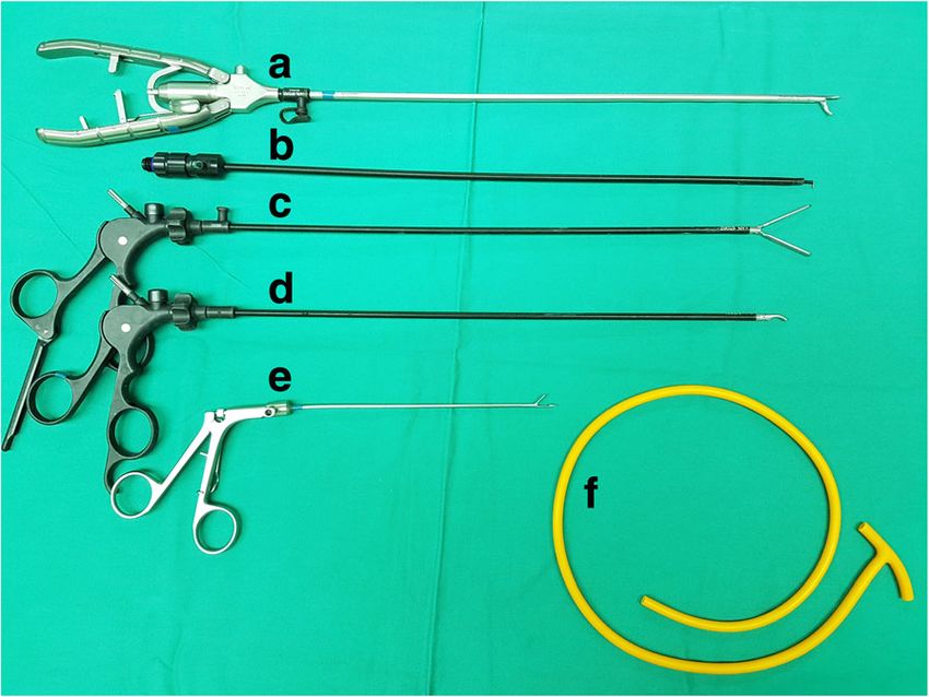
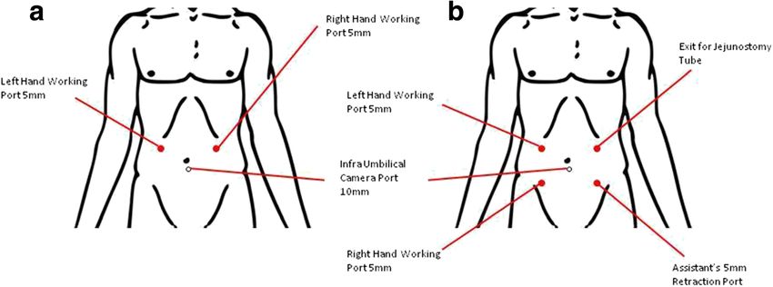
Siow et al. BMC Surgery (2017) 17:25                                                                                       Page 3 of 10

 Fig. 1 a Team position for staging laparoscopy. b Team position for laparoscopic feeding jejunostomy

polypropylene suture placed approximately 2 cm proximal                   30 ml/h. Feeding was gradually increased over the next 2–
and distal to the T-tube (Teleflex Medical, Kernen,                       3 days. A feeding pump was used during the initial phase
Germany), taking a seromuscular bite into the jejunal wall.               of enteral nutrition until bolus feeds were tolerated.
Subsequently, the needle is removed with both ends of
suture brought out onto the surface of abdomen using a                    Results
suture passer introduced through the same 2-mm stab                       Fifteen patients were enrolled in this study and the results
incision in a different track (Fig. 3g). Once the tube is                 are summarized in Table 1. The mean age of the patients
brought out through 5-mm port site, traction is applied to                was 63.3 ± 7.3 years, and 11 patients (73.3%) were male.
the two free ends of transabdominal fixation sutures to                   Eight patients (53.3%) had gastric carcinoma, four (26.7%)
approximate the jejunum onto the peritoneal surface of                    had oesophageal carcinoma and three (20%) had cardio-
the abdominal wall. The sutures are tied with the knot se-                oesophageal junction carcinoma. The indications for feeding
cured anterior to the fascia and buried in the subcutane-                 jejunostomy during staging laparoscopy were as follows: Pal-
ous tissue. Finally, the tube is flushed with normal saline               liative setting in non-resectable or metastatic carcinoma
solution to check the flow and ensure no leak (Fig. 3h).                  with obstructive symptoms (60.0%) and locally advanced
Figure 4 illustrates the final appearance of T-tube against               carcinoma for neo-adjuvant chemotherapy (40.0%). Laparo-
the abdominal wall.                                                       scopic feeding jejunostomy was performed successfully for
  The patients were reviewed by the nutritional support                   all patients. There were no intra-operative complications oc-
team postoperatively. Tube feeding commenced from                         curred as a consequence of the tube insertion technique.
postoperative day one via an infusion pump at a rate of                   The mean operative time was 66.0 ± 7.4 min. Enteral feeding

 Fig. 2 a Port placement for staging laparoscopy. b Port placement for laparoscopic feeding jejunostomy
Siow et al. BMC Surgery (2017) 17:25 Page 4 of 10
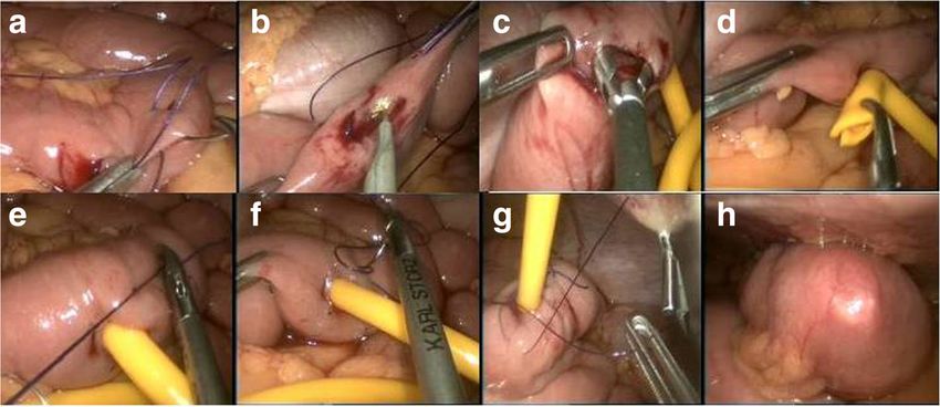
Fig. 3 Jejunostomy technique a First layer of purse-string suture of jejunostomy tube using polyglactin 910 3/0 suture. b Enterotomy done with
hook. c Enterotomy widened using Maryland dissector. d Insertion of T-tube into enterotomy. e First layer of purse-string suture knot secured. f
Second layer of purse-string made using the remaining polyglactin 910 3/0 suture. g Transfascial suturing with suture passer (thread grasper)
introduced through the same 2-mm stab incision in a different track. h T-tube flushed with normal saline to check for patency and leak
was commenced the next day for all patients after reviewed an adjunct to staging laparoscopy. The procedure requires
by nutrition support team and the appropriate polymeric only standard basic laparoscopic instruments such as
formula decided. The aim was to achieve and establish laparoscopic hook, Maryland forceps and monopolar
adequate enteral feeding (more than 50% total energy electrocautery. The use of T-tube is an inexpensive
requirement) within 3 days. Three patients did not achieve alternative to commercially available feeding tubes.
this target due to abdominal distension and was self- Since the first description of the use of jejunostomy tube
limiting. The mean postoperative stay was 5.6 ± 2.2 days. in 1891 by Witzel [4], a vast majority of patients with UGI
There were seven cases of minor late complications, includ- malignancies requiring nutritional support have successfully
ing four cases of minor leak and excoriation around the T-
tube and three cases of tube dislodgement. However, all Table 1 Demographics and surgical outcomes of patients who
were managed expectantly without the need for reoperation. underwent laparoscopic T-tube feeding jejunostomy
The mean duration of feeding tube was 127.3 ± 99.6 days. Variable Value Range
Number of patients 15
Discussion Age (years, mean ± SD) 63.3 ± 7.3 48.0–73.0
This study shows that laparoscopic feeding jejunostomy Gender (male:female) 11:4
can be performed safely with no significant morbidity as
Body mass index 19.8 ± 2.8 15.0–23.5
ASA
1 2
2 12
3 1
Operative time (minutes, mean ± SD) 66.0 ± 7.4 55.0–80.0
Postoperative hospital stay (days, mean ± SD) 5.6 ± 2.2 2.0–9.0
Operative-related complications 0
Conversion to laparotomy 0
Early complications
Minora 3
Majorb 0
Late complications
Minorc 7
b
Major 0
a
Minor early complications: 3 patients with feed intolerance
b
Fig. 4 Final appearance of the T-tube jejunostomy against the patient’s Major complications: tube-related complications requiring re-operation
c
abdominal wall Minor late complications: 4 minor leaks and skin excoriation & 3
tube dislodgementSiow et al. BMC Surgery (2017) 17:25 Page 5 of 10 undergone open jejunostomies. However, the open tech- jejunum while constructing Witzel tunnel could also lead nique is associated with increased operative morbidity and to luminal obstruction at the catheter insertion site [15]. hospital stay [5]. A laparoscopic approach is ideal as it not Dislodgement of the catheter from a Witzel tunnel only confers the advantages of minimally invasive tech- collapses the tunnel and does not allow easy placement of niques but avoids inflicting an additional surgical scar as catheter. On the contrary, a straight passage from the enter- one of the port sites can be used as the exit wound for the otomy to the anterior abdominal wall is important as it feeding tube with lower rates of surgical site infection. gives a straight trajectory that allows easy replacement of a There are considerable variations in techniques de- catheter in the event of dislodgement. The rationale of the scribed in the literature for performing laparoscopic feed- purse-string sutures is to create a seal around the jejunal ing jejunostomy. In general, either the total laparoscopic catheter. A second purse-string may be over-elaborative or or laparoscopic-assisted techniques have been employed unnecessary. However, we maintain the practice as we did [6]. The most common technique for total laparoscopic not encounter any case of intraperitoneal leakage of jejunal placement of feeding catheter is the Seldinger technique content. In addition, it is a much simpler procedure than [1, 7, 8]. It is a technique commonly involves a commer- the Stamm technique of additional inverting stitch. Trans- cial product [9–11], with percutaneous placement of feed- fascial suturing aligns the jejunum to the parietal periton- ing catheter performed after the bowel is secured to the eum and minimizes the risk of volvulus. abdominal wall, using a combination of needle, wire, dila- Different techniques of anchoring the jejunum to the an- tor, stent and feeding tubes [1, 7, 8]. Different combination terior abdominal wall have been described, either in the techniques have been described [12, 13]. Laparoscopic- forms of transfascial sutures (transabdominal sutures [8, assisted techniques involve exteriorizing the jejunum 10] or T-fasteners [5, 16]) or intracorporeal sutures [13, 17]. through a small abdominal incision or one of the trocar The transabdominal sutures (3–4 in number) are usually openings [14] to allow extracorporeal enterotomy and placed in a diamond configuration, incorporating the sero- placement of the catheter. Total laparoscopic technique is muscular layer of the jejunal wall and the anterior abdom- superior as it avoids minilaparotomy incision but has the inal wall [6]. The free ends of the suture are brought out disadvantage of requiring intracorporeal suturing [6]. Our onto the surface of abdomen using a thread gasper and the technique is a total laparoscopic technique that does not two threads are tied with the knot secured at the fascial requires percutaneous jejunostomy kit. It mimicks the layer [15]. Alternatively, the suture could be tied over bol- open technique with the initial placement of the feeding ster placed on the skin to prevent skin damage from the su- tube, followed by the withdrawal of the tube through a left ture [8, 11]. The T-fastener, originally developed for fixation abdominal port site and finally fixation of the jejunum to of stomach to the anterior abdomen in laparoscopic the abdominal wall via transabdominal sutures. gastrostomy, consists of a nylon suture attached to a metal T- There are three techniques described to secure the entry bar, is introduced percutaneously and dislodged into the je- of the feeding jejunostomy tube into the jejunum: a purse- junal lumen from the slotted needle by the stylet [5, 16, 18]. string suture, the Stamm inverting style or a Witzel tunnel Its placement over antimesenteric jejunal wall usually follows [13]. We adopt the purse-string suture method as we feel a diamond configuration [5, 16]. Our technique differs to the that it is an easier option laparoscopically as compared to conventional 3–4 sutures diamond or triangular configur- the Witzel and Stamm techniques. The Stamm technique, ation. We believe that 2 sutures suffice in aligning the je- initially described for gastric access and later adopted for junum against the abdominal wall after the purse-string enteral access, incorporates both the purse-string suture sutures have secured the tube snugly into the enterotomy. around the enterotomy site and inverting stitch of jejunal In terms of outcomes, our initial results demonstrate that wall around the tube to the overlying peritoneum. It the technique of laparoscopic T tube feeding jejunostomy requires some degree of finesse in order to place an invert- can be performed as an adjunct to staging laparoscopy ing stitch laparoscopically through the abdominal wall, a without any increase in peri-operative morbidity. The main maneuver that requires a pronounced supination-pronation technical challenge encountered during this procedure was of the wrist to drive the needle through the abdominal wall. the insertion of the T tube into the enterotomy. Prior to The Witzel technique involves creating a short serosal insertion, it is important to remove the back wall of the tunnel with imbricating sutures over the tube and along the horizontal limbs of the T-tube in order to prevent clogging long axis of the bowel. One study that favour Witzel tunnel as well as allowing guide-wire access for tube exchangei. indicated that such technique reduces the incidence of Additionally, we cut the horizontal limbs into two unequal severe surgical site infections and the rate of late jejuno- ends. Our insertion technique entails initial widening of the cutaneous fistulation [13]. Performing Stamm and Witzel enterotomy using Maryland forceps and inserting the long techniques laparoscopically, though feasible, can be tech- end first followed by the short end. We feel that the tech- nically challenging and time consuming when done exactly nical dexterity required for tube insertion will be improved like in the open technique [7]. Taking too much of the once the procedure is performed on a regular basis.
Siow et al. BMC Surgery (2017) 17:25                                                                               Page 6 of 10

   The complication rates reported for feeding jejunostomy       and antibiotics, and one had a change of catheter. Three
in the literature is variable, with an overall rate between      patients (20%) had catheter dislodgement which was suc-
1.5 and 37% [19]. We compared our data and complica-             cessfully replaced with Foley catheter at bedside.
tion with 12 selected series in the literature that report on       Feeding jejunostomy is a simple procedure yet an import-
the outcomes of laparoscopic feeding jejunostomy                 ant adjunct to staging laparoscopy. With the aim of achiev-
(Table 2). The rate for conversion to open surgery ranges        ing early enteral feeding and a reduction in postoperative
from 0 to 12.5% [11, 13], minor complications ranges from        morbidity, any complications arising from the procedure
5.3 to 32.1% [11, 20], major complications ranges from 0         will jeopardize its benefits as it will incur additional costs
to 10.7% [10, 11, 21] and mortality ranges from 0 to 11.1%       and delay subsequent oncologic treatment. Meticulous at-
[5]. The reported complications include wound infection          tention to tube placement technique remains a sine qua
[6, 22]; catheter dislodgement [6, 13, 23]; occlusion [13,       non to limit complication rates. Dislodgement of the tube
23]; pericatheter leak with generalized peritonitis [24]; as-    can be avoided by attention paid to the technique of secur-
piration pneumonia [22, 24]; small bowel necrosis [23,           ing and confirming catheter placement prior to usage [23].
25]; small bowel obstruction [13, 22, 23]; pneumatosis           Appropriate fixation of jejunum to the parietal peritoneum
intestinalis [23, 26]; abdominal wall infection [13, 23]; fis-   avoids migration of tube to the abdominal cavity [27], and
tula [13, 23]; volvulus [23]; and death [23, 25]. Myers JG et    the occurrence of small bowel volvulus or obstruction at
al. [23] presented an analysis of complications in a large       the jejunostomy site [5, 23]. Our technique of double purse-
series of patients involving insertion of needle catheter        string suturing ensures that the tube fits snugly in the small
jejunostomy at the time of laparotomy as an adjunct for a        bowel, eliminating the risk of leakage of jejunal content. In
variety of reasons. In their series of 2022 patients, the au-    addition, the T configuration of the tube prevents the risk of
thors found a low complication rate of 1.5% and con-             accidental tube dislodgement unless the tube is forcefully
cluded that learning curve and case volume, in addition to       jerked. Transfascial suturing aligns the jejunum to the par-
meticulous attention to operative details, are the import-       ietal peritoneum, preventing the bowel from falling away
ant factors accounting for it. Their observation was sub-        from the anterior abdominal wall. Our technique may ap-
stantiated by their review of series with more than 150          pear to be more demanding than those described in the lit-
catheter placements in which the reported complication           erature but it can be mastered from repeated practice. The
rate did not exceed 3% [23]. On the contrary, two retro-         time invested in perfecting the technique is rewarded with a
spective cohort studies on laparoscopic feeding jejunost-        favorable outcome as reflected in our series showing no leak
omy with case volume in excess of 150 reported an overall        or dislodgement. Prior to initiating enteral feeding, some
complication rate of 9.8–12.7% [13, 21]. In addition, most       authors perform a contrast study a day after the procedure
authors do not have series larger than 50 patients, and          prior to confirm the patency and intraluminal position of
such low complication rates could not be replicated.             the tube [12, 23]. However, we typically flush the feeding
Nevertheless, under-reporting of particularly minor com-         catheter with normal saline to check its position and for
plications as compared to major complications and mor-           any leak under laparoscopic visualization intra-operatively.
tality can occur in retrospective analysis and some authors         A T-tube has several advantages over other types of
argued that the safety of jejunostomy tube placement             tubes. Firstly, the T configuration of the tubing is resistant
should be assessed primarily in terms of major complica-         to accidental dislodgement of the tube, reducing the risk
tions requiring surgical intervention or resulting in death      of peritonitis. Secondly, the soft latex T-tube has less risk
[21]. Han-Geurts IJ et al. in their systematic review of lap-    of intestinal perforation as compared to stiffer jejunost-
aroscopic feeding jejunostomy involving a series of 384          omy tubes and encourages the early formation of a fistu-
patients detected a complication rate of 17% which was           lous tract [28]. This enables safe and easy replacement in
comparable to that of open surgery [6]. From their ana-          the event of dislodgement [28]. In addition, a T-tube obvi-
lysis, wound infection and tube dislodgement were the            ates the risk of bowel obstruction as it is generally smaller
most common complications [6]. Similar findings were             than other types of tube and it does not require an insuf-
observed in the current series that reported a late compli-      flated balloon to maintain its position in the bowel lumen.
cation rate of 46.7% (seven patients). However, no patients      The insertion of the tube under direct vision and confirm-
had serious complications that required surgical interven-       ation of position and non-leakage at the end of procedure
tion and there was no death associated with the proced-          eliminates the need for radiological confirmation. Balloon
ure. The mean hospital stay after the surgery was 5.5 days.      devices have been known to cause bowel obstruction due
The delay in discharge was mainly because of the institu-        to overfilling of the balloon which can also cause pressure
tion of the enteral feeding, awaiting referral to oncologists    necrosis of the bowel wall.
and logistic issues. Four patients (26.7%) developed late           Feeding intolerance is demonstrated when the patient
complication of skin excoriation around the tubing of            developed feeding-related abdominal symptoms such as
which three were managed conservatively with dressing            abdominal distension and diarrhea [19]. The degree of
Table 2 Comparison of selected studies on laparoscopic feeding jejunostomy in cohorts of 10 or more patients
Author         No.of Indication for              Operative Techniques       Tube-related                                              Feed-related            Conclusions
               Cases placement                   (total laparoscopic/       complications                                             gastrointestinal
                                                 laparoscopic aided)        (Minor/Major)                                             symptoms
Sangster W     23     Various indications        Total laparoscopic using   Minor complications (n = 2, 8.7%): superficial            NM                      No procedure related complications.
et al. [9]                                       a 10-French jejunostomy    skin breakdown around the tube (n = 2).                                           A valuable addition to the surgeon’s
                                                 catheter kit               Major complications (n = 1, 4.3%):                                                options for obtaining enteral access.
                                                                            superficial abscess around the tube requiring
                                                                            I & D. One unrelated death.
Grondona P     18     Part of staging            Total laparoscopic using   Minor complications (n = 3, 16.7%):                       NM                      A safe and reliable technique. A useful
et al. [10]           laparoscopy for            a dedicated feeding        tube dislodged (n = 1), leakage with wound                                        adjunct to staging laparoscopy for
                      esophagogastric            jejunostomy kit            infection (n = 1) & wound infection (n = 1).                                      esophagogastric cancer.
                                                                                                                                                                                                        Siow et al. BMC Surgery (2017) 17:25

                      cancer                                                No major complications.
Allen JW       35     Various indications        Total laparoscopic using   Minor complications (n = 4, 11.4%):                       NM                      Safe technique with no significant
et al. [15]                                      a 16 French T-tube         wound infection (n = 2) & leakage (n = 2)                                         morbidity or mortality
                                                                            Major complications (n = 1, 2.9%):
                                                                            intractable pain requiring laparotomy
Ben-David K    153    Prior to definitive        Total laparoscopic using   Minor complications (n = 15, 9.8%):                       NM                      A feasible and safe technique in one
et al. [21]           minimally invasive         a 16-French T-tube         superficial wound infection (n = 4),                                              of the largest series of laparoscopic
                      esophagectomy                                         dislodgement (n = 2), leak (n = 4) &                                              feeding jejunostomy tube for
                                                                            clogging (n = 5). No major complications.                                         esophageal cancer patients.
Mistry RC      19     Oesophageal resection      Total laparoscopic using   Minor complications (n = 1, 5.3%):                        NM                      An easy, inexpensive technique that
et al. [20]                                      a 12-French T-tube         extraperitoneal leakage of feeds due                                              does not require specialized
                                                                            to a damaged vertical limb of the T-tube.                                         equipment or feeding tubes.
Senkal M       80     Primary or recurrent       Total laparoscopic using   Minor complications (n = 7, 8.8%):                        NM                      A safe and effective technique. Does
et al. [12]           tumors of the upper        a 9-French jejunostomy     leakage (n = 2), tube occlusion (n = 3)                                           not require special equipment such
                      gastrointestinal tract     catheter kit               & dislodgement (n = 2).                                                           as T-fasteners, or transabdominal
                                                                            Major complications (n = 1, 1.3%):                                                suturing.
                                                                            abscess at the insertion site requiring drainage.
Heath EI       59     Part of the staging        Total laparoscopic using   Only major complications reported (n = 2, 3.4%):    NM                            Reported only two major complications
et al. [1]            laparoscopy for            a 10-French jejunostomy    perforation of the small bowel requiring laparotomy                               with only one related to the procedure
                      esophageal cancer          tube                       and small bowel resection (n = 1) & intraoperative                                of laparoscopic feeding jejunostomy.
                                                                            pulmonary oedema secondary to aortic valve                                        Minor complications were not reported.
                                                                            stenosis (n = 1).
Hotokezaka M 32       Various indications        Total laparoscopic using   Conversion to open (n = 4, 12.5%).                        Four patients (14.2%)   Safe procedure. High morbidity is
et al. [11]                                      an 18-French Silastic      Minor complications (n = 9, 32.1%):                       had nausea and one      usually related to preexisting disease.
                                                 duallumen feeding tube     dislodgement (n = 3), obstruction (n = 2) &               (3.6%) abdominal        Previous abdominal surgery is not
                                                                            leakage/wound erythema (n = 4).                           cramp.                  necessarily a contraindication.
                                                                            Major complications (n = 3, 10.7%):
                                                                            dislodgement (n = 1) & aspiration
                                                                            pneumonia (n = 2). Death within 30 days
                                                                            (n = 3, 10.7%): aspiration pneumonia and
                                                                            respiratory distress (n = 1) & unrelated death (n = 2).
Jenkinson AD   43     Part of the laparoscopic   Total laparoscopic using Minor complications (n = 11, 25.6%):                        NM                      A safe and simple technique that
et al. [17]           staging for                a 6-French infant feeding dislodgement (n = 5), blockage (n = 4) &                                           adds little to the morbidity and cost
                      esophagogastric cancer     catheter (Vygon)          connector breakage (n = 2).                                                        of managing patients with
                                                                           Major complications (n = 1, 2.3%):                                                 esophagogastric cancers.
                                                                           Dislodgement requiring laparoscopic replacement.
                                                                                                                                                                                                         Page 7 of 10
Table 2 Comparison of selected studies on laparoscopic feeding jejunostomy in cohorts of 10 or more patients (Continued)
Pili D, 25 Patients undergoing Total laparoscopic using Minor complications (n = 3, 12.0%): chronic NM No procedure related morbidity or
et al. [30] major surgery for 8- French jejunostomy catheter occlusion (n = 2) & slippage (n = 1). mortality. A feasible procedure with
esophageal cancer catheter kit. No major complications. the use of autoadjustable sutures to
overcome the limitation of the
laparoscopic handling.
Duh QY 36 Various indications Total laparoscopic using Conversion to open (n = 3, 8%). NM A safe and effective technique when
et al. [5] (a multicentre study) jejunostomy catheter kit Minor complications (n = 6, 16.7%): wound done by experienced laparoscopic
and T-fasteners. erythema or infection (n = 3) & dislodgement surgeons. Serious complications
(n = 3). Major complications (n = 3, 8.3%): are rare.
volvulus (n = 1) & dislodgement (n = 2).
Death (n = 4, 11.1%): unrelated to procedure.
Siow et al. BMC Surgery (2017) 17:25
Young MT 299 Various indications Total laparoscopic using No conversion to open surgery. NM A safe and feasible technique.
a
et al. [13] with majority for 10-French jejunostomy Early complications (n = 12, 4.0%): Associated with a low rate of
esophagogastric cancer catheter kit dislodgement (n = 3), clogging (n = 3), small bowel obstruction and no
intraperitoneal displacement (n = 2), intraabdominal catheter-related
broken tube (n = 1), rectus sheath hematoma infection.
(n = 1) & abdominal wall site infection (n = 2).
Late complications (n = 26, 8.7%): small bowel
obstruction (n = 1), jejunal fistula (n = 11),
dislodgement (n = 10) & broken or cogged tube
(n = 4). Mortality (n = 1, 0.3%):
unrelated to procedure.
Present series 15 Part of the staging Total laparoscopic using Minor complications (n = 7, 46.7%): Three patients (20.0%) A safe, cost-effective technique
laparoscopy for upper 18-French T-tube Skin excoriation around tubing (n = 4) had feed intolerance. with no procedure related
gastrointestinal malignancies & catheter dislodgement (n = 3). complications.
No major complications.
I & D incision & drainage, NM not mentioned. aThe authors divided the complications into early (30-day) and late (˃30 day), and did not fully specify the treatment action for each individual complications and hence
not able to differentiate between minor and major complications
Page 8 of 10Siow et al. BMC Surgery (2017) 17:25 Page 9 of 10
Abbreviations
ASA: American society of anesthesiologists; BMI: Body mass index;
CT: Computed tomography; TNM: Tumour nodes metastasis.; UGI: Upper
gastrointestinal
Acknowledgements
The authors thank the Director General of Health, Malaysia, for permission to
publish this paper.
Funding
No funding.
Availability of data and materials
Data will not be shared due to rules and regulations in Malaysia.
Authors’ contributions
SLS, HAM: Study conception and design. NKM: Acquisition of data. SLS, HAM:
Analysis and interpretation of data. SLS: Drafting of manuscript. HAM, CMW, MN:
Critical revision of manuscript. All authors read and approved the final manuscript.
Fig. 5 Surgical instruments and the T-tube device needed to perform
the procedure a Laparoscopic needle holder. b Laparoscopic L-hook. c Competing interests
Laparoscopic Johan grasper. d Laparoscopic Maryland dissecting The authors declare that they have no competing interest.
forceps. e Laparoscopic suture passer. f T-tube
Consent for publication
Not applicable.
Ethics approval and consent to participate
enteral tolerance varies in different studies and the This study was approved by the Institutional Review Board of the Sarawak
General Hospital.
frequency ranges from 5 to 35% [29]. However, it is
often self-limited and can be corrected by adjusting the
infusion rate and concentration of the feed or temporary Publisher’s Note
Springer Nature remains neutral with regard to jurisdictional claims in
cessation of feeding [29]. Three patients in our series published maps and institutional affiliations.
had feed intolerance and did not achieve the target cal-
Author details
orie and protein requirements within three days but the 1
Department of Surgery, Faculty of Medicine and Health Sciences, Universiti
abdominal distension was self-limiting and resolved. Malaysia Sarawak, 94300Kota Samarahan, Kuching, Sarawak, Malaysia.
2
The main limitations of this study are its retrospective Department of Surgery, Jalan Hospital, 93586 Kuching, Sarawak, Malaysia.
nature of review and small number of cases. However,
Received: 22 September 2016 Accepted: 10 March 2017
our innovative technique of totally laparoscopic place-
ment of jejunal T-tube is cost-effective since it requires
only standard basic laparoscopic instruments (Fig. 5) References
1. Heath EI, Kaufman HS, Talamini MA, Wu TT, Wheeler J, Heitmiller RF, et al.
and can be performed safely without procedure related The role of laparoscopy in preoperative staging of esophageal cancer. Surg
morbidity or mortality. In comparison to three other Endosc. 2000;14:495–9.
similar studies [15, 20, 21], our technique was unique in 2. D’Ugo DM, Persiani R, Caracciolo F, Ronconi P, Coco C, Picciocchi A.
Selection of locally advanced gastric carcinoma by preoperative staging
terms of the use of a larger-sized T-tube, the double laparoscopy. Surg Endosc. 1997;11:1159–62.
purse-string technique and the transfascial suturing with 3. O’Regan PJ, Scarrow GD. Laparoscopic jejunostomy. Endoscopy. 1990;22:39–40.
the suture passer for bowel alignment. 4. Witzel O. Zur teknik der magenfistelanlegung. Centralbl Chir. 1891;18:601–4.
5. Duh QY, Senokozlieff-Englehart AL, Siperstein AE, Pearl J, Grant JP, Twomey
PL, et al. Prospective evaluation of the safety and efficacy of laparoscopic
jejunostomy. West J Med. 1995;162:117–22.
Conclusions 6. Han-Geurts IJ, Lim A, Stijnen T, Bonjer HJ. Laparoscopic feeding
jejunostomy: a systematic review. Surg Endosc. 2005;19:951–7.
In conclusion, our experience with laparoscopic feeding 7. Allen JW, Spain DA. Open and laparoscopic surgical techniques for
jejunostomy as an adjunct to staging laparoscopy demon- obtaining enteral access. Tech Gastrointest Endosc. 2001;3:50–4.
strates that it is a safe and feasible technique. Our inexpen- 8. Schirmer BD. Laparoscopic placement of jejunostomy tube. In: Soper NJ
and Scott-Connor CEH, editors. The SAGES Manual: Volume 1 Basic
sive modification using a T-tube is safe with no immediate Laparoscopy and Endoscopy, Springer Science + Business Media. New York:
post-operative complications or mortality resulting from Springer-Verlag. 2012. p. 379–87.
the procedure. It enables nutritional supplementation for 9. Sangster W, Swanstrom L. Laparoscopic-guided feeding jejunostomy. Surg
Endosc. 1993;7:308–10.
patients with metastatic UGI malignancies as well as pa- 10. Grondona P, Andreani SM, Barr N, Singh KK. Laparoscopic feeding
tients who require neoadjuvant therapy to downstage their jejunostomy technique as part of staging laparoscopy. Surg Laparosc
tumours. The overall incidence of complications in our Endosc Percutan Tech. 2005;15:263–6.
11. Hotokezaka M, Adams RB, Miller AD, McCallum RW, Schirmer BD.
series may seem unacceptably high but the complications Laparoscopic percutaneous jejunostomy for long term enteral access. Surg
were all minor and were managed expectantly. Endosc. 1996;10:1008–11.Siow et al. BMC Surgery (2017) 17:25 Page 10 of 10
12. Senkal M, Koch J, Hummel T, Zumtobel V. Laparoscopic needle catheter
jejunostomy: modification of the technique and outcome results. Surg
Endosc. 2004;18:307–9.
13. Young MT, Troung H, Gebhart A, Shih A, Nguyen NT. Outcomes of
laparoscopic feeding jejunostomy tube placement in 299 patients. Surg
Endosc. 2016;30:126–31.
14. Gedaly R, Briceno P, Ravelo R, Weisinger K. Laparoscopic jejunostomy with
an 18-mm trocar. Surg Laparosc Endosc. 1997;7:420–2.
15. Allen JW, Ali A, Wo J, Bumpous JM, Cacchione RN. Totally laparoscopic
feeding jejunostomy. Surg Endosc. 2002;16:1802–5.
16. Duh QY, Way LW. Laparoscopic jejunostomy using T-fasteners as retractors
and anchors. Arch Surg. 1993;128:105–8.
17. Jenkinson AD, Lim J, Agrawal N, Menzies D. Laparoscopic feeding
jejunostomy in esophagogastric cancer. Surg Endosc. 2007;21:299–302.
18. Murayama KM, Johnson TJ, Thompson JS. Laparoscopic gastrostomy and
jejunostomy are safe and effective for obtaining enteral access. Am J Surg.
1996;172:591–5.
19. Han-Geurts IJ, Hop WC, Verhoef C, Tran KT, Tilanus HW. Randomized clinical
trial comparing feeding jejunostomy with nasoduodenal tube placement in
patients undergoing oesophagectomy. Br J Surg. 2007;94:31–5.
20. Mistry RC, Mehta SS, Karimundackal G, Pramesh CS. Novel cost-effective method
of laparoscopic feeding-jejunostomy. J Minim Access Surg. 2009;5:43–6.
21. Ben-David K, Kim T, Caban AM, Rossidis G, Rodriguez SS, Hochwald SN. Pre-
therapy laparoscopic feeding jejunostomy is safe and effective in patients
undergoing minimally invasive esophagectomy for cancer. J Gastrointest
Surg. 2013;17:1352–8.
22. Weltz CR, Morris JB, Mullen JL. Surgical jejunostomy in aspiration risk
patients. Ann Surg. 1992;215:140–5.
23. Myers JG, Page CP, Stewart RM, Schwesinger WH, Sirinek KR, Aust JB.
Complications of needle catheter jejunostomy in 2,022 consecutive
applications. Am J Surg. 1995;170:547–51.
24. Cogen R, Weinryb J, Pomerantz C, Fenstemacher P. Complications of
jejunostomy tube feeding in nursing facility patients. Am J Gastroenterol.
1991;86:1610–3.
25. Smith-Choban P, Max MH. Feeding jejunostomy: a small bowel stress test?
Am J Surg. 1988;155:112–7.
26. Smith CD, Sarr MG. Clinically significant pneumatosis intestinalis with
postoperative enteral feedings by needle catheter jejunostomy: an unusual
complication. JPEN J Parenter Enteral Nutr. 1991;15:328–31.
27. Tapia J, Murguia R, Garcia G, de los Monteros PE, Onate E. Jejunostomy:
techniques, indications, and complications. World J Surg. 1999;23:596–602.
28. Thodiyil PA, El-Masry NS, Peake H, Williamson RC. T-tube jejunostomy
feeding after pancreatic surgery: a safe adjunct. Asian J Surg. 2004;27:80–4.
29. Wani ML, Ahangar AG, Lone GN, Singh S, Dar AM, Bhat MA, et al. Feeding
jejunostomy: does the benefit overweight the risk (a retrospective study
from a single centre). Int J Surg. 2010;8:387–90.
30. Pili D, Ciotola F, Riganti JM, Badaloni A, Nieponice A. Autoadjustable sutures
and modified seldinger technique applied to laparoscopic jejunostomy.
World J Surg. 2015;39:325–7.
Submit your next manuscript to BioMed Central
and we will help you at every step:
• We accept pre-submission inquiries
• Our selector tool helps you to find the most relevant journal
• We provide round the clock customer support
• Convenient online submission
• Thorough peer review
• Inclusion in PubMed and all major indexing services
• Maximum visibility for your research
Submit your manuscript at
www.biomedcentral.com/submitYou can also read