International Journal of Veterinary Science
←
→
Page content transcription
If your browser does not render page correctly, please read the page content below
P-ISSN: 2304-3075; E-ISSN: 2305-4360

                       International Journal of Veterinary Science
                                                www.ijvets.com; editor@ijvets.com

    Short Communication                                                        https://doi.org/10.47278/journal.ijvs/2021.037

Bovine Papillomatosis in a One-Year-Old Kedah Kelantan Cross Cattle Calf
Mohammed Naji Odhah1,3*, Jasni Sabri2, Mohd Farhan Hanif Reduan2, Bashiru Garba4, Siti Nor Che
Yahya1, Eric Lim Teik Chung6, Siti Nurbakyah Khalid1, Faez Firdaus Abdullah Jesse5, Mohd Azam
Khan Gorimam2 and Nani Izreen MS2
1
 Department of Veterinary Clinical Studies, Faculty of Veterinary Medicine, Universiti Malaysia Kelantan (UMK),
Kampus Kota Locked Bag 36, Pengkalan Chepa, 16100, Kota Bharu, Kelantan, Malaysia; 2Department of Veterinary
Paraclinical Studies, Faculty of Veterinary Medicine, Universiti Malaysia Kelantan, Pengkalan Chepa, 16100 Kota
Bharu, Kelantan, Malaysia; 3Department of Public health, Faculty of Veterinary Medicine, Thamar University, 87246
Dhamar, Yemen; 4Department of Veterinary Public Health & Preventive Medicine, Faculty of Veterinary Medicine,
Usmanu Danfodiyo University, Sokoto, Nigeria; 5Department of Veterinary Clinical Studies, Faculty of Veterinary
Medicine, Universiti Putra Malaysia, 43400 UPM Serdang, Selangor; 6Department of Animal Science, Faculty of
Agriculture, Universiti Putra Malaysia, 43400 UPM Serdang, Selangor, Malaysia
*Corresponding author: naji.ao@umk.edu.my

    Article History: 20-222        Received: 23-Oct-2020            Revised: 28-Dec-2020         Accepted: 01-Jan-2021

    AB S T RA C T
    Bovine papillomatosis is an infectious disease, characterized by the presence of multiple benign mass that can regress
    spontaneously or progress into malignant neoplasia caused by bovine papillomavirus. Epidermal proliferation causes the
    lesion to have the keratotic surface that resembles a cauliflower. In this case report, bovine papillomatosis that was
    encountered in a farm at UMK Bachok, Kelantan will be discussed. A year-old male Kedah Kelantan (KK) cross cattle
    calf was presented with a presence of multiple, circular, around 1-2cm in diameter, wart-like lesion localized on the
    ventral part of the mandible and on the chin. A series of diagnostic approaches had been conducted to reach the definitive
    diagnosis, which includes biopsy for histopathology, polymerase chain reaction (PCR) and fecal examination.
    Key words: Bovine papillomatosis, Wart, Cauliflower-like lesion, Autogenous vaccine.
                                                                                                ©2021 IJVS - All Rights Reserved

                      INTRODUCTION                                  been identified, and they are classified in 4 different
                                                                    subgroups which are deltapapillomavirus, xipapiloma-
     Bovine papillomatosis is an infectious, contagious             virus, epsilonpapillomavirus and dyoxipapilomavirus
and a neoplastic disease, which is characterized by the             (Maeda et al. 2007). The classification was based on the
presence of multiple benign masses (papilloma) on the               homology within the genomic regions of L1 major
integument that can regress spontaneously or progress into          capsid protein and characteristic phylogenetic analysis.
malignant neoplasia (Araldi et al. 2015). In cattle,                BPV 1, 2, 13, and 14 in the delta class were associated
papilloma (also called warts), are most commonly found              with fibropapilloma (Araldi et al. 2017). BPV 3, 4, 6, 9,
on the head, neck shoulder and occasionally on the back             10, 11, and 12 in XI class were purely epitheliotropic
and abdomen. The lesions also have a keratotic surface              where they are associate with cutaneous papilloma. BPV 5
due to epidermal proliferation that resembles a cauliflower         and 8 in epsilon class share some similarities with the
(AL-Salihi et al. 2020); Timurkan and Alcigir 2017).                former two groups. BPV 7 in dyoxi class is a newly
Warts or papilloma would appear like a cauliflower, dry             found type. Papilloma or warts can occur anywhere on
and rough in appearance. It usually would have different            the body of an animal (Lyon 2011). It can be on the head,
shapes and sizes and varies in color; in the shade of grey          the muzzle, the limbs, the neck, as well as on the ventral
to black and it can be either be solitary in one place one or       part of the body (Sundberg 1987). A topography study
multiple (AL-Salihi et al. 2020).                                   was done by Jana and Mukherjee (2012) where wart most
     It is caused by bovine papillomavirus (BPV)                    commonly found on the anterior part of the body was
belonging to the family of Papillomaviridae. There are              52.25%, followed by posterior (32.50%), anteroposterior
15 different types of bovine papillomavirus that have               (12.01%) and the whole body (3.84%).

Cite This Article as: Odhah MN, Sabri J, Reduan MFH, Garba B, Yahya SNC, Chung ELT, Khalid SN, Jesse FFA,
Gorimam MAK and Izreen MSN, 2021. Bovine papillomatosis in a one-year-old Kedah Kelantan cross cattle calf.
International Journal of Veterinary Science x(x): xxxx. https://doi.org/10.47278/journal.ijvs/2021.037

                                                                1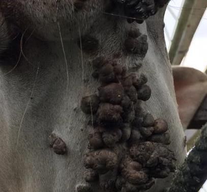
Int J Vet Sci, xxxx, x(x): xxx.
Clinical History result. The prognosis for this case is good as bovine
Janggut is a one-year-old male Kedah-Kelantan cross papillomatosis is a self-limiting disease. Meaning that
cattle that weighed around 200kg with a body condition with time, usually takes up in months, the lesion will
score of 3.5 and was manage extensively. The farm visit regress by its own. As in this case, the animal showed no
was on April 2018 for a routine deworming session. There systemic problem and was eating and drinking well.
were 23 other cattle that were also raised extensively. As
for Janggut and this case was an incidental finding. Upon
physical examination, Janggut appears alert and
responsive; his pulse, respiratory rate and temperature
were all within the normal range. The mucous membrane
was moist and pink, and there was no enlargement
involving the lymph nodes. On the integumentary system,
there was the presence of a wart-like lesion on the ventral
side to the mandible and on the chin (Fig. 1).
Based on the lesions, papillomatosis and
parapoxvirus infection were ruled in as differential
diagnosis. Also, Fig. 1 showed a presence of multiple,
circular, around 1-2cm in diameter, wart-like lesion
localized on the ventral part of the mandible and on the
chin.
Fig. 1: Papillomavirus lesions in the cow showing multiple
lesions on the ventral side to the mandible and the chin wall.
Diagnosis
On clinical examination, when the lesions are
multiple, and appear as a circular warty pedunculated
brownish keratinized lesion, they may be sufficient and
characteristic enough to confirm the diagnosis as
papillomatosis. However, a further test is needed to
confirm the diagnosis.
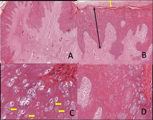
A biopsy on the wart was taken and fixed in a 10%
formalin and sent for histopathological analysis. On the
lowest magnification view, there was a well develop
finger-like projecting papillae (Fig. 2a). Similarly, Fig. 2b
shows hyper parakeratosis where there is the hyperplasia
of the stratum while retaining their nuclei (double yellow
arrowhead) while on the double black arrowhead, indicate
acanthosis as shown by hyperplasia of the stratum
spinosum with elongated rete pegs. Rete pegs are the
epithelial extensions that project into the underlying
connective tissue in the skin. Furthermore, the red
asterisk, there is ballooning degeneration of the Fig. 2: Histology section of warts. A photomicrograph of
keratinocyte and on the yellow arrow shows giant papilloma in a cattle calf. H & E Stain, 100X (A & B) and 400X
clumped pleomorphic keratohyalin granules. This (C & D).
condition is known as koilocytosis (Fig. 2c). There was
also marked hyperplasia of stratum granulosum with Treatment
prominent basophilic keratohyalin granules (Fig. 2d). Treatment approach was by giving the autogenous
Koilocytosis is a diagnostic marker for papillomavirus vaccine at 5ml subcutaneously and revaccinated at seven
infection and can be observed morphologically in days’ intervals for four weeks. Briefly, the papilloma
histological sections (Fig. 2). lesions were excised, finely ground and then filtered after
In addition to the histopathology, a polymerase chain resuspension in normal saline. Subsequently, the filtrate
reaction (PCR) was conducted after viral DNA was was treated with 0.5ml of 10% formaldehyde in order to
amplified by PCR assays using Promega Master Mix kit inactivate the virus. Finally, streptomycin-penicillin at
according to the instructions of the manufacturer. This 2mg/ml was added and stored in the refrigerator until
was achieved by screening for the presence of BPV DNA required (Mayilkumar et al. 2014).
using the FAP59/64 consensus primers followed by
amplification using BPV type-specific primers (BPV2– Client Education
5and 8-10 for skin lesions; Carvalho et al. 2012). The The client was educated on the need to isolate the
result came out positive for papillomavirus and negative animal from the rest of the herd to stop the spread of the
for parapoxvirus based on the positive and negative disease. He was also advised to disinfect the pens and
controls. There was no significant finding on the fecal equipment used with a 2-4% formaldehyde solution.
examination. Based on the diagnostic workup done, Improve feeding by giving more pasture and added
parapoxvirus was ruled out as the PCR result is negative. molasses to increase the appetite of the cattle. Vaccinate
Thus, a tentative diagnosis that can be deduced is remaining cows from the same herd with the prepared
papillomatosis based on the histopathology and also PCR autogenous vaccine.
2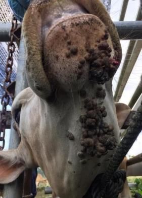
Int J Vet Sci, xxxx, x(x): xxx.

                      DISCUSSION                                   retardation. All of this will have an impact directly to the
                                                                   farmers (Araldi et al. 2017). The morbidity in this
     Bovine papillomavirus is a double-stranded, non-              condition is high while there is a low mortality rate.
envelope DNA virus. It is strictly species-specific with the       However, both of these can cause economic loss (Araldi
exception of BPV 3, which can cross species causing                et al. 2017).
sarcoid in equine. Pathogenesis, start when the virus is                Control and prevention are important to help in
able to enter the skin when there is microabrasion on the          reducing the loss faced by the farmers. As common as its
skin (Lunardi et al. 2013). The virus enters and infects the       sound “prevention is better than cure”. The commercial
keratinocyte of the stratum basale. Then the virus                 vaccine is used as prophylaxis, and the autogenous
amplifies, the basal keratinocyte at stratum basale undergo        vaccine is used as a treatment. The autogenous vaccine is
differentiation. This leads to the expression of viral             stated to be more effective than those commercially
protein that can stimulate cell growth (Stanley 2012). It          available (Turk et al. 2005). It is recommended to begin
results in hyperproliferation of the stratum spinosum and          vaccination in calves as early as 4-6 weeks of age in the
stratum granulosum. The hyperproliferation caused the              herd where this disease is circulating with a dose of about
formation of the exophytic mass, which is warts as a               0.4ml. intradermally given at two sites. The vaccination is
consequence of the acanthosis (Araldi et al. 2017). The            repeated within four to six weeks and at one year of age.
hyperproliferation also causes the keratinization process          Disinfect the equipment application inert material
to occur. The histological slides were characterized by the        surrounding the animal with 4-6% formaldehyde to kill
increase of the keratin granules in the granular layer. Then       the virus that are persistent in the environment (Suveges
the virus will assemble in the most differentiated                 and Schmidt, 2003; Turk et al. 2005). Disinfect also,
epithelium and the virion will be released via cell                instruments used while doing procedure such as
degeneration resulting in koilocyte formation that will            dehorning, tagging, tattooing, milking between animals.
later lead to cell apoptosis or cell death (Krawczyk et al.        Lastly, isolate the affected animal separately from the
2008).                                                             herd. As this disease is a contagious disease, it can easily
     There are several treatment options that have been            be transmitted to other naïve cattle (Turk et al. 2005).
reported, such as autohemotherapy, autogenous vaccine                   In conclusion, Janggut was positively diagnosed with
and surgical excision. A study by Kavithaa et al. (2014)           bovine papillomatosis based on clinical signs,
state that autohemotherapy was found to be the most                histopathology and polymerase chain reaction. The
effective therapy to cure papillomatosis. The procedure to         autogenous vaccine was given as treatment to stimulate
perform this therapy is first, by taking blood from the            the immune system to produce an antibody against the
jugular vein using an 18G hypodermic needle and syringe.           virus.
After that, the blood will be injected back intramuscularly.
This treatment is repeated at weekly interval for four             Acknowledgement
weeks continuously (Nehru et al. 2017). The mechanism                   The authors would like to acknowledge the
behind it is when the blood is re-injected back to the             Veterinary Officers of UMK Veterinary Clinic and
animal, it carries the fragment of viral particle and makes        laboratory staff of Histopathology and Molecular, Faculty
its way into the immune networks and the body produce              of Veterinary Medicine, Universiti Malaysia Kelantan for
appropriate antibodies (AL-Salihi et al. 2020).                    their technical assistance during the time of handling this
     For the autogenous vaccine, the procedure is by               case.
taking warts and put into normal saline and filter it, after
that 10% formalin and antibiotic is added and were left            Funding
overnight. After that, the vaccine is ready to be used                 This research did not receive any specific grant from
(Lesnik et al. 1999). A similar principle is used in the           funding agencies in the public, commercial, or not-for-
autogenous vaccine with the autohemotherapy where the              profit sectors.
body produce antibody through a series of an immune
response (Turk et al. 2005). Removal by surgical excision          Authors Contribution
is not recommended because it may lead to recurrence and                MNO, JS and MFHR, managed the case at the clinic,
stimulation of growth of the warts. However, it can be             ELTC, SNCY, worked together to produce the autogenous
done when the warts are solitary, mass size >5cm, small            vaccine, MAKG, NIMS conducted the histopathology and
in number and when it near their maximum size or when              interpretation of the slides, MNO, BG, SNK, FFAJ
regressing. In most cases, there are too many warts for            produced the initial manuscript draft. All authors revised
surgical removal (Carvalho et al. 2016).                           and granted approval for the final draft submission to the
     Bovine papillomatosis causes significant economic             journal.
losses due to the associated growth retardation, weight
loss, and decreased milk production in BPV-infected                                     REFERENCES
animals (Grindatto et al. 2015). For example, when the
papillomatosis occurs at the teat/udder area, this will            AL-Salihi KA, Al-Dabhawi AH, Ajeel AA, Erzuki IA and Ali
cause reduced milk production, the calf cannot suckle                  TAH, 2020. Clinico-Histopathological and immunohisto-
resulting in growth retardation for the calf, also the                 chemical study of ruminant’s cutaneous papillomavirus in
infection can occur when warts sloughed off and become                 Iraq. Veterinary Medicine International 2020: 1-11.
                                                                       https://doi.org/10.1155/2020/5691974.
a source of infection that cause mastitis (Grindatto et al.
                                                                   Araldi R, Melo T, Neves A, Spadacci-Morena D, Magnelli R,
2015). When warts occur on the mouth area, the cattle                  Modolo D and Beçak W, 2015. Hyperproliferative action of
will lose weight due to reduced appetite and cause growth

                                                               3
Int J Vet Sci, xxxx, x(x): xxx.
bovine papillomavirus: genetic and histopathological Lunardi M, de Alcântara BK, Otonel RAA, Rodrigues WB,
aspects. Genetics and Molecular Research 14: 12942- Alfieri AF and Alfieri AA, 2013. Bovine papillomavirus
12954. https://doi.org/10.4238/2015.October.21.15. type 13 DNA in equine sarcoids. Journal of Clinical
Araldi RP, Assaf SMR, Carvalho RF, Carvalho MAC, Souza Microbiology 51: 2167-2171. https://doi.org/10.1128/JCM.
JM, Magnelli RF and Beçak W, 2017. Papillomaviruses: a 00371-13.
systematic review. Genetics and Molecular Biology 40: 1- Maeda Y, Shibahara T, Wada Y, Kadota K, Kanno T, Uchida I
21. https://doi.org/10.1590/1678-4685-GMB-2016-0128. and Hatama S, 2007. An outbreak of teat papillomatosis in
Carvalho R, Araldi R, Lima T, Modolo D, Souza J and Beçak cattle caused by bovine papilloma virus (BPV) type 6 and
W, 2016. Synergic associations between the bovine unclassified BPVs. Veterinary Microbiology 121: 242-248.
papillomavirus infection and alimentary cofactors. https://doi.org/10.1016/j.vetmic.2006.12.015.
Reference Module in Food Bioscience 2016: 1-10. Mayilkumar K, Kokila S, Manimuthu P and Kuppusamy G,
https://doi.org/10.1016/B978-0-08-100596-5.21138-4. 2014. Formalin inactivated autogenous vaccine for
Carvalho CCR, Batista MVA, Silva MAR, Balbino VQ and treatment of papillomatosis in adult dairy cow. Indian
Freitas AC, 2012. Detection of bovine papillomavirus Journal of Veterinary Pathology 91: 13-15.
types, co‐infection and a putative new BPV11 subtype in Nehru A, Sunandhadevi S, Rama T and Muniyappan N, 2017.
cattle. Transboundary and Emerging Diseases 59: 441-447. Efficacy of auto-hemotherapy in bovine teat papillomatosis:
https://doi.org/10.1111/j.1865-1682.2011.01296.x. A case report. Advances in Animal and Veterinary Sciences
Grindatto A, Ferraro G, Varello K, Crescio MI, Miceli I, 5: 350. https://doi.org/10.17582/journal.aavs/2017/5.8.350.351
Bozzetta E and Nappi R, 2015. Molecular and histological Lyon F, 1994. IARC Monographs on the Evaluation of
characterization of bovine papillomavirus in North West Carcinogenic Risks to Humans. Some Industrial Chemicals
Italy. Veterinary Microbiology 180: 113-117. 60: 389-433.
https://doi.org/10.1016/j.vetmic.2015.08.001. Stanley MA, 2012. Epithelial cell responses to infection with
Jana D and Mukherjee SK, 2012. Management of bovine human papillomavirus. Clinical Microbiology Reviews 25:
cutaneous papillomatosis with ivermectin in farm bred calf 215-222. https://doi.org/10.1128/CMR.05028-11.
crops of West Bengal. Indian Journal of Field Veterinarians Sundberg J, 1987. Papillomavirus infections in animals. In:
8: 69-71. Papillomaviruses and Human Disease, Springer, pp: 40-103.
Kavithaa N, Rajkumar NV and Jiji R, 2014. Papillomatosis in Suveges T, and Schmidt J, 2003. Newer data on the occurrence in
jersey cows and its different medical treatment. Hungary of, losses caused by and ways of control of bovine
International Journal of Science, Environment and papillomatosis. Magyar Állatorvosok Lapja 125: 83-87.
Technology 3: 692-694. Timurkan MO, and Alcigir ME, 2017. Phylogenetic analysis of a
Krawczyk E, Suprynowicz FA, Liu X, Dai Y, Hartmann DP, partial L1 gene from bovine papillomavirus type 1 isolated
Hanover J and Schlegel R, 2008. Koilocytosis: a from naturally occurring papilloma cases in the
cooperative interaction between the human papillomavirus northwestern region of Turkey. Onderstepoort Journal of
E5 and E6 oncoproteins. The American Journal of Veterinary Research 84: 1-6. https://doi.org/10.4102/ojvr.
Pathology 173: 682-688. https://doi.org/10.2353/ajpath. v84i1.1450
2008.080280. Turk N, Zupancic Z, Staresina V, Kovac S, Babic T, Kreszinger
Lesnik F, Bires J, Suli J, Korim P, Posivak J, Mattova J and M, Milas Z, 2005. Severe bovine papillomatosis: detection
Levkut M, 1999. Autovaccination and metabolic profiles at of bovine papillomavirus in tumour tissue and efficacy of
bovine papillomatosis. Slovensky Veterinarsky Casopis treatment using autogenous vaccine and parammunity
(Slovak Republic) 24: 290-294. inducer. Veterinarski Arhiv 75: 391-397.
4You can also read


