Interaction Between Coxsackievirus B3 Infection and α-Synuclein in Parkinson's Disease
←
→
Page content transcription
If your browser does not render page correctly, please read the page content below
Interaction Between Coxsackievirus B3 Infection
and α-Synuclein in Parkinson’s Disease
Soo Jin Park
Ajou University School of Medicine and Graduate School of Medicine
Uram Jin
Ajou University School of Medicine and Graduate School of Medicine
Sang Myun Park ( sangmyun@ajou.ac.kr )
Ajou University School of Medicine https://orcid.org/0000-0002-1001-4743
Research article
Keywords: Parkinson’s disease, α-synuclein, Lewy body, coxsackievirus
Posted Date: January 19th, 2021
DOI: https://doi.org/10.21203/rs.3.rs-145815/v1
License: This work is licensed under a Creative Commons Attribution 4.0 International License.
Read Full License
Page 1/31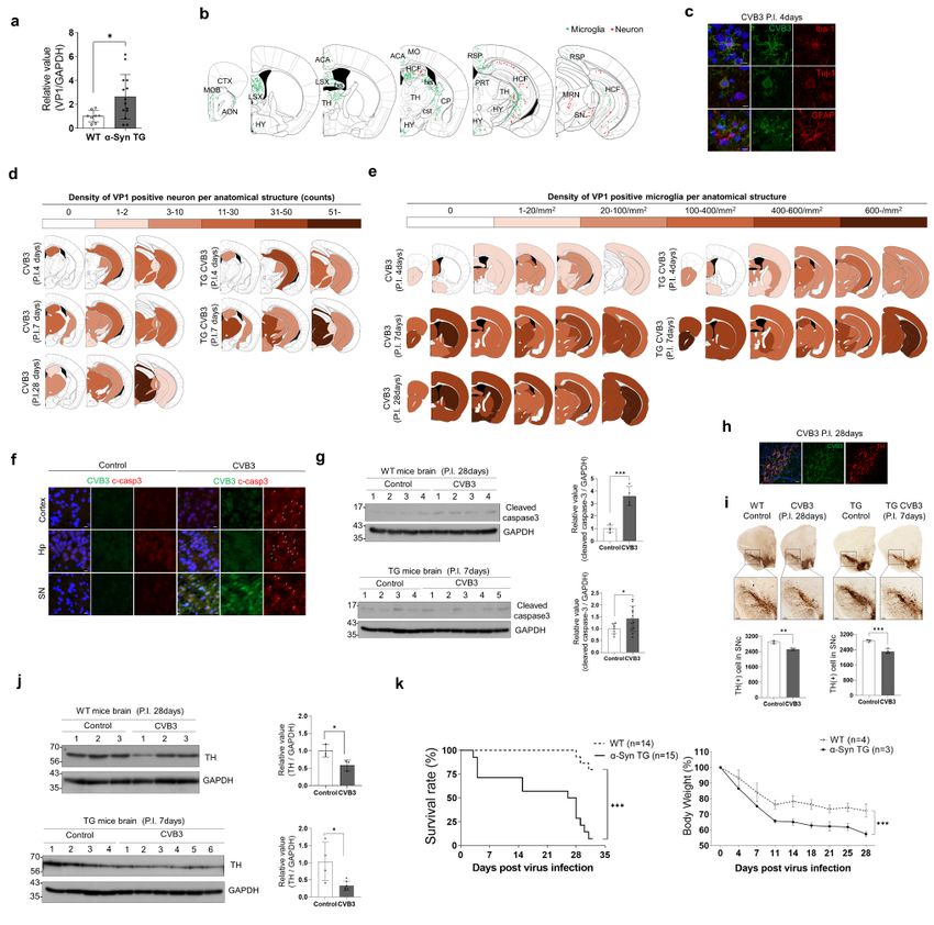
Abstract
Background

Parkinson's disease (PD) is one of the most common neurodegenerative disease. PD is pathologically
characterized by the death of midbrain dopaminergic neurons and the accumulation of intracellular
protein inclusions called Lewy bodies or Lewy neurites. The major component of Lewy bodies is α-
synuclein (α-syn). Prion-like propagation of α-syn has emerged as a novel mechanism in the progression
of PD. Targeting this mechanism could enable the development of disease-modifying therapies for
patients with PD. Nevertheless, the initial triggers of LB formation leading to acceleration of the process
remain elusive.

Methods

To evaluate α-syn function in viral replication, we infected coxsackievirus B3 (CVB3) to α-syn
overexpressed neurons or α-syn transgenic (TG) mice. We then performed biochemical and histological
analyses to evaluate interaction between CVB3 and α-syn in Lewy body formation.

Results

We demonstrated that CVB3 infection can induce α-syn-associated inclusion body formation in neurons
as a trigger. The inclusion bodies contained clustered organelles, including damaged mitochondria with
α-syn fibrils. α-Syn overexpression accelerated inclusion body formation and induced more concentric
inclusion bodies. In brains from CVB3 infected mice, α-syn aggregates in the cell body of midbrain
neurons were observed. Additionally, α-syn overexpression favored CVB3 replication and related
cytotoxicity. α-Syn transgenic mice had a low survival rate, enhanced CVB3 replication, and further
neuronal cell death, including dopaminergic neurons in the substantia nigra. These results may be due to
the different usage of autophagy between CVB3 and α-syn.

Conclusions

This study elucidated the mechanism of Lewy body formation and the pathogenesis of PD associated
with CVB3 infection.

Background
Parkinson's disease (PD) is one of the most common neurodegenerative disease. PD is pathologically
characterized by the death of midbrain dopaminergic neurons and the accumulation of intracellular
protein inclusions termed Lewy bodies (LBs) or Lewy neurites (LNs) [1, 2]. The major component of these
inclusions is α-synuclein (α-syn) [3]. Protein inclusions with α-syn aggregates have also been observed in
other neurodegenerative disorders, such as multiple system atrophy and dementia with Lewy body, which
are collectively referred to as α-synucleinopathies [4]. Multiplications and missense mutations of the α-
syn gene have been identified in patients with early onset familial PD [5]. Genome-wide association
                                                  Page 2/31
studies also demonstrated a strong association between α-syn gene and sporadic PD [6, 7], suggesting a
major role of α-syn in the pathogenesis of PD.

Lewy pathology first appears in the olfactory bulbs and dorsal motor nucleus of the vagus nerve, which is
connected to the enteric nervous system. The pathology progressively involves more regions of the
nervous system and then the cortical areas as the disease advances [8]. This pathology seems to be
present prior to the appearance of motor symptoms in PD and may be associated with gastrointestinal
and olfactory dysfunctions, which are frequently present in the prodromal phase of PD [9]. Substantial in
vitro and in vivo experimental evidence has implicated the prion-like propagation of α-syn as a novel
mechanism in the progression of PD [10–12]. Targeting this mechanism could enable the development of
disease-modifying therapies for patients with PD. Hampering this aim, the initial triggers of LB formation
leading to acceleration of the process remain elusive.

Viral infection is increasingly recognized as a risk factor for PD. A number of viruses have been
associated with both acute and chronic parkinsonism. These viruses include influenza virus,
coxsackievirus, Japanese encephalitis B virus, western equine encephalitis virus, and herpes virus [13]. It
has been proposed that peripheral infections, including viral infections accompanying inflammation, may
trigger PD [14].

Coxsackievirus is a single-stranded RNA virus belonging to the Picornaviridae family of viruses in the
genus termed Enterovirus [15]. More than 90% of coxsackieviruses infections are asymptomatic.
Clinically, infants or young adults are easily infected with this virus, and a small number develop severe
myocarditis [16] or meningitis [17]. Persistent coxsackievirus infection is also associated with chronic
myocarditis, dilated cardiomyopathy [18], and type I diabetes [19].

A recent report described virus-like particles and enterovirus antigen in the brainstem neurons of PD [20].
This finding prompted the proposal that enterovirus infection in PD may act as a seed for the aggregation
of α-syn in addition to the direct cytopathic effect of a virus infection on neurons. In addition, α-syn
inhibits West Nile virus (WNV) infection as a viral restriction factor [21], suggesting that α-syn expression
may affect viral infection in the central nervous system (CNS).

In the present study, we explored whether coxsackievirus B3 (CVB3) interacts with α-syn to induce
aggregation and further LB formation, and whether α-syn affects the replication of coxsackievirus.

Methods
Antibodies and reagents
Antibodies against α-syn were purchased from Abcam (#ab138501, Cambridge, UK), BD Biosciences
(#610786, Franklin Lakes, NJ), and Genetex (#GTX112799, Santa Barbara, CA). Antibodies against
pSer129 α-syn (#ab51253) and Tuj-1 (#ab18207) were obtained from Abcam. Antibody against VP1 of
CVB3 was purchased from Millipore (#MAB948, Danvers, MA). Antibody against LC3 was purchased
                                                  Page 3/31
from Sigma-Aldrich (#L8918, St. Louis, MO). Antibody against p62 was purchased from BD Biosciences
(#610832). Antibody against cleaved caspase-3 was purchased from Cell Signaling Technology (#9664,
Beverly, MA). Antibody against IBA-1 was purchased from Wako (#019-19741, Richmond, VA). Antibody
against glial fibrillary acidic protein (GFAP) was purchased from Neuromics (#RA22101, Montreal, QC).
Antibody against glyceraldehyde 3-phosphate dehydrogenase (GAPDH) was purchased from Santa Cruz
Biotechnology (#SC-32233, Santa Cruz, CA). LysoTracker (#L12492) and Lipofectamine2000 (#11668-
019) were obtained from Invitrogen (Carlsbad, CA). Retinoic acid (RA, #r2625), Bafilomycin A1 (Baf A1,
#B1793), polyinosine:polycytidylic acid (poly IC, #P0913), and Evans Blue Dye (EBD, #E2129) were
purchased from Sigma-Aldrich. Sudan Black B was purchased from Tokyo Chemical Industry (#4197 −
2505, Tokyo, Japan).

Animals
α-Syn transgenic (TG) mice overexpressing human α-syn under the control of neuron specific enolase
(NSE) promotor (C57BL/6N-Tg (NSE-h a Syn) Korl) were donated by the National Institute of Food and
Drug Safety Evaluation (NIFDS, Cheongju, Korea). Wild-type (WT) littermates or WT C57Bl/6N mice (DBL,
Eumseong, Korea) were used as controls. All animal procedures were conducted according to the
guidelines established by the Ajou University School of Medicine Ethics Review Committee (IACUC No.
2016-0047).

Cell culture
α-Syn, mCherry and α-syn-mCherry overexpressing SH-SY5Y cells were prepared as described previously
[22]. The cells were grown in Dulbecco’s modified Eagle’s medium (DMEM) containing 10% fetal bovine
serum (FBS) at 37 °C in a humidified atmosphere of 5% CO2 and 95% air. Primary cortical neurons were
cultured from 1-day-old pups of WT and α-syn TG mice in neurobasal medium (#21103-049, Invitrogen)
with GlutaMAXTM-I (#35050061, Thermo Fisher Scientific, Waltham, MA), and B-27 supplement (#17504-
044, Invitrogen) for 2 weeks on poly-D-lysine (#P7280, Sigma-Aldrich) coated cover-slides or cell culture
dishes. To differentiate SH-SY5Y cells, 50 µM of RA was added to the culture medium and incubated for 5
days.

Infection with coxsackievirus B3 (CVB3)
The H3 variant of CVB3, the Woodruff strain, and EGFP-CVB3 were a kind gift from Dr. E. Jeon (Samsung
Medical Center, Seoul, Korea). The virus was propagated in HeLa cells. Viral titers were determined using
plaque forming assay. SH-SY5Y cells were incubated with CVB3 at an indicated multiplicity of infection
(MOI) in serum-free DMEM for 1 h and further incubated in DMEM with 10% FBS for 24 h. For primary
neurons, cells were incubated with the indicated MOI of CVB3 in neuron culture medium for 24 h. In the in
vivo model, 8- to 11- week-old male mice were infected by intraperitoneal (IP) injection with 1.0 x 10⁶
plaque forming units (PFU) of CVB3 in 100 µl of phosphate-buffered saline (PBS).

Tissue preparation

                                                  Page 4/31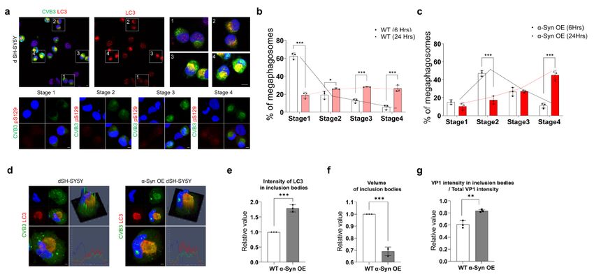
Mice were anesthetized and transcardially perfused first with perfusion solution containing 0.5% sodium
nitrate and 10 U/ml heparin, and then with 4% paraformaldehyde in 0.1 M phosphate buffer (PB; pH 7.2).
Brains were stored in 4% paraformaldehyde for 24 h at 4 °C, and then in a 30% sucrose solution until they
sank. For reverse transcription polymerase chain reaction (RT-PCR) and western blotting, mice were
perfused with only perfusion solution for 2 min and each organ was stored at − 70 °C until use. For
immunostaining, six separate series of 30 µm coronal brain sections were sectioned using a model
CM3050S cryostat (Leica, Wetzlar, Germany) and stored in an anti-freeze stock solution (PB containing
30% glycerol and 30% ethylene glycol, pH 7.2) at 4 °C before experiments.

Immunocytochemistry
Cells cultured on coverslips were washed three times with PBS and fixed with 4% paraformaldehyde for
10 min at room temperature. The fixed cells were washed several times with PBS and incubated in the
presence of permeabilization buffer (PBS containing 0.1% Triton X-100) for 5 min at room temperature.
After washing with PBS, the cells were blocked with blocking buffer (PBS containing 1% bovine serum
albumin [BSA]) for 1 h at room temperature, and then incubated with the indicated antibodies overnight at
4 °C. The samples were then incubated with Alexa 488- (#A21202, #A21206) or 568- (#A10037,
#A10042) conjugated secondary antibodies (all from Invitrogen) for 1 h and then exposed to Hoechst
stain for 5 min. The sections were mounted and observed by confocal microscopy (Carl Zeiss, Jena,
Germany). Live cells were incubated with 50 nM LysoTracker in DMEM with 10% FBS for 30 min. After
staining of nuclei with Hoechst for 5 min, the live cells were observed by confocal microscopy (Carl
Zeiss).
Western blot
Samples were lysed in an ice-cold RIPA buffer (50 mM Tris-HCl, pH 7.4, 0.5% sodium deoxycholate,
150 mM NaCl, 0.1% SDS, 1% Triton X-100) containing a protease inhibitor cocktail (#535140, Calbiochem,
Darmstadt, Germany) and a phosphatase inhibitor cocktail (#P3200-001, GenDEPOT, Baker, TX). After
brief sonication, the lysates were centrifuged at 16,000 x g for 30 min at 4 °C, and the supernatants were
collected. In case of mice tissues (brains and hearts), after lysing and homogenizing samples with TRIzol
(#TR118, Molecular Research Center Inc, Cincinnati, OH, USA), protein was isolated according to the
manufacturer's protocol. The protein concentrations were determined using a DC Protein Assay Reagents
Package (#5000116, Bio-Rad, Hercules, CA). Proteins were resolved by SDS-PAGE, transferred to a PVDF
or NC membrane, and immunoblotted with the indicated primary antibodies and horseradish peroxidase-
conjugated secondary antibody (#G-21040, Invitrogen or #111-035-003, Jackson ImmunoResearch, West
Grove, PA). They were then visualized using an enhanced chemiluminescence (ECL) system (#LF-
QC0101, AbFrontier, Seoul, Korea). The band intensities were determined using ImageJ software (NIH,
Bethesda, MD).

RT-PCR

                                                 Page 5/31
Total RNA was isolated from samples using TRIzol (#TR118, Molecular Research Center Inc.) according
to the manufacturer’s protocol. Total RNA was reverse transcribed using AMV Reverse Transcriptase
(#M0277L, New England Biolabs, Ipswich, MA). The transcript levels of target genes were quantified with
2×KAPA SYBR Fast Master Mix (#kk4602, Kapa Biosystems, Cape Town, South Africa) using the
StepOnePlus™ Real-Time PCR System (Applied Biosystems, Foster City, CA). For each target gene, the
transcript level was normalized to that of GAPDH and was calculated using the standard ∆∆CT method.
A complete list of primer sequences is provided in Supplementary Table 1.
Transmission electron microscopy (TEM)
Control and CVB3 infected samples were fixed with 0.1M sodium cacodylate buffer (pH 7.4) containing
1% formaldehyde / 2% glutaraldehyde for 30 min at 4 °C [23]. The samples were rinsed twice with cold
PBS, post-fixed with a mixture of 1% osmium tetroxide and 1% potassium ferricyanide, dehydrated in
graded alcohol and embedded in Durcupan ACM resin (Fluka, Yongin, Korea). Ultrathin sections were
obtained with resin, mounted on copper grids, and counterstained with uranyl acetate and lead citrate.
The specimens were observed using a Sigma 500 transmission electron microscope (Carl Zeiss).
Mitochondrial morphology analysis
Mitochondrial morphology in TEM images was analyzed as described previously [24]. Based on the type
of mitochondrial restructuring, four categories (type I-IV) were assigned. Type I comprised mitochondrial
cristae that were regular, tightly packed, and longitudinally oriented. Type II comprised mitochondria with
an abnormal shape or nonuniform size, and irregular cristae that lacked orientation and tightness. Type III
comprised mitochondria of varied shapes and sizes, with discontinuous outer membrane, fragmented
cristae, and swollen matrix. Type IV comprised mitochondria with a ruptured outer membrane, no cristae,
and myelin-like transformation.
Cytotoxicity assay
Cytotoxicity assays were performed using the LDH Cytotoxicity Assay Kit (#K311-400, BioVision;
Mountain View, CA) according to the manufacturer’s protocol. Following the CVB3 infection for 30 h, 50 µl
of medium and assay kit solution were mixed in wells of an optically clear 96-well plate. The absorbance
was measured within 10 min at 490 nm with an ELISA reader (Molecular Device, Wokingham, UK)
Flow cytometry
Flow cytometry analysis was performed as described previously [25]. After the indicated times of EGFP-
CVB3 infection, the cells were isolated and fixed by resuspension in 4% paraformaldehyde in PBS
overnight. After washing with PBS and resuspension in 1% BSA in PBS, the cells were analyzed using a
FACS Aria III (BD, Franklin Lakes, NJ). The data were analyzed using Flowing Software version 2.5.1
(Turku Bioscience, Turku, Finland).
Immunohistochemistry (IHC)
Page 6/31Every serial section in each set was collected and washed with PBS containing 0.2% Triton X-100 (PBST).
After blocking with 1% BSA in PBST, sections were incubated overnight at room temperature with the
indicated primary antibodies. The samples were incubated with Alexa 488- (#A21202, #A21206) or 568-
(#A10037, #A10042) conjugated secondary antibodies (all from Invitrogen) for 1 h and then stained
using Hoechst for 10 min. After mounting on slides, the sections were treated with Sudan Black B
solution (0.1% in 70% ethanol) to inhibit auto-fluorescence of mouse tissues. Images were captured using
a confocal microscope (Carl Zeiss) or a fluorescence microscope (Axioscan Z1, Carl Zeiss).
Cell density analysis
The hemispheres of mice were used for detecting VP1 by RT-PCR. The opposite hemispheres were used to
analyze cell density. The hemispheres from three mice whose brain VP1 levels were close to the average
were analyzed. We measured the number of cells (neurons) and the density of cells (microglia) per
specific anatomical structure. Tuj-1 merged VP1 positive cells were counted from three equivalent
locations for neurons and IBA-1 merged VP1 positive cells from six equivalent locations for microglia
along the rostrocaudal axis. The cells were marked with the suggested color. Microglia were counted
using the MetaMorph neurite outgrowth module (Molecular Devices). The density was obtained by
dividing the counted value by the area of each anatomical structure. The anatomical structure was
divided into cerebral cortex, cerebral nuclei, fimbria, internal capsule, thalamus, hypothalamus, and
midbrain. In the olfactory region, the cerebral cortex was divided into isocortex and olfactory areas, and
the hippocampus was separated from the cerebral cortex in the section including the hippocampus. In the
case of cerebral nuclei, the striatum and pallidum were largely divided, and the striatum was divided
again into the dorsal region, lateral septa complex, and ventral region.
Statistical analysis
All values of experimental data are expressed as mean ± SD. Statistical significance was evaluated using
the unpaired t-test, one-way ANOVA, or two-way ANOVA using Graphpad Prism software (Graphpad, La
Jolla, CA). Linear regression was used to analyze the correlation between mCherry and EGFP signal using
Graphpad Prism software.
Results
CVB3 infection regulates α-syn expression in neurons
To explore whether CVB3 affects α-syn, we infected differentiated SH-SY5Y cells (dSH-SY5Y cells) with
CVB3 (MOI 0.25) for 24 h. CVB3 VP1 colocalized with α-syn and the intensity of α-syn in infected cells
was increased. Interestingly, we observed that CVB3 infection induced large aggregates of α-syn that
completely filled the cytoplasm and pushed the nucleus aside, creating a half-moon appearance. This
appearance was clearer in dSH-SY5Y cells overexpressing α-syn (Fig. 1a). In primary cortical neurons, a
similar colocalization of VP1 with α-syn observed (Fig. 1b). In contrast, the mRNA level of α-syn was
decreased in dSH-SY5Y cells and primary cortical neurons infected with CVB3 (Fig. 1c). Analysis of an
Page 7/31open source database (GSE 19496) also showed that the mRNA level of α-syn in CVB3-infected mouse
heart was also decreased compared with that in the control (Supplementary Fig. 1a). Western blot
analysis showed that endogenous α-syn was decreased. Interestingly, ectopically overexpressed α-syn
was also decreased. It was likely to be more severe as the virus titer increased (Fig. 1d). When we
intraperitoneally infected WT mice with CVB3, decreased levels of α-syn mRNA and protein in the brain
were also observed (Fig. 1e-f). Given that CVB3 did not infect all cells, these results led us to speculate
that α-syn may be regulated differently in both CVB3 infected cells and neighboring cells. To explore this,
we compared α-syn levels in both non-infected and infected conditions. Increased α-syn level was
observed in CVB3-infected cells and the α-syn level was obviously decreased in neighboring cells near
CVB3-infected cells (Fig. 1g). Similar findings were observed in primary cortical neurons (Fig. 1h),
suggesting that α-syn was regulated differently in both CVB3-infected cells and neighboring cells. When
cells were treated with poly IC, an artificial analog to mimic RNA viral infection [26], α-syn was increased
at both the mRNA and protein levels, whereas α-syn aggregates were not observed (Fig. 1i-k). These
findings suggested that the observations were CVB3 specific. Analysis of another open source database
(GSE 7621) also revealed the decreased mRNA level of α-syn in the brains of patients with PD compared
with controls (Supplementary Fig. 1b). These results suggested that CVB3 infection induced large
cytosolic aggregates that colocalized with α-syn, and that α-syn expression was differentially regulated
between infected cells and neighboring cells.
CVB3 infection induces LB-like inclusion body formation in
neurons
CVB3 forms very large autophagy-related structures termed megaphagosomes in murine pancreatic
acinar cells as replication complexes [27]. To investigate these large aggregates colocalized with α-syn in
more detail, we stained with microtubule-associated protein 1A/1B light chain 3B (LC3), a marker for
autophagosomes [28]. These structures completely colocalized with LC3 in dSH-SY5Y cells, α-syn
overexpressing (OE) dSH-SY5Y cells and primary cortical neurons (Fig. 2a). These structures also co-
stained with pSer129 α-syn antibody in α-syn OE dSH-SY5Y cells and primary cortical neurons (Fig. 2b).
The colocalization with ubiquitin, another marker for LB [29], was more clearly observed in α-syn OE dSH-
SY5Y cells, suggesting that these structures may be LB-like inclusions (Fig. 2c). Next, we examined these
structures by TEM. In the absence of CVB3 infection, intracellular organelles spread throughout the
cytoplasm in both dSH-SY5Y cells and α-syn OE dSH-SY5Y cells, whereas the organelles of virus-infected
cells were gathered in spherical structures (Fig. 2d). These spherical structures contained various
disorganized organelles, consisting large amounts of vesicles, damaged mitochondria, and autophagic
components (Fig. 2d), similar to previously observed megaphagosomes [27]. These structures were also
similar to previously observed LBs [30]. In addition, replication particles of CVB3 were observed and were
more numerous in α-syn OE dSH-SY5Y cells than in dSH-SY5Y cells (Fig. 2e). They were also observed in
mouse primary neurons (Fig. 2f). Fibrillar structures were observed in CVB3-infected cells. The width and
length of these fibrillar structures in dSH-SY5Y cells were approximately 20 nm and 400 nm, respectively
(Fig. 2g). The fibrils were more numerous and longer in α-syn OE dSH-SY5Y cells than in dSH-SY5Y cells
Page 8/31(Fig. 2g). These patterns were also observed in primary cortical neurons from WT and α-syn TG mice
(Fig. 2h). These results suggested that CVB3 infection induced LB-like inclusions in neurons. In addition,
damaged mitochondria were analyzed as described previously [24]. In the resting condition, there were no
differences in mitochondrial morphology between dSH-SY5Y cells and α-syn OE dSH-SY5Y cells.
However, after infection with CVB3, damaged mitochondria were increased in dSH-SY5Y cells and even
more numerous in α-syn OE dSH-SY5Y cells (Fig. 2i-j), suggesting that mitochondrial damage by CVB3
infection was accelerated by α-syn overexpression.
α-Syn regulates maturation of LB-like inclusion bodies
induced by CVB3
We analyzed the relationship between the LB-like inclusion body formed by CVB3 and α-syn in more
detail. CVB3 induced different types of LB-like inclusion bodies over time when evaluated by LC3 staining
patterns. We classified them into four stages based on the staining pattern of LC3 (Fig. 3a). VP1 protein
of CVB3 VP1 was observed, but the state where intracellular LC3 morphology does not differ from
uninfected cells was defined as stage 1 (Fig. 3a1). The stage in which the intracellular arrangement of
LC3 began to show slight changes was defined as stage 2 (Fig. 3a2), and LC3 began to form a sphere
was defined as stage 3 (Fig. 3a3). Finally, the stage in which LC3 formed a complete sphere with strong
intensity was defined as stage 4 (Fig. 3a4). It also colocalized with pSer129 α-syn. Over time, the number
of inclusion body in stage 4 was increased (Fig. 3b,c), suggesting the maturation of LB-like inclusion
body. Interestingly, the number of inclusion bodies in stage 4 was higher in α-syn OE SH-SY5Y cells than
in the control (Fig. 3b,c). In addition, in α-syn OE SH-SY5Y cells, LC3 was colocalized to inclusion bodies
with higher intensity than control (Fig. 3d,e). Inclusion bodies were more condensed and the proportion of
CVB3 present in the inclusion body was also increased (Fig. 3d,e), suggesting that α-syn overexpression
accelerated the maturation of inclusion bodies induced by CVB3.
α-Syn regulates replication of CVB3 in neurons
Large autophagosomes induced by CVB3 are replication complexes [31]. Given that α-syn overexpression
accelerated the maturation of inclusion bodies induced by CVB3 and that α-syn OE dSH-SY5Y cells
displayed greater VP1 intensity in inclusion bodies than in dSH-SY5Y cells, we investigated whether α-syn
affected the replication of CVB3. CVB3 replication was increased in α-syn OE SH-SY5Y cells and primary
neurons from α-syn TG mice (Fig. 4a), consistent with the TEM analysis (Fig. 2e). Infection with the CVB3
variant co-expressing EGFP (CVB3-EGFP) produced increases in the number of EGFP-positive cells and
intensity of EGFP in α-syn OE SH-SY5Y cells (Fig. 4c-e). Additionally, when CVB3-EGFP was used to infect
SH-SY5Y cells overexpressing mCherry only or α-syn-mCherry, the intensity of EGFP was positively
proportional to that of mCherry in α-syn-mCherry OE SH-SY5Y cells, but not in mCherry only OE SH-SY5Y
cells (Fig. 4f,g). The cytotoxicity of CVB3 infection was more severe in α-syn OE SH-SY5Y cells (Fig. 4h),
suggesting that α-syn promotes CVB3 replication and related cytotoxicity.
Page 9/31CVB3 and α-syn differentially regulate autophagic activity
CVB3 inhibits the fusion of autophagosomes and lysosomes and uses autophagosomes as replication
complexes [31]. To confirm this, we monitored autophagic activity [32, 33]. LC3II levels were increased by
CVB3 infection. However, it was not further increased by treatment with bafilomycin A1 (BafA1) (Fig. 5a),
suggesting that CVB3 inhibited the late stage of the autophagic process. This was further supported by a
significant reduction of lysosomes evaluated by LysoTracker in CVB3-infected dSH-SY5Y cells (Fig. 5b).
On the other hand, LC3II levels were increased and treatment with BafA1 further increased LC3II levels in
α-syn OE dSH-SY5Y cells (Fig. 5c). p62 levels were decreased (Fig. 5d), suggesting that α-syn
overexpression increased autophagic flux, which agreed with a previous study [34]. We further analyzed
open source database information. Overexpression of human α-syn using a lentiviral vector in mouse
midbrain neurons revealed that the cluster of transcriptome was characterized by a "positive regulation of
macroautophagy" (GO:0016239) in gene ontology (GO) analysis compared to control (GSE70368)
(Supplementary Fig. 3a,b). In addition, "autophagosome maturation" (GO:0097352) in the induced
pluripotent stem cells (iPSC) of α-syn (SNCA) triplicated family (GSE30792) and "lysosome organization"
(GO:0007040) in the mouse striatum tissue of human α-syn TG mice (GSE116010) were also higher than
in controls (Supplementary Fig. 3a,b). These findings supported our data. Compared with the control,
CVB3 infection further increased LC3 II levels, and the treatment with BafA1 induced similar results in α-
syn OE dSH-SY5Y cells (Fig. 5e,f). These results suggested that α-syn overexpression promoted
autophagic flux and accelerated the formation of autophagosomes, which provided more replication
centers for CVB3.
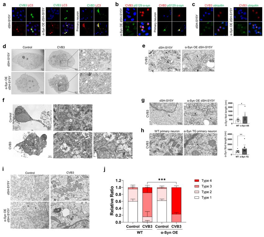
α-Syn regulates CVB3 replication in mouse brains
To confirm whether CVB3 also interacts with α-syn in the brain, CVB3 was intraperitoneally injected into
mice. At day 7 postinfection (PI), the detection of CVB3 was more pronounced in brains from α-syn TG
mice, compared with WT mice (Fig. 6a). The in vivo data were consistent with the in vitro data. IHC
analysis of the brain from WT mice detected CVB3 in the olfactory area, anterior cingulate area, lateral
septal nucleus, hippocampal region, fimbria, corticospinal tract, and hypothalamus, mainly along the
ventricles, which were colocalized with the IBA-1 microglial marker. In addition, CVB3 was also detected in
the hippocampus, lateral thalamus, and midbrain, which colocalized with the TUJ-1 neuronal marker.
CVB3 did not colocalize with the GFAP astrocyte marker (Fig. 6b,c), suggesting that CVB3 was detected in
the brain and infected with microglia and neurons, but not astrocytes.
CVB3 was observed in neurons located in the hippocampus, lateral thalamus, and midbrain at day 4 PI.
At day 7 PI, the number of neurons infected with CVB3 was increased compared to that in the mice at day
4 PI (Fig. 6d). At day 28 PI, the number of midbrain neurons infected with CVB3 was clearly increased
whereas the number of neurons located in other regions infected by CVB3 was relatively decreased
(Fig. 6d). The pattern of infected neurons was more accelerated in α-syn TG mice than in WT mice. At day
4 PI, the number of neurons infected with CVB3 was not significantly different between WT mice and α-
syn TG mice, but relatively more neurons infected with CVB3 were observed in the hippocampus of α-syn
Page 10/31TG mice, compared with that of WT mice (Fig. 6d). At day 7 PI, the pattern of infected neurons in TG mice
was comparable with that in WT mice at day 28 PI (Fig. 6d). The number of CVB3 infected microglia
increased at day 7 PI (Fig. 6e). At day 28 PI, the number of CVB3 infected microglia was similar to that at
day 7 PI (Fig. 6e). The number of CVB3 infected microglia was also higher in TG mice than in WT mice
during the same period (Fig. 6e).
At day 28 PI, the cleaved caspase 3 marker for apoptosis, was focally observed throughout the brain of
WT mice (Fig. 6f and Supplementary Fig. 3). This observation was also evident in TG mice at day 7 PI,
which was comparable to that in WT mice at day 28 PI (Fig. 6g). Additionally, CVB3 were infected
dopaminergic neurons located in the substantia nigra (Fig. 6h). The numbers of tyrosine hydroxylase
(TH)-positive cells were decreased in the substantia nigra from CVB3 infected mice at day 28 PI,
compared with the control, which was also comparable to that in TG mice at day 7 PI (Fig. 6i,j).
We then monitored the survival rate of both WT and α-syn TG mice. The survival of α-syn TG mice was
poorer than survival of WT mice. Weight loss was more severe in α-syn TG mice. The findings suggested
that α-syn TG mice were more susceptible to CVB3 infection (Fig. 6k). CVB3 is a cardiotropic virus that
induces myocarditis [16]. Therefore, we monitored myocardial damage and replication of CVB3 in the
heart. Heart damage and the level of VP1 in the myocardium from both mice groups were similar
(Supplementary Fig. 4a,b). The findings suggested that the difference in the survival rate of both mice
groups may not have been due to cardiac damage. These results suggested that CVB3 infection in the
mouse brain caused neuronal death, including dopaminergic neurons located in the substantia nigra, and
that α-syn accelerated the replication of CVB3 and neuronal death by CVB3.
CVB3 induces α-syn inclusions in mouse brains
Next, we examined the colocalization of α-syn and CVB3 VP1 in the brain of mice infected with CVB3. We
did not observe colocalization of α-syn and CVB3 VP1 in the brains of WT or α-syn TG mice. Instead, at
day 7 PI, a few α-syn inclusions in the cell body were observed in the interpeduncular nucleus from WT
mice where CVB3 was infected and the intensity of α-syn was lower than that of the control (Fig. 7a).
However, α-syn inclusions were not stained with the pSer129 α-syn antibody. In α-syn TG mice, more α-syn
inclusions were observed in the same region, although they were also not stained with the pSer129 α-syn
antibody (Fig. 7a). We performed western blotting to detect pSer129 α-syn. At day 28 PI in WT mice, pSer
129 α-syn was weakly detected and a decrease in α-syn expression was observed (Fig. 7b). Similar
observations were made in α-syn TG mice at day 7 PI, suggesting that α-syn inclusions were induced by
CVB3 infection and were accelerated by α-syn overexpression (Fig. 7c).
Discussion
Despite much research, the pathogenesis of PD remains elusive. Both genetic and environmental factors
in combination are suspected to contribute to the pathogenesis of PD [35]. LBs are the main pathological
hallmark of PD. Additionally, Lewy pathology progressively involves more regions of the nervous system
as the disease advances, and it is present prior to the appearance of motor symptoms in PD [8],
Page 11/31Accordingly, it is important to identify which factors initiate Lewy pathology to understand the
pathogenesis of PD. Several factors are suspected to be triggers of Lewy pathology. Several
environmental factors that contribute include pathogens, such as influenza virus, environmental
pollutants like pesticides, heavy metals, and head trauma [36]. In particular, viral infection has long been
considered a risk factor for neurodegenerative diseases [13]. The present data demonstrate that CVB3
interacts with α-syn to promote PD.
Concerning the influence of CVB3 on α-syn, CVB3 infection induces very large autophagy-related
structures ranging 10–20 µm in diameter in neurons. These structures colocalize with LC3 and have a
similar morphology to that of megaphagosomes shown in pancreatic acinar cells [27]. They also stain
with α-syn, pSer129 α-syn, and ubiquitin antibodies, suggesting a resemblance to LBs. LBs have
significant morphological diversity and are heterogeneous in their shape, biochemical composition, and
organization [29]. Examinations of the brains of patients with PD using super-high resolution microscopy
based on stimulated emission depletion (STED) revealed LBS with crowded organelles and lipid
membranes. This prompted the proposal that α-syn may modulate the compartmentalization and
function of membranes and organelles in LB-affected cells [30]. A recent report demonstrated the
formation of filament-like structures accompanying the sequestration of lipids, organelles, and
endomembrane structures using a seeding-based model of α-syn fibrillization, which recapitulated the
features of LBs observed in the brains of patients with PD [37]. Likewise, our TEM findings also
suggested that CVB3 infection resulted in the clustering of several organelles in the perinuclear space in
neurons. The crowded organelles contained damaged mitochondria and many fibrillar structures
surrounded the organelles. The number of fibrillar structures in α-syn OE dSH-SY5Y cells was higher than
that in dSH-SY5Y cells. These structures in α-syn OE dSH-SY5Y cells were also longer than those in dSH-
SY5Y cells. Given that α-syn fibrils exhibit 20 nm diameter in vitro [38], these observations suggested that
these fibrillar structures may be α-syn fibrils. Additionally, the process of LB formation consists of several
stages [39, 40]. CVB3 induced different types of LB-like inclusion bodies over time, which may reflect the
maturation of the inclusion bodies. In α-syn OE cells, the maturation of these inclusions was accelerated
and inclusion bodies were more condensed, suggesting that α-syn may regulate the maturation of
inclusion bodies as a major component. In addition, mitochondrial damage was induced by CVB3, which
was more in α-syn OE dSH-SY5Y cells. The observations are supported in part by previous studies
demonstrating that α-syn localizes in mitochondria and α-syn OE cells exhibit mitochondrial dysfunction
[41–43]. We demonstrated that CVB3 inhibited the late stage of the autophagic process in dSH-SY5Y
cells, consistent with a previous study [27]. It is well known that α-syn is somewhat degraded through
autophagy in cells [44–46], although other mechanisms have also been reported to be involved in α-syn
degradation [47]. Blocked late stage autophagy has been reported to cause α-syn accumulation [48].
Accordingly, the use of autophagy machinery by CVB3 may induce the formation of LB-like inclusions
associated with α-syn.
Although CVB3 induced large inclusion bodies containing α-syn, its expression was decreased. We
confirmed this using in vitro and in vivo model systems, and open source data from CVB3-infected hearts
of mice. In particular, neighboring cells may be more affected. How α-syn expression is regulated
Page 12/31differentially requires further investigation. A previous report demonstrated that WNV induced α-syn
expression, and α-syn was proposed as a viral restriction factor [21]. We also observed that treatment
with polyIC increased α-syn expression in dSH-SY5Y cells, suggesting that α-syn expression can be
regulated by viral infection. However, the decrease in α-syn expression may be CVB3 specific.
Interestingly, decreased α-syn mRNA in brains of patients with PD has been described [49, 50]. These
findings are contentious due to several technical issues, including sampling and normalization methods.
Our analysis of open data sources from patients with PD also confirmed it. Therefore, CVB3 infection
may reflect patients with PD.
When we infected mice with CVB3, CVB3 infection first appeared in the region of several anatomical
structures along the ventricles, suggesting the route of CVB3 into the CNS from the periphery. Neuron and
microglia infection progressed with time to other regions. In neurons, CVB3 was observed in the
hippocampus, lateral thalamus, and midbrain in the brain of mice at day 4 PI. Although we observed the
colocalization of CVB3 with α-syn in vitro, we could not observe the colocalization in the brain of mice
infected with CVB3. Instead, we observed α-syn aggregation in the cell bodies of neurons located in the
midbrain of WT mice at day 28 PI, and more neurons containing cytosolic α-syn aggregation were
observed in α-syn TG mice at day 7 PI. In addition, western blot analysis indicated that pSer129 α-syn
was slightly increased in the brain infected with CVB3. In a previous report [36], the authors proposed that
triggers alone are usually insufficient for PD to develop. Triggers often act transiently, with the triggering
event lasting a few weeks or months and occurring relatively early in the life of individuals that develop
PD. Accordingly, CVB3 infection may act transiently. It alone may not induce LB formation in the brain,
unlike in vitro. Alternatively, the neuronal α-syn aggregation observed in our in vivo system may not reach
the same mature stage as the PD LB, because our in vivo model system is not adequate for prolonged
observations due to the high mortality of CVB3 infection in C57BL/6 mice [51]. The long-term
consequences of CVB3 infection in the CNS are largely unknown. However, these viruses persist, and the
presence of viral RNA by itself is potentially pathogenic in some cases including schizophrenia [52] and
amyotrophic lateral sclerosis [53]. Interestingly, CVB3 infected BALB/c mice, which are more susceptible
to chronic CVB3 infection, reportedly showed TDP-43 aggregation in the hippocampal region at 90 days
PI [54]. In addition, it has been reported that cytosolic aggregates as well as soluble oligomers, which
were not observed in healthy controls, were observed in the heart of patients with dilated cardiomyopathy,
which is suspected to be caused by CVB3 infections [55, 56]. Congo red merged islet amyloid polypeptide
was also seen in pancreatic biopsies of patients with type 1 diabetes, suggesting that these aggregations
may be caused by enteroviruses stressing beta cells [57]. Accordingly, viral-induced intracellular protein
inclusions do not restrict neurons; rather, they are a general phenomenon.
In this study, we observed that α-syn expression regulated CVB3 replication. Overexpression of α-syn
increased CVB3 replication and CVB3 induced cytotoxicity. We confirmed this in vivo in brains of α-syn
TG mice. CVB3 replication was increased and the spread of CVB3 infected regions was also accelerated
in α-syn TG mice compared with control mice. In addition, CVB3 infection induced neuronal cell death,
especially dopaminergic neuronal cell death in the substantia nigra. Interestingly and supporting our
observation, patients with chronic EV71 encephalitis whose symptoms persisted for more than 2 months
Page 13/31displayed damage in most of the midbrain, including the substantia nigra [58]. Dopaminergic neuronal
cell death in the substantia nigra was also accelerated in α-syn TG mice. Furthermore, α-syn TG mice
survived less than WT mice. A previous report demonstrated that α-syn expression inhibits WNV growth
and replication, resulting in increased mortality of α-syn knock-out mice [21], which contradicts our
findings, demonstrating that α-syn TG mice show more CVB3 replication and a lower survival rate after
CVB3 infection. We cannot completely explain the discrepancy between these results. The balance of the
amount of α-syn in the brain may be important for the regulation of viral infection. Nevertheless, this
could be explained by the viral usage of autophagy. Previous studies have suggested that CVB3 uses
autophagosomes as their replication centers by inhibiting the binding of autophagosomes to lysosomes
[27, 59, 60]. The induction of autophagy in neurons was also associated with increased CVB3 replication
[59–61]. Given that an increase in α-syn expression accelerated autophagic flux, this environment favors
the replication of CVB3. In contrast, autophagy is known to inhibit WNV replication [62, 63]. Accordingly, it
may be virus-specific, and autophagy in both viruses may explain this discrepancy.
The epidemiologic links suggest that viral exposure over time may increase the risk for PD, although it is
unclear whether any specific viral infection causes PD. It could be related to direct virus-induced
cytotoxicity or virus-related inflammation [64–66]. Influenza viral infections induce parkinsonian
symptoms and a significant increase in phosphorylation and aggregation of α-syn [67, 68]. Repeated viral
infection may induce α-syn expression or/and α-syn aggregation, and chronic viral infection also induces
further inflammation, which may initiate and progress PD. Likewise, we observed that α-syn responded to
CVB3 infection. CVB3 infection regulated α-syn expression and aggregation. α-Syn may function as a
defense mechanism of the host cells against viral infection. The finding that infecting CVB3 interacted
with α-syn in various ways suggests an unexpected role of α-syn in the pathogenesis of PD. Further
studies are needed to explore this in more detail.
Conclusions
We investigated the relationship between CVB3 infection and α-syn. CVB3 infection induced α-syn
associated inclusion body formation in neurons as a trigger. These inclusion bodies contained clustered
organelles including damaged mitochondria with α-syn fibrils. α-Syn overexpression accelerated inclusion
body formation and induced more concentric inclusion bodies. Brains from CVB3 infected mice harbored
α-syn aggregates in the cell body of the midbrain. The data indicate that CVB3 infection blocks the late
stage of autophagy, inducing inclusion body formation containing α-syn fibrils. Overexpression of α-syn
favors CVB3 replication and related cytotoxicity. The survival of α-syn TG mice was poor. CVB3
replication was more extensive in these mice, with further neuronal cell death, including dopaminergic
neurons. α-Syn overexpression accelerated autophagic flux, which favored the replication of CVB3. The
collective findings clarify the mechanism of LB formation and the pathogenesis of PD associated with
CVB3 infection.
Abbreviations
Page 14/31PD : Parkinson's disease
α-Syn : α-Synuclein
CVB3 : coxsackievirus B3
LBs : Lewy bodies
AD : Alzheimer’s disease
WNV : West Nile virus
LC3 : microtubule-associated protein 1A/1B light chain 3B
GFAP : glial fibrillary acidic protein
GAPDH : glyceraldehyde 3-phosphate dehydrogenase
RA : retinoic acid
Baf A1 : bafilomycin A1
poly IC : polyinosine:polycytidylic acid
EBD : Evans blue dye
TG : transgenic
NSE : neuron specific enolase
WT : wild-type
KO : knockout
DMEM : Dulbecco’s modified Eagle’s medium
FBS : fetal bovine serum
MOI : multiplicity of infection
IP injection : intraperitoneal injection
PFU : plaque forming units
PBS : phosphate-buffered saline
RT-PCR : reverse transcription polymerase chain reaction
Page 15/31ICC : immunocytochemistry
BSA : bovine serum albumin
ECL : enhanced chemiluminescence system
h : human
m : mouse
SNCA : α-Synuclein
TEM : transmission electron microscopy
LDH : lactate dehydrogenase
IHC : immunocytochemistry
PBST : PBS containing 0.2% Triton X‐100
GO : gene ontology
DAPI : 4′,6-diamidino-2-phenylindole
CVB3-EGFP : CVB3 variant co-expressing EGFP
DEG : differential expressed genes
GEO : gene expression omnibus
iPSC : induced pluripotent stem cells
PI : postinfection
CTX : cortex
MOB : main olfactory bulb
AON : anterior olfactory nucleus
LSX : lateral septal complex
HY : hypothalamus
ACA : anterior cingulate area
fxs : fornix system
Page 16/31MO : somatomotor area
HCF : Hippocampal formation
TH : thalamus
CP : caudoputamen
Cst : corticospinal tract
RSP : retrosplenial area
PRT : pretectal region
MRN : midbrain reticular nucleus
SN : substantia nigra
c-caspase3 : cleaved caspase3
Hp - hippocampus
TH : tyrosine hydroxylase
DAB : diaminobenzidine
SNc : substantia nigra pars compacta
STED : stimulated emission depletion
MAVS : mitochondrial antiviral signaling protein
EV71 : enterovirus71
Declarations
Ethics approval and consent to participate
All animal procedures were conducted according to the guidelines established by the Ajou University
School of Medicine Ethics Review Committee (IACUC No. 2016-0047).
Consent for publication
Not applicable
Page 17/31Availability of data and materials
The datasets used and/or analysed during the current study are available from the corresponding author
on reasonable request.
Competing interests
The authors declare that they have no competing interests
Funding
This research was supported by the National Research Foundation of Korea (NRF) grants funded by the
Korean government (Ministry of Science and ICT) (Grant No. NRF-2017R1E1A1A01073713, NRF-
2019R1A5A2026045).
Authors' contributions
SJP, UJ, and SMP designed the study. SJP and UJ performed the experiments. SJP, UJ, and SMP
analyzed and discussed the data. SJP and SMP wrote and edited the manuscript. All authors read and
approved the final manuscript.
Acknowledgements
We thank to Dr. E. Jeon (Samsung Medical Center, Seoul, Korea) for kindly providing the H3 variant of
CVB3, the Woodruff strain, and EGFP-CVB3. We also thank NIFDS for providing C56BL/6-Tg (NSE-
haSyn)Korl mice and their information.
References
1. Poewe W, Seppi K, Tanner CM, Halliday GM, Brundin P, Volkmann J, Schrag A-E, Lang AE: Parkinson
disease. Nature reviews Disease primers 2017, 3:17013.
2. Kalia LV, Lang AE: Parkinson's disease. Lancet 2015, 386:896-912.
3. Spillantini MG, Schmidt ML, Lee VM, Trojanowski JQ, Jakes R, Goedert M: Alpha-synuclein in Lewy
bodies. Nature 1997, 388:839-840.
4. Eschbach J, Danzer KM: α-Synuclein in Parkinson's disease: pathogenic function and translation into
animal models. Neurodegenerative Diseases 2014, 14:1-17.
5. Mullin S, Schapira A: The genetics of Parkinson's disease. British medical bulletin 2015, 114:39-52.
Page 18/316. Nalls MA, Pankratz N, Lill CM, Do CB, Hernandez DG, Saad M, DeStefano AL, Kara E, Bras J, Sharma
M: Large-scale meta-analysis of genome-wide association data identifies six new risk loci for
Parkinson's disease. Nature genetics 2014, 46:989.
7. Satake W, Nakabayashi Y, Mizuta I, Hirota Y, Ito C, Kubo M, Kawaguchi T, Tsunoda T, Watanabe M,
Takeda A: Genome-wide association study identifies common variants at four loci as genetic risk
factors for Parkinson's disease. Nature genetics 2009, 41:1303.
8. Braak H, Del Tredici K, Rub U, de Vos RA, Jansen Steur EN, Braak E: Staging of brain pathology
related to sporadic Parkinson's disease. Neurobiology of aging 2003, 24:197-211.
9. Braak H, Del Tredici K: Invited Article: Nervous system pathology in sporadic Parkinson disease.
Neurology 2008, 70:1916-1925.
10. Volpicelli-Daley L, Brundin P: Prion-like propagation of pathology in Parkinson disease. Handb Clin
Neurol 2018, 153:321-335.
11. Vargas JY, Grudina C, Zurzolo C: The prion-like spreading of alpha-synuclein: From in vitro to in vivo
models of Parkinson's disease. Ageing Res Rev 2019, 50:89-101.
12. Tofaris GK, Goedert M, Spillantini MG: The Transcellular Propagation and Intracellular Trafficking of
alpha-Synuclein. Cold Spring Harb Perspect Med 2017, 7.
13. Jang H, Boltz DA, Webster RG, Smeyne RJ: Viral parkinsonism. Biochimica et Biophysica Acta (BBA)-
Molecular Basis of Disease 2009, 1792:714-721.
14. Tulisiak CT, Mercado G, Peelaerts W, Brundin L, Brundin P: Can infections trigger alpha-
synucleinopathies? Prog Mol Biol Transl Sci 2019, 168:299-322.
15. Garmaroudi FS, Marchant D, Hendry R, Luo H, Yang D, Ye X, Shi J, McManus BM: Coxsackievirus B3
replication and pathogenesis. Future microbiology 2015, 10:629-653.
16. Tam PE: Coxsackievirus myocarditis: interplay between virus and host in the pathogenesis of heart
disease. Viral immunology 2006, 19:133-146.
17. Muir P, Van Loon A: Enterovirus infections of the central nervous system. Intervirology 1997, 40:153-
166.
18. Chapman NM, Kim KS: Persistent coxsackievirus infection: enterovirus persistence in chronic
myocarditis and dilated cardiomyopathy. Curr Top Microbiol Immunol 2008, 323:275-292.
19. Alidjinou EK, Sané F, Engelmann I, Geenen V, Hober D: Enterovirus persistence as a mechanism in the
pathogenesis of type 1 diabetes. Discov Med 2014, 18:273-282.
20. Dourmashkin RR, McCall SA, Dourmashkin N, Hannah MJ: Virus-like particles and enterovirus
antigen found in the brainstem neurons of Parkinson’s disease. F1000Research 2018, 7.
21. Beatman EL, Massey A, Shives KD, Burrack KS, Chamanian M, Morrison TE, Beckham JD: Alpha-
synuclein expression restricts RNA viral infections in the brain. Journal of virology 2016, 90:2767-
2782.
22. Choi YR, Cha SH, Kang SJ, Kim JB, Jou I, Park SM: Prion-like Propagation of alpha-Synuclein Is
Regulated by the FcgammaRIIB-SHP-1/2 Signaling Pathway in Neurons. Cell Rep 2018, 22:136-148.
Page 19/3123. Yoon S, Park S, Han J, Kang J, Kim J, Lee J, Park S, Shin H, Kim K, Yun M: Caspase-dependent cell
death-associated release of nucleosome and damage-associated molecular patterns. Cell death &
disease 2014, 5:e1494-e1494.
24. Shults NV, Kanovka SS, Ten Eyck JE, Rybka V, Suzuki YJ: Ultrastructural changes of the right
ventricular myocytes in pulmonary arterial hypertension. Journal of the American Heart Association
2019, 8:e011227.
25. Robinson SM, Tsueng G, Sin J, Mangale V, Rahawi S, McIntyre LL, Williams W, Kha N, Cruz C,
Hancock BM: Coxsackievirus B exits the host cell in shed microvesicles displaying autophagosomal
markers. PLoS pathogens 2014, 10:e1004045.
26. Alexopoulou L, Holt AC, Medzhitov R, Flavell RA: Recognition of double-stranded RNA and activation
of NF-κB by Toll-like receptor 3. Nature 2001, 413:732.
27. Kemball CC, Alirezaei M, Flynn CT, Wood MR, Harkins S, Kiosses WB, Whitton JL: Coxsackievirus
infection induces autophagy-like vesicles and megaphagosomes in pancreatic acinar cells in vivo.
Journal of virology 2010, 84:12110-12124.
28. Shibutani ST, Saitoh T, Nowag H, Münz C, Yoshimori T: Autophagy and autophagy-related proteins in
the immune system. Nature immunology 2015, 16:1014-1024.
29. Wakabayashi K, Tanji K, Odagiri S, Miki Y, Mori F, Takahashi H: The Lewy body in Parkinson’s disease
and related neurodegenerative disorders. Molecular neurobiology 2013, 47:495-508.
30. Shahmoradian SH, Lewis AJ, Genoud C, Hench J, Moors TE, Navarro PP, Castaño-Díez D,
Schweighauser G, Graff-Meyer A, Goldie KN: Lewy pathology in Parkinson’s disease consists of
crowded organelles and lipid membranes. Nature Neuroscience 2019, 22:1099.
31. Wong J, Zhang J, Si X, Gao G, Mao I, McManus BM, Luo H: Autophagosome supports coxsackievirus
B3 replication in host cells. Journal of virology 2008, 82:9143-9153.
32. Klionsky DJ, Abdelmohsen K, Abe A, Abedin MJ, Abeliovich H, Acevedo Arozena A, Adachi H, Adams
CM, Adams PD, Adeli K, et al: Guidelines for the use and interpretation of assays for monitoring
autophagy (3rd edition). Autophagy 2016, 12:1-222.
33. Yoshii SR, Mizushima N: Monitoring and Measuring Autophagy. Int J Mol Sci 2017, 18:1865.
34. Xilouri M, Vogiatzi T, Vekrellis K, Park D, Stefanis L: Abberant α-synuclein confers toxicity to neurons
in part through inhibition of chaperone-mediated autophagy. PloS one 2009, 4:e5515.
35. Klein C, Schlossmacher MG: Parkinson disease, 10 years after its genetic revolution: multiple clues to
a complex disorder. Neurology 2007, 69:2093-2104.
36. Johnson ME, Stecher B, Labrie V, Brundin L, Brundin P: Triggers, Facilitators, and Aggravators:
Redefining Parkinson's Disease Pathogenesis. Trends Neurosci 2019, 42:4-13.
37. Mahul-Mellier AL, Burtscher J, Maharjan N, Weerens L, Croisier M, Kuttler F, Leleu M, Knott GW,
Lashuel HA: The process of Lewy body formation, rather than simply alpha-synuclein fibrillization, is
one of the major drivers of neurodegeneration. Proc Natl Acad Sci U S A 2020, 117:4971-4982.
Page 20/3138. Lashuel HA: Do Lewy bodies contain alpha-synuclein fibrils? and Does it matter? A brief history and
critical analysis of recent reports. Neurobiology of disease 2020:104876.
39. Wakabayashi K, Hayashi S, Kakita A, Yamada M, Toyoshima Y, Yoshimoto M, Takahashi H:
Accumulation of alpha-synuclein/NACP is a cytopathological feature common to Lewy body disease
and multiple system atrophy. Acta Neuropathol 1998, 96:445-452.
40. Katsuse O, Iseki E, Marui W, Kosaka K: Developmental stages of cortical Lewy bodies and their
relation to axonal transport blockage in brains of patients with dementia with Lewy bodies. J Neurol
Sci 2003, 211:29-35.
41. Devi L, Raghavendran V, Prabhu BM, Avadhani NG, Anandatheerthavarada HK: Mitochondrial import
and accumulation of alpha-synuclein impair complex I in human dopaminergic neuronal cultures
and Parkinson disease brain. J Biol Chem 2008, 283:9089-9100.
42. Nakamura K, Nemani VM, Azarbal F, Skibinski G, Levy JM, Egami K, Munishkina L, Zhang J, Gardner
B, Wakabayashi J, et al: Direct membrane association drives mitochondrial fission by the Parkinson
disease-associated protein alpha-synuclein. J Biol Chem 2011, 286:20710-20726.
43. Park JH, Burgess JD, Faroqi AH, DeMeo NN, Fiesel FC, Springer W, Delenclos M, McLean PJ: Alpha-
synuclein-induced mitochondrial dysfunction is mediated via a sirtuin 3-dependent pathway. Mol
Neurodegener 2020, 15:5.
44. Paxinou E, Chen Q, Weisse M, Giasson BI, Norris EH, Rueter SM, Trojanowski JQ, Lee VM,
Ischiropoulos H: Induction of alpha-synuclein aggregation by intracellular nitrative insult. The
Journal of neuroscience : the official journal of the Society for Neuroscience 2001, 21:8053-8061.
45. Vogiatzi T, Xilouri M, Vekrellis K, Stefanis L: Wild type α-synuclein is degraded by chaperone-
mediated autophagy and macroautophagy in neuronal cells. Journal of Biological Chemistry 2008,
283:23542-23556.
46. Alvarez-Erviti L, Rodriguez-Oroz MC, Cooper JM, Caballero C, Ferrer I, Obeso JA, Schapira AH:
Chaperone-mediated autophagy markers in Parkinson disease brains. Archives of neurology 2010,
67:1464-1472.
47. Stefanis L, Emmanouilidou E, Pantazopoulou M, Kirik D, Vekrellis K, Tofaris GK: How is alpha-
synuclein cleared from the cell? Journal of neurochemistry 2019, 150:577-590.
48. Ejlerskov P, Hultberg JG, Wang J, Carlsson R, Ambjorn M, Kuss M, Liu Y, Porcu G, Kolkova K, Friis
Rundsten C, et al: Lack of Neuronal IFN-beta-IFNAR Causes Lewy Body- and Parkinson's Disease-like
Dementia. Cell 2015, 163:324-339.
49. Neystat M, Lynch T, Przedborski S, Kholodilov N, Rzhetskaya M, Burke RE: Alpha-synuclein
expression in substantia nigra and cortex in Parkinson's disease. Mov Disord 1999, 14:417-422.
50. Kingsbury AE, Daniel SE, Sangha H, Eisen S, Lees AJ, Foster OJ: Alteration in alpha-synuclein mRNA
expression in Parkinson's disease. Mov Disord 2004, 19:162-170.
51. Blyszczuk P: Myocarditis in Humans and in Experimental Animal Models. Front Cardiovasc Med
2019, 6:64.
Page 21/31You can also read