Integrin-linked kinase tunes cell-cell and cell-matrix adhesions to regulate the switch between apoptosis and EMT downstream of TGFβ1
←
→
Page content transcription
If your browser does not render page correctly, please read the page content below
MBoC | ARTICLE

Integrin-linked kinase tunes cell–cell and cell-
matrix adhesions to regulate the switch between
apoptosis and EMT downstream of TGFβ1
Ayse Nihan Kilinca, Siyang Hanb, Lena A. Barretta, Niroshan Anandasivama,
and Celeste M. Nelsona,b,*
a
Departments of Chemical & Biological Engineering and bMolecular Biology, Princeton University, Princeton, NJ 08544

ABSTRACT Epithelial-mesenchymal transition (EMT) is a morphogenetic process that en-                                            Monitoring Editor
dows epithelial cells with migratory and invasive potential. Mechanical and chemical signals                                    Valerie Marie Weaver
                                                                                                                                University of California,
from the tumor microenvironment can activate the EMT program, thereby permitting cancer
                                                                                                                                San Francisco
cells to invade the surrounding stroma and disseminate to distant organs. Transforming
growth factor β1 (TGFβ1) is a potent inducer of EMT that can also induce apoptosis depend-                                      Received: Feb 3, 2020
ing on the microenvironmental context. In particular, stiff microenvironments promote EMT                                       Revised: Dec 17, 2020
while softer ones promote apoptosis. Here, we investigated the molecular signaling down-                                        Accepted: Dec 29, 2020
stream of matrix stiffness that regulates the phenotypic switch in response to TGFβ1 and
uncovered a critical role for integrin-linked kinase (ILK). Specifically, depleting ILK from mam-
mary epithelial cells precludes their ability to sense the stiffness of their microenvironment. In
response to treatment with TGFβ1, ILK-depleted cells undergo apoptosis on both soft and
stiff substrata. We found that knockdown of ILK decreases focal adhesions and increases
cell–cell adhesions, thus shifting the balance from cell–matrix to cell–cell adhesion. High cell–
matrix adhesion promotes EMT whereas high cell–cell adhesion promotes apoptosis down-
stream of TGFβ1. These results highlight an important role for ILK in controlling cell pheno-
type by regulating adhesive connections to the local microenvironment.

INTRODUCTION
Epithelial-mesenchymal transition (EMT), a morphological and tran-                        metastatic dissemination (Friedl and Alexander, 2011). This transi-
scriptional program that permits epithelial cells to transdifferentiate                   tion is regulated by both mechanical and chemical signals from the
into motile mesenchymal cells, occurs during normal embryonic de-                         surrounding microenvironment. For example, the extracellular ma-
velopment and tissue regeneration. When activated aberrantly,                             trix (ECM) is stiffer within breast tumors than within the surrounding
EMT can contribute pathologically to the metastasis of cancer                             normal mammary tissue, which correlates with poor survival (Paszek
(Varga and Greten, 2017). During the EMT process, cells down-reg-                         et al., 2005). In response to increased ECM stiffness, the transcrip-
ulate intercellular adhesions, detach from their neighbors, and                           tion factor Twist1 acts as a mechanomediator to activate EMT (Wei
up-regulate cell–matrix adhesions to facilitate local invasion and                        et al., 2015). Moreover, stiff matrices induce the localization of
                                                                                          Rac1b to the plasma membrane, thereby promoting the assembly
                                                                                          of the NADPH oxidase complex, which activates EMT in response to
This article was published online ahead of print in MBoC in Press (http://www             matrix metalloproteinases (MMPs) (Lee et al., 2012). Stiff matrices
.molbiolcell.org/cgi/doi/10.1091/mbc.E20-02-0092) on January 6, 2021.
Competing interests: The authors declare no competing or financial interest.
                                                                                          have been found to promote EMT in several different epithelial cell
*Address correspondence to: Celeste M. Nelson (celesten@princeton.edu).                   types (Lee et al., 2012; Wei et al., 2015; Rice et al., 2017).
Abbreviations used: ECM, extracellular matrix; EMT, epithelial-mesenchymal                    Transforming growth factor β1 (TGFβ1) is the most well-studied
transition; ILK, integrin-linked kinase; MMP, matrix metalloproteinase; PBS, phos-        regulator of EMT within the cellular microenvironment and can also
phate-buffered saline; PDMS, polydimethylsiloxane; shRNA, short hairpin RNA;
TGF, transforming growth factor.                                                          induce apoptosis under some microenvironmental conditions. How
© 2021 Kilinc et al. This article is distributed by The American Society for Cell Biol-   cells ultimately decide to undergo EMT or apoptosis in response to
ogy under license from the author(s). Two months after publication it is available        TGFβ1 remains unclear, but several studies have suggested context-
to the public under an Attribution–Noncommercial–Share Alike 3.0 Unported
Creative Commons License (http://creativecommons.org/licenses/by-nc-sa/3.0).
                                                                                          dependent mechanisms. Mouse mammary epithelial cells were
“ASCB®,” “The American Society for Cell Biology®,” and “Molecular Biology of              found to become resistant to TGFβ1-induced apoptosis after chronic
the Cell®” are registered trademarks of The American Society for Cell Biology.            exposure to the ligand, which suppresses pro-apoptotic pathways

402 | A. N. Kilinc et al.                                                                                                            Molecular Biology of the Cell
(Gal et al., 2008). In mouse liver epithelial cells, TGFβ1 induces          that has a stiffness similar to that of the normal mammary gland
apoptosis or EMT depending on the stage of the cell cycle, with             (E∼130 Pa), shcntl cells are rounded and form multicellular clusters
apoptosis and EMT taking place during G2/M and G1/S, respec-                that resemble the structure of mammary acini. Although depleting
tively (Yang et al., 2006). In human retinal pigment epithelial cells,      ILK (shILK) does not alter cell morphology on soft substrata, this
TGFβ1 was shown to regulate the cell cycle through survivin and,            manipulation causes cells to exhibit a rounded morphology on stiff
thus, the switch between EMT and apoptosis (Lee et al., 2013). ECM          substrata. These morphologies strongly resemble the epithelial-like
stiffness also regulates cell fate downstream of TGFβ1 in both canine       morphology before EMT and the mesenchymal-like morphology
kidney and mouse mammary epithelial cells—soft matrices promote             after, which inspired us to examine the role of ILK in EMT.
apoptosis, whereas stiff matrices promote EMT (Leight et al., 2012).            To understand how ILK expression affects the EMT process, we
     Mechanical signals from the cellular microenvironment are trans-       performed time-lapse imaging analysis of shcntl and shILK cells cul-
lated into biochemical signaling cascades through cellular mecha-           tured on soft or stiff substrata in the presence or absence of TGFβ1.
notransduction, which enables cells to sense, respond, and adapt to         We found that on soft substrata, cells start to detach from the matrix
their physical surroundings. This process is largely mediated by inte-      within 24 h of exposure to TGFβ1, irrespective of ILK levels (Figure
grin signaling. Integrins bind to specific motifs in ECM proteins such      1C). Most cells have completely detached from soft substrata within
as fibronectin, whereas their cytoplasmic tails associate with linker       48 h of exposure to TGFβ1 (Figure 1C). However, on stiff substrata,
proteins including talin, vinculin, and integrin-linked kinase (ILK).       treatment with TGFβ1 only induces detachment of shILK cells. In
ILK, interacting with parvin (which binds to paxillin and actin) and        contrast, shcntl cells cultured on stiff substrata adopt an elongated
PINCH, takes part in transmitting forces from integrins to the actin        and spindle-like morphology within 24 h of TGFβ1 exposure, consis-
cytoskeleton and in forming functional focal adhesion complexes             tent with an EMT phenotype. Surprisingly, we observed a similar
(Elad et al., 2013). ILK plays a fundamental role in many different         spindle-like morphology in the few shILK cells that remain anchored
signaling pathways and cell behaviors including EMT. Overexpres-            on stiff substrata after 48 h of exposure to TGFβ1 (Figure 1C). To
sion of ILK causes human kidney and mouse mammary epithelial                determine whether the detachment of shILK cells is the result of
cells to acquire a mesenchymal morphology, in part by disrupting            apoptosis at earlier time points, we used a fluorometric assay to
cell–cell adhesions (Somasiri et al., 2001; Li et al., 2003). Conversely,   examine the activity of caspase-3 after 24 h of TGFβ treatment. As
we recently found that depleting ILK abolishes the ability of cells to      expected, this analysis confirmed that depletion of ILK in cells on
change their morphology in response to ECM stiffness—mammary                both soft and stiff microenvironments synergistically promotes
epithelial cells that normally spread out on stiff substrata exhibit a      apoptosis, instead of EMT, downstream of TGFβ1 (Figure 1D).
rounded morphology when ILK is down-regulated (Han et al., 2018).
     Here, we examined the mechanism by which matrix stiffness con-         ILK depletion prevents TGFβ1-induced EMT in mammary
trols the fate of mammary epithelial cells in response to treatment         epithelial cells in a time- and stiffness-dependent manner
with TGFβ1. Specifically, we focused on signaling from the ECM              Because its depletion results in the detachment of cells and a de-
through ILK. We used engineered synthetic substrata to mimic the            layed EMT morphology in the cells that remain anchored after pro-
mechanical stiffnesses of the normal mammary gland and of breast            longed TGFβ1 treatment, we speculated that ILK may regulate the
tumors. We found that ILK modulates how NMuMG mouse mam-                    switch between EMT and apoptosis. To test this hypothesis, we cul-
mary epithelial cells respond to TGFβ1 in a stiffness-dependent and         tured shcntl and shILK cells on soft or stiff substrata in the presence
time-dependent manner: knockdown of ILK prevents EMT in the                 or absence of TGFβ1 for 24 h and assayed for the expression of
first 24 h of exposure to TGFβ1 in mammary epithelial cells cultured        several EMT markers using immunoblotting and immunofluores-
on both soft and stiff substrata; after 48 h of treatment with TGFβ1,       cence analysis. Depletion of ILK itself promotes an increase in the
a small population of ILK-depleted mammary epithelial cells un-             expression of the epithelial marker E-cadherin and decreases in that
dergo EMT only on stiff substrata while the majority of these cells         of the mesenchymal markers αSMA, Snail, and vimentin in cells on
undergo apoptosis. Moreover, depletion of ILK and culture on soft           stiff substrata (Figure 2, A and B). Treatment of shcntl cells with
microenvironments synergistically promote TGFβ1-induced apop-               TGFβ1 for 24 h induces changes in expression consistent with an
tosis. We further found that depletion of ILK increases the mem-            EMT program, including down-regulation of E-cadherin (Figure 2,
brane localization of cell–cell adhesion proteins and decreases the         A–C) and ZO1 (Supplemental Figure S1) and up-regulation of
activation of focal adhesion proteins, suggesting that ILK regulates        αSMA, Snail, and vimentin (Figure 2, A–C) on stiff substrata, as well
the balance between cell–cell and cell–ECM adhesion. Through bal-           as in the few cells that remain attached on soft substrata. In contrast,
ancing adhesion to the microenvironment, ILK controls the func-             depletion of ILK abolishes the induction of EMT by TGFβ1; cell–cell
tional switch between EMT and apoptosis downstream of TGFβ1.                contacts remain intact and the expression levels of E-cadherin,
Taken together, our results suggest a central role for ILK in tuning        αSMA, and Snail remain relatively unchanged. Consistently, shILK
the relative strength of cell–cell and cell–matrix adhesions, which         cells are less migratory than controls (Supplemental Figure S2).
represents critical input in cell-fate decisions downstream of TGFβ1.       These data show that the expression of ILK is required for TGFβ1 to
                                                                            induce EMT in NMuMG mouse mammary epithelial cells.
RESULTS                                                                          After 48 h of exposure to TGFβ1, shcntl cells continue to show
Soft substrata and loss of ILK promote apoptosis in                         decreased expression of E-cadherin and ZO1 and increased expres-
response to TGFβ1                                                           sion of Snail and αSMA (Figure 3, A and B) on both soft and stiff
We previously found that stably depleting ILK using short hairpin           substrata, as expected. The few shILK cells that remain attached on
RNA (shRNA) leads to a striking change in the morphology of                 stiff substrata show a decrease in E-cadherin localization to cell–cell
NMuMG mouse mammary epithelial cells (Figure 1, A and B) (Han               contacts and increased levels of Snail and αSMA (Figure 3). The low
et al., 2018). Specifically, control NMuMG cells (shcntl) exhibit an        numbers of shcntl cells that remain anchored on soft substrata and
elongated, well-spread, and spindle-like morphology when cultured           the similarly low numbers of shILK cells that remain on both
on stiff substratum with an elastic modulus similar to that of              microenvironments precluded us from performing immunoblotting
mammary tumors (E∼4020 Pa). When cultured on a soft substratum              analysis at this late time point.

Volume 32 March 1, 2021                                                                            ILK switches between EMT and apoptosis    | 403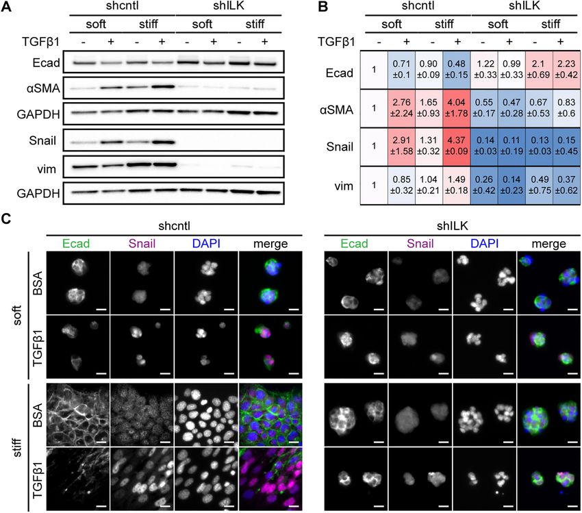
FIGURE 1: Depletion of ILK and culture on soft substrata synergistically promote TGFβ1-induced apoptosis in NMuMG
mouse mammary epithelial cells. (A) Phase-contrast images of shcntl and shILK cells cultured on soft (E∼130 Pa) or stiff
(E∼4020 Pa) fibronectin-coated polyacrylamide substrata. Scale bars, 50 μm. (B) Immunoblotting analysis for ILK
normalized to GAPDH in lysates from shcntl and shILK cells. (C) Phase-contrast images of shcntl and shILK cells cultured
on soft or stiff substrata and treated with TGFβ1 (2 ng/ml). Scale bars, 100 μm. (D) Quantification of caspase-3 activity in
shcntl and shILK cells cultured on soft or stiff substrata and treated with or without TGFβ1 for 24 h. Error bars represent
SEM of three (B) or six (D) independent replicates; ns, not significant; *P < 0.05; **P < 0.01; ***P < 0.001; ****P < 0.0001.

404 | A. N. Kilinc et al.                                                                                                 Molecular Biology of the Cell
FIGURE 2: Depletion of ILK prevents TGFβ1-induced EMT in NMuMG mouse mammary epithelial cells. (A) Immuno blotting analysis for E-cadherin, αSMA, Snail, vimentin, or GAPDH in shcntl and shILK cells cultured on soft or stiff substrata and treated with or without TGFβ1 for 24 h; immunoblotting replicates are shown in Supplemental Figure S1. (B) Quantification of immunoblotting analysis; shown are mean ± SD of three independent replicates. Color coding indicates relative increase (red) or decrease (blue) in expression, compared with shcntl cells on soft substrata in the absence of TGFβ1. (C) Immunofluorescence analysis for E-cadherin (green), Snail (magenta), and nuclei (blue) in shcntl and shILK cells cultured on soft or stiff substrata and treated with or without TGFβ1 for 24 h. Scale bars, 10 μm. Depletion of ILK decreases the activation of focal adhesion activated FAK (pY397-FAK) and activated paxillin (pY118-paxillin) proteins (Figure 4, A and B; Supplemental Figure S4). Immunoblotting analy- Our data suggest that the switch between EMT and apoptosis sis also revealed that FAK and paxillin are phosphorylated and acti- downstream of TGFβ1 may be associated with ILK-mediated vated in control cells on stiff substratum and that this regulation is changes in adhesion and mechanosensing. The likelihood of this abolished in shILK cells (Figure 4, A and B; Supplemental Figure S4). possibility was strengthened by following the shapes of cells retro- The levels of total FAK and total paxillin are not affected by substra- spectively in response to treatment with TGFβ1. We carried out tum stiffness (Figure 4, A and B). Consistently, immunofluorescence time-lapse imaging analysis, identified cells that underwent EMT or analysis of cells cultured on plastic revealed that depletion of ILK apoptosis, and then followed those cells retrospectively in time. We causes an enrichment of cortical actin and decreases the formation found that cells that eventually underwent apoptosis maintained a of cell–matrix adhesions containing activated FAK and paxillin rounded morphology, while those that eventually underwent EMT (Figure 4, C–F). Depleting ILK thus reduces cell–matrix adhesion in exhibited a more irregular shape, generating protrusions through- mammary epithelial cells. out the 48 h of TGFβ1 treatment (Supplemental Figure S3). These data indicate that cells that maintain cell–ECM adhesions are able to Depletion of ILK increases cell–cell adhesion undergo EMT in response to TGFβ1. Cortical actin is often associated with cadherin-based cell–cell adhe- To determine the effects of ILK on cell–matrix adhesion, we ex- sions (Ratheesh and Yap, 2012; Bachir et al., 2017). That we found amined the expression and localization of focal adhesion proteins an increase in cortical actin in shILK cells suggested that ILK expres- by immunoblotting and immunofluorescence analysis. We found sion may regulate adherens junctions. Indeed, immunofluorescence that depletion of ILK does not affect the total levels of FAK (Figure analysis revealed elevated levels of α-catenin, β-catenin, and corti- 4A) or paxillin (Figure 4B), but significantly decreases the levels of cal actin at sites of intercellular contact in shILK cells cultured on Volume 32 March 1, 2021 ILK switches between EMT and apoptosis | 405
The balance between cell–ECM and
cell–cell adhesion regulates the switch
between EMT and apoptosis
downstream of TGFβ1
Since shILK cells show enhanced cell–cell
adhesion and preferentially undergo apop-
tosis instead of EMT in response to TGFβ1,
we hypothesized that experimentally de-
creasing cell–cell adhesion may permit
TGFβ1 to induce EMT in these cells. Previ-
ous studies have reported that disrupting
cell–cell adhesion using function-blocking
anti-E-cadherin antibodies or shRNA-medi-
ated knockdown promotes the expression
of mesenchymal markers and the acquisition
of a mesenchymal morphology in mammary
and kidney epithelial cells (Behrens et al.,
1989; Qin et al., 2005; Lehembre et al.,
2008). Conversely, manipulating cell–cell
adhesion by plating cells at different densi-
ties also affects the expression of epithelial
and mesenchymal markers in mammary epi-
thelial cells, with decreased E-cadherin and
increased vimentin at low densities (Cichon
et al., 2015). We therefore needed an alter-
native approach to test our hypothesis. We
cultured shcntl and shILK cells on substrata
micropatterned with single-cell-sized islands
to promote the isolation of individual cells
from their neighbors and thus achieve low
cell–cell adhesion in both cell types at the
same density. We used islands sufficiently
large (900 μm2) to permit cells to spread to
the same extent as those on stiff substrata in
the presence of TGFβ1 (650 μm2), but small
enough that we were able to capture indi-
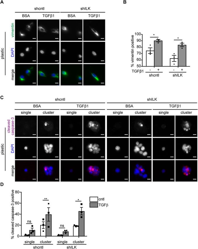
vidual cells on a fraction of the islands. Curi-
ously, we observed that a fraction of the
FIGURE 3: On stiff substrata, depletion of ILK leads to either cell detachment or a delayed EMT
isolated cells express the mesenchymal
after prolonged exposure to TGFβ1. Immunofluorescence analysis for (A) E-cadherin (green),
Snail (magenta), and nuclei (blue), or (Β) ZO1 (green), αSMA (magenta), and nuclei (blue) in shcntl marker vimentin, even before exposure to
and shILK cells cultured on soft or stiff substrata and treated with or without TGFβ1 for 48 h. TGFβ1, regardless of ILK levels. Nonethe-
Scale bars, 10 μm. less, we found that treating both micropat-
terned shcntl and shILK cells with TGFβ1 for
24 h increases the fraction of cells that ex-
plastic compared with controls (Figure 5, A and B). Consistent with press vimentin, consistent with the promotion of an EMT phenotype
our findings on stiff substrata (Figure 2A), immunoblotting analysis (Figure 6, A and B). In contrast, we found low levels of apoptosis in
revealed an increase in the levels of E-cadherin and α-catenin in single micropatterned cells, as inferred from staining for cleaved
shILK cells cultured on plastic as compared with controls (Figure 5C). caspase-3 (Figure 6, C and D). Instead, we found that TGFβ1 in-
The association of adherens junction proteins with the actin cyto- duces high levels of apoptosis in cells located within multicellular
skeleton correlates with strong cell–cell interactions (Mege and Ishi- clusters on the micropatterned islands (Figure 6, C and D). Our re-
yama, 2017). To quantify the extent to which adherens junction com- sults suggest that low cell–cell adhesion promotes EMT in response
ponents are associated with the cytoskeleton in shILK cells, we to TGFβ1, rather than apoptosis, irrespective of ILK expression.
measured the distribution of E-cadherin, α-catenin, and β-catenin in Conversely, to determine whether low cell–ECM adhesion pro-
Triton X–100-fractionated cell lysates. We found a robust increase in motes apoptosis in response to TGFβ1, we pharmacologically inhib-
the level of cytoskeleton-associated β-catenin, as indicated by the ited FAK in NMuMG cells cultured on soft or stiff substrata in the
Triton X-100-insoluble fraction, in shILK cells as compared with con- presence or absence of TGFβ1. We treated cells with increasing
trols (Figure 5D). However, the total level of β-catenin is relatively concentrations of the FAK inhibitor PF-573228 and ‘as expected’
unchanged in response to depletion of ILK (Figure 5C). Therefore, found that the levels of pY397-FAK decrease as inhibitor concentra-
depleting ILK expression enhances adherens junctions in NMuMG tion increases (Figure 7A). Caspase-3 activity analysis revealed sig-
cells. These data reveal that the decrease in cell–matrix adhesion nificantly higher levels of apoptosis in FAK-inhibited cells that were
associated with loss of ILK is balanced or compensated by an in- treated with TGFβ1 than in vehicle-treated controls (Figure 7B).
crease in cell–cell adhesion. Therefore, low cell–cell and high cell–ECM adhesion favors EMT,
406 | A. N. Kilinc et al. Molecular Biology of the Cell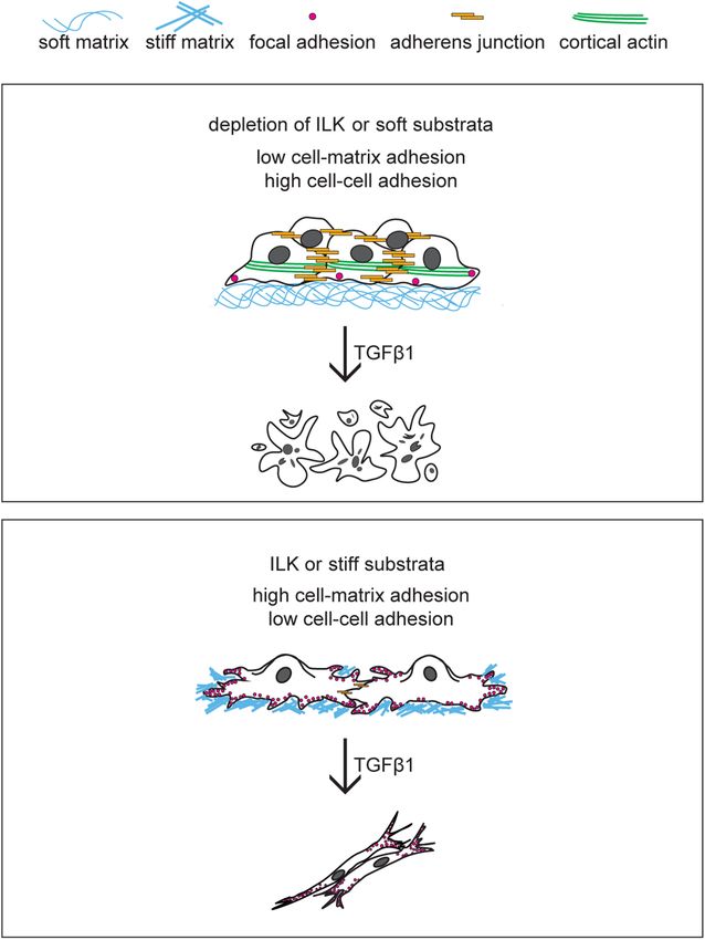
FIGURE 4: Depletion of ILK decreases the activation and assembly of focal adhesion proteins. Immunoblotting analysis
for GAPDH and (A) total FAK, pY397-FAK (pFAK), (B) total paxillin (Pax), and pY118-paxillin (pPax) in shcntl and shILK
cells cultured on soft or stiff substrata. Quantification shows mean ± SD for three independent experiments;
immunoblotting replicates and statistical analysis are shown in Supplemental Figure S4. Immunofluorescence analysis for
F-actin (green), nuclei (blue), and (C) FAK (magenta), (D) pFAK (magenta), (E) Pax (magenta), or (F) pPax (magenta) in
shcntl and shILK cells cultured on plastic. Scale bars, 20 μm.

whereas high cell–cell and low cell–ECM adhesion favors apoptosis          adherens junction protein E-cadherin and reorganize cortical actin
in response to TGFβ1. Surprisingly, previous work found that reduc-        into stress fibers (Somasiri et al., 2001). Similarly, soft matrices pro-
ing FAK activity, which would be expected to decrease mechano-             mote apoptosis downstream of TGFβ1 while stiff matrices promote
sensing, is not sufficient to alter apoptosis on stiff substrata (Leight   EMT, consistent with previous findings (Leight et al., 2012), suggest-
et al., 2012); however, that study used lower concentrations of the        ing that depletion of ILK disrupts the ability of cells to sense the
FAK inhibitor, which may explain their observation. Our data show          mechanical stiffness of their microenvironment. This is not surprising
that the mechanical stiffness of the microenvironment signals              given the essential role for ILK in integrin signaling and focal adhe-
through ILK to tune the relative balance of cell–cell to cell–matrix       sion assembly. However, when ILK-deficient mammary epithelial
adhesion, which in turn defines the phenotypic response to TGFβ1.          cells are cultured as individual cells on micropatterned islands and
                                                                           thus prevented from forming cell–cell adhesions, exposure to
DISCUSSION                                                                 TGFβ1 increases the fraction of cells that express the mesenchymal
Here we demonstrate that ILK controls the fate of mammary epithe-          marker vimentin. Our results highlight the role of ILK in regulating
lial cells downstream of TGFβ1 by modulating the balance between           the cooperation between cell–cell and cell–matrix adhesions in re-
cell–cell and cell–matrix adhesion. Specifically, depletion of ILK         sponse to the stiffness of the microenvironment, which governs cell-
leads to increased cortical actin and adherens junctions and de-           fate decisions downstream of TGFβ1 (Figure 8).
creased focal adhesions. This promotes apoptosis and suppresses/               Stiff microenvironments were previously found to promote EMT.
delays EMT in response to TGFβ1, regardless of matrix stiffness.           Increased ECM stiffness regulates the PI3K/Akt signaling pathway to
These results are consistent with a previous study, which found that       activate EMT downstream of TGFβ1 in mammary epithelial cells
overexpression of ILK causes mammary epithelial cells to lose the          (Leight et al., 2012). In addition, increased matrix stiffness induces

Volume 32 March 1, 2021                                                                           ILK switches between EMT and apoptosis     | 407
FIGURE 5: Depletion of ILK strengthens cell–cell adhesion. Immunofluorescence analysis for F-actin (green), nuclei (blue), and (A) α-catenin (αcat; magenta) or (B) β-catenin (βcat; magenta) in shcntl and shILK cells. Scale bars, 20 μm. (C) Immunoblotting analysis for E-cadherin (Ecad), αcat, and βcat in shcntl and shILK cells. (D) Immunoblotting analysis for Triton X-100-insoluble or soluble fractions of Ecad, αcat, and βcat in lysates from shcntl and shILK cells. Quantification shows mean ± SD for three independent experiments; immunoblotting replicates and statistical analysis are shown in Supplemental Figure S5. the nuclear translocation of Twist1, an EMT-associated transcription from apoptosis and able to undergo EMT after prolonged exposure factor, by releasing it from its cytoplasmic binding partner G3BP2 to TGFβ1. In line with this, signaling through FAK is required for (Wei et al., 2015). In a mouse model of pancreatic cancer, fibrotic transcriptional up-regulation of mesechymal markers and delocal- stiffening increases vimentin expression, decreases E-cadherin ex- ization of membrane-associated E-cadherin downstream of TGFβ1 pression, and induces a mesenchymal morphology, consistent with in murine hepatocytes (Cicchini et al., 2008). These results suggest an EMT program (Rice et al., 2017). Our data add to the underlying that the promotion of EMT by substratum stiffness is not a binary mechanism of this regulation. Even in the absence of TGFβ1, deple- decision—the rate at which the transition takes place may depend tion of ILK reduces the expression of Snail, suggesting that ILK (and on the relative extent of focal adhesion engagement. cell–ECM adhesion) positively regulates Snail. Snail has been shown Apoptosis and EMT are two fundamental cellular behaviors that to confer resistance to apoptosis downstream of TGFβ1 and is suf- are tightly controlled by TGFβ1. In the context of cancer, TGFβ1 can ficient to trigger EMT (Franco et al., 2010). Given that stiff microen- suppress tumor progression by inducing cell death (Guasch et al., vironments up-regulate the expression of ILK (Han et al., 2018), it is 2007) or promote cancer cell invasion and dissemination by stimulat- possible that stiff matrices prime cells to undergo EMT through ILK- ing EMT (Heldin et al., 2012). The diverse downstream consequences mediated signaling through Snail. Moreover, we found that deplet- of exposure to TGFβ1 raise several questions: why does TGFβ1 ing ILK prevents EMT on stiff substrata after 24 h of treatment with stimulate apoptosis in some cells and EMT in others? Is there a de- TGFβ1, consistent with previous work (Serrano et al., 2013). Of inter- fault cellular fate downstream of TGFβ1 and, if so, under what cir- est, after 48 h of exposure to TGFβ1, the majority of cells that lack cumstances will the other fate be activated? Some studies have sug- ILK have detached and the remaining cells have undergone EMT. gested that cell-fate decisions downstream of TGFβ1 are cell Based on our analysis of focal adhesions and our single-cell mi- cycle-dependent (Yang et al., 2006; Iordanskaia and Nawshad, 2011; cropatterning experiments, we speculate that the few shILK cells Lee et al., 2013). Our results suggest an alternative but not mutually that remain anchored might have relatively stronger focal adhesions exclusive mechanism, in which high cell–matrix adhesion promotes (but still weaker than those in control cells), and are thus rescued EMT whereas high cell–cell adhesion promotes apoptosis. 408 | A. N. Kilinc et al. Molecular Biology of the Cell
FIGURE 6: Low cell–cell adhesion promotes TGFβ1-induced EMT. (A) Immunofluorescence analysis for vimentin (green)
and nuclei (blue) in shcntl and shILK cells on micropatterns treated with or without TGFβ1 for 24 h. Scale bars, 10 μm.
(B) Quantification of the percentage of vimentin-positive single shcntl and shILK cells cultured on micropatterned
substrata treated with or without TGFβ1 for 24 h. (C) Immunofluorescence analysis for cleaved caspase-3 (magenta) and
nuclei (blue) in shcntl and shILK cells on micropatterns treated with or without TGFβ1 for 24 h. Scale bars, 10 μm.
(D) Quantification of the percentage of cleaved caspase-3-positive shcntl and shILK cells cultured individually or as
clusters on micropatterned substrata and treated with or without TGFβ1 for 24 h. Error bars represent SEM of three
independent replicates; each replicate contains at least 200 cells per condition. *P < 0.05; **P < 0.01.

   Cadherin-based adherens junctions and integrin-based focal ad-         adhesive network that regulates several cellular functions (Weber
hesions are intrinsically linked by the actin cytoskeleton. In response   et al., 2011; Mui et al., 2016). Engagement of one type of adhesion
to mechanical stimuli, these two classes of adhesions engage com-         may alter the other type of adhesion. This cross-talk facilitates mor-
mon signaling proteins to remodel the actin cytoskeleton and coor-        phogenesis in embryonic development and enables tensional ho-
dinate intracellular and intercellular force transmission, forming an     meostasis in healthy tissue. However, this interdependence may

Volume 32 March 1, 2021                                                                         ILK switches between EMT and apoptosis   | 409
confirmed to be free of mycoplasma using a
                                                                                                       commercially available kit (Lonza). NMuMG
                                                                                                       cells were seeded at a density of 50,000
                                                                                                       cells/cm2 on plastic or fibronectin-coated
                                                                                                       polyacrylamide substrata of soft (E∼130 Pa)
                                                                                                       or stiff (E∼4020 Pa) compliances and cul-
                                                                                                       tured at 37°C and 5% CO2 as described pre-
                                                                                                       viously (Lee et al., 2012). Cells were cultured
                                                                                                       for 8–10 h before treatment with TGFβ1 (2
                                                                                                       ng/ml; Peprotech) and/or FAK inhibitor (PF-
                                                                                                       573228; Sigma), which were added to the
                                                                                                       culture medium. Cells were subsequently
                                                                                                       cultured for 6 and 12 h (immunofluores-
                                                                                                       cence), 24 h (caspase-3 activity, immunob-
                                                                                                       lotting, immunofluorescence), or 48 h
FIGURE 7: Low cell–matrix adhesion promotes TGFβ1-induced apoptosis. (A) Immunoblotting
                                                                                                       (immunofluorescence).
analysis for pFAK, total FAK, and GAPDH in cells treated with different concentrations of the
FAK inhibitor PF-573228. Quantification shows mean ± SD for three independent experiments;
                                                                                                           To knock down the expression of ILK,
immunoblotting replicates are shown in Supplemental Figure S6. (B) Quantification of caspase-3         NMuMG cells were transduced with lentivi-
activity in cells cultured on soft or stiff substrata in the presence or absence of PF-573228 (5 μM)   ral particles carrying two shRNAs against ILK
and treated with or without TGFβ1 for 24 h. Error bars represent SEM of three independent              (sc-35667-V and sc-35666-V; Santa Cruz) or
replicates. **P < 0.01; ***P < 0.001.                                                                  control lentivirus expressing a scrambled
                                                                                                       shRNA sequence. Stable shRNA-expressing

also promote malignant transformation. The degree of E-cadherin
engagement directs the spatial arrangement of cell–matrix traction
forces (Mertz et al., 2013), and force transmission through E-cad-
herin adhesions requires coordinated modulation of local cell–ECM
adhesion (Ng et al., 2014). During branching morphogenesis of the
salivary gland, fibrillar fibronectin can increase cell–matrix adhesion
concomitantly with a loss of cell–cell adhesion to promote the for-
mation of epithelial clefts (Sakai et al., 2003). Furthermore, planar
cell polarity signaling regulates the spatiotemporal assembly of fi-
bronectin by transfering tension from cadherin-based adhesions to
integrins (Dzamba et al., 2009). In colon cancer cells, integrin signal-
ing is required for Src-induced internalization of E-cadherin (Avizie-
nyte et al., 2002). In ovarian carcinoma cells, integrin clustering in-
duces MMP-dependent shedding of the E-cadherin ectodomain,
which allows cells to migrate independently (Symowicz et al., 2007).
Here we found that ILK modulates the balance between adherens
junctions and focal adhesions in mammary epithelial cells, which
regulates cell shape and acts as an essential component in cell-fate
decisions downstream of TGFβ1. Of note, depleting ILK not only
results in decreased levels of phosphorylated FAK but also abolishes
the ability of stiff matrices to induce the phosphorylation of this focal
adhesion protein. These results suggest that ILK may be required for
stiffness-dependent assembly and activation of focal adhesions. Ex-
amining how ILK mediates the cross-talk between cell–cell and cell–
ECM adhesions may contribute to a more comprehensive under-
standing of the cellular adhesive network. Future work should also
focus on elucidating how signaling inputs from cell–cell and cell–
ECM adhesion are integrated to trigger the switch between EMT
and apoptosis.

MATERIALS AND METHODS
Request a protocol through Bio-protocol.

Cell culture and reagents
                                                                             FIGURE 8: ILK controls cell fate downstream of TGFβ1 by modulating
NMuMG mouse mammary epithelial cells (passed at a 1:10 ratio                 the balance between cell–cell and cell–matrix adhesion. Similar to soft
and used before passage 20) were obtained from the American                  matrix, depletion of ILK increases cell–cell adhesion and decreases
Type Culture Collection and cultured in high glucose DMEM                    cell–matrix adhesion, promoting apoptosis downstream of TGFβ1. In
(HyClone) supplemented with 10% fetal bovine serum (Atlanta Bio-             contrast, a stiff microenvironment leads to elevated cell–matrix
logicals) and 50 μg/ml gentamicin (Life Technologies); cells were            adhesion and promotes EMT downstream of TGFβ1.

410 | A. N. Kilinc et al.                                                                                                 Molecular Biology of the Cell
clones were produced according to the manufacturer’s instructions concentrations were measured in each sample, followed by im-
and selected using puromycin. munoblotting analysis.
Micropatterned substrata Immunoblotting analysis
Single-cell-sized islands (30 × 30 μm2) of fibronectin were prepared Samples were lysed in RIPA lysis buffer (Thermo Scientific) supple-
as described previously (Gomez et al., 2010). Briefly, sterile stamps mented with Complete protease inhibitor cocktail (Roche) and pro-
of polydimethylsiloxane (PDMS; Sylgard 184) were coated with 25 tein concentrations were measured using the detergent-compatible
μg/ml fibronectin (BD Biosciences, Franklin Lakes, NJ) in phosphate- Protein Assay Kit (Bio-Rad). Samples were then mixed with Laemmli
buffered saline (PBS) overnight at 4°C and dried under compressed sample buffer, boiled at 95°C for 5 min, resolved by SDS–PAGE, and
nitrogen. Fibronectin was microcontact-printed onto custom-made, transferred to nitrocellulose membranes. Membranes were then
UV/ozone-treated, PDMS-coated glass-bottom tissue culture blocked in 5% nonfat milk in 0.1% Tween-20 in Tris-buffered saline
dishes, and unstamped regions were blocked with 1% Synperonic and incubated overnight at 4°C in blocking buffer containing anti-
F108 (Fluka, Buchs, Germany) in PBS. Cells were then seeded at a bodies specific for ILK (3862, 1:1000; Cell Signaling), E-cadherin
density of 50,000 cells per sample and allowed to adhere to the re- (3195, 1:1000; Cell Signaling), vimentin (V5255, 1:1000; Cell Signal-
sulting islands of fibronectin. ing), Snail (3895, 1:500; Cell Signaling), αSMA (A5228, 1:500;
Sigma), FAK (3285S, 1:500; Cell Signaling), pY397-FAK (44625G,
Time-lapse imaging 1:500; Invitrogen), paxillin (ab32084, 1:1000; Abcam), pY118-paxil-
Time-lapse movies were acquired using a Hamamatsu C4742-95 lin (44722G, 1:1000; Invitrogen), α-catenin (ab51032, 1:3500; Ab-
camera attached to a Nikon Ti-U inverted microscope and fitted cam), β-catenin (ab32572, 1:3500; Abcam), or GAPDH (3683S,
with an environmental chamber held at 90% humidity, 37°C, and 5% 1:1000; Cell Signaling). Bands were detected using horseradish-
CO2 (Pathology Devices, Westminster, MD). Images were acquired peroxidase–conjugated secondary antibodies (1:5000; Cell Signal-
at 10× magnification every 30 min for a total of 48 h. ing) and Amersham ECL Western Blotting Reagent (GE Healthcare)
as a chemiluminescent substrate. Densitometry analysis was per-
Immunofluorescence analysis formed using ImageJ.
Samples were fixed with 4% paraformaldehyde for 15 min, washed
with PBS, and then permeabilized with 0.3% Triton X-100 for 30 min. Scratch wound assay
After blocking for 1 h with 5% goat serum (Sigma) and 2% bovine Confluent monolayers of shcntl or shILK cells were treated with
serum albumin, the samples were incubated with primary antibody TGFβ1 (2 ng/ml; Peprotech) for 24 h. Monolayers were then
against E-cadherin (3195, 1:300; Cell Signaling), Snail (3895, 1:300; scratched with pipette tips and washed once with culture medium
Cell Signaling), αSMA (A5228, 1:200; Sigma), ZO1 (40-2200, 1:300; to remove cellular debris. Time-lapse images were acquired at 10×
Thermo Fisher Scientific), vimentin (V2258, 1:200; Sigma), cleaved magnification every 30 min for a total of 40 h. Wound areas were
caspase-3 (9961, 1:200; Cell Signaling), FAK (ab40794, 1:200; Ab- calculated using the ImageJ Wound Healing Tool plugin (Volker
cam), pY397-FAK (44-625G, 1:50; Invitrogen), paxillin (ab32084, Baeker) for at least three independent experiments.
1:200; Abcam), pY118-paxillin (44-722G, 1:200; Invitrogen), α-
catenin (ab51032, 1:200; Abcam), or β-catenin (D10A8, 1:100; Cell Cell tracking analysis
Signaling). Samples were then washed with PBS and incubated with Cell tracking analysis was conducted on ImageJ software using the
Alexa Fluor–conjugated secondary antibodies (1:200; Invitrogen). Manual Tracking plugin (Fabrice Cordeli). For each of 20 tracks per
Nuclei were counterstained with Hoechst 33342 (1:1000; Invitro- condition, one cell was manually followed through a series of half-
gen). After additional washes with PBS, samples were visualized hour or 1-h interval stacks. Once the cell remained relatively station-
using a 20×/0.45 NA air objective on a Nikon Eclipse Ti-U inverted ary in the frame, the track was completed. Cell speed data were
fluorescence microscope (Nikon) equipped with a Hamamatsu collected from the output of the plugin for analysis.
ORCA charge-coupled device camera. Image analysis was per-
formed on at least 200 cells for each experimental group over three Statistical analysis
independent experiments using ImageJ. Data represent the results of at least three independent experi-
ments; sample number and sizes are reported in the figure legends.
Caspase-3 activity assay All statistical analyses were performed using GraphPad Prism 5.0,
Caspase-3 activity was determined using an EnzChek Caspase-3 As- following the recommendations in the Prism statistics guide. Statisti-
say Kit #2 (Invitrogen) according to the manufacturer’s instructions. cal comparisons of means for normally distributed data were con-
Caspase-3 activity was normalized to the total cell number as deter- ducted using an unpaired Student’s t test (equal variances; Figure
mined using a hemacytometer. 6B; Supplemental Figure S5), paired t test (Gaussian distribution;
Figure 6D), Welch’s t test (unequal variances; Supplemental Figure
Soluble and insoluble protein fractionation assay S2), one-way ANOVA with Tukey–Kramer multiple comparisons test
To separate insoluble (membrane-bound) and soluble (intracellu- (Supplemental Figure S4), or two-way ANOVA with Tukey multiple
lar) fractions of adherens junction proteins, cells were lysed in cy- comparisons test (Figures 1D and 7B). Normality was determined
toskeletal buffer (300 mM sucrose, 10 mM PIPES, pH 6.8, 50 mM using either the D’Agostino–Pearson test (for large samples) or the
NaCl, 3 mM MgCl2, 0.5% Triton X-100) containing complete pro- Shapiro–Wilk test (for all others). P < 0.05 was considered to repre-
tease inhibitors (Roche). The lysates were incubated on ice for 15 sent a significant difference between experimental groups.
min, passed through a 27-gauge needle four times, and then cen-
trifuged at 14,000 rpm for 20 min at 4°C. The supernatant was ACKNOWLEDGMENTS
collected as the soluble fraction. The pellet was solubilized in 8% This work was supported in part by grants from the National Insti-
SDS lysis buffer, followed by sonication and centrifuged at 14,000 tutes of Health (CA187692, CA214292), the David & Lucile Packard
rpm for 10 min and collected as the insoluble fraction. Protein Foundation, the Camille & Henry Dreyfus Foundation, and a Faculty
Volume 32 March 1, 2021 ILK switches between EMT and apoptosis | 411Scholars Award from the Howard Hughes Medical Institute. A.N.K. Lehembre F, Yilmaz M, Wicki A, Schomber T, Strittmatter K, Ziegler D, Kren
was supported in part by a postdoctoral fellowship from the New A, Went P, Derksen PW, Berns A, et al. (2008). NCAM-induced focal
adhesion assembly: a functional switch upon loss of E-cadherin. EMBO
Jersey Commission on Cancer Research. J 27, 2603–2615.
Leight JL, Wozniak MA, Chen S, Lynch ML, Chen CS (2012). Matrix rigid-
REFERENCES ity regulates a switch between TGF-beta1-induced apoptosis and
Avizienyte E, Wyke AW, Jones RJ, McLean GW, Westhoff MA, Brunton VG, epithelial-mesenchymal transition. Mol Biol Cell 23, 781–791.
Frame MC (2002). Src-induced de-regulation of E-cadherin in colon Li Y, Yang J, Dai C, Wu C, Liu Y (2003). Role for integrin-linked kinase in me-
cancer cells requires integrin signalling. Nat Cell Biol 4, 632–638. diating tubular epithelial to mesenchymal transition and renal interstitial
Bachir AI, Horwitz AR, Nelson WJ, Bianchini JM (2017). Actin-based adhe- fibrogenesis. J Clin. Invest 112, 503–516.
sion modules mediate cell interactions with the extracellular matrix and Mege RM, Ishiyama N (2017). Integration of cadherin adhesion and cyto-
neighboring cells. Cold Spring Harb Perspect Biol 9, a023234. skeleton at adherens junctions. Cold Spring Harb Perspect Biol 9.
Behrens J, Mareel MM, Van Roy FM, Birchmeier W (1989). Dissecting tumor Mertz AF, Che Y, Banerjee S, Goldstein JM, Rosowski KA, Revilla SF, Niessen
cell invasion: epithelial cells acquire invasive properties after the loss of CM, Marchetti MC, Dufresne ER, Horsley V (2013). Cadherin-based in-
uvomorulin-mediated cell-cell adhesion. J Cell Biol 108, 2435–2447. tercellular adhesions organize epithelial cell-matrix traction forces. Proc
Cicchini C, Laudadio I, Citarella F, Corazzari M, Steindler C, Conigliaro A, Natl Acad Sci USA 110, 842–847.
Fantoni A, Amicone L, Tripodi M (2008). TGFbeta-induced EMT requires Mui KL, Chen CS, Assoian RK (2016). The mechanical regulation of integrin-
focal adhesion kinase (FAK) signaling. Exp Cell Res 314, 143–152. cadherin crosstalk organizes cells, signaling and forces. J Cell Sci 129,
Cichon MA, Nelson CM, Radisky DC (2015). Regulation of epithelial-mes- 1093–1100.
enchymal transition in breast cancer cells by cell contact and adhesion. Ng MR, Besser A, Brugge JS, Danuser G (2014). Mapping the dynamics
Cancer Inform 14, 1–13. of force transduction at cell-cell junctions of epithelial clusters. eLife 3,
Dzamba BJ, Jakab KR, Marsden M, Schwartz MA, DeSimone DW (2009). e03282.
Cadherin adhesion, tissue tension, and noncanonical Wnt signaling Paszek MJ, Zahir N, Johnson KR, Lakins JN, Rozenberg GI, Gefen A,
regulate fibronectin matrix organization. Dev Cell 16, 421–432. Reinhart-King CA, Margulies SS, Dembo M, Boettiger D, et al. (2005).
Elad N, Volberg T, Patla I, Hirschfeld-Warneken V, Grashoff C, Spatz JP, Tensional homeostasis and the malignant phenotype. Cancer Cell 8,
Fassler R, Geiger B, Medalia O (2013). The role of integrin-linked 241–254.
kinase in the molecular architecture of focal adhesions. J Cell Sci 126, Qin Y, Capaldo C, Gumbiner BM, Macara IG (2005). The mammalian
4099–4107. Scribble polarity protein regulates epithelial cell adhesion and migration
Franco DL, Mainez J, Vega S, Sancho P, Murillo MM, de Frutos CA, Del through E-cadherin. J Cell Biol 171, 1061–1071.
Castillo G, Lopez-Blau C, Fabregat I, Nieto MA (2010). Snail1 suppresses Ratheesh A, Yap AS (2012). A bigger picture: classical cadherins and the
TGF-beta-induced apoptosis and is sufficient to trigger EMT in hepato- dynamic actin cytoskeleton. Nat Rev Mol Cell Biol 13, 673–679.
cytes. J Cell Sci 123, 3467–3477. Rice AJ, Cortes E, Lachowski D, Cheung BCH, Karim SA, Morton JP, Del Rio
Friedl P, Alexander S (2011). Cancer invasion and the microenvironment: Hernandez A (2017). Matrix stiffness induces epithelial-mesenchymal
plasticity and reciprocity. Cell 147, 992–1009. transition and promotes chemoresistance in pancreatic cancer cells.
Gal A, Sjoblom T, Fedorova L, Imreh S, Beug H, Moustakas A (2008). Sus- Oncogenesis 6, e352–e352.
tained TGF beta exposure suppresses Smad and non-Smad signalling in Sakai T, Larsen M, Yamada KM (2003). Fibronectin requirement in branching
mammary epithelial cells, leading to EMT and inhibition of growth arrest morphogenesis. Nature 423, 876–881.
and apoptosis. Oncogene 27, 1218–1230. Serrano I, McDonald PC, Lock FE, Dedhar S (2013). Role of the integrin-
Gomez EW, Chen QK, Gjorevski N, Nelson CM (2010). Tissue geometry linked kinase (ILK)/Rictor complex in TGFbeta-1-induced epithelial-
patterns epithelial-mesenchymal transition via intercellular mechano- mesenchymal transition (EMT). Oncogene 32, 50–60.
transduction. J Cell Biochem 110, 44–51. Somasiri A, Howarth A, Goswami D, Dedhar S, Roskelley CD (2001). Over-
Guasch G, Schober M, Pasolli HA, Conn EB, Polak L, Fuchs E (2007). Loss expression of the integrin-linked kinase mesenchymally transforms mam-
of TGFbeta signaling destabilizes homeostasis and promotes squamous mary epithelial cells. J Cell Sci 114, 1125–1136.
cell carcinomas in stratified epithelia. Cancer Cell 12, 313–327. Symowicz J, Adley BP, Gleason KJ, Johnson JJ, Ghosh S, Fishman DA,
Han S, Pang MF, Nelson CM (2018). Substratum stiffness tunes proliferation Hudson LG, Stack MS (2007). Engagement of collagen-binding integrins
downstream of Wnt3a in part by regulating integrin-linked kinase and promotes matrix metalloproteinase-9-dependent E-cadherin ectodo-
frizzled-1. J Cell Sci 131. main shedding in ovarian carcinoma cells. Cancer Res 67, 2030–2039.
Heldin CH, Vanlandewijck M, Moustakas A (2012). Regulation of EMT by Varga J, Greten FR (2017). Cell plasticity in epithelial homeostasis and
TGFbeta in cancer. FEBS Lett 586, 1959–1970. tumorigenesis. Nat Cell Biol 19, 1133–1141.
Iordanskaia T, Nawshad A (2011). Mechanisms of transforming growth factor Weber GF, Bjerke MA, DeSimone DW (2011). Integrins and cadherins join
beta induced cell cycle arrest in palate development. J Cell Physiol 226, forces to form adhesive networks. J Cell Sci 124, 1183–1193.
1415–1424. Wei SC, Fattet L, Tsai JH, Guo Y, Pai VH, Majeski HE, Chen AC, Sah RL,
Lee J, Choi JH, Joo CK (2013). TGF-beta1 regulates cell fate during Taylor SS, Engler AJ, Yang J (2015). Matrix stiffness drives epithelial-mes-
epithelial-mesenchymal transition by upregulating survivin. Cell Death enchymal transition and tumour metastasis through a TWIST1-G3BP2
Dis 4, e714. mechanotransduction pathway. Nat Cell Biol 17, 678–688.
Lee K, Chen QK, Lui C, Cichon MA, Radisky DC, Nelson CM (2012). Matrix Yang Y, Pan X, Lei W, Wang J, Song J (2006). Transforming growth factor-
compliance regulates Rac1b localization, NADPH oxidase assembly, and beta1 induces epithelial-to-mesenchymal transition and apoptosis via a
epithelial-mesenchymal transition. Mol Biol Cell 23, 4097–4108. cell cycle-dependent mechanism. Oncogene 25, 7235–7244.
412 | A. N. Kilinc et al. Molecular Biology of the CellYou can also read