Innovative light sources for phototherapy# - De Gruyter
←
→
Page content transcription
If your browser does not render page correctly, please read the page content below
Biomolecular Concepts 2022; 13: 256–271
Research Article
Giovanni Romano, Giacomo Insero*, Santi Nonell Marrugat, Franco Fusi
Innovative light sources for phototherapy#
https://doi.org/10.1515/bmc-2022-0020
received April 10, 2022; accepted May 3, 2022
Introduction
Abstract: The use of light for therapeutic purposes dates The use of light for therapeutic purposes is an ancient
back to ancient Egypt, where the sun itself was an inno- practice. We have news of this through hieroglyphic,
vative source, probably used for the first time to heal skin cuneiform, and alphabetical writings datable between
diseases. Since then, technical innovation and advance- 3000 and 500 BC. The first light source used was the
ment in medical sciences have produced newer and more sun, without any other means in between.
sophisticated solutions for light-emitting sources and The applications of heliotherapy changed over time,
their applications in medicine. Starting from a brief his- but the sun remained the only source of light until 1700.
torical introduction, the concept of innovation in light In the second half of the eighteenth century, lenses or
sources is discussed and analysed, first from a technical filters were introduced between the sun and the body,
point of view and then in the light of their fitness to thus manufacturing the first instruments for phototherapy.
improve existing therapeutic protocols or propose new It was only with the development of electricity that the
ones. If it is true that a “pure” technical advancement first artificial light sources were born. In 1880, Thomas
is a good reason for innovation, only a sub-system of Edison’s company started the commercial production of
those advancements is innovative for phototherapy. To the first filament bulbs. In 1897, Niels Ryberg Finsen
illustrate this concept, the most representative examples created the first artificial lamp for phototherapy (arc
of innovative light sources are presented and discussed, lamp with carbon electrodes) to replace sunlight filtered
both from a technical point of view and from the per- through glass media for his experiments on Lupus (Figure 1).
spective of their diffusion and applications in the clin- These studies earned him the Nobel Prize in 1903 in
ical field. recognition of his contribution to the treatment of dis-
eases, especially lupus vulgaris, with concentrated light
Keywords: light source, phototherapy, innovation, photo-
radiation, whereby he has opened a new avenue for med-
dynamic therapy, photomedicine.
ical science. In 1927, a patent paved the way to produce
fluorescent lamps filled with mercury vapours and an
inner coating of Beryllium, soon replaced by fluorescent
oxides. This technology has had great prominence in the
phototherapy field up to the present day, with intensive
use for various pathologies of dermatological impor-
tance. Nevertheless, the great innovation of the last cen-
tury was the advent of coherent light sources, namely
# This manuscript is dedicated to Professor Silvia E. Braslavsky on the invention of the ruby laser in 1960. Only 10 years
the occasion of her 80th birthday. later did the first semiconductor light sources appear
(light-emitting diodes [LEDs]); together with lasers, LEDs
represent the key technologies for the application of
* Corresponding author: Giacomo Insero, Department of
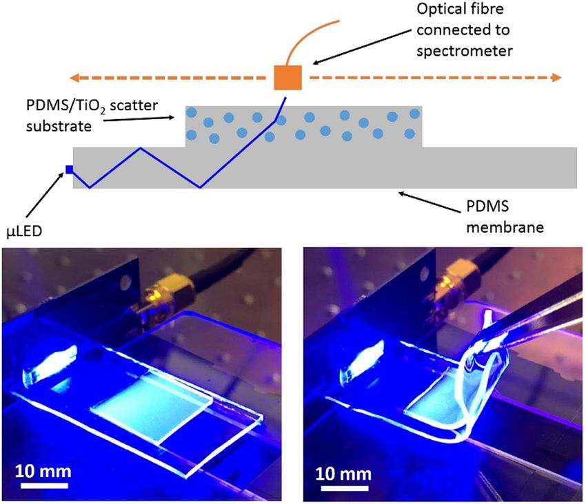
Experimental and Clinical Biomedical Sciences “Mario Serio”,
optical radiation to biomedicine. Today, light sources
University of Florence, Viale G. Pieraccini 6, 50139 Florence, emit pulsed or continuous wave wavelengths, from UVC
Italy; National Research Council, National Institute of Optics to infrared, for both therapeutic and diagnostic purposes.
(CNR-INO), Via Carrara 1, 50019 Sesto Fiorentino, FI, Italy, Since the first lamps, great progress has been made: in a
e-mail: giacomo.insero@unifi.it century, we have gone from rudimentary instrumenta-
Giovanni Romano, Franco Fusi: Department of Experimental and
tion where metal and mechanics were predominant to
Clinical Biomedical Sciences “Mario Serio”, University of Florence,
Viale G. Pieraccini 6, 50139 Florence, Italy
the current semiconductor light sources and wearable
Santi Nonell Marrugat: Institut Quimic de Sarria, Universidad illuminators. Improvements, of course, are remarkable
Ramon Llull, Via Augusta 390, 08017 Barcelona, Spain also from the patient’s perspective.
Open Access. © 2022 Giovanni Romano et al., published by De Gruyter. This work is licensed under the Creative Commons Attribution 4.0
International License.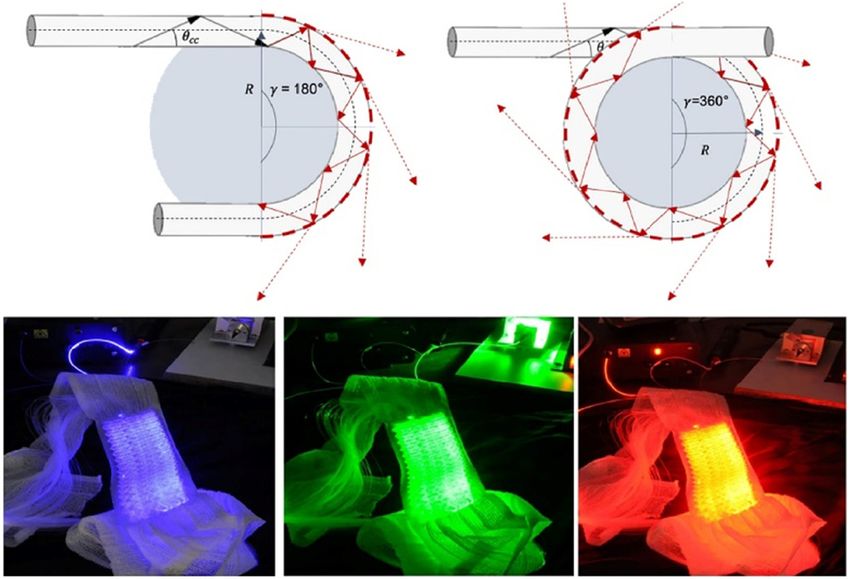
Innovative light sources for phototherapy 257
of contractility in cardiac tissue [7], detection of indivi-
dual viral particles [8], and advancement of in vivo sen-
sing [9,10].
On the other hand, organic LEDs (OLEDs) are prob-
ably the emitter type associated with the largest light
sources ever realized (like the OLED screen display
installed at the Dubai Aquarium & Underwater Zoo
(UAE) at Dubai Mall), even if with no medical applica-
tions for the moment. In the future, these source types
could be considered for performing total body photo-
therapy exposure. This is currently performed using
LED sources and is especially applied to the case of
neonatal jaundice [11,12].
The above light sources are extracorporeal or need an
Figure 1: Phototherapy with a Finsen lamp at the Institut Finsen, extracorporeal optical excitation source, which precludes
Copenhagen, Denmark, circa 1900 (from https://rarehistoricalphotos. their use for the treatment of internal diseases or deeply
com/).
seated lesions within internal organs unless invasive
systems, for example, optical fibres, are used for light
What makes a light source delivery. As such, there is a great interest in the develop-
ment of light sources that can illuminate the diseased
innovative? region from within. One approach towards this goal is
Bioluminescence Resonant Energy Transfer, which takes
In the first instance, we could think that innovation
advantage of the natural phenomenon of biolumines-
in light sources can be associated with improvements in
cence to provide a cell-based light source, for example,
one of the following interconnected characteristics: size,
based on the firefly luciferase bioluminescence [13,14].
working principle, light quality, and the aggregation
Likewise, the development of nanotechnology has
state of the light emitter. Let us analyse them briefly.
provided several types of inorganic nanomaterials that
can effectively act as light sources for phototherapy
applications [15]. Besides being often functionalized to
Size acquire further targeting and/or photosensitizing proper-
ties, some of these targeted nanomaterials present a long-
Since their invention, lasers have made a tremendous lasting luminescence afterglow or persistent luminescence,
impact on modern science and technology. Of current such as in the case of zinc gallogermanate or silicate hosts
specific interest are the numerous advantages brought doped with different elements [16,17]. In most applications,
about by their miniaturization. In recent years, micro- a narrow particle size distribution is desirable; particle
laser design and applications have greatly increased, dimensions in the range of 50–300 nm are commonly pre-
especially in the case of whispering-gallery microlasers pared to prevent possible long-term toxicity [18].
(WGMs) [1]. WGMs are generally made by dispersing dye
molecules into a polymeric microstructure with a typical
size of the order of tens of microns: laser emission is
obtained upon optical excitation. A similar condition Aggregation state
can be realized in certain organisms in nature that can
synthesize fluorescent proteins (like the Green Fluores- Nowadays, the most widespread light source types in
cent Protein). In these organisms, lasing within living medicine are based on solid or gaseous emitters, classi-
cells can be carried out when an external optical excita- fied according to the aggregation state of the radiating
tion is provided, resulting in a “biological cellular laser” species. Among the solid emitters, we can mention those
[2,3]. Remarkably, these cells remained alive even after based on semiconductors (e.g. diode lasers, LEDs, and
prolonged action of laser operation. OLEDs) as well as the solid-state lasers (e.g. Nd:YAG
WGMs have found several applications in biosensing and Ti:Sapphire lasers). Among the gas-based sources,
[4,5]: light amplification and lasering to and from biolog- we mention the gas-discharge lamps and gas-based lasers
ical systems enable intracellular sensing [6], monitoring (e.g. CO2 and excimer lasers).
258 Giovanni Romano et al.
Innovation can be found in solid or gaseous sources Working principle
covering new wavelengths or wavelength ranges, such
as the case of far-ultraviolet C [far-UVC] [19] or mid- The very first lamps were based on an incandescent fila-
infrared [20]. ment, exploiting black body emission. These so-called
Regarding liquid sources, the state-of-the-art is prob- thermal sources are still present (halogen and infrared-/
ably represented by liquid-dye lasers, but they are gen- near-infrared lamps) even if no significant technological
erally difficult to maintain and operate, and therefore, innovations are noticeable in the medical application
their usage is limited to very few cases. However, inno- field. Non-thermal sources generally include different
vative and easy to operate optoelectronic devices based working principles, among which the most widespread
on liquid materials have recently been reported, such as ones are based on atomic, molecular, or interband transi-
light-emitting electrochemical cells (ECLs) [21] and liquid tions: fluorescent, excimer or arc lamps, LEDs, OLEDs,
OLEDs [22]. The first ones are based on electrochemilu- and coherent laser emission. The working principle itself
minescence, where the active layer consists of a mixture could justify innovation in phototherapy if it represents
of an emitter and an ionic polyelectrolyte (Figure 2). For the reason to overcome limitations of use in the clinical
OLEDs, light emission is obtained by applying a voltage practice. This is especially true in all those cases where
to a flux of liquid OLEDs inside a microchannel. Com- internal organs must be targeted. For example, as also
pared to solid ones, liquid emitters could allow the devel- illustrated in the following chapters, phosphorescence-
opment of more flexible and effective protocols for light based sources could be proposed as an alternative to
delivery, like in the case of cave organ illumination. “tethered sources” (e.g. fibreoptic delivered light): lever-
This could be performed either by direct injection of the aging the delayed-emission principle, these sources are
light-emitting liquid (provided it is biocompatible) or by intrinsically non-invasive or minimally invasive, with
use of ad hoc and transparent catheters with purposely limitations arising from the capacity to efficiently store
designed geometry. Possible innovative sources based energy and release it in due time. In the last few years,
on a liquid emitter could also derive from aerosolized materials science has greatly advanced in designing and
sources, presented later in the text. synthesizing new phosphorescent materials and in parti-
cular room-temperature phosphorescent materials, with
many possible medical applications ranging from bio-
imaging to cancer and antibacterial therapy [23]. Similar
considerations could be applied to bio-derived sources
based on bioluminescence and using a “biochemical
fuel” (e.g. ATP), which is or can be made available in
situ. Ultimately, these sources are based on chemilumi-
nescence, another case of an “old” working principle
with possible new applications. A recent example can
be found in the European project Lumiblast, where mito-
chondria-powered chemiluminescence is exploited to treat
inaccessible tumours non-invasively [24,25]. A similar idea
is being recently developed with applications in both
therapy and detection of infectious agents and considering
as well luminol-based self-luminescent systems [26].
Finally, other physical phenomena could be exploited
in the coming years, namely light emission by tribolumi-
nescence (upon mechanical stimulation), acoustolumi-
nescence (upon acoustic vibration of single crystals or
granular media), and sonoluminescence (upon imploding
bubbles in a liquid when excited by sound) [27]. Tribolu-
minescence is generally considered an umbrella term for
light emission resulting from mechanical stress, and as
Figure 2: Sketch of ECLs and the chemical structures of different
types of emitters and ionic additives. CP, conjugated polymer; ITMC, such, it includes both acousto- and sonoluminescence
ionic transition-metal complex; PL, polyelectrolyte; and IL, ionic [28–30]. Its applications are now mainly in the fields
liquid. Image from ref. [21]. of sensors, displays, and bioimaging devices [30,31].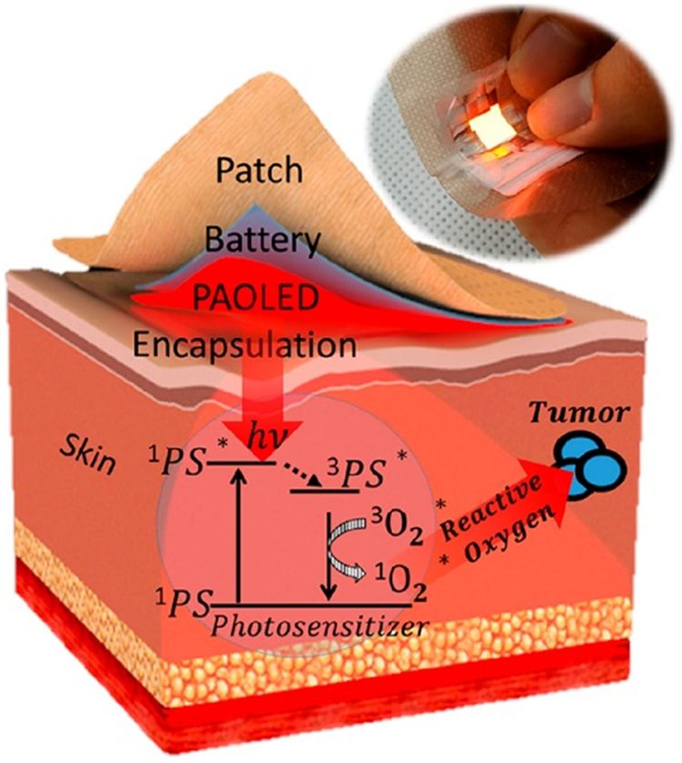
Innovative light sources for phototherapy 259
Nevertheless, sonoluminescence has recently seen inter- the light source and its emission. Fortunately, there are
esting and innovative developments in the therapeutic as many variables to play with. Thinking of light–tissue
field [32,33], also due to the much greater penetration interaction [36], the rationale is to consider the plethora
depth of sound in tissues with respect to optical radiation. of light-induced effects (Figure 3) and maximize the
desired one while minimizing the downsides, also con-
sidering the physics of light penetration in the biological
Medical needs matter and the presence of different absorbers (Figure 4)
[37–39] to answer the following questions: Which main
Over time, both technological and medical advancements effect is sought (Figure 3)? Which wavelength or spec-
have generated new needs to satisfy and new possibilities trum? Pulsed or continuous emission? Coherent or inco-
for further applications. As it is difficult to understand if herent light? Which power and/or irradiance? Which illu-
and which of the two fields drives progress first, we must
choose a perspective in which to frame our analysis. With
this logic in mind, the needs coming from the clinical
world are certainly a strong driving force to produce
innovation and a good reason to define innovation as
well: if a light source responds to an unmet clinical
need, that source is innovative. Talking about therapy,
examples of new medical needs are the following: decrease
invasiveness, reach difficult districts, address new targets,
define alternative therapeutic schemes, increase the
facility of use and/or flexibility, increase the emitted
power and/or available wavelengths, etc. A similar scheme,
in principle, could also be applied to diagnosis.
In this perspective, attention is first driven to the
application and then bounces back to the principles of
light–tissue interaction and the technology producing
the desired light emission characteristics to respond to
that specific application. For example, thinking of the
photochemical reactions at the basis of the photo-
dynamic inactivation (PDI) of certain bacteria types,
the infection localization in the stomach or the lungs Figure 3: Light-induced effects in tissue. Image from ref. [36].
calls for a light source that should be delivered inside
those organs, effectively and possibly non-invasively. In
dermatology, patient’s pain is often one of the drawbacks
of photodynamic therapy (PDT) treatments, which gen-
erates the need for a lower light dose rate and longer
treatments, at the same time compatible with efficient
point-of-care management. The result, in some cases, is
the recommendation for daylight therapy, which makes
the sun itself a (newly) innovative light source. In tox-
icology, preliminary research has considered a transe-
sophageal fibreoptic illumination of the lungs for the
photo-dissociation of carboxy-haemoglobin as a pos-
sible alternative therapy in the case of CO intoxication
[34,35]. An increased CO elimination rate and a decreased
CO uptake were demonstrated during poisoning in a
murine model, together with a survival improvement
in ongoing CO poisoning. Figure 4: In human skin, a spectral region of minimal light
It is evident that each new request (“medical need”) attenuation, commonly referred to as the “optical windows,” is
limits the number of the possible variables characterizing observed between 600 and 1,300 nm. Image from ref. [39].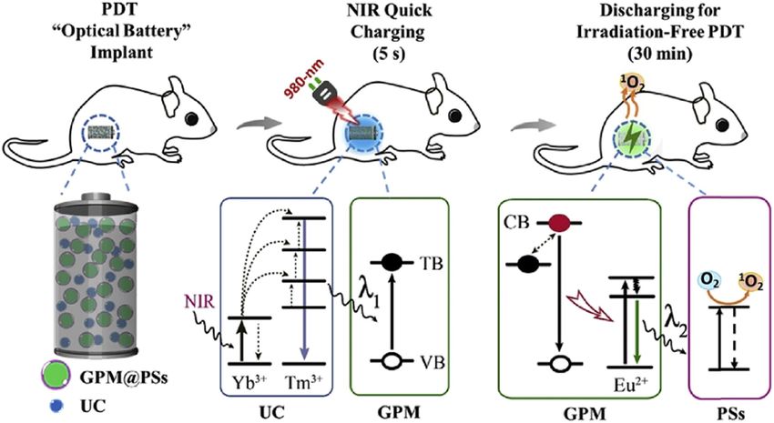
260 Giovanni Romano et al.
mination dimensions and/or depths? Which collateral Wearable sources
effect? How to convey the light (e.g. free in the air, via
an optical fibre, ingested, inhaled)? Wearable photonic devices have many applications in
Phototherapies have nowadays numerous thera- healthcare, including sensors for physiological moni-
peutic applications, starting from dermatology where toring, and light sources for phototherapy [46]. The first
light delivery is only limited by penetration issues in marketed devices consisted of LED arrays that directly
the target tissue; this corresponds to the best possible irradiate the skin, but the rigid structure makes it diffi-
scenario for an in vivo application. For many years, cult to determine the treatment dosage and avoid illu-
other disciplines have been added such as ophthal- mination hot spots. The development of flexible and
mology, gastroenterology, pneumology, urology, and conformable lighting technologies has allowed max-
gynaecology, besides various applications in surgery imum treatment efficiency at lower power and longer
(anticancer therapies) and clinical microbiology (micro- treatment time. Liu et al. [47] reported a wearable,
bial infections). The point here is to find where innovation flexible, and lightweight array of 55 LEDs (Figure 5)
is present and how it is driven by (old or new) medical designed to be worn on a knee for osteoarthritis photo-
needs. From a physical point of view, solving those needs therapy with a 0–13 mW/cm2 irradiance at 630 nm. A
means affording two main topics, namely the study of more innovative approach has been developed by Far-
light-induced effects (Figure 3) and the role of light absor- rell et al. [48] proposing a conformable device made by
bers in tissues (Figure 4). For that, the light wavelength is a 1 mm thick elastomeric membrane edge-lit by specially
one of the main parameters to care about. In therapy, its fabricated micro sized LEDs. Nanoparticle-based scat-
choice depends on the target chromophore(s) and desired tering films (polydimethylsiloxane membrane) are uti-
penetration depth, in diagnostics on the molecule(s) to lized to extract the light from the membrane; a uniform
be investigated. In both cases, we must consider which emission of 15 μW/cm2 is reported over an area of 2 cm2.
depth in the tissues we want to reach. Then, light emis- Compared to LEDs, OLEDs are very thin (thickness of
sion parameters must be chosen to define the various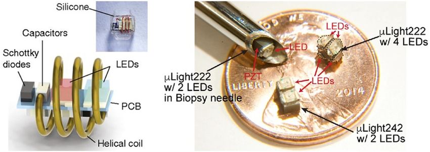
Innovative light sources for phototherapy 261
Figure 5: The top figure shows a simple sketch of the device: a thin elastomeric membrane with a scattering film on it is edge-lit by micro-
sized LEDs. On the bottom part, two pictures of the device are shown. In particular, the uniform light emission and the device flexibility are
clearly shown. Adapted from ref. [48].
A different approach to wearable devices consists
of using light-emitting fabrics based on optical fibres.
Generally, optical fibres are used in telecommunication
or transmission power applications, where they are req-
uested to guide the radiation with minimal losses from
one end to the other, thanks to the internal total reflec-
tion condition that takes place in the fibre core. Unlike
most cases, optical fibre losses are rather requested to
obtain the desired emission (illumination) geometry in
phototherapy applications. Light leakage along the fibre
is generally obtained via processing or macro bending of
the fibre cladding [55–57]. In the first case, cladding
micro-perforations allow light propagation from the fibre
core to the environment. With macro bending (Figure 7,
top), the fibre radius of curvature exceeds a specific cri-
tical value; this prevents total internal reflection from
occurring in the core, leading to light emission along
Figure 6: Parallel-stacked OLED (PAOLED) designed in the form of
the fibre. Multiple bending can be kept in place while
plaster. The schematic illustration of PDT treatment principle is also
shown. Adapted from ref. [52]. embedding the fibres in a stable structure, such as a
textile (Figure 7, bottom). Mordon et al. [58] reported
convert mechanical energy harvested from the environ- several applications of light-emitting fabrics by in vitro
ment into electricity. and in vivo experiments aimed at evaluating their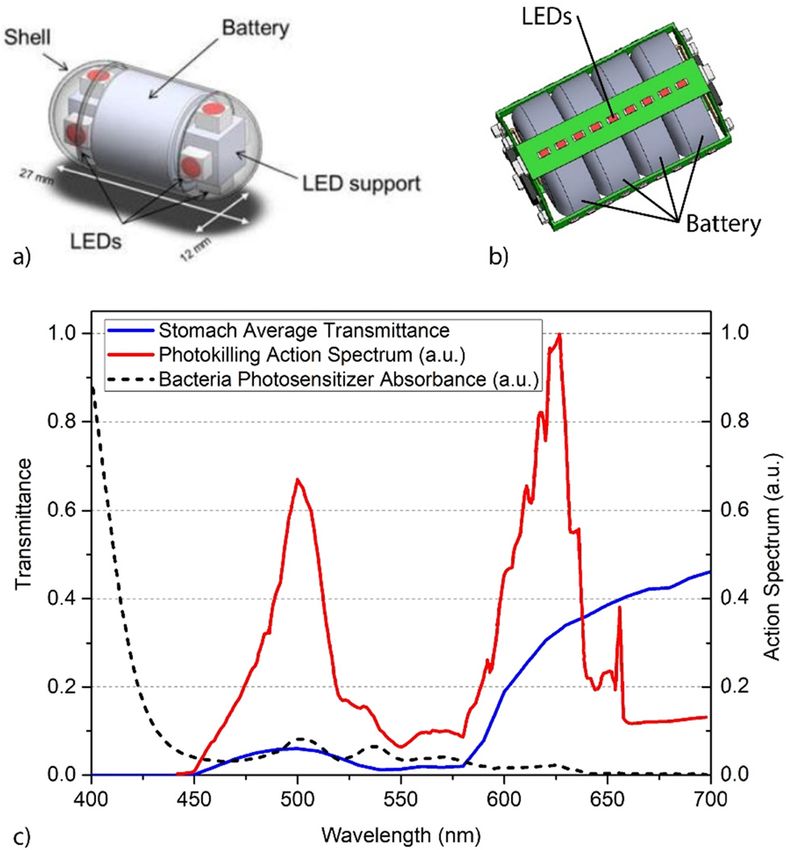
262 Giovanni Romano et al.
Figure 7: Top: when the radius of curvature R of a bended-optical fibre exceeds a specific critical value, leaking of the light propagating into
the fibre is achieved. Bottom: bended fibre can be embedded in a textile structure, thus realizing a light-emitting fabric. Adapted from
ref. [58].
efficacy for antitumoral PDT [59,60]. These light-emit- optical access to deep layers [62–64]. A different innova-
ting and plastic fabrics emit in the range 400–1,200 nm: tive approach to delivering light up to a 4 mm depth under
using a 2.6 W 635 nm laser, they obtain an average irra- the skin has been proposed by Hu et al. [65] with the
diance of approximately 1 mW/cm2 over a surface of development of a near-infrared rechargeable “optical
about 660 cm2 with almost 91% homogeneity. Other battery” implant for irradiation-free continuous PDT
clinical applications include the treatment of actinic (Figure 8). The battery is fabricated by a combination
keratosis and the primary extramammary Paget’s disease of an upconversion material with typical ultraviolet
of the vulva [58]. Quandt et al. [61] developed a textile to (UV)/blue emission, a UV rechargeable, persistent phos-
be used as a wearable, long-term phototherapy device in phor and a photosensitizer, assembled into a biocompa-
the treatment of neonatal jaundice (hyperbilirubinemia), tible material that can be solidified in any size and
producing a homogeneous light emission within 4% var- shape. Excitation of the upconversion material with
iations. By designing a network of polymer optical fibres, just 5 s of 980 nm radiation allows the generation of
they present the first example of direct manufacturing of 30 min persistent luminescence at 520 nm, after which
phototherapeutic clothing realized without any post- a new “recharge” can be applied for the next 30 min
processing of the textile material. The broad transmis- treatment. In vivo experiments were performed on
sion spectrum of polymeric optical fibre, together with HT29 tumour implanted subcutaneously into mice,
an adequate light source, should allow obtaining the demonstrating the inhibition of tumour proliferation.
needed power density requested for specific phototherapy
applications.
One of the major limitations of the light delivery Sources for interstitial phototherapy
approach based on external, with respect to the body,
sources is related to the limited light penetration depth In interstitial phototherapy, one or more optical fibres are
in tissues. To overcome this restriction, percutaneous inserted into the target tissue to deliver in situ therapeutic
light delivery systems have been designed. The idea is light, for example, by their insertion into catheters. Most
to use a light-transparent material to create an array of applications are in interstitial PDT (I-PDT): light sources
microneedles, with a typical length of up to 2 mm, that are generally lasers, whose wavelength matches the
can guide or focalize light through the skin, thus providing absorption peak of the photosensitizing molecule;
Innovative light sources for phototherapy 263
Figure 8: Schematic illustration of the “optical battery” implant for PDT. GPM: green persistent luminescence materials; PSs: photosen-
sitizers; UC: upconversion materials; TB: trapping band; VB: valence band. Figure from ref. [65].
applications are mainly antitumoral, with different pos- This can be realized by adding roughness or scattering
sible organs to address [66]. To completely exploit the particles in the core-cladding borders. Diffuser balloons
therapeutic scheme of fibre-driven light delivery, innova- can also be employed and eventually filled with scattering
tion has focused on the design of various light-emission liquid (typically a lipid suspension) to improve light
geometries (e.g. point emission from the fibre tip and delivery and uniformity to the tissue under treatment.
cylindrical emission along the fibre). Together, treatment Dupont et al. [71] developed a new device for intrao-
planning software and procedures have been developed perative PDT dedicated to glioblastoma treatment, con-
for fibre positioning and dose release protocols both in sisting of a balloon coupled with a cylindrical diffuser
space and in time, resembling the more established prac- fibre and mounted on a trocar. After tumour resection,
tices of treatment planning in radiotherapy. Intrinsically, the balloon is positioned in the surgical cavity and inflated
I-PDT and the associated sources are more invasive than with a diffusing solution to conform its shape to the
the therapeutic approach with wearable devices, though anatomy of the operative cavity. The light source is set
much less invasive than intraoperative phototherapy (see to provide a therapeutic fluence value at a 5 mm depth
next paragraph). Besides, sources for I-PDT are adaptable within surrounding tissues. The device allows homoge-
to the specific organ and anatomical region [59,67–70] neous irradiation of the resection area, improving the
and are now being considered as an alternative (or adju- treatment efficacy [72]. A novel mini-invasive approach
vant) to more established techniques such as surgery and in the glioblastoma multiforme treatment was proposed
radiation therapy. by Leroy et al. [73]. Cylindrical diffusing optical fibres
are introduced inside the tumour, without large cra-
niotomy, to illuminate the cancer cells, previously sen-
Sources for intraoperative phototherapy sitized via a photosensitizer. Other recent works focused
on the treatment of breast cancer [74,75]. In a different
The most straightforward approach to delivering light in work, Chamberlain et al. [76] developed an intraopera-
deep tissue is to take advantage of the presence of exposed tive flexible optical fibre-based surface applicator able
areas during surgical intervention, allowing for direct light to administer controlled and homogeneous light irradi-
delivery. Similar to interstitial phototherapy, light is gen- ance during surgery within the thoracic cavity. The main
erally guided through optical fibres, possibly inserted into drawback of intraoperative sources is that they do not
needles, catheters, balloons, or other devices. Generally, allow chronic implantation, thus reducing the light delivery
optical fibres are modified to emit diffuse light (Figure 9). to a single dose.
264 Giovanni Romano et al.
(1–1.5 GHz). The device consists of a helical coil and a
micro-printed circuit board that also includes LEDs
encapsulated in medical-grade silicone. When suffi-
cient radiant power is sent to the device, part of its
energy is extracted by the incident field via the coil
and used to turn on the LEDs (660 and 400 nm), produ-
cing a total radiant power of 1.3 mW. The device can
also communicate the effective light dose delivered to
the surrounding tissue, thus providing a real-time wire-
less dosimetry system. The efficacy of this device has
been tested in vivo for in situ photosensitizer activation
through more than 3 cm thick tissue to suppress tumour
activity in a murine cancer model. A similar approach
has also been reported by Kim et al. [78] proposing an
implantable micro source powered by ultrasound and
enabling light delivery in deep-seated solid tumours
(Figure 10). The device has red (655 nm) and blue
(470 nm) LEDs and can produce up to 6.5 mW/cm2 of
optical power when powered with an acoustic wave at
720 kHz. In vivo tests in mice with 4T1-induced tumour
(breast cancer) show light delivery capability at the
therapeutic dose levels. Multiple implants can be pow-
ered simultaneously on the same tumour.
Figure 9: Fibres with (a) microlens, (b) spherical and (c) cylindrical
diffusor, and (d) balloon applicator with integrated fibre surrounded
by scattering medium. Image from ref. [42].
Ingestible light sources for the eradication of
Implantable sources stomach infections
A very innovative and interesting approach that can Ingestible medical devices have been developed, patented,
overcome the limitations discussed above is represented and used in the clinical practice for several years, to pro-
by implantable and wireless photonic devices, generally vide advancement in the diagnosis and treatment of gas-
powered via external ultrasound or electromagnetic trointestinal tract-related conditions [79]. Most of them
waves: their tiny dimensions allow direct implantation have a diagnostic purpose (sensing and/or sampling),
at the target size with minimally invasive procedures, and some release a payload (e.g. insulin) to perform
such as incisional biopsy (Figure 10). Bansal et al. [77] therapy. One of the very few examples of ingestible devices
reported miniaturized wireless powered photonic imp- for phototherapy is represented by a luminous pill for
lants activated by an external radiofrequency source the control and eradication of Helicobacter pylori (Hp)
Figure 10: Examples of implantable wireless light sources powered by an external radiofrequency source (left) and by ultrasound emitter
(right). Images adapted from refs [77] and [78], respectively.
Innovative light sources for phototherapy 265
infection in the stomach, whose elective therapy is based green and red being the best-expected excitation wave-
on antibiotics and associated drugs like pump inhibitors. lengths to optimize PDI [88]. The device is provided with
Building on previous research on Hp photo-inactivation a temperature and pH sensor; this last sensing the pas-
via endogenous porphyrin excitation [80–82] and subse- sage from the stomach to the intestine where photo-
quent clinical trials with modified gastroscopes [83,84], a therapy is avoided for safety reasons. In 2016, a similar
new medical device was designed, patented, and proto- device based on LED sources was designed and charac-
typed in the form of a smooth cylinder-shaped capsule, terized, provided with a pH sensor and a wireless com-
provided with visible LEDs powered by a battery and munication module, being anyway limited by blue-light
driven by a programmable board [85–87] (Figure 11a and emission only [91]. These devices are good examples of
b). LED emission covers the biggest possible solid angle, light delivery instead of drug delivery, especially con-
being photokilling based on the passive capsule posi- sidering the rise in antibiotic-resistance rates at a world-
tioning inside the stomach cavity and its displacements wide level. The similarity in the therapeutic approach
associated with the physiological peristaltic movements. with respect to antibiotics (both imply the use of “pills”)
The light emission parameters have been defined fol- makes these devices a very promising alternative also
lowing an in vivo action spectrum study for Hp photo- from the point of view of the patient’s compliance, the
killing (Figure 11c) obtained by semi-theoretical methods final important passage between the concept of innova-
[88,89], together with safety studies on in vitro models [90] tion and the release of the therapeutic principle, light in
and more recently in vivo on minipigs (work under sub- our case.
mission). Interestingly, absorption by the gastric mucosa
tissue seems to have a crucial role in this case: due to Hp
positioning on both the mucosa surface and in between
the gastric plicae and rugae, the excitation light is pre- Inhalable light sources for controlling lung
ferentially absorbed by the surrounding tissue before infections
reaching the bacterial target. This is compatible with
Since the beginning, most of the research on lung photo-
therapy has been applied to cancer treatment [92]. Light
delivery is accomplished thanks to modified fiberscopes,
possibly integrating both illumination and irradiation
fibres [93,94] and the synergy between different photo-
induced effects, like in the case of combined photothermal
and photodynamic therapies [95]. These approaches cer-
tainly contain innovative aspects, mainly from a technical
point of view linked to the modification of existing diag-
nostic instrumentation to provide them with a therapeutic
purpose.
Towards the definition of less invasive approaches,
an inhalable light source based on phosphorescence has
been recently proposed to perform PDI for the control of
antibiotic-resistant and recalcitrant lung infections [96,97].
Through a bottom-up approach, the project encom-
passes the following steps (Figure 12): (i) synthesis of
biocompatible and phosphorescent nanoparticles, (ii) their
aerosolization and (iii) activation by external light excita-
tion, (iv) luminous aerosol inhalation, and (v) therapeutic
action via bacterial PDI. This therapeutic scheme repre-
sents a further step in an ideal path of light source
“dematerialization” and towards minimal invasiveness,
Figure 11: (a) First and (b) second prototype version of the ingestible
also considering the absence of externally administered
light source for the eradication of stomach infections. In (c) the
action spectrum for in vivo photokilling of H. pylori and the average photosensitizers. Therefore, the aerosol particles are at
stomach antrum wall transmittance spectrum. Figures (a) and (b) the same time the vehicle and the source of therapeutic
adapted from refs [85] and [87], respectively. photons into the various lung regions by way of their266 Giovanni Romano et al.
Figure 12: Light4Lungs: five-step concept.
phosphorescence properties. This does not exclude the activities [101]. The key point here is represented by
possible and contemporary administration of exogenous reaching a compromise between a low radiation pene-
photosensitizers, possibly incorporated in the aerosol tration into human tissues (to minimize negative effects)
particles themselves, to enhance the photokilling effect. and a good absorption by the pathogen of interest. Given
The aerosol-mediated delivery of photons has a more the typical bacterium and virus sizes (∼1 and 0.1 μm,
“classical” (still innovative) counterpart in the aerosol- respectively), the penetration of UVC radiation into their
based delivery of drugs into the lungs, especially in the respective structures does not constitute a limit factor,
case of lung infections [98]. paving the way to an effective biocidal action, which as
This approach starts from state-of-the-art technology usual depends on the light dose. These considerations
for aerosolization, which must be modified to be com- bring our attention to the far UVC region. Between 170
patible with aerosol excitation prior to its delivery. and 240 nm, the only sources of a certain efficiency are
Therefore, innovation stems from both new technolo- excimer lamps and lasers (Figure 13), where excimer
gical solutions and the synthesis of new biocompatible refers to a diatomic excited molecule generally com-
light-emitting materials with a long afterglow. Corres- posed of a noble gas and a halogen atom. As an alter-
pondingly, innovative therapeutic protocols will be native, UVC LEDs are still limited by low efficiency and
defined in the direction of a self-administration of the poor emitted power especially in the far UVC region [102],
light. making them unsuitable for clinical applications. Excimer
lamps, and in particular those emitting at λ = 308 nm,
have long been used in dermatology for targeted photo-
therapy [103]. Recent studies show that the antimicrobial
Antiviral far UVC source efficacy of 222 nm radiation is high and comparable with
254 nm radiation. This has been demonstrated in vitro on
Light per se has long been used as a powerful antimicro- both bacterial [104] and virus models [105], including
bial. UV radiation kills viruses and bacteria by chemically
altering the genetic material. The most effective wave-
lengths for inactivation correspond to DNA and RNA
absorption peaks, located in the UVC range (100–280 nm).
The most widespread technology to produce UVC radia-
tion is based on mercury vapour lamps, emitting a signif-
icant component at 254 nm. This radiation is mutagenic
and can induce skin cancers [99], cataracts, and corneal
damage [100]. Therefore, although economical and effi-
cient, these sources are not suitable for areas frequented
by people, unless their use is restricted to night-time or
purposely dedicated time. Innovation in this respect is
especially desirable to extend the source compatibility
for a “real time disinfection,” applicable to a myriad of Figure 13: Picture of commercial UVC sources emitting at 222 nm
different contexts from working spaces to public trans- (www.firstuvc.com). The sketch shows the internal components of a
port, public/private offices, and cultural or recreational cylindrical excimer UVC source (adapted from ref. [111]).Innovative light sources for phototherapy 267
coronavirus [106]. Antibacterial efficacy of 222 nm radia- emitted radiant power. This is very much linked to the
tion has also been demonstrated in human patients’ development of new and more effective solutions to
sacral and gluteal pressure ulcers. Its effectiveness also store energy and efficiently exploit it. Fortunately, there
spans numerous bacterial species including methicillin- are many different forms of energy to exploit.
resistant Staphylococcus aureus, Pseudomonas aeruginosa,
and Klebsiella pneumoniae [107]. At the same time, the Acknowledgments: The authors would like to thank Dr
risks to human health associated with exposure to UVC Maria Méndez (ICIQ, Tarragona) for useful discussions
radiation at λ = 222 nm seem to be much lower than about nanoemitters and all the partners of the Light4Lungs
those associated with UV radiation at longer wave- consortium.
lengths [108,109]. The reason for this lies in the very small
penetration depth in tissues (a few μm at 222 nm). In the Funding information: This work was supported by the
skin, far UVC radiation is absorbed by the superficial projects “Device endoscopico per fototerapia antibat-
stratum corneum containing dead cells, with negligible terica intragastrica” and “Terapia fotodinamica contro il
relative presence in the underlying tissues (e.g. the batterio pseudomonas Savastanoi agente della rogna del-
dermis). Similarly, corneal damage is negligible or absent l’olivo” by Fondazione Cassa di Risparmio di Firenze.
even at high UVC doses, compared to the damage induced This research project is also funded by the Tuscany
by 254 nm radiation [110]. The net result is that the muta- Region, through the “Suppression of Airborne Viral
genicity of far UVC seems to be substantially negligible, as Epidemic Spread by Ultraviolet light barriers” (SAVES
is the ability to induce inflammatory reactions compared US) project and by the project “Light4Lungs” H2020-
to UVC at longer wavelengths [108]. FETOPEN-2018-2020, grant agreement no. 863102.
Authors contributions: G.R., G.I., and F.F. wrote the ori-
ginal paper. All authors revised the manuscript.
Conclusion
Conflict of interest: F.F. and G.R. acknowledge being
We have shown examples of innovative light sources for also Probiomedica srl. Other authors state no conflict
phototherapy, describing and justifying the reasons for of interest.
their innovation content. Probably, the doubt remains
about the “best” definition of source and of innovation: Data availability statement: The data sets generated
how is a therapeutic light source defined? Does it corre- during and/or analysed during the current study are
spond merely to the light emitter itself or should it also available from the corresponding author on reasonable
comprise the whole device, including all the technical request.
solutions to deliver light to the region or district of
interest? Should we then refer to “innovative light-emit-
ting devices and protocols” instead? This would probably
be more suitable if the clinical application context is References
recalled. Talking about innovation: is it necessarily linked
to new emitter solutions or, on the opposite side, to new [1] Manzo M, Cavazos O. Solid state optical microlasers fabri-
solutions for using state-of-the-art emitters? For example, cation via microfluidic channels. Optics. 2020;1:88–96.
let us consider that the sun has come back again as an doi: 10.3390/opt1010007.
innovative source with the advent of daylight photo- [2] Pile D. Cellular lasers. Nat Photonics. 2011;5:438.
doi: 10.1038/nphoton.2011.126.
therapy in dermatology.
[3] Gather MC, Yun SH. Single-cell biological lasers. Nat
Among all possible novelties, non-invasiveness could Photonics. 2011;5:406–10. doi: 10.1038/nphoton.2011.99.
be considered as the feature most research is aiming at [4] Toropov N, Cabello G, Serrano MP, Gutha RR, Rafti M,
(e.g. ingestible and inhalable sources, self-luminescent Vollmer F. Review of biosensing with whispering-gallery
therapies, and wearable devices). Considering the results mode lasers. Light Sci Appl. 2021;10:42. doi: 10.1038/
obtained up to now, one of the main goals of future work s41377-021-00471-3.
[5] Jiang X, Qavi AJ, Huang SH, Yang L. Whispering-gallery
will be to avoid the loss of therapeutic effectiveness that
sensors. Matter. 2020;3:371–92. doi: 10.1016/
comes at the price of non-invasiveness: wearable sources j.matt.2020.07.008.
are limited by light penetration issues while un-tethered [6] Prasetyanto EA, Wasisto HS, Septiadi D. Cellular lasers
sources (inhalable and ingestible) are limited by the for cell imaging and biosensing. Acta Biomater.268 Giovanni Romano et al.
2022;143:S1742706122001623–51. doi: 10.1016/ light emission. Sci Rep. 2020;10:14528. doi: 10.1038/
j.actbio.2022.03.031. s41598-020-70838-w.
[7] Schubert M, Woolfson L, Barnard IRM, Dorward AM, [23] Zhi J, Zhou Q, Shi H, An Z, Huang W. Organic room tem-
Casement B, Morton A, et al. Monitoring contractility in perature phosphorescence materials for biomedical appli-
cardiac tissue with cellular resolution using biointegrated cations. Chem – Asian J. 2020;15:947–57. doi: 10.1002/
microlasers. Nat Photonics. 2020;14:452–8. doi: 10.1038/ asia.201901658.
s41566-020-0631-z. [24] Lumiblast Project n.d. https://www.lumiblast.eu/.
[8] Toropov N, Osborne E, Joshi LT, Vollmer F. Direct single-par- [25] Grigalavicius M, Berg K, Theodossiou TA. Detection of che-
ticle detection and sizes recognition of adenovirus with miluminescence-induced photosensitizer activation through
whispering-gallery mode resonances. Optical sensors and fluorescence and concomitant singlet oxygen generation.
sensing congress 2021. Washington, DC: OSA; 2021. Proc. SPIE 11786, Optical Methods for Inspection,
p. SW5H.5. doi: 10.1364/SENSORS.2021.SW5H.5. Characterization, and Imaging of Biomaterials V; 2021.
[9] Humar M, Hyun Yun S. Intracellular microlasers. Nat p. 117860B. doi: 10.1117/12.2600610.
Photonics. 2015;9:572–6. doi: 10.1038/nphoton.2015.129. [26] Zhang E, Huang Y, Wang S. Self-luminescent photodynamic
[10] Chen Y-C, Chen Q, Fan X. Lasing in blood. Optica. therapy and pathogen detection for infectious diseases. Drug
2016;3:809–15. doi: 10.1364/OPTICA.3.000809. Deliv Transl Res. 2021;11:1451–5. doi: 10.1007/s13346-021-
[11] Kumar S. Developing an effective low cost blue-green led 00989-4.
phototherapy method for neonatal jaundice treatment. Int J [27] Prevenslik TV. Acoustoluminescence and sonoluminescence.
Eng Appl Sci Technol. 2021;6:104–9. J Lumin. 2000;87–89:1210–2. doi: 10.1016/S0022-2313(99)
[12] Amadi HO, Abdullahi RA, Mokuolu OA, Ezeanosike OB, 00513-X.
Adesina CT, Mohammed IL, et al. Comparative outcome of [28] Zink JI. Triboluminescence. Acc Chem Res. 1978;11:289–95.
overhead and total body phototherapy for treatment of doi: 10.1021/ar50128a001.
severe neonatal jaundice in Nigeria. Paediatr Int Child [29] Olawale DO, Okoli OOI, Fontenot RS, Hollerman WA, editors.
Health. 2020;40:16–24. doi: 10.1080/ Triboluminescence. Cham: Springer International Publishing;
20469047.2019.1610607. 2016. doi: 10.1007/978-3-319-38842-7.
[13] Yuan H, Chong H, Wang B, Zhu C, Liu L, Yang Q, et al. [30] Monette Z, Kasar AK, Menezes PL. Advances in tribolumines-
Chemical molecule-induced light-activated system for cence and mechanoluminescence. J Mater Sci Mater Electron.
anticancer and antifungal activities. J Am Chem Soc. 2019;30:19675–90. doi: 10.1007/s10854-019-02369-8.
2012;134:13184–7. doi: 10.1021/ja304986t. [31] Xie Y, Li Z. Triboluminescence: recalling interest and new
[14] Yang Y, Hou W, Liu S, Sun K, Li M, Wu C. Biodegradable aspects. Chem. 2018;4:943–71. doi: 10.1016/
polymer nanoparticles for photodynamic therapy by biolu- j.chempr.2018.01.001.
minescence resonance energy transfer. Biomacromolecules. [32] Canaparo R, Foglietta F, Giuntini F, Francovich A, Serpe L. The
2018;19:201–8. doi: 10.1021/acs.biomac.7b01469. bright side of sound: perspectives on the biomedical appli-
[15] Chitgupi U, Qin Y, Lovell JF. Targeted nanomaterials cation of sonoluminescence. Photochem Photobiol Sci.
for phototherapy. Nanotheranostics. 2017;1:38–58. 2020;19:1114–21. doi: 10.1039/D0PP00133C.
doi: 10.7150/ntno.17694. [33] Beguin E, Shrivastava S, Dezhkunov NV, McHale AP,
[16] Wang J, Li Y, Mao R, Wang Y, Yan X, Liu J. Persistent lumi- Callan JF, Stride E. Direct evidence of multibubble sonolu-
nescent nanoparticles as energy mediators for enhanced minescence using therapeutic ultrasound and microbubbles.
photodynamic therapy with fractionated irradiation. J Mater ACS Appl Mater Interfaces. 2019;11:19913–9. doi: 10.1021/
Chem B. 2017;5:5793–805. doi: 10.1039/C7TB00950J. acsami.9b07084.
[17] Bessière A, Durand JO, Noûs C. Persistent luminescence [34] Zazzeron L, Liu C, Franco W, Nakagawa A, Farinelli WA,
materials for deep photodynamic therapy. Nanophotonics. Bloch DB, et al. Pulmonary phototherapy for treating carbon
2021;10:2999–3029. doi: 10.1515/nanoph-2021-0254. monoxide poisoning. Am J Respir Crit Care Med.
[18] Yang J, Zhao Y, Meng Y, Zhu H, Yan D, Liu C, et al. Irradiation- 2015;192:1191–9. doi: 10.1164/rccm.201503-0609OC.
free photodynamic therapy in vivo induced by enhanced [35] Rose JJ, Xu Q, Wang L, Gladwin MT. Shining a light on carbon
deep red afterglow within NIR-I bio-window. Chem Eng J. monoxide poisoning. Am J Respir Crit Care Med.
2020;387:124067. doi: 10.1016/j.cej.2020.124067. 2015;192:1145–7. doi: 10.1164/rccm.201508-1579ED.
[19] Bergman RS. Germicidal UV sources and systems. Photochem [36] Zam A. Laser–tissue interaction. In Stübinger S, Klämpfl F,
Photobiol. 2021;97:466–70. doi: 10.1111/php.13387. Schmidt M, Zeilhofer H-F, editors. Lasers oral maxillofac.
[20] Isensee K, Kröger-Lui N, Petrich W. Biomedical applications Surg. Cham: Springer International Publishing; 2020. p.
of mid-infrared quantum cascade lasers – a review. Analyst. 25–34. doi: 10.1007/978-3-030-29604-9_3.
2018;143:5888–911. doi: 10.1039/C8AN01306C. [37] Bigio IJ, Fantini S. Quantitative biomedical optics: theory,
[21] Fresta E, Costa RD. Beyond traditional light-emitting elec- methods, and applications. Cambridge: Cambridge
trochemical cells – a review of new device designs and University Press; 2016.
emitters. J Mater Chem C. 2017;5:5643–75. doi: 10.1039/ [38] Vo-Dinh T, editor. Biomedical photonics handbook: thera-
C7TC00202E. peutics and advanced biophotonics. CRC Press; 2019.
[22] Kawamura M, Kuwae H, Kamibayashi T, Oshima J, [39] Scholkmann F, Kleiser S, Metz AJ, Zimmermann R, Mata
Kasahara T, Shoji S, et al. Liquid/solution-based microfluidic Pavia J, Wolf U, et al. A review on continuous wave functional
quantum dots light-emitting diodes for high-colour-purity near-infrared spectroscopy and imaging instrumentation andInnovative light sources for phototherapy 269
methodology. NeuroImage. 2014;85:6–27. doi: 10.1016/ optical fibers. Textiles. 2021;1:337–60. doi: 10.3390/
j.neuroimage.2013.05.004. textiles1020017.
[40] Patrice T, editor. Photodynamic therapy. Cambridge: Royal [56] Cinquino M, Prontera CT, Pugliese M, Giannuzzi R, Taurino D,
Society of Chemistry; 2003. doi: 10.1039/9781847551658. Gigli G, et al. Light-emitting textiles: device architectures,
[41] Hamblin MR, Jori G, editors. Photodynamic inactivation of working principles, and applications. Micromachines.
microbial. Pathogens: medical and environmental applica- 2021;12:652. doi: 10.3390/mi12060652.
tions. Cambridge: Royal Society of Chemistry; 2011. [57] Gong Z, Xiang Z, OuYang X, Zhang J, Lau N, Zhou J, et al.
doi: 10.1039/9781849733083. Wearable fiber optic technology based on smart textile: a
[42] Kim MM, Darafsheh A. Light sources and dosimetry techni- review. Materials. 2019;12:3311. doi: 10.3390/ma12203311.
ques for photodynamic therapy. Photochem Photobiol. [58] Mordon S, Thécua E, Ziane L, Lecomte F, Deleporte P, Baert G,
2020;96:280–94. doi: 10.1111/php.13219. et al. Light emitting fabrics for photodynamic therapy:
[43] Casas A. Clinical uses of 5-aminolaevulinic acid in photody- Technology, experimental and clinical applications. Transl
namic treatment and photodetection of cancer: A review. Biophotonics. 2020;2:2. doi: 10.1002/tbio.202000005.
Cancer Lett. 2020;490:165–73. doi: 10.1016/ [59] Quilbe A, Moralès O, Baydoun M, Kumar A, Mustapha R,
j.canlet.2020.06.008. Murakami T, et al. An efficient photodynamic therapy treat-
[44] Simões JCS, Sarpaki S, Papadimitroulas P, Therrien B, ment for human pancreatic adenocarcinoma. J Clin Med.
Loudos G. Conjugated photosensitizers for imaging and 2020;9:192. doi: 10.3390/jcm9010192.
PDT in cancer research. J Med Chem. 2020;63:14119–50. [60] Baydoun M, Moralès O, Frochot C, Ludovic C, Leroux B,
doi: 10.1021/acs.jmedchem.0c00047. Thecua E, et al. Photodynamic therapy using a new folate
[45] Sato K. The “light” guide for surgery. EBioMedicine. receptor-targeted photosensitizer on peritoneal ovarian
2020;56:102808. doi: 10.1016/j.ebiom.2020.102808. cancer cells induces the release of extracellular vesicles
[46] Iqbal SMA, Mahgoub I, Du E, Leavitt MA, Asghar W. Advances with immunoactivating properties. J Clin Med. 2020;9:1185.
in healthcare wearable devices. Npj Flex Electron. 2021;5:9. doi: 10.3390/jcm9041185.
doi: 10.1038/s41528-021-00107-x. [61] Quandt BM, Pfister MS, Lübben JF, Spano F, Rossi RM,
[47] Liu K, Chen H, Wang Y, Wang M, Tang J. Wearable flexible Bona G-L, et al. POF-yarn weaves: controlling the light out-
phototherapy device for knee Osteoarthritis. Electronics. coupling of wearable phototherapy devices. Biomed Opt
2021;10:1891. doi: 10.3390/electronics10161891. Express. 2017;8:4316–30. doi: 10.1364/BOE.8.004316.
[48] Farrell F, Xie E, Guilhabert B, Haughey A-M, Connolly P, [62] Kim M, An J, Kim KS, Choi M, Humar M, Kwok SJ, et al. Optical
Dawson MD, et al. A wearable phototherapy device utilizing lens-microneedle array for percutaneous light delivery.
micro-LEDs. St Annual International Conference of the IEEE Biomed Opt Express. 2016;7:4220–7. doi: 10.1364/
Engineering in Medicine and Biology Society. EMBC, Berlin, BOE.7.004220.
Germany: IEEE; 2019; 2019 41. p. 67–70. doi: 10.1109/ [63] Wu X, Park J, Chow SYA, Kasuya MCZ, Ikeuchi Y, Kim B.
EMBC.2019.8857880. Localised light delivery on melanoma cells using optical
[49] Keum C, Murawski C, Archer E, Kwon S, Mischok A, microneedles. Biomed Opt Express. 2022;13:1045–60.
Gather MC. A substrateless, flexible, and water-resistant doi: 10.1364/BOE.450456.
organic light-emitting diode. Nat Commun. 2020;11:6250. [64] Zhang H, Zhao H, Zhao X, Xu C, Franklin D, Vázquez‐
doi: 10.1038/s41467-020-20016-3. Guardado A, et al. Biocompatible light guide‐assisted
[50] Song YJ, Kim J-W, Cho H-E, Son YH, Lee MH, Lee J, et al. wearable devices for enhanced UV light delivery in deep skin.
Fibertronic organic light-emitting diodes toward fully Adv Funct Mater. 2021;31:2100576. doi: 10.1002/
addressable, environmentally robust, wearable displays. adfm.202100576.
ACS Nano. 2020;14:1133–40. doi: 10.1021/ [65] Hu L, Wang P, Zhao M, Liu L, Zhou L, Li B, et al. Near-infrared
acsnano.9b09005. rechargeable “optical battery” implant for irradiation-free
[51] Murawski C, Gather MC. Emerging biomedical applications photodynamic therapy. Biomaterials. 2018;163:154–62.
of organic light‐emitting diodes. Adv Opt Mater. doi: 10.1016/j.biomaterials.2018.02.029.
2021;9:2100269. doi: 10.1002/adom.202100269. [66] Shafirstein G, Bellnier D, Oakley E, Hamilton S, Potasek M,
[52] Jeon Y, Noh I, Seo YC, Han JH, Park Y, Cho EH, et al. Parallel- Beeson K, et al. Interstitial photodynamic therapy – a
stacked flexible organic light-emitting diodes for wearable focused review. Cancers. 2017;9:12. doi: 10.3390/
photodynamic therapeutics and color-tunable optoelectro- cancers9020012.
nics. ACS Nano. 2020;14:15688–99. doi: 10.1021/ [67] Swartling J, Axelsson J, Ahlgren G, Kälkner KM, Nilsson S,
acsnano.0c06649. Svanberg S, et al. System for interstitial photodynamic
[53] Jeon Y, Choi H-R, Kwon JH, Choi S, Nam KM, Park K-C, et al. therapy with online dosimetry: first clinical experiences of
Sandwich-structure transferable free-form OLEDs for wear- prostate cancer. J Biomed Opt. 2010;15:058003. doi: 10.1117/
able and disposable skin wound photomedicine. Light Sci 1.3495720.
Appl. 2019;8:114. doi: 10.1038/s41377-019-0221-3. [68] Komolibus K, Fisher C, Swartling J, Svanberg S, Svanberg K,
[54] Xiao X, Chen G, Libanori A, Chen J. Wearable triboelectric Andersson-Engels S. Perspectives on interstitial photody-
nanogenerators for therapeutics. Trends Chem. namic therapy for malignant tumors. J Biomed Opt.
2021;3:279–90. doi: 10.1016/j.trechm.2021.01.001. 2021;26:070604. doi: 10.1117/1.JBO.26.7.070604.
[55] Kallweit J, Pätzel M, Pursche F, Jabban J, Morobeid M, Gries T. [69] Osuchowski M, Bartusik-Aebisher D, Osuchowski F,
An overview on methods for producing side-emitting polymer Aebisher D. Photodynamic therapy for prostate cancer – A270 Giovanni Romano et al.
narrative review. Photodiagnosis Photodyn Ther. violet light phototherapy: A pilot clinical trial: VIolet
2021;33:102158. doi: 10.1016/j.pdpdt.2020.102158. Phototherapy For H. Pylori. Infection. Lasers Surg Med.
[70] Kubrak T, Karakuła M, Czop M, Kawczyk-Krupka A, 2009;41:337–44. doi: 10.1002/lsm.20770.
Aebisher D. Advances in Management of bladder cancer – the [85] Tortora G, Orsini B, Pecile P, Menciassi A, Fusi F, Romano G.
role of photodynamic therapy. Molecules. 2022;27:731. An ingestible capsule for the photodynamic therapy of
doi: 10.3390/molecules27030731. helicobacter pylori infection. IEEEASME Trans Mechatron.
[71] Dupont C, Mordon S, Deleporte P, Reyns N, Vermandel M. 2016;21:1935–42. doi: 10.1109/TMECH.2016.2536944.
A novel device for intraoperative photodynamic therapy [86] Romano G, Calusi S, Gnerucci A, Orsini B, Faraoni P,
dedicated to glioblastoma treatment. Future Oncol. Tortora G, et al. Optical modelling of the gastric tissue to
2017;13:2441–54. doi: 10.2217/fon-2017-0261. optimize the phototherapy efficacy against H. pylori infec-
[72] Dupont C, Vermandel M, Leroy H-A, Quidet M, Lecomte F, tion. 18th Italian National Conference on Photonics
Delhem N, et al. INTRaoperative photodynamic therapy for Technologies. Fotonica 2016. Rome, Italy: Institution
glioblastomas (INDYGO): study protocol for a phase i clinical of Engineering and Technology; 2016. p. 86 (4).
trial. Neurosurgery. 2019;84:E414–9. doi: 10.1093/neuros/ doi: 10.1049/cp.2016.0946.
nyy324. [87] Luzzi A, Tortora G. An intelligent wired capsule for the
[73] Leroy H-A, Baert G, Guerin L, Delhem N, Mordon S, Reyns N, treatment of helicobacter pylori. Appl Sci. 2021;12:28.
et al. Interstitial photodynamic therapy for glioblastomas: a doi: 10.3390/app12010028.
standardized procedure for clinical use. Cancers. [88] Gnerucci A, Faraoni P, Calusi S, Fusi F, Romano G. Influence
2021;13:5754. doi: 10.3390/cancers13225754. of stomach mucosa tissue on the efficacy of intragastric
[74] Banerjee SM, El-Sheikh S, Malhotra A, Mosse CA, Parker S, antibacterial PDT. Photochem Photobiol Sci. 2020;19:34–9.
Williams NR, et al. Photodynamic therapy in primary breast doi: 10.1039/C9PP00315K.
cancer. J Clin Med. 2020;9:483. doi: 10.3390/jcm9020483. [89] Morici P, Battisti A, Tortora G, Menciassi A, Checcucci G,
[75] Ismael FS, Amasha H, Bachir W. Optimized cylindrical dif- Ghetti F, et al. The in vitro Photoinactivation of Helicobacter
fuser powers for interstitial PDT breast cancer treatment pylori by a novel LED-based device. Front Microbiol.
planning: a simulation study. BioMed Res Int. 2020;11:283. doi: 10.3389/fmicb.2020.00283.
2020;2020:1–11. doi: 10.1155/2020/2061509. [90] Faraoni P, Gnerucci A, Ranaldi F, Orsini B, Romano G, Fusi F.
[76] Chamberlain S, Bellnier D, Yendamuri S, Lindenmann J, Side effects of intra-gastric photodynamic therapy: an in
Demmy T, Nwogu C, et al. An optical surface applicator for vitro study. J Photochem Photobiol B. 2018;186:107–15.
intraoperative photodynamic therapy. Lasers Surg Med. doi: 10.1016/j.jphotobiol.2018.07.010.
2020;52:523–9. doi: 10.1002/lsm.23168. [91] Li Z, Ren B, Tan H, Liu S, Wang W, Pang Y, et al. Capsule
[77] Bansal A, Yang F, Xi T, Zhang Y, Ho JS. In vivo wireless design for blue light therapy against Helicobacter pylori.
photonic photodynamic therapy. Proc Natl Acad Sci. PLOS ONE. 2016;11:e0147531. doi: 10.1371/
2018;115:1469–74. doi: 10.1073/pnas.1717552115. journal.pone.0147531.
[78] Kim A, Zhou J, Samaddar S, Song SH, Elzey BD, [92] Schweitzer VG, Cortese DA, editors. Photodynamic
Thompson DH, et al. An implantable ultrasonically-powered therapy – 1994: treatment of benign and malignant upper
micro-light-source (µLight) for photodynamic therapy. Sci aerodigestive tract disease. Amelia Island, FL: 1994.
Rep. 2019;9:1395. doi: 10.1038/s41598-019-38554-2. p. 403–17. doi: 10.1117/12.203384.
[79] Mandsberg NK, Christfort JF, Kamguyan K, Boisen A, [93] Kinoshita T, Effat A, Gregor A, Inage T, Ishiwata T, Motooka Y,
Srivastava SK. Orally ingestible medical devices for gut et al. A novel laser fiberscope for simultaneous imaging and
engineering. Adv Drug Deliv Rev. 2020;165–166:142–54. phototherapy of peripheral lung cancer. Chest.
doi: 10.1016/j.addr.2020.05.004. 2019;156:571–8.
[80] Hamblin MR, Viveiros J, Yang C, Ahmadi A, Ganz RA, [94] Ishiwata T, Seki T, Gregor A, Aragaki M, Motooka Y,
Tolkoff MJ. Helicobacter pylori accumulates photoactive Kinoshita T, et al. A preclinical research platform to evaluate
porphyrins and is killed by visible light. Antimicrob Agents photosensitizers for transbronchial localization and photo-
Chemother. 2005;49:2822–7. doi: 10.1128/AAC.49.7.2822- therapy of lung cancer using an orthotopic mouse model.
2827.2005. Transl Lung Cancer Res. 2021;10:243–51.
[81] Battisti A, Morici P, Ghetti F, Sgarbossa A. Spectroscopic [95] Liu B, Qiao G, Han Y, Shen E, Alfranca G, Tan H, et al. Targeted
characterization and fluorescence imaging of Helicobacter theranostics of lung cancer: PD-L1-guided delivery of gold
pylori endogenous porphyrins. Biophys Chem. nanoprisms with chlorin e6 for enhanced imaging and
2017;229:19–24. doi: 10.1016/j.bpc.2017.05.010. photothermal/photodynamic therapy. Acta Biomater.
[82] Battisti A, Morici P, Signore G, Ghetti F, Sgarbossa A. 2020;117:361–73.
Compositional analysis of endogenous porphyrins from [96] Light4Lungs FET Project n.d. https://light4lungs.eu/and
Helicobacter pylori. Biophys Chem. 2017;229:25–30. https://cordis.europa.eu/project/id/863102.
doi: 10.1016/j.bpc.2017.06.006. [97] Treghini C, Dell’Accio A, Fusi F, Romano G. Aerosol-based
[83] Ganz RA, Viveiros J, Ahmad A, Ahmadi A, Khalil A, Tolkoff MJ, antimicrobial photoinactivation in the lungs: an action
et al. Helicobacter pylori in patients can be killed by visible spectrum study. Photochem Photobiol Sci. 2021;20:985–96.
light. Lasers Surg Med. 2005;36:260–5. doi: 10.1002/ [98] Douafer H, Andrieu V, Brunel JM. Scope and limitations
lsm.20161. on aerosol drug delivery for the treatment of infectious
[84] Lembo AJ, Ganz RA, Sheth S, Cave D, Kelly C, Levin P, et al. respiratory diseases. J Controlled Rel. 2020;325:276–92.
Treatment of Helicobacter pylori infection with intra-gastric doi: 10.1016/j.jconrel.2020.07.002.You can also read