Inhibition of Russell's Viper Venom using Silver Nanoparticle-Bovine Serum Albumin-Curcumin Conjugates
←
→
Page content transcription
If your browser does not render page correctly, please read the page content below
Research Paper
Inhibition of Russell’s Viper Venom using Silver
Nanoparticle-Bovine Serum Albumin-Curcumin Conjugates
D. PANGAM, V. JAISWAL AND P. DONGRE*
Department of Biophysics, University of Mumbai, Vidyanagari, Santacruz, Mumbai, Maharashtra 400098, India

 Pangam et al.: Anti-Venom Effect of Nanoparticle-Bovine Serum Albumin-Curcumin Complex

 Russell’s viper being one of the most venomous snake species in Asia causes significant amount of snake
 bites and deaths due to neurotoxicity, coagulopathy, haemorrhage, cardiotoxicity, myotoxicity, hypotension,
 oedema, tissue damage and convulsion. Treatment with monoclonal antibodies, anti-snake venom is the
 only medically approved treatment available for snake envenomation and has many limitations and
 side effects such as anaphylactic shock, skin rashes, nausea to vomiting and fever, chills, hypotension,
 seizures, serum sickness, pruritus, urticaria, arthralgia, lymphadenopathy, encephalopathy etc. Recently,
 nanoscience and nanotechnology are playing an important role in medical field and has proven to be
 quite beneficial. In present study, chemically synthesized silver nanoparticles (40±3 nm) are used to inhibit
 Russell’s viper venom activity. The silver nanoparticles and albumin conjugated with curcumin was
 prepared and characterized using spectroscopic techniques. Venom inhibitory potency of the prepared
 conjugate was analysed using acidimetry, protease assay and whole blood clotting test. The ultraviolet-
 visible spectra showed enhancement in absorbance of venom with significant hyperchromic shift and thus
 confirmed modification of venom by silver nanoparticles. This also led to the enhanced interaction with
 bovine serum albumin-curcumin conjugates. Fluorescence data also showed strong interaction between
 the conjugates and venom. Interestingly the activity of Russell’s viper venom was reduced to 75 %-96 %.
 The possible mechanism of action of bovine serum albumin-curcumin complex on venom treated with
 silver nanoparticles is explained at molecular level and biocompatibility of the same was also analysed.
 Blood agglutination by bovine serum albumin-curcumin complex showed its in vitro biocompatibility.
 Key words: Russell’s viper venom, silver nanoparticle, curcumin, fluorescence spectroscopy, blood clotting

Snake envenomation is common and unfortunate event basic and neutral Phospholipase A2 (basic PLA2 being
in rural areas of tropical and subtropical countries of the most toxic), serine proteases, metalloproteases, RVV
Asia and Africa[1]. Populations that suffer from this factor X activator (RVV-X), RVV factor V activator
hazard mainly include farmers, plantation workers, (RVV-V), thrombin-like enzymes, cysteine-rich
forest guards, labourers etc. India accounts for one of secretory proteins, L-Amino Acid Oxidases (LAAO),
the highest number of fatalities due to snake bites with C-type lectin-like proteins, Kunitz-type serine protease
11 000 deaths per year[2]. This problem ensues due to inhibitor, disintegrin, nucleotidase, phosphodiesterase,
the lack of knowledge of first aid measures, reliance on vascular endothelial growth factor and vascular nerve
harmful tradition practices and myths, lack of medical growth factor families[5].
emergency facilities, untimely administration of
 Irrespective of the presence of lot of many enzymes and
appropriate Anti-Snake Venom (ASV) dose and finally
 components, PLA2 gains major attention owing to
non-specific reactions of ASV.
 its ability to induce a wide range of pharmacological
Russell’s viper, one of the most venomous snake found effects, which includes neurotoxicity, coagulopathy,
in South-East Asia[3], accounts for 50 % of snake bites[4]. haemorrhage, cardiotoxicity, myotoxicity, hypotension,
Russell’s Viper Venom (RVV) varies in composition oedema, tissue damage and convulsion[6]. However,
depending on geographical variation. Despite this
variation, basic composition remains the same. Analysis This is an open access article distributed under the terms of the Creative
 Commons Attribution-NonCommercial-ShareAlike 3.0 License, which
by tandem mass spectrometry has revealed the presence allows others to remix, tweak, and build upon the work non-commercially,
of approximately 63 different proteins belonging to 12 as long as the author is credited and the new creations are licensed under
 the identical terms
families. Amongst these, few have been proven to be
responsible of venom intoxication. This includes acidic, Accepted 22 July 2022
 Revised 11 February 2022
 Received 26 July 2021
*Address for correspondence
 Indian J Pharm Sci 2022;84(4):938-949
E-mail: drpmdongre@yahoo.co.in
938 Indian Journal of Pharmaceutical Sciences July-August 2022
www.ijpsonline.com
metalloprotease, RVV-X and serine protease, RVV-V are its venom neutralizing potency. Herbs including
the prime lead of coagulopathy induced by viper venom. Aristolochia bracteolata, Tylophora indica, Leucas
They may act as pro-coagulant, anticoagulant, platelet aspera have been proven to be potent anti-venom
aggregating and fibrinolytic proteinases[7]. LAAO is against Russell’s viper and Cobra snake venom[17].
also one of the major components of viper venom. Postsynaptic neurotoxin of Thai Cobra venom was
LAAO contains flavin, which contributes to the yellow neutralized by rhizomes of Curcuma spp.[18]. Such
color of venom, generates oxidative stress producing herbal compounds have been hypothesized to act by
Hydrogen peroxide (H2O2). It induces apoptosis, following mechanisms viz. protein precipitation, enzyme
cytotoxicity, interferes with platelet aggregation, inactivation hypothesis, chelation hypothesis, adjuvant
haemorrhage, haemolysis, oedema etc.[8]. Nucleotidase action hypothesis, antioxidant action hypothesis, protein
acts by hydrolyzing Adenosine Triphosphate (ATP) to folding hypothesis and combination hypothesis, among
Adenosine Diphosphate (ADP) and ADP to Adenosine which protein precipitation-inactivation hypothesis
Monophosphate (AMP). This action of nucleotidases being most accepted one[19]. Nanotechnology has
induces shock symptoms by depleting ATP abundance shown promising outcomes in the field of biomedicine.
in the prey[9]. Vascular nerve growth factor has been Nanoparticle C60 fullerene prolonged survival of Acheta
suggested to induce hypotension through the liberation domesticus (Cricket) envenomed with Crotalus venom[20].
of nitric oxide and histamine[10]. It also enhances the Also green synthesized Silver Nanoparticles (SNPs)
expression of vascular endothelial growth factor which showed neutralizing efficiency against Cobra venom[21].
increases the permeability of blood vessels for venom[7,11]. In recent study, SNPs have been shown to be potent
 against RVV[22]. Moreover, conjugation of nanoparticles
ASV is medically approved treatment against snake
 to biomolecules outcomes the production of conjugates
envenomation. ASV is enzyme (pepsin) refined Fragment
 with altered functions (of both nanoparticles and
antigen-binding (Fab) fragments of Immunoglobulin G
 biomolecules being conjugated to it). This was proved in
(IgG) antibodies isolated from serum of horse or sheep
 a study where soy protein nanoparticles conjugated with
that has been immunized with venom of one or more
 ASV immunoglobulins Fab’2 fragment showed efficient
species. ASV can be monovalent neutralizing venom
 neutralization of protease, phospholipase, hyaluronidase
of only one species or polyvalent neutralizing several
 enzymes of Bungarus caeruleus than free ASV[23].
species of snake venom[12]. The fundamental principle
behind the interaction of venom with ASV is antigen- Present investigation explores the antisnake venom
antibody reaction. Usually, antibodies form immune activity of Curcumin (CUR) isolated from Curcuma
complexes with antigens and are rapidly phagocytized. longa. This study aims to understand ASV activity of CUR
Venom components in absence of ASV interact with and SNP-Bovine Serum Albumin (BSA) conjugates.
surface antigen present on host cell forming agglutinins. Biophysical (Ultraviolet (UV)-visible spectroscopy,
However, antibodies present in ASV forms immune fluorescence spectroscopy) and biochemical (acidimetric
complexes with venom components, thereby inhibiting assay, protease assay, whole blood clotting assay)
its contact with cell surface antigens. So formed immune aspects were used to investigate ASV activity of these
complexes are either eliminated through renal route or conjugates.
phagocytised[13]. Unfortunately 20 % of ASV treated
patients suffer early or late reactions of ASV[14]. Adverse MATERIALS AND METHODS
reactions of ASV are usually seen in those who are not
 Chemicals:
previously exposed to equine proteins[15]. These reactions
of ASV include both acute and delayed responses. Lyophilized Doboia russelli (Russell’s viper) crude
Acute adverse reactions are anaphylactic starting from venom was purchased from Haffkine institute, Parel,
rashes, nausea to vomiting and also pyrogenic i.e. fever, Mumbai.
chills, hypotension, seizures etc. Delayed reaction
 BSA (HiMedia laboratories), CUR (from Curcuma
includes serum sickness, pruritus, urticaria, arthralgia,
 longa) (Sigma-Aldrich Company Ltd), silver nitrate
lymphadenopathy, encephalopathy etc.[16].
 (Qualigens Pharma Pvt. Ltd.), casein (SDFCL Sd Fine
Many attempts have been taken to develop alternative Chem Limited), tri-sodium citrate (SRL Chemicals),
therapy in order to antagonize snake venom. Folk calcium chloride (SDFCL Sd Fine Chem Limited),
medicines which are used in the preliminary treatments sodium chloride (HiMedia laboratories), Sodium
of snake bite in rural areas have been surveyed for deoxycholate (HiMedia laboratories), Trichloroacetic

July-August 2022 Indian Journal of Pharmaceutical Sciences 939

www.ijpsonline.com
Acid (TCA) (Loba Chemie Pvt. Ltd.), Sodium hydroxide recorded in the range of 450-700 nm. Excitation as
(NaOH) (SDFCL Sd Fine Chem Limited), Sodium well as emission slit width was set to 10 nm and PMT
carbonate (Loba Chemie Pvt. Ltd.), copper sulphate (SRL voltage at 570 V. VS conjugate was also studied for its
Chemicals), sodium potassium tartarate (SDFCL Sd Fine interaction with varying CUR concentrations (0-5 µM);
Chem Limited), Folin-Ciocalteu reagent (SDFCL Sd and BC conjugates (BC1-BC4). Venom was also studied
Fine Chem Limited) were used. All chemicals used were for its interaction with BC conjugates.
of Analytical Reagent (AR) grade.
 Venom neutralizing assay:
10 mg/ml of stock solution of RVV was prepared in
double distilled water and 200 µg/ml working solution Acidimetric assay: Acidimetric assay was performed
was used. CUR (1 mM) was prepared in ethanol. using a method described by Tan et al.[25] with slight
Phosphate buffer (pH 7.4) was used as a diluent for the modification. PLA2 activity was studied using egg
experiments; however whole blood clotting test was yolk which is a rich source of phospholipids. Egg yolk
performed using saline. All experiments were performed suspension was prepared by mixing equal volumes
in triplicates. of egg yolk, calcium chloride (18 mM) and sodium
 deoxycholate (8.1 mM). The pH of this suspension was
Synthesis and characterization of SNPs: adjusted to 8.00 using 0.1 N NaOH.
Chemical reduction method was used for synthesis of 15 ml of egg yolk suspension was added to 100 µl of
SNPs[24]. 1 mM silver nitrate was heated to 80°, with reaction mixture containing venom in order to initiate
continued stirring on hot plate magnetic stirrer. At hydrolysis and pH change was recorded as a function
80°, 1 % tri-sodium citrate was added drop wise until of time. Unit decrease in pH corresponds to 133 µM
the colorless solution tuned pale yellow. Synthesized fatty acid release. Enzyme activity was calculated using
SNPs were cooled and stored in dark. Characterization equation 1.
was carried out using UV-visible spectroscopy
 Enzyme activity (µM/min=Fatty acid release/Time)
(NanoPhotometer®, Implen) and dynamic light scattering
 (Eq. 1)
system (Zetasizer nano ZS90, Malvern).
 Appropriate control was used (100 µl saline) in place of
UV-visible spectroscopy: reaction mixture. Inhibition studies were carried for all
 complexes containing venom.
UV measurements were recorded for following
interactions: CUR alone; venom-CUR; Venom-SNP Protease assay: Proteolysis of casein protein by venom
(VS) and equimolar BSA-CUR (BC). Optimized VS proteases[26] was performed at pH 7.4. Casein (200 µg/
conjugate was further studied for its interaction with ml) and venom (200 µg/ml) were incubated in 2 ml
varying CUR concentration and various equimolar BC phosphate buffer for an hour at 37°. The reaction was
conjugates [1.875 µM (BC1); 3.75 µM (BC2); 7.5 µM terminated by adding 100 µl TCA. The mixture was
(BC3) and 15 µM (BC4)]. UV-visible measurements filtered and 1 ml of filtrate was used to determine the
were recorded in the range of 200-600 nm. concentration of tyrosine released in the mixture.
 Tyrosine concentration was determined by Folin-Lowry
Fluorescence spectroscopy: method using L-tyrosine as a standard. Enzyme activity
The fluorescence spectroscopy (Varian, Cary Eclipse) was expressed in unit/h where 1 unit corresponds to 0.02
was performed using 10 mm path length cuvette. µM tyrosine release. Assay was performed for all venom
 complexes to understand their inhibition profile.
Fluorescence quenching of venom was studied for
venom-CUR interaction and VS interaction. Following Whole blood clotting test: Pro-coagulant activity of
parameters were set to understand venom-CUR viper venom was studied using whole blood clotting
interaction and VS interaction. Excitation wavelength test by determining recalcification time[27]. Blood was
(of venom) was set at 280 nm. Emission spectra were collected from a healthy volunteer in 3.8 % sodium
recorded in the range of 310-500 nm. Excitation and citrate containing vacutainer tube. About 100 µl whole
emission slit width was set to 10 nm with Photomultiplier blood was incubated with 100 µl venom (200 µg/ml)
Tubes (PMT) voltage at 530 V. (pH=7.4) and clotting time was recorded upon addition
 of 50 µl calcium chloride (0.25 M). Inhibition studies
Fluorescence enhancement of CUR was studied by were performed for all venom complexes.
exciting it at 425 nm. The emission spectra were

940 Indian Journal of Pharmaceutical Sciences July-August 2022
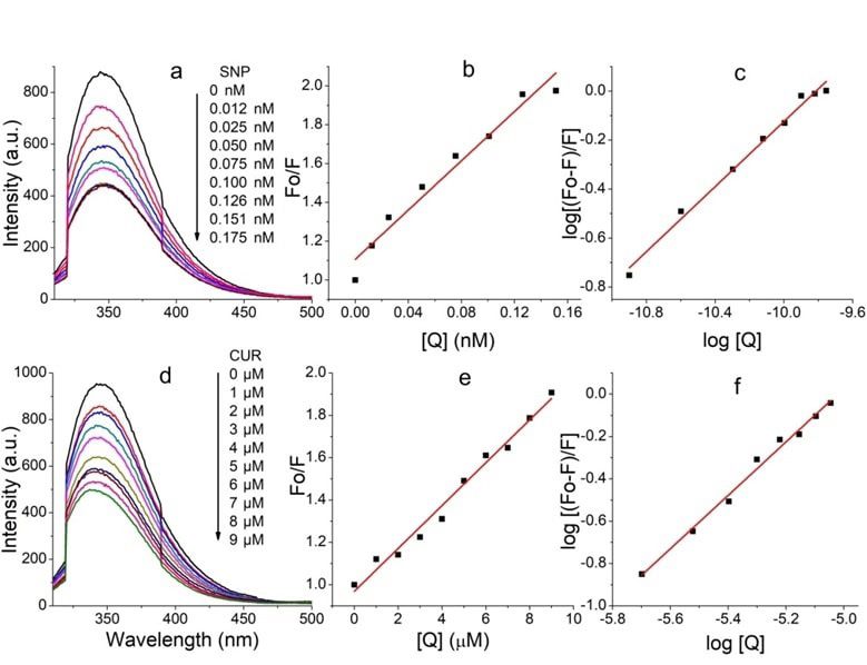
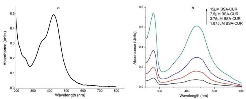
www.ijpsonline.com
RESULTS AND DISCUSSION around 424 nm. This indicates that venom provide less
SNPs were synthesized using bottom-up approach of stability to CUR as compared to BSA.
nanomaterials synthesis. The method involves reduction Absorption spectra of VS conjugate with increasing
of silver ions by citrate ions forming particle seeds to concentrations of CUR exhibited an increase in
which more and more citrate ion accumulates. This absorbance intensity of venom (fig. 2c). Absorbance
results in decrease in citrate ions availability, due to intensity of CUR at 432 nm was obtained; however at
which larger seeds take the help of smaller one in order high concentration the peak shifts towards blue region
to grow via Ostwald ripening[28]. Here, tri-sodium citrate i.e. at 428 nm. This indicates the incapability of venom
works as reducing agent as well as capping agent, to provide stability to CUR.
preventing nanoparticles from aggregation. Change
 Venom-BC conjugate interaction and VS-BC conjugate
in colour of the solution indicates the formation of
 interaction is shown in fig. 2d and fig. 2e respectively.
SNPs which were further characterized by UV-visible
 Unlike venom-CUR, CUR peak (430 nm) in both
spectroscopy, showing an absorbance maximum at 420
 these absorption spectra confirms its stability in the
nm. Hydrodynamic size of SNP was found to be 40±3
 hydrophobic core of the BSA. Stability was further
nm. Using equation 2, the concentration of SNP was
 improved as suggested by the red shift of 6 nm of CUR
found to be 0.50 nM.
 peak as its concentration was increased. Similar shifts
Concentration of SNP= NTotal/NVNA (Eq. 2) were observed by Barik et al.[30] and Yang et al.[31].
Where NTotal is the total number of silver atoms added to Such a small spectral shifts owe majorly to the ionic
the solution, N=Number of silver atoms per nanoparticle, interactions between protein and CUR along with some
V=Volume of the solution and NA=Avogadro’s number. hydrophobic interactions[32]. Significant hyper chromic
 shift in the intensities of the protein and CUR peak was
Spectral characteristics of equimolar BC conjugates are observed in VS-BC conjugate interaction when compared
shown in fig. 1. CUR shows absorption maxima at 424 nm to corresponding venom-BC conjugate interaction. This
(fig. 1a). However in the presence of BSA, CUR becomes could be attributed to the SNP modified venom that leads
stabilize as evident by its peak shift to 434 nm (fig. 1b). to an enhanced interaction between two conjugates.
Peak at 278 nm represents BSA peak. UV characteristic
of venom complexes are shown in fig. 2. Venom shows Fluorescence quenching plots were shown in fig. 3.
a peak at 277 nm which upon SNP addition undergoes Fluorescence characteristics of venom-CUR interaction
hyper chromic shift with prominent widening (fig. 2a). (fig. 3a) and VS interaction (fig. 3d) were studied.
This hyper chromic shift reveals the formation of ground Fluorescence emission intensity of venom at 341 nm was
state complexes of venom components with SNP[29]. significantly quenched by SNP. Fluorescence spectral
Fig. 2b represents the interaction between venom and shift gives knowledge about the microenvironment of
CUR. An increase in the absorption of venom at 277 nm the fluorescence[33]. Here the red shift can be attributed to
with a blue shift of 2 nm was observed with increasing the exposure of fluorophore to the aqueous environment.
CUR concentration, indicating hydrophobic interaction Similar quenching was observed in venom-CUR
between CUR and venom components. Unlike BC interaction; however the blue shift indicates the
interaction, venom-CUR interaction shows CUR peak at hydrophobic interaction between venom and CUR.

Fig. 1: UV-visible spectra of (a) CUR and (b) BSA with CUR
July-August 2022 Indian Journal of Pharmaceutical Sciences 941
www.ijpsonline.com

Fig. 2: UV-visible spectra of venom with conjugate. UV-visible spectra of venom with (a) SNP; (b) CUR; (c) VS-CUR and UV-visible spec-
tra of VS complex with (d) CUR (e) Equimolar BC conjugates

Fig. 3: Fluorescence quenching plots. Fluorescence quenching of venom by (a) SNPs; (b) With respective Stern-Volmer; (c) Double loga-
rithmic plot; (d) Fluorescence quenching of venom by CUR; (e) With respective Stern-Volmer and (f) Double logarithmic plot

Quenching of fluorescence relates the decrease in proteins by polar or charged quencher. Since quencher
fluorescence intensity of fluorophore due to number molecules do not freely penetrate the hydrophobic core
of reasons including ground state complex formation, of proteins, thus only those tryptophan residues which
excited state reactions, molecular rearrangements, energy are present on the surface of the protein are quenched.
transfer and collisional encounters etc. This quenching is Quenching is of two types: Dynamic quenching, where
mainly due the quenching of tryptophan fluorescence in the fluorescence is ceased upon the contact of fluorophore
942 Indian Journal of Pharmaceutical Sciences July-August 2022
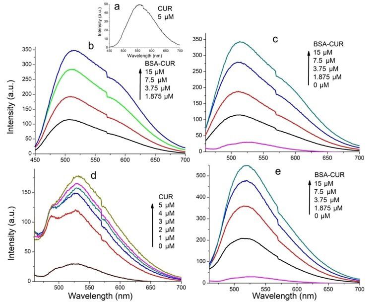
www.ijpsonline.com
and quencher at excited state and static quenching, where Fluorescence data showed that the fluorescence of venom
non-fluorescent complex is formed between fluorophore was greatly quenched by CUR at the concentration range
and quencher. For both interactions to occur, it needs of 1-9 µM. This result infers that the microenvironment
contact between fluorophore and quencher. The possible of the fluorophore was affected by CUR. Also venom
mechanism of quenching was studied using Stern- fluorescence was found to be completely quenched
Volmer plot (fig. 2b and fig. 2e) and KSV was calculated by SNP, showing a saturation point at 0.175 nM
with the help of following Stern-Volmer equation. concentration of SNP. Thus 20 µM CUR and 0.175 nM
F0/F=1+kq 0 [Q]=1+KSV [Q] (Eq. 3) SNP were used to study biochemical activities of venom.

Where F0 and F are fluorescence intensities in Also, fluorescence enhancement of CUR was studied for
absence and presence of quencher respectively, kq is following interaction: CUR; equimolar BC conjugates;
the biomolecular quenching rate constant, 0 is the life venom-BC conjugates; VS-CUR conjugates and VS-BS
time of fluorophore, [Q] is concentration of the quencher conjugates.
and KSV is constant. Average fluorescence life time i.e. 0 Fluorescence enhancement plots were shown in fig. 4.
for biopolymers is 10-8 s. The Stern-Volmer quenching Fluorescence emission intensity of CUR was observed at
constant is given by KSV=kq 0 thus biomolecular 567 nm when excited at 425 nm (fig. 4a); it undergoes a
quenching rate constant can be calculated using formula, blue shift upon BSA interaction (fig. 4b). This suggests
kq=KSV/ 0. that CUR is being localized in hydrophobic pockets of
Generally a linear Stern-Volmer plot indicates the BSA, resulting in increased stability of CUR in aqueous
presence of only one class of fluorophore, all having environment. This localization can be attributed to the
equal access to the quencher. Deviation towards X-axis interaction of aryl group of CUR with hydrophobic
from linearity infers the presence of more than one spaces of BSA[31].
class of fluorophore with unequal accessibility. In case Fluorescence characteristics of VS complex with CUR
of dynamic quenching the biomolecular quenching (fig. 4d) was studied by exciting CUR at 425 nm.
constant is near 1×10¹⁰ M-1s-1 and for static quenching Enhancement in the fluorescence intensity was seen
this value becomes higher. Current investigation shows when CUR was added to the VS complex. Fluorescence
the quenching mechanism of venom components by both intensity was further enhanced upon addition of more
CUR and SNP is static. This was confirmed by the value and more CUR. In comparison with the fluorescence
of biomolecular quenching constant, which was found spectra of CUR with venom, increased fluorescence
to be 1.01×10¹⁴ M-1s-1 and 6.35×10¹⁷ M-1s-1 for CUR and enhancement was observed in the spectra of CUR with
SNP respectively (Table 1). VS. This observation expects increased protection
The number of binding sites (n) and binding constant against venom in presence of SNP and CUR.
(K) of venom for both CUR and SNP was calculated by Moreover, interaction of venom and BC complexes was
plotting a graph of Log [(F0–F)/F] vs. log [Q]. This graph investigated. Fluorescence spectra were recorded by
gives a straight line with an equation given below. exciting CUR (fig. 4c). Emission intensity at 520 nm was
Log [(F0–F)/F]=logK+nlog [Q] (Eq. 4) not changed upon addition of increasing concentration of
 BC complexes to venom. Finally VS complex incubated
Where n is number of binding sites and K is binding with BC complex and fluorescence spectra were recorded
constant. by exciting CUR (fig. 4e). A great increase in emission
The values of binding number and binding constants intensity was observed at 520 nm with a blue shift of
suggest that, though venom have less binding sites for about 1 nm. Such a huge increase entails an effective
SNP, their binding affinity is more. interaction predicting higher protection against venom
 toxicity.
TABLE 1: BINDING PROPERTIES OF VENOM CONJUGATES
Samples KSV kq (M-1s-1) Binding sites (n) Binding constant
Venom-CUR (1.01±0.09)×10 5
 (1.01±0.09)×10 14
 1.261±0.12 (2.14±0.23)×106
VS (6.35±0.26)×109 (6.35±0.26)×1017 0.664±0.04 (3.28±0.15)×106
Note: Stern-Volmer quenching constant, binding constant and number of binding sites of venom for SNP and CUR. The results are expressed
as mean±Standard Deviation (SD)

July-August 2022 Indian Journal of Pharmaceutical Sciences 943
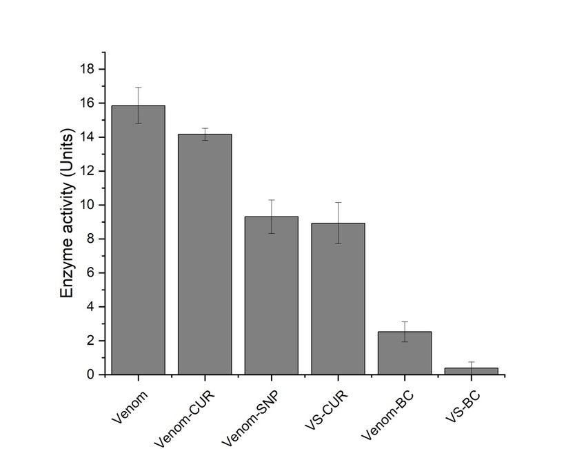
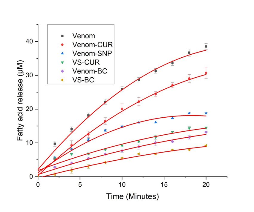
www.ijpsonline.com

Fig. 4: Fluorescence enhancement plots. Fluorescence spectra of (a) CUR; (b) Equimolar BC complexes; (c) Venom with increasing equi-
molar BC concentration; (d) VS complex with CUR and (e) VS complex with equimolar BC concentration

Venom neutralizing potential of CUR complexes presence of different inhibitory complexes is shown in
is shown here. For all biochemical assays saturating Table 2.
concentration of CUR (20 µM), SNP (0.18 nM) and
 In a report by Kadali et al.[23], soy protein nanoparticles
equimolar BC (15 µM) were used.
 were conjugated with anti-venom and neutralization
Acidimetry was performed to measure the activity potential of this formulation was studied. Complete
of PLA2 of RVV. PLA2 with Asp49 at its active site neutralization of PLA2 activity of Bungarus caeruleus
hydrolyses ester bond at Nucleophilic Substitution venom was achieved by this formulation at ratio 1:400
bimolecular (SN2) position of phospholipids[34] present in (venom conjugated soy nanoparticles). Also activity of
the membrane of various cells. Upon the action of PLA2, Doboia russelli venom was found to be antagonized
lysophospholipids and fatty acids, mostly arachidonic up to 40 folds by methanolic extract of Curcuma
acids[35] are released. Released fatty acids were measured aromatica[37]. Polyphenols may inhibit snake venom
acidimetrically on pH meter as a drop in pH of the egg either by precipitating venom proteins or by chelating
yolk suspension (i.e. one unit change in pH corresponds cofactor required for these enzymes to work, however
to 133 µM fatty acids released). Egg yolk which is a rich CUR interacts with active site of PLA2[38]. Molecular
source of lecithin wherein phospholipids are present in docking experiment on PLA2 of RVV and compounds
great amount[36] was used as a substrate. such as CUR suggests their interaction with amino
 acids and ions present in active site of PLA2. These
Activity of viper venom and viper venom incubated with
 inhibitory components have been shown to interact with
different test complexes was studied using this method
 Asp49, Gly30 substrate binding[39]. CUR exhibit strong
(fig. 5).
 antioxidant activity which can help to prevent oxidative
Venom showed increased fatty acids released with time, damage caused by PLA2 by specifically interacting
whereas its activity with various complexes was found to with conserved residues of PLA2 that is critical for its
be serially reduced in following order, VS-BC>Venom- catalysis[40].
BC>VS-CUR>VS>Venom-CUR. Activity of venom in
944 Indian Journal of Pharmaceutical Sciences July-August 2022
www.ijpsonline.com

Fig. 5: Fatty acids release with time by venom and venom complexes, ( ) Venom; ( ) Venom-CUR; ( ) VS; ( ) VS-CUR; ( )
Venom-BC and ( ) VS-BC

TABLE 2: PERCENTAGE REDUCTION IN THE PLA2 ACTIVITY OF VENOM COMPLEXES
Samples % Reduction
Venom-CUR 21.37±1.59
VS 51.72±0.56
VS-CUR 62.07±0.93
Venom-BC 65.51±0.59
VS-BC 75.86±0.46
Note: The table shows percentage reduction in the PLA2 activity of various venom complexes. Venom treated with SNP and BC complex
showed highest reduction in venom phospholipase activity. The results are expressed as mean±SD

Protease activity was assessed using the method given residues along with fragmented peptides in the system.
by Greenberg with slight modification. Protease activity Reaction terminator TCA stops the reaction and it also
by this method involved incubation of protease with forms complexes with undigested casein which can be
substrate, resulting in release of tyrosine residues separated from free tyrosine by filtration. The filtrate
which can be quantified spectrometrically using Folin- containing released tyrosine residues were quantified
Lowry method. Viper venom contains many serine using Folin’s method. Phenolic group of tyrosine residues
and metalloproteinases[41]. Venom metalloproteases are complexes with copper increasing its reactivity with
known to be major lead in the pathogenesis of venom- phosphomolybdate present in Folin’s reagent. Copper
induced local tissue damage[42]. These proteases cleave complexed phenol group reduces phosphomolybdate
proteins releasing amino acids and several peptides. giving rise to a blue colored compound which absorbs
Zinc dependent metalloproteinases from viper venom maximum at 660 nm. This color intensity is directly
are major cause of hemorrhagic damage[43], disrupting proportional to the concentration of tyrosine residues.
microvessels that further causes uncontrolled bleeding[44]. Enzyme activity of viper venom in presence of different
In current investigation, milk casein was used as substrate inhibitory complexes was studied (fig. 6).
on which venom proteases acts, releasing tyrosine

July-August 2022 Indian Journal of Pharmaceutical Sciences 945
www.ijpsonline.com

Fig. 6: Protease activity of venom and venom complexes

Protease activity of venom in presence of various CUR tissue phospholipids, platelet phospholipids and calcium
and SNP complexes was found to be reduced. Percentage form a catalytic complex called prothrombinase which
reduction of venom activity by CUR conjugates are convert prothrombin to thrombin. Thrombin intern
shown in Table 3. converts fribrinogen to insoluble fibrins which are cross
 linked by activated factor-XIII forming a plug at of
CUR is a well-known protease inhibitor having ability to
 intertwined fibrin polymers. On the other hand intrinsic
inhibit chymotrypsin-like activity of purified rabbit 20S
 pathway starts with factor-XII which activates factor-
proteasome and cellular 26S proteasome[45]. It is also
 XI. Factor-XI further stimulate factor-IX to activate
known to inhibit matrix metalloproteinase activity[46]. It
 its cofactor i.e. factor VIII. This activated cofactor acts
has a profound metal chelating ability[47] that can make
 on factor-X and the cascade is continued in similar
it a good metalloprotease inhibitor. RVV proteases thus
 way to that of extrinsic pathway[51]. Since RVV-X and
could be inhibited by chelation of metal ions that are
 RVV-V from viper venom directly activates factor-X
required for enzyme catalysis.
 and factor-V, lengthy extrinsic and intrinsic routes are
Viperidae family is very well known for its procoagulant skipped. This results in procoagulation of blood. Clotting
and anticoagulant activities. Among these species time of venom with several inhibitory complexes was
Doboia russelli is a strong procoagulant owing to the found to be prolonged (Table 4).
presence of RVV-X[48] and RVV-V[49]. RVV-X, a calcium
 Stability of RVV-X is due to disulphide linkages between
dependent metalloproteinase, digests heavy chain of
 one heavy and two light chains. The proposed mechanism
factor-X of blood coagulation cascade and cleaves the
 of interaction states that light chain recognizes Gamma
bond between Arg52 and Ile53. However, serine protease
 (γ)-Carboxyglutamic acid (Gla) domain of factor-X
RVV-V acts on factor-V which is a component of blood
 and heavy chain cleaves it, leading to the activation
coagulation cascade. It is acted upon by RVV-V and gets
 of factor-X[52]. CUR on the other hand is supportive
cleaved at two positions i.e. at Arg1545 and Arg1018[50].
 for hemostasis, anticoagulation and fibrinolysis[53]. It
Normal blood coagulation cascade is classified in two
 has been observed that CUR inhibits both intrinsic
pathways, intrinsic and extrinsic, both converging at
 and extrinsic pathways of coagulation by inhibiting
factor-X activation. Extrinsic pathway starts with the
 factor-Xa and thrombin generation[54]. Hence in current
exposure of Tissue Factor (TF) to coagulation cascade of
 investigation inhibition of procoagulant activity may be
plasma due to any injury to vascular system. TF interacts
 due the disruption of this stable conformation of RVV-X
with factor-VIIa and calcium converting factor-X to Xa.
 by CUR and SNP complexes.
Factor-X has a cofactor i.e. factor-V. Factor-Xa, factor-V,
946 Indian Journal of Pharmaceutical Sciences July-August 2022
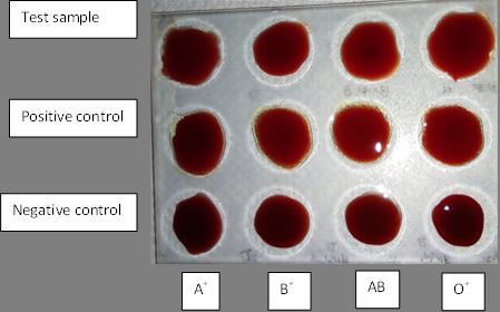
www.ijpsonline.com
TABLE 3: PERCENTAGE REDUCTION IN THE PROTEASE ACTIVITY OF VENOM COMPLEXES
Samples % Reduction
Venom-CUR 10.99±2.35
VS 37.41±3.9
VS-CUR 40.20±4.5
Venom-BC 83.79±3.28
VS-BC 96.94±2.44
Note: The table shows percentage reduction in the protease activity of various venom complexes. Venom treated with SNP and BC
complex showed highest reduction in venom protease activity. The results are expressed as mean±SD

TABLE 4: PROCOAGULANT ACTIVITY OF VENOM AND VENOM COMPLEXES
Samples Clotting time (min)
Venom 0.47±0.035
Venom-CUR 1.08±0.02
VS 1.14±0.02
VS-CUR 1.20±0.09
Venom-BC 1.36±0.06
VS-BC 1.40±0.021
Saline control 2.54±0.155
Note: The table shows reduction in the procoagulant activity of various venom complexes. Procoagulant activity of venom was reduced
greatly when treated with SNP and BC complex. The results are expressed as mean±SD

Venom PLA2 acts on membrane phospholipids by binding characterized using UV-visible and dynamic light
to its substrate through hydrophobic channels, which scattering. RVV-SNP and BC conjugates were prepared
suggests that the activity of venom can be inhibited and analysed for anti-venom activity. UV-visible
by blocking this hydrophobic channel[55]. Structural and fluorescence spectra of BC conjugate showed
and molecular chemistry of CUR gives it an advantage hydrophobic and hydrophilic interaction between
to proteins with high affinity. Phenyl rings of CUR can CUR and BSA, hence making CUR stable in aqueous
make pi-pi (ᴫ-ᴫ) interaction with aromatic amino acids. environment. Fluorescence intensity of RVV was found
Phenolic and carbonyl functional groups can further to be significantly quenched by SNP and by CUR;
share hydrogen bond with proteins[56]. Simulation studies however RVV was found to have more binding site for
have also shown how CUR adopts different conformation CUR. Enhancement of fluorescence intensity in VS-BC
by making hydrophobic contacts with protein. In docked complex was significantly higher than other complexes.
complex of PLA2 and CUR it was shown that one half Inhibitory effect of these complexes was observed using
portion of the CUR was bound to the active site of the biochemical assays. VS-BC complex showed major
enzyme and the other half was protruding outside. This inhibition. PLA2 activity and protease activity of venom
indicates that CUR might be blocking the substrate from was reduced by 75 % and 96 % respectively. Procoagulant
entering the active site of PLA2[57]. activity of venom was also inhibited and clotting time
 of blood was increased to normal. This complex was
Direct agglutination test is one of the preliminary means
 also confirmed to be biocompatible in vitro, which was
to identify biocompatibility of any component to be used
 studied by simple agglutination test.
in medicine. It is a simple test that involves mixing of
test compound with a drop of blood on a cavity slide and The inhibitory effects of this complex can be attributed
the results could be observed in few seconds. SNP+BC to the interaction of active sites of venom enzymes
complex was analysed for its biocompatibility of all four with SNP and CUR as well. Moreover BSA involved
blood group. Appropriate positive and negative controls in BC conjugates improves solubility, stability of CUR
were used. The complex showed no agglutination for and protect it from photodamage. CUR has greatest
any of the blood group (fig. 7) suggesting it’s in vitro therapeutic properties and the current report suggests its
biocompatibility. anti-venom activity against RVV, This complex needs to
 be further analysed for the same in vivo.
In the present study, SNPs were synthesized and
July-August 2022 Indian Journal of Pharmaceutical Sciences 947
www.ijpsonline.com

Fig. 7: Agglutination test. Test sample: SNPs-BC; Positive control: Antisera for respective blood group and Negative control: Saline

Acknowledgements: 9. Kalita B, Patra A, Jahan S, Mukherjee AK. First report of the
 characterization of a snake venom apyrase (Ruviapyrase) from
The financial assistance of Chhatrapati Shahu Maharaj Indian Russell's viper (Daboia russelii) venom. Int J Biol
Research, Training and Human Development Institute Macromol 2018;111:639-48.
(SARTHI), Pune is greatly acknowledged. 10. Aird SD, Watanabe Y, Villar-Briones A, Roy MC, Terada
 K, Mikheyev AS. Quantitative high-throughput profiling of
Conflict of interests: snake venom gland transcriptomes and proteomes (Ovophis
 okinavensis and Protobothrops flavoviridis). BMC Genomics
The authors declared no conflict of interest. 2013;14(1):1-27.
 11. Julio-Pieper M, Lozada P, Tapia V, Vega M, Miranda C,
REFERENCES Vantman D, et al. Nerve growth factor induces vascular
 endothelial growth factor expression in granulosa cells via a
1. Gupta YK, Peshin SS. Snake bite in India: Current scenario of trkA receptor/mitogen-activated protein kinase-extracellularly
 an old problem. J Clin Toxicol 2014;4(1):1-9. regulated kinase 2-dependent pathway. J Clin Endocrinol Metab
2. Kasturiratne A, Wickremasinghe AR, de Silva N, Gunawardena 2009;94(8):3065-71.
 NK, Pathmeswaran A, Premaratna R, et al. The global burden 12. Sani I, Umar RA, Hassan SW, Faruq UZ. Antisnake venoms and
 of snakebite: A literature analysis and modelling based on their mechanisms of action: A review. Saudi J Med Pharm Sci
 regional estimates of envenoming and deaths. PLoS Med 2018;4(5):512-20.
 2008;5(11):1591-604.
 13. León G, Stiles B, Alape A, Rojas G, Gutiérrez JM. Comparative
3. Jayanthi GP, Gowda TV. Geographical variation in India in study on the ability of IgG and F (ab') 2 antivenoms to neutralize
 the composition and lethal potency of Russell's viper (Vipera lethal and myotoxic effects induced by Micrurus nigrocinctus
 russelli) venom. Toxicon 1988;26(3):257-64. (coral snake) venom. Am J Trop Med Hyg 1999;61(2):266-71.
4. Pe T, Aye B, Myint AA, Swe TN, Warrell DA. Bites by Russell’s 14. Warrell DA. WHO/SEARO Guidelines for the clinical
 viper (Daboia russelli siamensis) in Myanmar: Effect of the management of snakebite in the Southeast Asia region. SE Asian
 snake’s length and recent feeding on venom antigenaemia J Trop Med Public Health 1999;30(1):1-85.
 and severity of envenoming. Trans R Soc Trop Med Hyg
 15. de Silva HA, Ryan NM, de Silva HJ. Adverse reactions to
 1991;5(6):804-8.
 snake anti-venom and their prevention and treatment. Br J Clin
5. Sharma M, Das D, Iyer JK, Kini RM, Doley R. Unveiling the Pharmacol 2016;81(3):446-52.
 complexities of Daboia russelii venom, a medically important
 16. Poovazhagi V, Ravikumar SA, Jagadeeshwari A, Arulganesh P,
 snake of India, by tandem mass spectrometry. Toxicon
 Prabhu RS, Anupama S. Anti-snake venom induces reactions
 2015;107:266-81.
 among children with snake envenomation. Int J Contemp
6. Kini RM. Excitement ahead: Structure, function and mechanism Pediatr 2017;4(2):629-34.
 of snake venom phospholipase A2 enzymes. Toxicon
 17. Sakthivel G, Dey A, Nongalleima K, Chavali M, Rimal Isaac
 2003;42(8):827-40.
 RS, Singh NS, et al. In vitro and in vivo evaluation of polyherbal
7. Tan NH, Fung SY, Tan KY, Yap MK, Gnanathasan CA, Tan formulation against Russell’s viper and cobra venom and
 CH. Functional venomics of the Sri Lankan Russell's viper screening of bioactive components by docking studies. Evid
 (Daboia russelii) and its toxinological correlations. J Proteomics Based Complement Alternat Med 2013;2013:1-13.
 2015;128:403-23.
 18. Ratanabanangkoon K, Cherdchu C, Chudapongse P. Studies on
8. Costa TR, Burin SM, Menaldo DL, de Castro FA, Sampaio the cobra neurotoxin inhibiting activity in an extract of Curcuma
 SV. Snake venom L-amino acid oxidases: An overview on sp. (Zingiberaceae) rhizome. Southeast Asian J Trop Med Public
 their antitumor effects. J Venom Anim Toxins Incl Trop Dis Health 1993;24(1):178-85.
 2014;20(23):1-7.

948 Indian Journal of Pharmaceutical Sciences July-August 2022
www.ijpsonline.com
19. Gomes A, Das R, Sarkhel S, Mishra R, Mukherjee S, 39. Nirmal N, Praba GO, Velmurugan D. Modeling studies on
 Bhattacharya S, et al. Herbs and herbal constituents active phospholipase A2-inhibitor complexes. Indian J Biochem
 against snake bite. Indian J Exp Biol 2010;48:865-78. Biophys 2008;45:256-62.
20. Karain BD, Lee MK, Hayes WK. C60 Fullerenes as a novel 40. Gómez-Betancur I, Gogineni V, Salazar-Ospina A, León F.
 treatment for poisoning and envenomation: A proof-of-concept Perspective on the therapeutics of anti-snake venom. Molecules
 study for snakebite. J Nanosci Nanotechnol 2016;16(7):7764-71. 2019;24(18):3276.
21. Suman B, Eswari B, Divya BJ, Pallavi C, Venkataswamy M, 41. Matsui T, Fujimura Y, Titani K. Snake venom proteases
 Kemparaj K, et al. Effect of silver nano particles synthesized of affecting hemostasis and thrombosis. Biochim Biophys Acta
 Trichodesma indicum against Naja Naja (cobra) venom. Int J 2000;1477(2):146-56.
 Pharm Sci Res 2018;9(8):3291-6. 42. Aung HT, Nikai T, Komori Y, Nonogaki T, Niwa M, Takaya
22. Hingane VC, Pangam D, Dongre PM. Inhibition of crude Y. Biological and pathological studies of rosmarinic acid as
 viper venom action by silver nanoparticles: A biophysical and an inhibitor of hemorrhagic Trimeresurus flavoviridis (habu)
 biochemical study. Biophys Physicobiol 2018;15:204-13. venom. Toxins 2010;2(10):2478-89.
23. Renu K, Gopi K, Jayaraman G. Formulation and characterisation 43. Gutiérrez JM, Rucavado A, Escalante T, Díaz C. Hemorrhage
 of antibody-conjugated soy protein nanoparticles-Implications induced by snake venom metalloproteinases: Biochemical
 for neutralisation of snake venom with improved efficiency. and biophysical mechanisms involved in microvessel damage.
 Appl Biochem Biotechnol 2014;174(7):2557-70. Toxicon 2005;45(8):997-1011.
24. Lee PC, Meisel D. Adsorption and surface-enhanced Raman of 44. Bjarnason JB, Fox JW. Hemorrhagic metalloproteinases from
 dyes on silver and gold sols. J Phys Chem 1982;86(17):3391-5. snake venoms. Pharmacol Ther 1994;62(3):325-72.
25. Tan NH, Tan CS. Acidimetric assay for phospholipase A using egg 45. Milacic V, Banerjee S, Landis-Piwowar KR, Sarkar FH,
 yolk suspension as substrate. Anal Biochem 1988;170(2):282-8. Majumdar AP, Dou QP. Curcumin inhibits the proteasome
26. Greenberg DM. Plant proteolytic enzymes. In: Colowick SP activity in human colon cancer cells in vitro and in vivo. Cancer
 and Kalpan SP, editors. Methods in Enzymology. 2nd ed. USA: Res 2008;68(18):7283-92.
 Academic press Inc; 1955. 46. Cheng TS, Chen WC, Lin YY, Tsai CH, Liao CI, Shyu HY, et al.
27. Vineetha MS, Janardhan B, More SS. Biochemical and Curcumin-targeting pericellular serine protease matriptase role
 pharmacological neutralization of Indian saw-scaled viper snake in suppression of prostate cancer cell invasion, tumor growth
 venom by Canthium parviflorum extracts. Indian J Biochem and metastasis curcumin-suppressing matriptase, prostate cancer
 Biophys 2017;54:173-85. cell invasion, and metastasis. Cancer Prev Res 2013;6(5):495-
28. Pacioni NL, Borsarelli CD, Rey V, Veglia AV. Synthetic routes 505.
 for the preparation of silver nanoparticles: A mechanistic 47. Ferrari E, Benassi R, Sacchi S, Pignedoli F, Asti M, Saladini M.
 perspective. In: Alarcon EI, Griffith M, Udekwwu KI, editors. Curcumin derivatives as metal-chelating agents with potential
 Silver nanoparticle applications: In the fabrication and design of multifunctional activity for pharmaceutical applications. J Inorg
 medical and biosensing devices; Cham: Springer 2015. p. 13-46. Biochem 2014;139:38-48.
29. Kathiravan A, Renganathan R, Anandan S. Interaction of 48. Suntravat M, Nuchprayoon I, Pérez JC. Comparative study of
 colloidal AgTiO2 nanoparticles with bovine serum albumin. anticoagulant and procoagulant properties of 28 snake venoms
 Polyhedron 2009;28(1):157-61. from families Elapidae, Viperidae and purified Russell’s viper
30. Barik A, Priyadarsini KI, Mohan H. Photophysical studies on venom-factor X activator (RVV-X). Toxicon 2010;56(4):544-53.
 binding of curcumin to bovine serum albumin. Photochem 49. Kini RM, Rao VS, Joseph JS. Procoagulant proteins form snake
 Photobiol 2003;77(6):597-603. venoms. Hemostasis 2001;31(3):218-24.
31. Yang M, Wu Y, Li J, Zhou H, Wang X. Binding of curcumin 50. Kalafatis M, Beck DO, Mann KG. Structural requirements for
 with bovine serum albumin in the presence of i-carrageenan expression of factor Va activity. J Biol Chem 2003;278(35):33550-
 and implications on the stability and antioxidant activity of 61.
 curcumin. J Agric Food Chem 2013;61(29):7150-5. 51. Palta S, Saroa R, Palta A. Overview of the coagulation system.
32. Mitra SP. Binding and stability of curcumin in presence of Indian J Anaesth 2014;58(5):515-23.
 bovine serum albumin. J Surf Sci Technol 2007;23(3):91. 52. Liu X, Atwater M, Wang J, Huo Q. Extinction coefficient of gold
33. Kar T, Basak P, Sen S, Ghosh RK, Bhattacharyya M. Analysis nanoparticles with different sizes and different capping ligands.
 of curcumin interaction with human serum albumin using Colloids Surf B Biointerfaces 2007;58(1):3-7.
 spectroscopic studies with molecular simulation. Front Biol 53. Keihanian F, Saeidinia A, Bagheri RK, Johnston TP, Sahebkar
 2017;12(3):199-209. A. Curcumin, hemostasis, thrombosis, and coagulation. J Cell
34. Stafford RE, Dennis EA. Lysophospholipids as biosurfactants. Physiol 2018;233(6):4497-511.
 Colloids Surf 1987;30(1):47-64. 54. Kim DC, Ku SK, Bae JS. Anticoagulant activities of curcumin
35. Urs NA, Yariswamy M, Joshi V, Nataraju A, Gowda TV, and its derivative. BMB Rep 2012;45(4):221-6.
 Vishwanath BS. Implications of phytochemicals in snakebite 55. Dileep KV, Tintu I, Mandal PK, Karthe P, Haridas M, Sadasivan
 management: Present status and future prospective. Toxin Rev C. Crystal structure of porcine pancreatic phospholipase A2 in
 2014;33(3):60-83. complex with 2-methoxycyclohexa-2-5-diene-1, 4-dione. Front
36. Blesso CN. Egg phospholipids and cardiovascular health. Life Sci 2011;5(3-4):135-9.
 Nutrients 2015;7(4):2731-47. 56. Gupta SC, Prasad S, Kim JH, Patchva S, Webb LJ, Priyadarsini
37. Alam MI. Inhibition of toxic effects of viper and cobra venom IK, et al. Multitargeting by curcumin as revealed by molecular
 by Indian medicinal plants. Pharmacol Pharm 2014;5(8):828-37. interaction studies. Nat Prod Rep 2011;28(12):1937-55.
38. Pereanez JA, Nunez V, Patino AC, Londono M, Quintana JC. 57. Dileep KV, Tintu I, Sadasivan C. Molecular docking studies
 Inhibitory effects of plant phenolic compounds on enzymatic and of curcumin analogs with phospholipase A2. Interdiscip Sci
 cytotoxic activities induced by a snake venom phospholipase A2. 2011;3(3):189-97.
 Vitae 2011;18(3):295-304.

July-August 2022 Indian Journal of Pharmaceutical Sciences 949
You can also read