Inhibition of RANKL and Sema4D improves residual ridge resorption in mice
←
→
Page content transcription
If your browser does not render page correctly, please read the page content below
www.nature.com/scientificreports
OPEN Inhibition of RANKL and Sema4D
improves residual ridge resorption
in mice
Meri Hisamoto1*, Shunsuke Kimura2,3*, Kai Iwata1, Toshihiko Iwanaga2 & Atsuro Yokoyama1
Residual ridge resorption (RRR) is a chronic and progressive bone resorption following tooth loss.
It causes deterioration of the oral environments and leads to the pathogenesis of various systemic
diseases. However, the molecular mechanisms and risk factors for RRR progression are still unclear
and controversial. In this study, we developed a tooth extraction model using mice for analyzing
long-term morphological and gene expression changes in the alveolar bone. We further applied
ovariectomy to this model to elucidate the effects of osteoporosis on RRR progression. As a result,
the alveolar bone loss was biphasic and consisted of rapid loss in the early stages and subsequently
slow and sustained bone loss over a long period. Histological analysis indicated that ovariectomy
prolonged the activation of osteoclasts in the alveolar bone. Furthermore, the expressions of Tnfsf11
and Sema4d kept increasing for a long time in OVX mice. Administration of neutralization antibodies
for receptor activator of NF-κB ligand (RANKL) effectively suppressed RRR. Similarly, inhibition of
Semaphorin 4D (Sema4D) also improved alveolar bone loss. This study demonstrated that reduced
ovarian function may be a risk factor for RRR and that RANKL and Sema4D suppression are potential
treatments.
Residual ridge resorption (RRR) is a continuous, often lifelong, alveolar bone resorption occurring after tooth
loss. Losing teeth in adult is the result of injury or disease, such as dental avulsion, tooth decay, and periodontal
disease. The proportion of people who lost their teeth increases with age. In Japan, 40% of people in their late
40 s and 60% of people in their early 50 s have lost at least one t ooth1. In the United States. 26% of adults aged 65
or older have eight or fewer teeth and about 17% in them have lost all of their t eeth2. RRR therefore can occur
in anyone with a high probability.
RRR follows initial wound healing, which includes epithelial integrity restoration accompanied by bone
formation within the extraction socket and bone resorption at the edge of the socket. After the initial rapid
healing stage, bone resorption slows down but sometimes persists for a long time. As RRR progresses, dentures
become unstable, and dental implant placement becomes difficult, thus worsening the oral health-related quality
of life and social activity. Several studies have suggested that prolonged alveolar bone resorption is caused by
aging, excessive mechanical stress, and p eriodontitis3–5. However, the risk factors of RRR progression are still
controversial.
Osteoporosis is a metabolic disease that affects postmenopausal women. This disease is commonly character-
ized by low bone mass and bone tissue deterioration, which can lead to increased risk of bone fractures. However,
not all individuals with osteoporosis develop RRR. Some animal studies have demonstrated positive correlations
between alveolar bone loss or delayed healing of extraction sockets and systemic osteoporosis6,7. Contrarily,
others have shown a weak relationship or no relevance at all8,9. Thus, the relationship between osteoporosis and
jawbone resorption has not been elucidated.
The treatment for osteoporosis mainly involves two drug types. Most of the currently available drugs for
osteoporosis are bisphosphonates or anti-RANKL monoclonal antibody drugs. These drugs inhibit osteoclastic
bone resorption; however, they can sometimes, but not necessarily, induce drug-related osteonecrosis of the jaw
(ARONJ) following tooth e xtraction10,11. Another type of drug promoting osteoblastic bone formation is parathy-
roid hormone (PTH). Intermittent administration of PTH can treat osteoporosis of long bones and vertebrae12,
and some studies have indicated that intermittent PTH therapy could increase the mineral density of the jawbone
in animal m odels13,14. However, intermittent PTH treatment only initially increases bone formation and promotes
1
Department of Oral Functional Prosthodontics, Division of Oral Functional Science, Faculty of Dental Medicine,
Hokkaido University, Sapporo 060‑8586, Japan. 2Laboratory of Histology and Cytology, Graduate School of
Medicine, Hokkaido University, Sapporo 060‑8638, Japan. 3Division of Biochemistry, Faculty of Pharmacy, Keio
University, Tokyo 105‑8512, Japan. *email: merimeri@den.hokudai.ac.jp; kimura-sn@pha.keio.ac.jp
Scientific Reports | (2022) 12:4094 | https://doi.org/10.1038/s41598-022-08016-3 1
Vol.:(0123456789)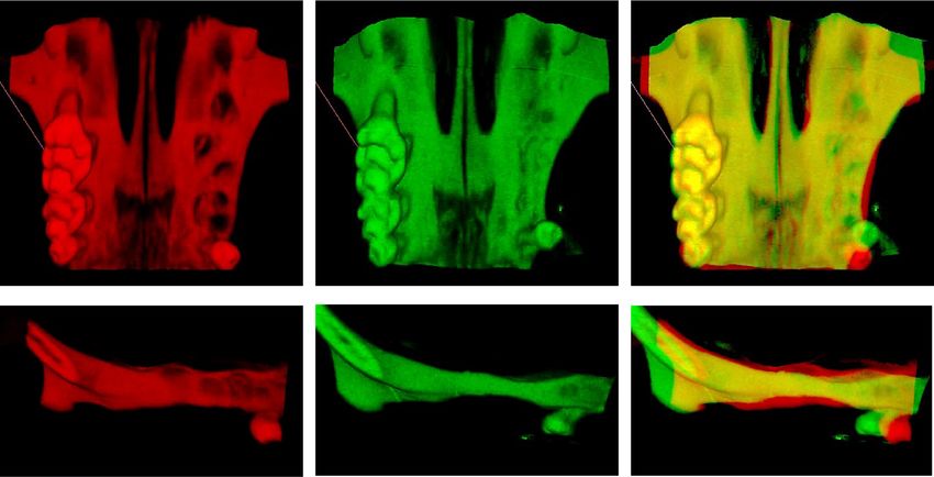
www.nature.com/scientificreports/
Figure 1. Resorption of the maxillary alveolar bone after teeth extractions in mice. (a) Bone volumes of both
the extracted and non-extracted sides were determined with μCT analysis. The measurement range of bone
volume is indicated in purple in the µCT-3D image of the upper images, which is between the M1 mesial
buccal root and M2 distal buccal root. M1, M2, and M3 denote the first molar, second molar, and third molar,
respectively. The graph in the lower panel shows the time-dependent changes in bone volume of the teeth-
extracted and the non-extracted sides and are expressed as a ratio of the bone volume on day 0 after tooth
extraction in the same individual. The data are expressed as mean ± S.D. (n = 3) and are representative of three
independent experiments. (b) The selected regions of interest (ROIs) in or areas near the extraction socket of the
first molar are displayed in the circled areas of the µCT image in the upper panel. The graph in the lower panel
shows the time-dependent changes in the ratio of the CT values of the maxilla alveolar bone of the extracted
region (ROI-2) to the non-extracted buccal side (ROI-1). Each symbol represents an individual mouse. The data
shown in the graphs are representative of three independent experiments. ***P < 0.005; calculated using two-way
ANOVA.
bone resorption; therefore, the treatment period is limited. A Sost-specific antibody treatment, which promotes
bone formation and inhibits bone resorption, was developed and used clinically. An animal study suggested
that sclerostin inhibition increased the alveolar bone volume and architecture in rats with alveolar bone l oss15.
Semaphorin 4D (Sema4D) is expressed by osteoclasts and is a mediator of osteoclast–osteoblast communica-
tion. Moreover, it inhibits osteoblastic bone formation. Injection of Sema4D-specific antibodies into ovariecto-
mized mice was shown to promote osteoblastic bone formation without affecting osteoclastic bone resorption
in the femur16. However, there have only been a few reports on the therapeutic effect of Sema4D inhibition on
jawbone loss following tooth extraction.
This study aimed to (1) elucidate the factor of alveolar bone resorption using a murine tooth extraction model
and (2) investigate whether injection of RANKL-specific antibodies or Sema4D-specific antibodies could be a
useful approach to the suppression of alveolar bone resorption in an ovariectomized mouse model of postmeno-
pausal osteoporosis.
Results
Maxillary teeth extractions in mice does not induce long‑term loss of maxillary alveolar bone
volume. RRR in humans is characterized by sustained loss of the mandible and maxillary bone mass for
prolonged periods following tooth extraction. However, there is little information about bone morphological
changes after extraction in other mammals, including rodents. Therefore, we investigated the morphological
changes of the maxillary alveolar bones of mice following teeth extractions using µCT imaging. At 16 weeks after
teeth extractions, the maxillary bone was absorbed on the maxillary sinus side and the maxilla alveolar crest side
but was only absorbed slightly on the buccal side (Supplementary Fig. S1 online).
We examined the long-period changes of BV on the extracted and non-extracted sides of the maxillary
bone from day 0 to 24 weeks following teeth extractions. The BV of the extracted side rapidly decreased to
73.6% ± 1.51% (mean ± standard deviation) by 9 weeks post-extraction (Fig. 1a). Subsequently, although the
alveolar BV slightly decreased, there was no significant difference between 9 and 24 weeks (P = 0.27 calculated
Scientific Reports | (2022) 12:4094 | https://doi.org/10.1038/s41598-022-08016-3 2
Vol:.(1234567890)
www.nature.com/scientificreports/
using Student’s t-test). This result suggested that alveolar bone resorption following teeth extractions is rarely
long-lasting in healthy mice.
The CT value at an extraction socket of the first molar recovered to 80.0% ± 0.06% of the amount of the adja-
cent alveolar bone of the buccal side at 5 weeks post-extraction, indicating that the extraction socket of the first
molar was almost filled with new bone by 5 weeks post-extraction (Fig. 1b).
These results indicated that the sharp decrease in alveolar BV in the weeks following teeth extractions was
related to the activation of bone remodeling during healing of the extraction s ocket17,18.
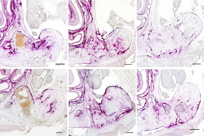
Ovariectomy promotes long‑term resorption of the maxillary bone after teeth extrac-
tions. Postmenopausal osteoporosis could be one of the possible risk factors for ridge r esorption19. We ana-
lyzed bone morphology following teeth extractions using OVX mice as an animal model for osteoporosis. The
images obtained via superimposition of CT-3D images revealed that the height and width of the alveolar bone
were reduced after 16 weeks post-extraction in OVX mice (Fig. 2a).
The temporal observation of alveolar BV with µCT indicated that the BV in OVX mice rapidly decreased to
69.7% ± 2.60% at 5 weeks post-extraction and subsequently decreased gradually to 55.3% ± 2.63% by 17 weeks fol-
lowing teeth extractions. This result indicated that alveolar bone resorption in OVX mice continues over the long
term. Meanwhile, the BV in the sham mice decreased to 84.5% ± 0.30% at 5 weeks and then slightly decreased, but
was not statistically significant (P = 0.267 calculated using Student’s t-test), to 79.5% ± 0.49% at 17 weeks (Fig. 2b).
This kinetic BV change in the sham mice was almost identical to the non-treated mice (Fig. 1a).
The ratio of the CT values in the extraction socket of sham mice recovered to 84.2% ± 3.05% at 5 weeks after
extraction and reached 96.7% ± 0.89% at 17 weeks, whereas the CT value in that of OVX mice only recovered to
55.5% ± 1.33% at five weeks and subsequently remained almost unchanged during our observation period up to
17 weeks (66.1% ± 3.06%) following teeth extractions (Fig. 2c). This result indicated that the extraction socket
was not entirely restored in the OVX mice.
Prolonged activation of bone remodeling during the restoration of extraction sockets in OVX
mice. We examined the temporal distribution of osteoclasts during healing of the extraction sockets using
enzymatic histochemistry of TRAP, which demonstrated that osteoclasts increased around the extraction sock-
ets immediately after teeth extractions (Fig. 3a). High TRAP activity persisted in the tooth extraction area,
especially on the surface of the buccal side of the alveolar bone of OVX mice even at 12 weeks following teeth
extractions. Quantitative image analysis revealed that there was prolonged activation of osteoclasts around the
tooth extraction site of OVX mice (Fig. 3b).
Quantitative PCR analysis indicated persistent increases in the expressions of genes encoding proinflamma-
tory cytokines, including Tnf and Il1a in OVX mice (Fig. 4 and supplementary Fig. S3 online). Similar increased
expressions in OVX mice were observed for genes encoding osteoblast-related molecules, including Alpl, Bglap,
Tnfsf11, Dkk1, Slc2a1, Sema4d, Sost, and Runx2, except for Tnfrsf11b, in OVX mice (Fig. 4 and supplementary
Fig. S3 online). On the other hands, osteoclast-associated genes were increased at 5 weeks after teeth extrac-
tions in OVX mice but at 12 weeks they fell to the same level as the sham mice (Supplementary Fig. S3 online).
Neutralization of RANKL or Sema4d can prevent bone resorption of alveolar bone. RANKL
is expressed on the surface of osteoblasts and enhances o steoclastogenesis20. Sema4d derived from osteoclasts
16
inhibits osteoblastic bone formation . Consequently, both molecules may promote bone resorption. We con-
firmed that the increased expressions of genes Tnfsf11 and Sema4d encoding RANKL and Sema4D, respectively,
in alveolar bone were sustained at least until 12 weeks after teeth extractions (Fig. 4).
Therefore, we hypothesized that inhibition of RANKL and Sema4D slows the progression of alveolar bone
resorption after tooth extraction; then, we conducted experiments to inhibit RANKL and Sema4D with neutral-
izing antibodies against each. The neutralizing monoclonal antibody against mouse RANKL, clone OYC1, has
been reported as an antibody that stabilizes in the body for at least 4 weeks and suppresses bone resorption21.
We tried a single injection of anti-mouse RANKL-neutralizing monoclonal antibody intraperitoneally after
teeth extraction of OVX mice (Fig. 5a). As the result, the administration of this antibody effectively inhibited
the reduction of the alveolar bone volume (Fig. 5b). And the bone in the extraction socket also recovered faster
by RANKL inhibition (Fig. 5c).
For the inhibition of Sema4D, anti-Sema4D antibody was intraperitoneally injected once every 3 days for
8 weeks as described previous r eport16 (Fig. 5d). And bone volume of the alveolar bone were measured by µCT
images at 12 weeks after teeth extractions of OVX mice (Fig. 5e,f). As the results, the inhibition of Sema4D
increased the volumes of alveolar bone after teeth extractions; the BV was increased to 130.0% in the mice
administered with anti-Sema4D antibodies compared with that of the control experiment (Fig. 5g). These data
indicated the possibility that neutralization of RANKL or Sema4D inhibits long-time bone resorption and sus-
tainably increases bone formation in alveolar bone after teeth extractions.
Discussion
Bone loss in the mandibular or maxillary bones, which lasts long following tooth loss, is characteristic of RRR
in humans. In this study, we used mice as an experimental model to observe long-term maxillary BV for up to
24 weeks after teeth extractions. We found that, in healthy mice, a sharp decrease in BV was observed in the
short period after teeth extractions, and subsequently, the loss of BV was slight or almost none. Conversely,
ovariectomized mice, used as animal model of postmenopausal osteoporosis, had long-lasting gradual loss of
BV after a short and sharp decrease. The decrease in BV observed in ovariectomized mice was similar to that
during RRR in humans, which shows a biphasic decline, with a sharp reduction in the early stage and a gradual
Scientific Reports | (2022) 12:4094 | https://doi.org/10.1038/s41598-022-08016-3 3
Vol.:(0123456789)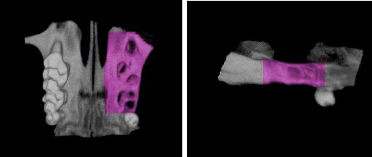
www.nature.com/scientificreports/
Figure 2. Ovariectomy prolongs maxillary bone resorption after teeth extractions. (a) Ventral views (upper
images) and lateral views (lower images) of μCT-3D images of the maxilla at day 0 (red) and 16 weeks (green)
post-extraction of OVX mice are shown in the left and the middle images, respectively. The right image shows
a superimposed illustration of day 0 and 16 weeks post-extraction. The arrows indicate decreased regions
of the alveolar bone after 16 weeks post-extraction. (b) The graph shows the time-dependent changes in the
bone volumes of the teeth-extracted side of the alveolar bone as described in Fig. 1a. Closed squares represent
OVX mice and closed circles represent sham mice as the control. Data are expressed as mean ± S.D. (n = 3). (c)
The graph shows the time-dependent changes in the ratio of the CT values of the maxilla alveolar bone of the
extracted region (ROI-2) to the non-extracted buccal side (ROI-1) of the alveolar bone of OVX mice (closed
square) and sham mice (closed circle), as described in Fig. 1b. The data shown in the graphs are representative of
two independent experiments. ***P < 0.005; calculated using two-way ANOVA.
long-lasting decrease t hereafter22. The long-lasting loss of maxilla BV after teeth extractions may be promoted
by risk factors such as osteoporosis but is not observed in healthy conditions.
Postmenopausal osteoporosis leads to micro-architectural deterioration of bone tissue and low bone mineral
density23. Some clinical studies reported that the RRR increased in osteoporotic edentulous p atients24,25. However,
other studies demonstrated that there was no statistical relationship between edentulous jaw resorption and
osteoporosis26,27. Thus, the relationship between RRR and osteoporosis is still controversial and has not been
sufficiently elucidated. Our experimental study on a novel animal model revealed the possibility that reduced
ovarian function exacerbates RRR. However, clarifying the relationship between human postmenopausal osteo-
porosis and RRR is insufficient in only our murine model. Additional clinical studies are needed in the future.
We focused on Sema4d, which functions as an inhibitor of bone formation by suppressing osteoblast differ-
entiation and modulating osteoblast m obility28,29. Sema4d derived from osteoclasts played a role as an inhibitor
of bone formation, and mice with targeted deletion of gene encoding Sema4d or its receptor, Plexin-B1, had
Scientific Reports | (2022) 12:4094 | https://doi.org/10.1038/s41598-022-08016-3 4
Vol:.(1234567890)www.nature.com/scientificreports/
Figure 3. Prolonged increase of osteoclasts in the maxillary bone of OVX mice. (a) Enzymatic histochemistry
staining for TRAP (red) on the decalcified maxillary alveolar bone at day 1, 3 weeks, and 12 weeks of sham
or OVX mice following teeth extractions. The arrows indicate TRAP-positive signals inside and around the
extraction socket. The arrowheads indicate TRAP-positive signals on the buccal side of the maxillary alveolar
bone. Bars: 100 μm. (b) The graph shows the time-dependent changes in the ratio of the total area of TRAP-
positive cells to the surface area of the maxilla alveolar bone of the extracted side in sham or OVX mice. The
bone of the extracted side was around M2. Data are expressed as mean ± S.D. (sham mice: n = 4–5. OVX mice:
n = 4–6), *P < 0.05, ***P < 0.005; calculated using calculated using Student’s t-test.
Scientific Reports | (2022) 12:4094 | https://doi.org/10.1038/s41598-022-08016-3 5
Vol.:(0123456789)www.nature.com/scientificreports/
Figure 4. Ovariectomy sustains prolonged high expression of several mRNAs encoding bone metabolism and
proinflammatory cytokines after teeth extractions. The expressions of mRNA encoding bone metabolism in the
maximally alveolar bone were examined via quantitative PCR in both OVX mice (closed circle) and sham mice
(open circle). The graphs show relative expressions of indicated genes normalized to the expression level of day
0 of the sham mice. Data are expressed as mean ± S.E. Three mice were used in each group. *P < 0.05; calculated
using Student’s t-test.
increased BV. We further demonstrated that administration of a neutralization antibody for Sema4d efficiently
prevented femur bone loss in ovariectomized mice16. In our experimental model, administration of Sema4d
neutralization antibodies into OVX mice significantly suppressed the loss of alveolar BV after teeth extractions.
This result suggested that Sema4d is a factor involved in the promotion of bone resorption after tooth extrac-
tion and that inhibition of Sema4d may be a useful treatment for RRR. Consistent with our study, a recent study
reported the therapeutic effects of small interfering RNA for silencing Sema4d mRNA, which increased the BV
over the total volume of the mandibular bone and prevented alveolar bone height loss in an osteoporotic m odel30.
RANKL is a tumor necrosis factor (TNF) cytokine family and functions as a key factor for osteoclast differen-
tiation and activation. Human anti-RANKL neutralizing antibodies are utilized for the treatment of osteoporosis
and cancer-induced bone diseases. Administration of RANKL antibodies into OVX mice also suppressed long-
lasting alveolar bone resorption after teeth extractions in our experimental model. Noteworthy, this antibody
was sufficiently effective with a single dose immediately after tooth extraction.
It has been pointed out that RANKL-neutralizing antibodies have the risk of causing antiresorptive agent-
related osteonecrosis of the jaw (ARONJ), which is local osteonecrosis of the maxilla and mandible with no
symptoms in other b ones31,32. In our experimental model, however, no significant signs of maxilla bone necrosis
were found. This result indicated that some additional factors, such as chronic inflammation caused by peri-
odontal disease or excessive stimulation from dentures, may be required for the onset of ARONJ in addition to
the suppression of osteoclasts by inhibiting RANKL function.
Ovariectomy increases osteoclast formation and the lifespan by producing osteoclastogenic cytokines and
stimulating bone resorption, resulting in rapid bone loss. We found transient increases in several osteoclasto-
genic cytokines after teeth extractions. In healthy mice, these transient increases immediately returned to near
their original level. Contrarily, in OVX mice, higher levels of expression of osteoclastogenic cytokines, includ-
ing Tnfsf11, Sema4d, Il1a, and Tnf, were found to persist for more extended periods, at least until 12 weeks
after teeth extractions. On the other hand, quantitative PCR analysis could not detect persistent upregulation
of osteoclast-related genes. In this study, we used wide region of alveolar bones including extraction sockets as
samples for quantitative PCR. As the result, the number of osteoclasts contained may be reduced, and changes
Scientific Reports | (2022) 12:4094 | https://doi.org/10.1038/s41598-022-08016-3 6
Vol:.(1234567890)www.nature.com/scientificreports/
Figure 5. Administration of neutralization antibodies for RANKL or Sema4D can prevent prolonged bone resorption of the maxillary
alveolar bone of OVX mice. (a) Experimental scheme of administration of the neutralization antibody and µCT observation. T.Ext;
teeth extractions. (b) The graph shows the time-dependent changes in the bone volumes of the teeth-extracted side of the alveolar
bone of OVX mice administrated with anti-RANKL antibody (open circle) or PBS as control (closed circle), as described in Fig. 1a.
Data are expressed as mean ± S.D. (n = 3). (c) The graph shows the time-dependent changes in the ratio of CT values of the maxillary
alveolar bone of OVX mice as described in Fig. 1b. Open circles represent the mice administrated with anti-RANKL antibody and
closed circles represent PBS as control (closed circle). The data shown in the graphs are representative of two independent experiments.
***P < 0.005; calculated using two-way ANOVA. (d) Experimental scheme of administration of the neutralization antibody and µCT
observation. T.Ext; teeth extraction. (e and f) Ventral views (upper left images) and lateral views (lower images) of μCT-3D images of
the maxillary bone, and the µCT images of extraction socket of the first molar (right images) at 12 weeks post-extraction of OVX mice.
The images are the alveolar bone of OVX mice administrated with PBS as the control (e) and anti-Sema4D antibody (f). (g) The graphs
show maxillary alveolar bone volumes at 12 weeks after teeth extractions of OVX mice administrated with anti-Sema4D antibodies.
Controls are the volume of alveolar bone of mice that have had ovariectomy and tooth extraction but have not been administered
antibodies. The mice were euthanized at 12 weeks following teeth extractions. The heads were removed and subjected to µCT analysis,
and the BV were measured. The data shown in the graphs are representative of two independent experiments. **P < 0.01 (calculated
using Student’s t-test).
Scientific Reports | (2022) 12:4094 | https://doi.org/10.1038/s41598-022-08016-3 7
Vol.:(0123456789)www.nature.com/scientificreports/
in osteoclast-related genes may not have been detected. Nevertheless, enzymatic histochemistry clearly revealed
that the activity of osteoclasts persists in the maxilla of OVX mice after teeth extractions. Our data suggest that
RRR progression is caused by prolonged osteoclast activation with reduced ovarian function.
With long-term observation of the maxillary bone in this study, we demonstrated that suppression of bone
resorption by administration of anti-RANKL or anti-Sema4D antibodies could improve long-lasting alveolar
bone resorption following teeth extractions. The mechanism underlying the development of RRR is still unclear.
Our experimental model, therefore, will be a good tool for studying ridge resorption and developing therapeutic
drugs.
In conclusion, (1) bone resorption after tooth extraction did not progress without a risk factor in our murine
model; (2) Reduced ovarian function delayed the healing of extraction sockets and may be a risk factor for RRR;
and (3) administration of anti-RANKL antibodies or anti-Sema4d antibodies may be a good therapeutic method
to delay bone loss by RRR.
Materials and methods
Animals. BALB/cAJcl female mice were used in all experiments and were purchased from the CLEA Japan,
Inc. The mice were maintained in conventional conditions under standard condition of 12/12 h of light/dark
cycle at temperature 25˚C ± 3˚C and 35% to 60% humidity at the animal facility of Graduate School of Medicine,
Hokkaido University. One-week acclimation period was provided before the start of the experiment. All animal
experiments followed ARRIVE guidelines and approved by the animal care guidelines for the Care and Use of
Laboratory Animals in Hokkaido University Graduate School of Medicine, Japan (approval number: 150139).
The teeth extractions of mice. The left maxillary first molar (M1) and second molar (M2) were extracted
from 7-week-old mice under anesthesia via intraperitoneal injection of an anesthesia cocktail of 0.75 mg/kg
medetomidine (Nippon Zenyaku Kogyo Co., Ltd.), 4 mg/kg midazolam (Maruishi Pharmaceutical Co., Ltd.),
and 5 mg/kg butorphanol (Meiji Seika Pharma Co., Ltd.; supplementary Fig. S2a online). After the teeth extrac-
tions, the mice were awakened via injection of 0.75 mg/kg atipamezole (Nippon Zenyaku Kogyo Co., Ltd.),
which is an antagonistic regent for medetomidine. The group of mice without teeth extractions were used as an
experimental control. Three independent experiments were performed using three mice in each experimental
group. The heads of mice at day 0 to 24 weeks post-extraction were scanned using micro x-ray computed tomog-
raphy (µCT; Latheta LC-200, HITACHI, Japan) at 24-μm voxel resolution with an energy level of 50 kV under
anesthesia (supplementary Fig. S2b online). We weighed the mice daily to confirm that no significant weight
loss occurred.
The µCT images were imported into the ImageJ software and then processed into three dimensional images to
measure the maxillary alveolar bone volume (BV) and CT value. The BVs were compared with the non-extracted
sides. The measurement range of BV was between the M1 mesial buccal root and M2 distal buccal root (Fig. 1a).
The CT value of the extraction socket of M1 was compared with the alveolar bone of the buccal side of the same
side. The region of interest (ROI)-1 was placed in the maxilla alveolar bone of the buccal side, and ROI-2 was
placed in the extraction socket (Fig. 1b). Mice were sacrificed at 24 weeks post-extraction.
Changes in the morphology of maxillary alveolar bones after teeth extractions in OVX
mice. Five-week-old female mice were divided into two groups, sham-operated and ovariectomized (OVX),
with each group consisting of three mice. Two independent experiments were performed. Ovariectomies were
performed under anesthesia via intraperitoneal injection with a cocktail of 0.75 mg/kg medetomidine, 4 mg/kg
midazolam, and 5 mg/kg butorphanol. Ovaries were removed using small bilateral dorsal flank incisions after
the hair was shaved. Sham-operated mice received similar incisions under anesthesia without ovary removal
and used as an experimental control. The maxillary molars (M1 and M2) of the sham mice and OVX mice were
extracted at 2 weeks following surgery. The changes in the maxillary bone by µCT were assessed, and the BV
and CT values of the maxillary extraction sockets were measured. We monitored the weight of the mice daily
to confirm that no significant weight loss occurred. Mice were sacrificed and maxillary bones were harvested at
16 weeks post-extraction.
Tissue preparation for enzymatic histochemistry of tartrate‑resistant acid phosphatase
(TRAP). OVX and sham-operated mice were euthanized by intraperitoneal injection with an overdose of
pentobarbital sodium 1 day, 1 week, 3 weeks, 5 weeks, and 12 weeks after tooth extraction. They were perfused
with physiological saline through the heart, followed by 4% paraformaldehyde in 0.1 M phosphate buffer (pH
7.4). The heads of the mice were removed, immersed in the same fixative for 24 h, and decalcified with 5% EDTA
for 4 weeks at 4 °C. The decalcified tissues were dipped in 30% sucrose solution overnight at 4 °C, embedded in
OCT compound (Sakura Finetek, Tokyo, Japan), and quickly frozen in liquid nitrogen. Frozen sections, about
16 μm in thickness, were mounted on MAS-coated glass slides.
Histological sections were stained with TRAP Staining Kit in accordance with the manufacturer’s protocol
(Wako, Japan). The nuclei were stained with hematoxylin. The stained sections were observed and captured by
a light microscope with digital camera (BX51 with DP-80, Olympus, Tokyo, Japan). The ratio of the total area of
TRAP-positive cells to the surface area of the bone on the extracted side in both sham mice and OVX mice was
calculated using the ImageJ software (version 1.53f51, http://imagej.nih.gov/ij).
Quantitative PCR analysis. For the RNA preparation from the maxillary bone, the tissues were snap-
frozen in liquid nitrogen and homogenized in liquid nitrogen using a mortar and pestle to powderize it. TRIzol
Reagent (Life Technologies) was added to the homogenized tissues, and the total RNA was purified with a
Scientific Reports | (2022) 12:4094 | https://doi.org/10.1038/s41598-022-08016-3 8
Vol:.(1234567890)www.nature.com/scientificreports/
high salt solution (Nippon Gene). First-strand cDNA synthesis was completed using ReverTraAce (TOYOBO).
Quantitative PCR reactions were conducted using Rotor-Gene 6000 equipment (Qiagen) or StepOnePLUS™
(Thermo Fisher Scientific K.K.) using KAPA SYBR Green Fast PCR Kit (KAPA Biosystems). Most of the specific
primers were designed by Primer Bank33 and are presented in Supplementary Table S online.
Administration of neutralized antibodies into OVX mice. The molars (M1 and M2) were extracted
at 2 weeks after ovariectomy. Ovariectomized 7-week-old mice were divided into three groups (n = 3 per group).
OVX mice were intraperitoneally administered PBS or 1 mg/kg anti-Sema4D antibody (clone BMA-12, Bio-
Legend) once every 3 days for 8 w eeks16. The mice were euthanized at 12 weeks following teeth extractions.
The heads were removed and subjected to µCT analysis (ScanXmate-A080, COMSCAN TECNO CO., LTD.
Yokohama Japan), and the BV were measured. For the administration of anti-RANKL antibody (clone OYC1,
ORIENTAL YEAST CO., LTD.), the antibody was intraperitoneally injected with 5 mg/kg only once on the next
day following teeth extractions. The µCT images of the mouse heads were taken under anesthesia at 0-, 3-, 5-,
and 12-weeks post teeth extractions as described in the paragraph of teeth extractions of mice.
Statistics. Statistical analyses in certain time point were conducted using Student’s t-test in Figs. 3b, 4, and
5g. Differences between groups in time-dependent changes were analyzed statistically using two-way analysis of
variance (ANOVA) in Figs. 1a,b, 2b,c, 5b,c. All statistical analyses were calculated with Prism software (Prism 9
for macOS).
Ethical approval. The experimental protocols used in the present study were approved by the animal care
guidelines for the Care and Use of Laboratory Animals in Hokkaido University Graduate School of Medicine,
Japan (approval number: 150139).
Received: 28 December 2021; Accepted: 28 February 2022
References
1. Survey of Dental Disease in 2016 by the ministry of Health, Labor and Welfare in Japan. https://w ww.m hlw.g o.j p/t oukei/l ist/6 2-2 8.
html (2016).
2. Center for Disease Control and Prevention, US Dept of Health and Human Services https://w ww.cdc.gov/oralhealth/fast-facts/
tooth-loss/index.html (2019).
3. Mogi, M., Otogoto, J., Ota, N. & Togari, A. Differential expression of RANKL and osteoprotegerin in gingival crevicular fluid of
patients with periodontitis. J. Dent. Res. 83(2), 166–169 (2004).
4. Tsutsumi, T. et al. Micro-computed tomography for evaluating alveolar bone resorption induced by hyperocclusion. J. Prosthodont.
Res. 62(3), 298–302 (2018).
5. Yoshinaga, Y., Ukai, T. & Abe, Y. H. Expression of receptor activator of nuclear factor kappa B ligand relates to inflammatory bone
resorption, with or without occlusal trauma in rats. J. Periodontal. Res. 42(5), 402–409 (2007).
6. Pereira, M. C., Zecchin, K. G., Campagnoli, E. B. & Jorge, J. Ovariectomy delays alveolar wound healing after molar extractions
in rats. J. Oral Maxillofac. Surg. 65(11), 2248–2253 (2007).
7. Chen, C. H. et al. An osteopenic/osteoporotic phenotype delays alveolar bone repair. Bone 112, 212–219 (2018).
8. Esteves, C. M. et al. Ovariectomy-associated changes in interradicular septum and in tibia metaphysis in different observation
periods in rats. Pathol. Res. Pract. 211(2), 125–129 (2015).
9. Moriya, Y., Ito, K. & Murai, S. Effects of experimental osteoporosis on alveolar bone loss in rats. J. Oral Sci. 40(4), 171–175 (1998).
10. Marx, R. E. Pamidronate (Aredia) and zoledronate (Zometa) induced avascular necrosis of the jaws: a growing epidemic. J. Oral
Maxillofac. Surg. 61(9), 1115–1117 (2003).
11. Ruggiero, S. L. et al. American Association of Oral and Maxillofacial Surgeons position paper on medication-related osteonecrosis
of the jaw–2014 update. J. Oral Maxillofac. Surg. 72(10), 1938–1956 (2014).
12. Hodsman, A. B. et al. Parathyroid hormone and teriparatide for the treatment of osteoporosis: A review of the evidence and sug-
gested guidelines for its use. Endocr. Rev. 26(5), 688–703 (2005).
13. Hunziker, J., Wronski, T. J. & Miller, S. C. Mandibular bone formation rates in aged ovariectomized rats treated with anti-resorptive
agents alone and in combination with intermittent parathyroid hormone. J. Dent. Res. 79(6), 1431–1438 (2000).
14. Bellido, M. et al. PTH increases jaw mineral density in a rabbit model of osteoporosis. J. Dent. Res. 89(4), 360–365 (2010).
15. Liu, M. et al. Sclerostin and DKK1 inhibition preserves and augments alveolar bone volume and architecture in rats with alveolar
bone loss. J. Dent. Res. 97(9), 1031–1038 (2018).
16. Negishi-Koga, T. et al. Suppression of bone formation by osteoclastic expression of semaphorin 4D. Nat. Med. 17(11), 1473–1480
(2011).
17. Pagni, G., Pellegrini, G., Giannobile, W. V. & Rasperini, G. Postextraction alveolar ridge. Preservation biological basis and treat-
ments. Int. J. Dent. 2012, 151030. https://doi.org/10.1155/2012/151030 (2012).
18. Politis, C., Schoenaers, J., Jacobs, R. & Agbaje, J. O. Wound healing problems in the mouth. Front. Physiol. 7, 507. https://doi.org/
10.3389/fphys.2016.00507 (2016).
19. Devlin, H. & Ferguson, M. W. Alveolar ridge resorption and mandibular atrophy. A review of the role of local and systemic factors.
Br. Dent. J. 170(3), 101–104 (1991).
20. Kim, J. M. et al. Osteoblast-Osteoclast communication and bone homeostasis. Cells 9(9), 2073 (2020).
21. Furuya, Y. et al. Increased bone mass in mice after single injection of anti-receptor activator of nuclear factor-kappaB ligand-
neutralizing antibody: evidence for bone anabolic effect of parathyroid hormone in mice with few osteoclasts. J. Biol. Chem.
286(42), 37023–37031 (2011).
22. Atwood, D. A. Reduction of residual ridges: a major oral disease entity. J. Prosthet. Dent. 26(3), 266–279 (1971).
23. Zhang, S. et al. Felodipine blocks osteoclast differentiation and ameliorates estrogen-dependent bone loss in mice by modulating
p38 signaling pathway. Exp. Cell Res. 387(2), 111800 (2020).
24. Hirai, T., Ishijima, T., Hashikawa, Y. & Yajima, T. Osteoporosis and reduction of residual ridge in edentulous patients. J. Prosthet.
Dent. 69(1), 49–56 (1993).
Scientific Reports | (2022) 12:4094 | https://doi.org/10.1038/s41598-022-08016-3 9
Vol.:(0123456789)www.nature.com/scientificreports/
25. Singhal, S. et al. The effect of osteoporosis on residual ridge resorption and masticatory performance in denture wearers. Gero-
dontology https://doi.org/10.1111/j.1741-2358.2011.00610.x (2012).
26. Springe, B., Slaidina, A., Soboleva, U. & Lejnieks, A. Bone mineral density and mandibular residual ridge resorption. Int. J. Pros-
thodont. 27(3), 270–276 (2014).
27. Ozola, B. et al. The influence of bone mineral density and body mass index on resorption of edentulous jaws. Stomatologija 13(1),
19–24 (2011).
28. Negishi-Koga, T. & Takayanagi, H. Bone cell communication factors and Semaphorins. Bonekey Rep. 1, 183. https://doi.org/10.
1038/bonekey.2012.183 (2012).
29. Kim, B. J. & Koh, J. M. Coupling factors involved in preserving bone balance. Cell Mol. Life Sci. 76(7), 1243–1253 (2019).
30. Zhang, Y. et al. Prevention of alveolar bone loss in an osteoporotic animal model via interference of Semaphorin 4d. J. Dent. Res.
93(11), 1095–1100 (2014).
31. Baba, A. et al. CT imaging features of antiresorptive agent-related osteonecrosis of the jaw/medication-related osteonecrosis of
the jaw. Dentomaxillofac. Radiol. 47(4), 20170323. https://doi.org/10.1259/dmfr.20170323 (2018).
32. Bagan, L. et al. Serum levels of RANKL and OPG, and the RANKL/OPG ratio in bisphosphonate-related osteonecrosis of the jaw:
Are they useful biomarkers for the advanced stages of osteonecrosis?. Med. Oral. Patol. Oral. Cir. Bucal. https://doi.org/10.4317/
medoral.22128 (2017).
33. Wang, X. & Seed, B. A. PCR primer bank for quantitative gene expression analysis. Nucleic Acids Res. https://doi.org/10.1093/nar/
gng154 (2003).
Acknowledgements
We would like to thank Prof. Masahiko Watanabe, Hokkaido University, for valuable suggestions; Dr. Junko
Nio-Kobayashi, Hokkaido University, for teaching the technique of ovariectomy; Prof. Nobuyuki Udagawa and
Masanori Koide, Matsumoto Dental University, for μCT analysis.
Author contributions
M.H. and S.K. contributed to conception, design, data acquisition, analysis, and interpretation, performed sta-
tistical analysis, drafted and critically revised the manuscript. K.I. contributed to conception, data analysis, and
critically revised the manuscript. T.I. contributed to conception, data analysis, and interpretation, and critically
revised the manuscript. A.Y. contributed to conception and design, and interpretation of data, and critically
revised the manuscript. All authors reviewed the manuscript.
Funding
This study was supported by grants from Fusion-H program from Hokkaido University.
Competing interests
The authors declare no competing interests.
Additional information
Supplementary Information The online version contains supplementary material available at https://doi.org/
10.1038/s41598-022-08016-3.
Correspondence and requests for materials should be addressed to M.H. or S.K.
Reprints and permissions information is available at www.nature.com/reprints.
Publisher’s note Springer Nature remains neutral with regard to jurisdictional claims in published maps and
institutional affiliations.
Open Access This article is licensed under a Creative Commons Attribution 4.0 International
License, which permits use, sharing, adaptation, distribution and reproduction in any medium or
format, as long as you give appropriate credit to the original author(s) and the source, provide a link to the
Creative Commons licence, and indicate if changes were made. The images or other third party material in this
article are included in the article’s Creative Commons licence, unless indicated otherwise in a credit line to the
material. If material is not included in the article’s Creative Commons licence and your intended use is not
permitted by statutory regulation or exceeds the permitted use, you will need to obtain permission directly from
the copyright holder. To view a copy of this licence, visit http://creativecommons.org/licenses/by/4.0/.
© The Author(s) 2022
Scientific Reports | (2022) 12:4094 | https://doi.org/10.1038/s41598-022-08016-3 10
Vol:.(1234567890)You can also read