Aqueous-Deficient Dry Eye Exacerbates Signs and Symptoms of Allergic Conjunctivitis in Mice
←
→
Page content transcription
If your browser does not render page correctly, please read the page content below
International Journal of
              Molecular Sciences

Article
Aqueous-Deficient Dry Eye Exacerbates Signs and Symptoms of
Allergic Conjunctivitis in Mice
Tatsuma Kishimoto                  , Waka Ishida      , Isana Nakajima, Ken Fukuda *            and Kenji Yamashiro

                                           Department of Ophthalmology and Visual Science, Kochi Medical School, Kochi University,
                                           Kochi 783-8505, Japan; t.kishimoto@kochi-u.ac.jp (T.K.); wakai@kochi-u.ac.jp (W.I.);
                                           jm-i-nakajima@kochi-u.ac.jp (I.N.); yamashk@kochi-u.ac.jp (K.Y.)
                                           * Correspondence: k.fukuda@kochi-u.ac.jp; Tel.: +81-88880-2391

                                           Abstract: Dry eye disease (DED) and allergic conjunctivitis affect a large number of patients, and
                                           many patients usually have both symptoms. We investigated the interactions between DED and
                                           allergic conjunctivitis in mice. Four experimental groups were compared: control, DED, allergy,
                                           and allergy with DED. DED was induced by removing the extraorbital lacrimal glands of the mice.
                                           Allergic conjunctivitis was induced by intraperitoneal administration of ovalbumin and antigen eye
                                           drops. The early phase reaction of the allergy was evaluated using the clinical score, scratching
                                           behavior, and vascular permeability in the conjunctiva. Epithelial barrier function was assessed
                                           by an LC-biotin assay. Tear fluid volume and corneal fluorescein staining decreased in the DED
                                           and allergy with DED groups. LC-biotin penetrated the entire epithelium of both the cornea and
                                           conjunctiva in DED mice. The clinical score of the early phase reaction was higher in allergy-induced
                                           mice than in non-allergy mice. Edema of the eyelid and conjunctiva were aggravated in mice with
                                           DED. The number of scratching episodes and leakage of Evans blue into the conjunctiva were higher
                                           in allergy-induced DED mice than in control mice. The presence of aqueous-deficient dry eye caused
                                           ocular surface epithelial damage and exacerbated allergic signs and symptoms.
Citation: Kishimoto, T.; Ishida, W.;
Nakajima, I.; Fukuda, K.; Yamashiro,
                                           Keywords: dry eye; allergic conjunctivitis; tear fluid; conjunctiva; cornea; barrier function; mouse
K. Aqueous-Deficient Dry Eye
Exacerbates Signs and Symptoms of
Allergic Conjunctivitis in Mice. Int. J.
Mol. Sci. 2022, 23, 4918. https://
doi.org/10.3390/ijms23094918
                                           1. Introduction
                                                 Allergic conjunctival disease (ACD) is an allergen-induced inflammatory disease of
Academic Editors: Murat Dogru and
                                           the conjunctiva. Several types of ACD have been identified: seasonal and perennial allergic
Takashi Kojima
                                           conjunctivitis (AC), atopic keratoconjunctivitis, vernal keratoconjunctivitis, and giant papil-
Received: 29 March 2022                    lary conjunctivitis [1]. Allergic diseases are very common worldwide, and their prevalence
Accepted: 26 April 2022                    is increasing. In a recent survey, the prevalence of allergic conjunctival diseases (ACDs)
Published: 28 April 2022                   was reported to be 48.7% in Japan [2]. Allergic conjunctivitis is the most common type of
Publisher’s Note: MDPI stays neutral       ACD and is mediated by IgE-dependent type I hypersensitivity. Atopic keratoconjunctivitis
with regard to jurisdictional claims in    and vernal keratoconjunctivitis, which are more severe forms of ACD, are caused by both
published maps and institutional affil-    IgE-dependent reactions and non-IgE-mediated chronic inflammation. Conjunctival mast
iations.                                   cells play critical roles in the conjunctival response in allergic conjunctivitis. Allergens
                                           that penetrate the conjunctiva cross-link the antigen-specific IgE on mast cells, causing
                                           mast cell degranulation and inducing itching, edema, and hyperemia. Therefore, reduced
                                           permeability of the epithelium and clearance of ocular surface antigens may affect signs
Copyright: © 2022 by the authors.          and symptoms of allergic conjunctivitis.
Licensee MDPI, Basel, Switzerland.               Dry eye disease (DED) is a multifactorial chronic disease characterized by loss of
This article is an open access article     homeostasis in the tear film [3]. The prevalence of DED is also high worldwide, and its
distributed under the terms and
                                           frequency is reported to be higher in Asia, including Japan, than in other continents [4].
conditions of the Creative Commons
                                           Large epidemiological studies have shown that the prevalence of DED in Japan is 17.4%
Attribution (CC BY) license (https://
                                           among men and 30.3% among women [5]. In an epidemiological survey of office workers,
creativecommons.org/licenses/by/
                                           the prevalence of DED, including probable cases, was very high at 65.6% [6]. Using visual
4.0/).

Int. J. Mol. Sci. 2022, 23, 4918. https://doi.org/10.3390/ijms23094918                                          https://www.mdpi.com/journal/ijms
Int. J. Mol. Sci. 2022, 23, 4918                                                                                               2 of 11

                                   display terminals for >4 h is associated with an increased risk of DED [7], and may be
                                   related to the high prevalence of dry eye in Japan. Etiologically, DED can be classified
                                   as aqueous-deficient dry eye (ADDE) or evaporative dry eye (EDE). Recent reports have
                                   proposed that ADDE and EDE are not mutually exclusive and may exist on a continuum [3].
                                   Older patients, or those with inflammation of the lacrimal gland, androgen deficiency, or
                                   presence of systemic drugs, can also be predisposed to lacrimal gland dysfunction. Both
                                   ADDE and EDE cause epithelial cell loss and damage that results in the disruption of the
                                   epithelial barrier.
                                         Patients with DED and ACD show similar signs and symptoms. For example, the
                                   simultaneous occurrence of itchiness and dryness of the eye has been observed in many
                                   patients [8], and one report showed that many subjects with moderate-to-severe ocular itch
                                   symptoms also had severe symptoms of DED [9]. These studies suggest that DED and ACD
                                   not only occur simultaneously but that they also mutually influence onset and severity. The
                                   causes of DED in patients with ACD have been studied in detail. Ocular surface inflamma-
                                   tion in ACD reportedly affects tear volume, tear film stability, mucin expression, meibomian
                                   gland dysfunction, and epithelial phenotype, resulting in DED [10–18]. Whereas many
                                   studies have demonstrated the impact of ACD on DED, there are few direct studies of
                                   the impact of DED on ACD. Researchers have speculated that altered epithelial barrier,
                                   decreased surface clearance, and inflammation may be factors that exacerbate ACD in
                                   patients with DED [19]. However, no direct studies have demonstrated that DED worsens
                                   the ocular allergic symptoms. The aim of this study was to investigate whether ADDE
                                   affects or exacerbates the signs and symptoms of ACD in mice.

                                   2. Results
                                   2.1. Effects of AC on Dry Eye
                                        First, we examined the effects of AC on dry eye signs. Before lacrimal gland removal,
                                   there was no difference in tear fluid volume between the lacrimal gland removal group and
                                   control group (data not shown), but the amount of tear fluid was significantly decreased
                                   in mice with excised lacrimal glands regardless of the induction of AC (Figure 1). The
                                   number of goblet cells in the conjunctiva tended to increase in DED mice, but there were
                                   no significant differences between any of the groups (Figure 2). Corneal fluorescein scores
                                   were significantly high in mice with excised lacrimal glands, but no further increase was
                                   observed in allergy-induced DED mice (Figure 3). Corneal fluorescein scores were similar
                                   before and after the allergy challenge.

                                   Figure 1. Tear volume before and after induction of AC in mice with or without dry eye (DED).
                                   The amount of tear fluid was measured at day 21 before antigen challenge (A) and at day 28 (B).
                                   The length of the wet thread is shown as means ± standard error of the means in each group. Four
                                   experimental groups were compared: control (n = 6), DED (n = 6), allergy (n = 6), and allergy with
                                   DED (n = 5). ** p < 0.01 (Tukey–Kramer test) versus non-allergy control group. PBS, mice challenged
                                   with phosphate-buffered saline (PBS) (non-allergy group); OVA, mice challenged with ovalbumin
                                   (OVA) (allergy-induced group); DED, mice with excised extraorbital lacrimal glands.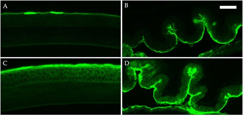
Int. J. Mol. Sci. 2022, 23, 4918                                                                                                   3 of 11

                                   Figure 2. Number of goblet cells in the conjunctiva. The eyes were isolated for histological analysis;
                                   the number of goblet cells in the epithelium of the conjunctiva was counted on day 29. Representative
                                   photo of conjunctiva and goblet cells showing a purple-magenta periodic acid–Schiff-positive reaction
                                   (A). Data are shown as means ± standard error of the means in each group (B). Four experimental
                                   groups were compared: control (n = 6), DED (n = 6), allergy (n = 6), and allergy with DED (n = 5). PBS,
                                   mice challenged with phosphate-buffered saline (PBS) (non-allergy group); OVA, mice challenged
                                   with ovalbumin (OVA) (allergy-induced group); dry eye disease (DED), mice with excised extraorbital
                                   lacrimal glands. Bar, 50 µm.

                                   Figure 3. Corneal staining score before and after the induction of AC with or without dry eye
                                   (DED). Corneal fluorescein score was classified at day 21 before antigen challenge (A) and at day 28
                                   (B). The sum of the scores is shown as means ± standard error of the means in each group. Four
                                   experimental groups were compared: control (n = 6), DED (n = 6), allergy (n = 6), and allergy with
                                   DED (n = 5). ** p < 0.01 (Tukey–Kramer test) versus non-allergy control group. PBS, mice challenged
                                   with phosphate-buffered saline (PBS) (non-allergy group); OVA, mice challenged with ovalbumin
                                   (OVA) (allergy-induced group); DED, mice with excised extraorbital lacrimal glands.

                                         We also evaluated the barrier function of the ocular surface in DED mice using an
                                   LC-biotin assay. Fluorescent immunostaining after the administration of LC-biotin eye
                                   drops showed that LC-biotin remained on the superficial epithelial layer of the cornea,
                                   and the dye did not penetrate the epithelium of mice without DED. In contrast, the dye
                                   infiltrated the entire corneal epithelium layer of DED mice. Similarly, whereas LC-biotin
                                   was observed only in the superficial layer of the conjunctival epithelium of mice without
Int. J. Mol. Sci. 2022, 23, 4918 4 of 11
Int. J. Mol. Sci. 2022, 23, 4918 DED, dye infiltration was observed in the entire conjunctival epithelium of DED 4mice
of 11
(Figure 4).
Figure4.4. Epithelial
Figure Epithelial barrier
barrierfunction
functionofofocular
ocularsurface byby
surface LC-biotin assay.
LC-biotin Epithelial
assay. barrier
Epithelial was eval-
barrier was
uated by LC-biotin assay. The eyes were challenged with LC-biotin in phosphate-buffered
evaluated by LC-biotin assay. The eyes were challenged with LC-biotin in phosphate-buffered saline 30
saline
min prior to harvest. Frozen sections of the cornea (A,C) and conjunctiva (B,D) were stained
30 min prior to harvest. Frozen sections of the cornea (A,C) and conjunctiva (B,D) were stained with with
streptavidin Alexa Fluor 488 conjugate in mice without (A,B) and with dry eye (C,D). Bar, 50 μm.
streptavidin Alexa Fluor 488 conjugate in mice without (A,B) and with dry eye (C,D). Bar, 50 µm.
2.2.Effects
2.2. EffectsofofDry
DryEye
Eyeon
onAC
AC
Wethen
We thenexamined
examinedthe theeffects
effectsofofDED
DEDononAC.
AC.Systemic
Systemicimmune
immuneresponses
responseswere
werenotnot
differentininmice
different micewith
withoror without
without DED;DED; serum
serum levels
levels of total
of total IgE IgG1
IgE and and IgG1 were signifi-
were significantly
cantly increased,
increased, andwas
and IgG2a IgG2a was significantly
significantly decreased
decreased in all sensitized
in all sensitized mice, regardless
mice, regardless of the
of the presence
presence of DEDof DED (Figure
(Figure 5). 5).
Figure 5. Serum IgE (A), IgG1 (B), and IgG2a (C) levels of mice. Serum IgE and IgG levels were
Figure 5. Serum IgE (A), IgG1 (B), and IgG2a (C) levels of mice. Serum IgE and IgG levels were evaluated
evaluated
24 h after 24
thehlast
after the last
antigen antigen Data
challenge. challenge. Dataas
are shown are shown
means ± standard
as meanserrors
± standard errors in
of the means of each
the
means
group.in each
Four group. Fourgroups
experimental experimental groups were
were compared: compared:
control control
(n = 6), DED (n = (n
6),=allergy
6), DED(n =(n6),
= and
6), allergy
allergy
(nwith
= 6),DED
and allergy
(n = 5). with
* pInt. J. Mol. Sci. 2022, 23, 4918 5 of 11
Figure 6. Clinical signs were measured at 20 min after first antigen challenge at day 21. Scores are
shown as means ± standard errors in each group. Four experimental groups were compared: control
(n = 6), DED (n = 6), allergy (n = 6), and allergy with DED (n = 5). ** p < 0.01 (Tukey–Kramer test)
versus non-allergy control group; †† p < 0.01 (Tukey–Kramer test) versus allergy-induced group;
‡‡ p < 0.01 (Tukey–Kramer test) versus non-allergy dry eye disease (DED) group.
Figure 7. Number of scratching episodes. The scratching behavior of the mice was counted for
10 min after first antigen challenge at day 21, and the number of scratching episodes by the hindlimb
was counted. The number of scratching episodes is shown as means ± standard error in each group.
Four experimental groups were compared: control (n = 6), DED (n = 6), allergy (n = 6), and allergy
with DED (n = 5). ** p < 0.01 (Tukey–Kramer test) versus non-allergy control group. PBS, mice
challenged with phosphate-buffered saline (PBS) (non-allergy group); OVA, mice challenged with
ovalbumin (OVA) (allergy-induced group); dry eye disease (DED), mice with excised extraorbital
lacrimal glands.Int. J. Mol. Sci. 2022, 23, 4918 6 of 11
Figure 8. Evans blue dye leakage in conjunctiva. Dye leakage was evaluated 30 min after antigen chal-
lenge at day 28, shown as arbitrary units ± standard error in each group. Four experimental groups
were compared: control (n = 6), DED (n = 6), allergy (n = 6), and allergy with DED (n = 5). * p < 0.05
(Tukey–Kramer test) versus non-allergy control group. PBS, mice challenged with phosphate-buffered
saline (PBS) (non-allergy group); OVA, mice challenged with ovalbumin (OVA) (allergy-induced
group); dry eye disease (DED), mice with excised extraorbital lacrimal glands.
3. Discussion
In this study, we demonstrated that tear deficiency exaggerates the signs and symp-
toms of AC in mice. The signs and symptoms of AC, including edema of the eyelid and
conjunctiva, eye scratching behavior, and vascular permeability in the conjunctiva after
antigen challenge, were significantly augmented in mice that underwent lacrimal gland
removal. Decreased tear clearance and epithelial barrier function of the ocular surface
may result in longer retention and higher penetration of antigens to the ocular surface,
leading to exaggeration of the symptoms of AC. Epithelial barrier dysfunction can lead
to pathogenesis of both DED and ACD [19,20]. Many reports have demonstrated that
ACD affects the status of tear fluid and ocular surface epithelia, resulting in DED [10–18].
However, there are no basic or clinical studies that directly show the effect of DED on the
clinical signs and symptoms of ACD. The present study demonstrates, for the first time in
an animal study, that lacrimal ADDE may exaggerate signs and symptoms of ACD. Our
study may be a first step toward the development of therapeutic agents and the elucidation
of the pathophysiology of patients with combined DED and ACD.
Various ocular surface diseases manifest as impairment of the epithelial barrier, in-
cluding DED, ACDs, infection, and chemical injuries [21]. Yokoi et al. demonstrated that
the corneal epithelial barrier function had significantly decreased in accordance with the
severity of superficial punctate keratopathy in DED patients, as measured by fluorescein
uptake [22]. In addition, treatment with hyaluronan eye drops alleviated the decline in the
corneal barrier in patients with DED [23]. Furthermore, corneal barrier function reportedly
decreased even in patients with DED who did not exhibit visible corneal epithelial damage
on slit-lamp examination [24]. These studies suggest that epithelial tight junctions of the
ocular surface may be impaired in patients with DED regardless of the presence of visibleInt. J. Mol. Sci. 2022, 23, 4918 7 of 11
corneal lesions on slit-lamp examination. In mice, dry eye experimentally induced by
low humidity environmental challenge also decreased the levels of tight junction proteins
and impaired corneal epithelial barrier function [25,26]. In patients with seasonal AC, the
expression of the adherens junction protein E-cadherin in the conjunctival epithelium is
reportedly downregulated, regardless of the season [1]. Conjunctival allergen challenge
also reportedly decreased the expression of the tight junction proteins zonula occludens-1
and E-cadherin in the conjunctival epithelium in a mouse model of AC [11]. Decreased
epithelial barrier function is an important factor for the sensitization and exacerbation of al-
lergic diseases. In this study, we demonstrated that mice with tear deficiency had impaired
epithelial barriers in the conjunctiva and cornea, as estimated by LC-biotin penetration.
Therefore, it is possible that the reduced epithelial barrier function of the conjunctiva allows
antigens to penetrate the conjunctiva easily, possibly exacerbating allergic symptoms.
In the present study, because the antigen was systemically sensitized with an adjuvant,
a strong Th2-type immune response could be induced; thus, the systemic immune response
did not differ between mice with and without dry eye. Fujishima et al. reported that
tear volume and clearance were lower in patients with AC who tested negative for serum
antigen-specific IgE than in those who tested positive for serum antigen-specific IgE [27]. In
a recent study, AC was observed in patients who tested negative for serum antigen-specific
IgE [28]. Therefore, in patients with aqueous-deficient dry eye, the antigens may remain on
the ocular surface for a long period owing to poor tear clearance, possibly contributing to
local sensitization and exacerbation of symptoms.
The influence of different types of ACDs on tear function has been extensively in-
vestigated. Most studies have reported that in patients with AC, tear volume does not
change, but tear stability estimated by tear breakup time (TBUT) is reduced [10,15,18,27].
In patients with atopic keratoconjunctivitis (AKC), the tear volume has been reported
to be unchanged [11,12,16,17], while some studies have reported a decrease in the tear
volume [13,14]. However, all those studies reported a decrease in the TBUT [11–14,16,17].
Onguchi et al. revealed that the onset time of AKC affects tear function and ocular surface
findings, and tear volume and epithelial damage in childhood-onset adult AKC patients
were considerably worse than those in adult-onset adult AKC patients, pediatric patients,
and controls. These results suggest that prolonged ocular surface inflammation may be
important for the disruption of the healthy ocular surface, including the tear film [16]. In
patients with vernal keratoconjunctivitis (VKC), the tear volume is increased, but the TBUT
is reportedly decreased [14,29]. Interestingly, decreased tear film stability has been observed
even in the quiescent phases of VKC [29]. Conjunctival goblet cells have also been reported
to decrease in patients with various ACDs, including AC [18,30], AKC [13,14,16,17,31], and
VKC [14,17]. In particular, a decrease in MUC5 AC, a secreted mucin, has been found along
with a decrease in goblet cells in patients with AKC [13,14,16]. In contrast, the density of
goblet cells and the severity of corneal staining were not affected in mice with AC in the
present study, which may change in the future depending on the duration of ocular surface
inflammation, as reported by Onguchi [16].
When cells become necrotic, intracellular inflammatory substances called alarmins,
such as IL-1α and IL-33, are released outside the cells, resulting in sterile inflammation [32,33].
Recent studies have reported the involvement of alarmins in both ocular allergies and
DED. Alarmins released from necrotic corneal epithelial cells act on corneal fibroblasts and
epithelial cells, causing enhanced production of chemokines and cytokines, such as eotaxin,
and decreased epithelial barrier function [34,35]. IL-33, an epithelial cell-derived alarmin,
does not degranulate mast cells by itself but synergistically degranulates mast cells when
acting simultaneously with antigens [36]. Alarmin may be involved in the exacerbation
of allergic inflammation [37]. The concentration of IL-33 is also reportedly elevated in
the tear fluid of DED patients [38,39]. In addition, the tear levels of IL-33 in patients with
DED were positively correlated with the tear levels of IL-4 and IL-5 [38]. These results
may be related to the fact that many patients have overlapping symptoms of itching and
dryness of the eye [8] and that dry eye is severe in patients with itching [9]. Therefore,Int. J. Mol. Sci. 2022, 23, 4918 8 of 11
alarmins released by epithelial injury caused by dry eye may exacerbate allergic symptoms
by promoting mast cell degranulation. The role of alarmins in DED and allergies requires
further investigation.
We evaluated the epithelial barrier function with the use of the LC-biotin assay. The
limitation of the present study is the lack of detailed analysis of each component of the
epithelial barrier, such as the adhesion proteins and the mucin expression. Further immuno-
histochemical analysis should be performed in the future to evaluate the ocular surface
epithelia and the mucin expression in more detail.
In conclusion, the presence of ADDE exacerbated the clinical signs and symptoms of
ACD, possibly through ocular surface epithelial barrier disruption and reduced allergen
clearance.
4. Materials and Methods
4.1. Ethical Treatment of Animals
This study was approved by the Kochi University Animal Care and Use Committee
(permit number J-70). BALB/c mice were purchased from Japan SLC Inc. (Hamamatsu,
Shizuoka, Japan) and maintained under specific pathogen-free conditions at the animal
facility of Kochi Medical School. Eight-week-old specific-pathogen-free female mice were
used in the experiments. All the procedures were performed in accordance with the
Association for Research in Vision and Ophthalmology Statement for the Use of Animals in
Ophthalmic and Vision Research.
4.2. Experimental Procedure for Lacrimal Grand Excision and Experimental Allergic Conjunctivitis
The mice were anesthetized with an intraperitoneal injection of an anesthetic combina-
tion of 0.45 µg/g medetomidine (Domitor; Nippon Zenyaku Kogyo, Tokyo, Japan), 6 µg/g
midazolam (Sandoz, Yamagata, Japan), and 7.5 µg/g butorphanol (Vetorphale; Meiji Seika
Pharma, Tokyo, Japan). The exorbital lacrimal glands of both eyes were exposed through
a careful incision near the anterior ear and excised [40]. Subsequently, talibid ophthalmic
ointment 0.3% (Santen Pharmaceutical Co., Osaka, Japan) was applied.
Experimental AC was induced using a previously described protocol with slight
modifications [41] (Figure 9). Briefly, mice were injected intraperitoneally three times with
30 µg ovalbumin (OVA) (Worthington Biochemical Corp, NJ, USA) mixed with 1 mg of
Imject Alum (Thermo Fisher Scientific, MA, USA) at intervals of 7 days. Seven days after
the third sensitization, both eyes of each mouse were challenged with OVA in PBS (1 mg
per 5 µL per eye) from day 21 to day 28.
Figure 9. Timeline for induction of dry eye and experimental AC. To induce dry eye, the extraorbital
lacrimal glands of the mice were removed 1 day before sensitization. To induce experimental AC, a
mixture of ovalbumin (OVA) and alum (Al(OH)3 ) was administered intraperitoneally three times
at an interval of 7 days. Seven days after the third sensitization, corneal fluorescein score and the
amount of tear fluid were measured; then, both eyes of each mouse were challenged with OVA in
phosphate-buffered solution (PBS) from day 21 to day 28.Int. J. Mol. Sci. 2022, 23, 4918 9 of 11
4.3. Evaluation of Dry Eye
To assess dry eye signs, we evaluated corneal fluorescein staining and tear fluid
volume as described previously [40]. Fluorescein solution was extracted from one sheet
of Fluores Ocular Examination Test Paper 0.7 mg (AYUMI Pharmaceutical Corporation,
Tokyo, Japan) with 500 µL of sterile saline. The concentration of the fluorescein sodium
salt was approximately 1.4 mg/mL. Then, 1 µL of fluorescein solution was applied to each
eye, wiped off, and observed using a portable slit lamp with a blue filter (Kowa Company,
Ltd., Tokyo, Japan). Corneal fluorescein scores were classified as 0–3 (0, no fluorescence; 1,
sparse spot fluorescence; 2, dense spot fluorescence; and 3, very dense spot fluorescence).
Five locations were observed (center, upper left, lower left, upper right, and lower right),
and the sum of the scores obtained was calculated.
The volume of tear fluid was measured using phenol red threads (Zone Quick, Ayumi
Pharmaceutical Co. Tokyo, Japan). The mice were fixed for 15 s with the thread inserted
into the lower eyelid under no anesthesia, and the length of the wet thread was measured.
4.4. Evaluation of Clinical Conjunctival Allergic Reaction
Clinical signs were assessed on day 21, as previously described [42]. The scratching
behavior of the mice was counted for 10 min after the first eye drop, and the number of
scratching episodes by the hindlimb was counted.
To investigate vascular permeability, Evans blue dye leakage was evaluated as previ-
ously described, with slight modifications [43]. The conjunctivas were harvested 30 min
after the OVA or PBS challenge. Evans blue was extracted for 48 h in 400 µL of 0.5% Na2
SO4 and acetone (3:7). After centrifugation, absorbance (620 nm) of the supernatant was
measured using a spectrophotometer.
Twenty-four hours after the last challenge, serum IgE, IgG1, and IgG2a levels were
evaluated using ELISA, as described previously [41]. OVA-specific IgE and IgG1 were
assayed using an anti-OVA IgE ELISA kit or an anti-OVA IgG1 ELISA kit (Cayman Chemical
Company, MI, USA) according to the manufacturer’s protocol.
4.5. Histological Analysis of Periodic Acid-Schiff Staining, and LC-Biotin Assay
Paraffin sections were cut to 2 µm slices, deparaffinized, and hydrated. The sections
were dipped in 0.5% periodate solution (FUJIFILM WakoPure Chemical Corporation, Osaka,
Japan), oxidized for 5 min, and then washed with distilled water, dipped in Cold Schiff’s
Reagent (FUJIFILM WakoPure Chemical Corporation) for 15 min, washed in water for
5 min, and then placed in sulfurous acid water (FUJIFILM WakoPure Chemical Corporation)
for 3 min three times. After rinsing, the sections were dipped in Mayer’s hematoxylin
(Muto Pure Chemicals Co., Ltd., Tokyo, Japan) for 5 min. The sections were washed in
water for 5 min, dehydrated, and mounted using a mounting medium. The number of
goblet cells in the epithelium of the conjunctiva showing a purple-magenta PAS-positive
reaction was counted by two observers in a blinded manner.
To assess the epithelial barrier function, we performed an LC-biotin assay. LC-biotin
has been reported to cross-link proteins and not penetrate intact tight junctions, and we
followed a previously reported method [44]. LC-biotin (Thermo Fisher Scientific, Waltham,
MA, USA) in PBS (1 mg/mL; 5 µL) was administered 30 min prior to eye harvest. Frozen
sections were cut into 7 µm slices and fixed with 4% paraformaldehyde (Nacalai Tesque,
Inc., Kyoto, Japan) for 10 min at room temperature. The cells were then washed with
PBS and incubated with streptavidin Alexa Fluor 488 conjugate (Thermo Fisher Scientific)
for 1 h at room temperature, washed with PBS, and mounted with the VECTIRSHIELD
mounting medium containing DAPI (Vector Laboratories, Inc., Burlingame, CA, USA).
4.6. Statistical Analysis
Data were analyzed using Dunnett’s test or the Tukey–Kramer test using Statcel 4
software (OMS, Saitama, Japan).Int. J. Mol. Sci. 2022, 23, 4918 10 of 11
Author Contributions: Conceptualization, T.K. and K.F.; methodology, T.K., W.I. and K.F.; investi-
gation, T.K., W.I. and I.N.; writing—original draft preparation, T.K. and K.F.; writing—review and
editing, T.K., W.I., K.F. and K.Y.; supervision, K.F. and K.Y.; funding acquisition, T.K. All authors have
read and agreed to the published version of the manuscript.
Funding: This work was supported by JSPS KAKENHI (grant number 21K16896).
Institutional Review Board Statement: This study was approved by the Committee for the Care
and Use of Laboratory Animals at Kochi University (permit no. J-70) and was performed in strict
accordance with the Statement on the Use of Animals in Ophthalmic and Vision Research of the
Association for Research in Vision and Ophthalmology.
Informed Consent Statement: Not applicable.
Data Availability Statement: Not applicable.
Conflicts of Interest: The authors declare no conflict of interest.
References
1. Miyazaki, D.; Takamura, E.; Uchio, E.; Ebihara, N.; Ohno, S.; Ohashi, Y.; Okamoto, S.; Satake, Y.; Shoji, J.; Namba, K.; et al.
Japanese guidelines for allergic conjunctival diseases 2020. Allergol. Int. 2020, 69, 346–355. [CrossRef] [PubMed]
2. Miyazaki, D.; Fukagawa, K.; Fukushima, A.; Fujishima, H.; Uchio, E.; Ebihara, N.; Shoji, J.; Takamura, E.; Namba, K.; Ohashi, Y.;
et al. Air pollution significantly associated with severe ocular allergic inflammatory diseases. Sci. Rep. 2019, 9, 18205. [CrossRef]
[PubMed]
3. Craig, J.P.; Nichols, K.K.; Akpek, E.K.; Caffery, B.; Dua, H.S.; Joo, C.K.; Liu, Z.; Nelson, J.D.; Nichols, J.J.; Tsubota, K.; et al. TFOS
DEWS II Definition and Classification Report. Ocul. Surf. 2017, 15, 276–283. [CrossRef]
4. Stapleton, F.; Alves, M.; Bunya, V.Y.; Jalbert, I.; Lekhanont, K.; Malet, F.; Na, K.S.; Schaumberg, D.; Uchino, M.; Vehof, J.; et al.
TFOS DEWS II Epidemiology Report. Ocul. Surf. 2017, 15, 334–365. [CrossRef]
5. Hanyuda, A.; Sawada, N.; Uchino, M.; Kawashima, M.; Yuki, K.; Tsubota, K.; Tanno, K.; Sakata, K.; Yamagishi, K.; Iso, H.; et al.
Relationship between unhealthy sleep status and dry eye symptoms in a Japanese population: The JPHC-NEXT study. Ocul. Surf.
2021, 21, 306–312. [CrossRef]
6. Yokoi, N.; Uchino, M.; Uchino, Y.; Dogru, M.; Kawashima, M.; Komuro, A.; Sonomura, Y.; Kato, H.; Tsubota, K.; Kinoshita, S.
Importance of tear film instability in dry eye disease in office workers using visual display terminals: The Osaka study. Am. J.
Ophthalmol. 2015, 159, 748–754. [CrossRef]
7. Uchino, M.; Schaumberg, D.A.; Dogru, M.; Uchino, Y.; Fukagawa, K.; Shimmura, S.; Satoh, T.; Takebayashi, T.; Tsubota, K.
Prevalence of dry eye disease among Japanese visual display terminal users. Ophthalmology 2008, 115, 1982–1988. [CrossRef]
8. Hom, M.M.; Nguyen, A.L.; Bielory, L. Allergic conjunctivitis and dry eye syndrome. Ann. Allergy Asthma Immunol. 2012, 108,
163–166. [CrossRef]
9. Galor, A.; Small, L.; Feuer, W.; Levitt, R.C.; Sarantopoulos, K.D.; Yosipovitch, G. The Relationship Between Ocular Itch, Ocular
Pain, and Dry Eye Symptoms (An American Ophthalmological Society Thesis). Trans. Am. Ophthalmol. Soc. 2017, 115, T5.
10. Dogru, M.; Gunay, M.; Celik, G.; Aktas, A. Evaluation of the tear film instability in children with allergic diseases. Cutan. Ocul.
Toxicol. 2016, 35, 49–52. [CrossRef]
11. Ibrahim, O.M.; Matsumoto, Y.; Dogru, M.; Adan, E.S.; Wakamatsu, T.H.; Shimazaki, J.; Fujishima, H.; Tsubota, K. In vivo
confocal microscopy evaluation of meibomian gland dysfunction in atopic-keratoconjunctivitis patients. Ophthalmology 2012, 119,
1961–1968. [CrossRef] [PubMed]
12. Wakamatsu, T.H.; Okada, N.; Kojima, T.; Matsumoto, Y.; Ibrahim, O.M.; Dogru, M.; Adan, E.S.; Fukagawa, K.; Katakami, C.;
Tsubota, K.; et al. Evaluation of conjunctival inflammatory status by confocal scanning laser microscopy and conjunctival brush
cytology in patients with atopic keratoconjunctivitis (AKC). Mol. Vis. 2009, 15, 1611–1619. [PubMed]
13. Dogru, M.; Matsumoto, Y.; Okada, N.; Igarashi, A.; Fukagawa, K.; Shimazaki, J.; Tsubota, K.; Fujishima, H. Alterations of the
ocular surface epithelial MUC16 and goblet cell MUC5AC in patients with atopic keratoconjunctivitis. Allergy 2008, 63, 1324–1334.
[CrossRef] [PubMed]
14. Hu, Y.; Matsumoto, Y.; Dogru, M.; Okada, N.; Igarashi, A.; Fukagawa, K.; Tsubota, K.; Fujishima, H. The differences of tear
function and ocular surface findings in patients with atopic keratoconjunctivitis and vernal keratoconjunctivitis. Allergy 2007, 62,
917–925. [CrossRef]
15. Suzuki, S.; Goto, E.; Dogru, M.; Asano-Kato, N.; Matsumoto, Y.; Hara, Y.; Fujishima, H.; Tsubota, K. Tear film lipid layer alterations
in allergic conjunctivitis. Cornea 2006, 25, 277–280. [CrossRef]
16. Onguchi, T.; Dogru, M.; Okada, N.; Kato, N.A.; Tanaka, M.; Takano, Y.; Fukagawa, K.; Shimazaki, J.; Tsubota, K.; Fujishima, H.
The impact of the onset time of atopic keratoconjunctivitis on the tear function and ocular surface findings. Am. J. Ophthalmol.
2006, 141, 569–571. [CrossRef]Int. J. Mol. Sci. 2022, 23, 4918 11 of 11
17. Dogru, M.; Asano-Kato, N.; Tanaka, M.; Igarashi, A.; Shimmura, S.; Shimazaki, J.; Okada, N.; Takano, Y.; Fukagawa, K.; Tsubota,
K.; et al. Ocular surface and MUC5AC alterations in atopic patients with corneal shield ulcers. Curr. Eye Res. 2005, 30, 897–908.
[CrossRef]
18. Dogru, M.; Ozmen, A.; Erturk, H.; Sanli, O.; Karatas, A. Changes in tear function and the ocular surface after topical olopatadine
treatment for allergic conjunctivitis: An open-label study. Clin. Ther. 2002, 24, 1309–1321. [CrossRef]
19. Leonardi, A.; Modugno, R.L.; Salami, E. Allergy and Dry Eye Disease. Ocul. Immunol. Inflamm. 2021, 29, 1168–1176. [CrossRef]
20. Singh, N.; Diebold, Y.; Sahu, S.K.; Leonardi, A. Epithelial barrier dysfunction in ocular allergy. Allergy 2021. [CrossRef]
21. Leong, Y.Y.; Tong, L. Barrier function in the ocular surface: From conventional paradigms to new opportunities. Ocul. Surf. 2015,
13, 103–109. [CrossRef] [PubMed]
22. Yokoi, N.; Kinoshita, S. Clinical evaluation of corneal epithelial barrier function with the slit-lamp fluorophotometer. Cornea 1995,
14, 485–489. [CrossRef] [PubMed]
23. Yokoi, N.; Komuro, A.; Nishida, K.; Kinoshita, S. Effectiveness of hyaluronan on corneal epithelial barrier function in dry eye. Br.
J. Ophthalmol. 1997, 81, 533–536. [CrossRef]
24. Gobbels, M.; Spitznas, M. Influence of artificial tears on corneal epithelium in dry-eye syndrome. Graefes. Arch. Clin. Exp.
Ophthalmol. 1989, 227, 139–141. [CrossRef]
25. Setala, N.L.; Metso, J.; Jauhiainen, M.; Sajantila, A.; Holopainen, J.M. Dry eye symptoms are increased in mice deficient in
phospholipid transfer protein (PLTP). Am. J. Pathol. 2011, 178, 2058–2065. [CrossRef]
26. Pelegrino, F.S.; Pflugfelder, S.C.; De Paiva, C.S. Low humidity environmental challenge causes barrier disruption and cornification
of the mouse corneal epithelium via a c-jun N-terminal kinase 2 (JNK2) pathway. Exp. Eye Res. 2012, 94, 150–156. [CrossRef]
[PubMed]
27. Fujishima, H.; Toda, I.; Shimazaki, J.; Tsubota, K. Allergic conjunctivitis and dry eye. Br. J. Ophthalmol. 1996, 80, 994–997.
[CrossRef]
28. Yamana, Y.; Fukuda, K.; Ko, R.; Uchio, E. Local allergic conjunctivitis: A phenotype of allergic conjunctivitis. Int. Ophthalmol.
2019, 39, 2539–2544. [CrossRef]
29. Villani, E.; Dello Strologo, M.; Pichi, F.; Luccarelli, S.V.; De Cilla, S.; Serafino, M.; Nucci, P. Dry Eye in Vernal Keratoconjunctivitis:
A Cross-Sectional Comparative Study. Medicine 2015, 94, e1648. [CrossRef]
30. Borazan, M.; Karalezli, A.; Akova, Y.A.; Akman, A.; Kiyici, H.; Erbek, S.S. Efficacy of olopatadine HCI 0.1%, ketotifen fumarate
0.025%, epinastine HCI 0.05%, emedastine 0.05% and fluorometholone acetate 0.1% ophthalmic solutions for seasonal allergic
conjunctivitis: A placebo-controlled environmental trial. Acta Ophthalmol. 2009, 87, 549–554. [CrossRef]
31. Nelson, J.D.; Wright, J.C. Conjunctival goblet cell densities in ocular surface disease. Arch. Ophthalmol. 1984, 102, 1049–1051.
[CrossRef]
32. Matzinger, P. Tolerance, danger, and the extended family. Annu. Rev. Immunol. 1994, 12, 991–1045. [CrossRef] [PubMed]
33. Oppenheim, J.J.; Yang, D. Alarmins: Chemotactic activators of immune responses. Curr. Opin. Immunol. 2005, 17, 359–365.
[CrossRef]
34. Fukuda, K.; Ishida, W.; Miura, Y.; Kishimoto, T.; Fukushima, A. Cytokine expression and barrier disruption in human corneal
epithelial cells induced by alarmin released from necrotic cells. Jpn. J. Ophthalmol. 2017, 61, 415–422. [CrossRef] [PubMed]
35. Fukuda, K.; Ishida, W.; Tanaka, H.; Harada, Y.; Matsuda, A.; Ebihara, N.; Fukushima, A. Alarmins from corneal epithelial cells
upregulate CCL11 and VCAM-1 in corneal fibroblasts. Investig. Ophthalmol. Vis. Sci 2013, 54, 5817–5823. [CrossRef]
36. Tsuji, K.; Fukuda, K.; Fukushima, A. Inhibition by Tranilast of the Synergistic Induction of Degranulation and IL-13 Expression by
IL-33 and FcvarepsilonRI Cross-linking in Mast Cells. Ocul. Immunol. Inflamm. 2017, 25, 841–843. [CrossRef]
37. Fukuda, K.; Ishida, W.; Kishimoto, T.; Nakajima, I.; Miura, Y.; Sumi, T.; Yamashiro, K. Role of Damage-Associated Molecular
Patterns (DAMPs/Alarmins) in Severe Ocular Allergic Diseases. Cells 2022, 11, 1051. [CrossRef] [PubMed]
38. Luo, G.; Xin, Y.; Qin, D.; Yan, A.; Zhou, Z.; Liu, Z.C. Correlation of interleukin-33 with Th cytokines and clinical severity of dry
eye disease. Indian J. Ophthalmol. 2018, 66, 39–43. [CrossRef]
39. Na, K.S.; Mok, J.W.; Kim, J.Y.; Rho, C.R.; Joo, C.K. Correlations between tear cytokines, chemokines, and soluble receptors and
clinical severity of dry eye disease. Investig. Ophthalmol. Vis. Sci. 2012, 53, 5443–5450. [CrossRef] [PubMed]
40. Stevenson, W.; Chen, Y.; Lee, S.M.; Lee, H.S.; Hua, J.; Dohlman, T.; Shiang, T.; Dana, R. Extraorbital lacrimal gland excision: A
reproducible model of severe aqueous tear-deficient dry eye disease. Cornea 2014, 33, 1336–1341. [CrossRef]
41. Ishida, W.; Fukuda, K.; Harada, Y.; Sumi, T.; Taguchi, O.; Tsuda, M.; Yagita, H.; Fukushima, A. Oral administration of Ag
suppresses Ag-induced allergic conjunctivitis in mice: Critical timing and dose of Ag. Br. J. Ophthalmol. 2013, 97, 492–497.
[CrossRef] [PubMed]
42. Magone, M.T.; Chan, C.C.; Rizzo, L.V.; Kozhich, A.T.; Whitcup, S.M. A novel murine model of allergic conjunctivitis. Clin.
Immunol. Immunopathol. 1998, 87, 75–84. [CrossRef] [PubMed]
43. Takahashi, A.; Sumi, T.; Tada, K.; Mibu, H.; Shii, D.; Kayasuga, A.; Fukushima, A. Evaluation of histamine-induced conjunctival
oedema in guinea pigs by means of image analysis. Br. J. Ophthalmol. 2010, 94, 1657–1661. [CrossRef] [PubMed]
44. Shimazaki, J.; Higa, K.; Kato, N.; Satake, Y. Barrier function of cultivated limbal and oral mucosal epithelial cell sheets. Investig.
Ophthalmol. Vis. Sci. 2009, 50, 5672–5680. [CrossRef] [PubMed]You can also read