In-depth analysis reveals complex molecular aetiology in a cohort of idiopathic cerebral palsy - Oxford Academic Journals
←
→
Page content transcription
If your browser does not render page correctly, please read the page content below
Brain Page 2 of 440
In-depth analysis reveals complex molecular aetiology in a
cohort of idiopathic cerebral palsy
Na Li,1,† Pei Zhou,1,† Hongmei Tang,2,† Lu He,2,† Xiang Fang,1,† Jinxiang Zhao,3 Xin Wang,3
Yifei Qi,4 Chuanbo Sun,1 Yunting Lin,5 Fengying Qin,1 Miaomiao Yang,1 Zhan Zhang,1
Downloaded from https://academic.oup.com/brain/advance-article/doi/10.1093/brain/awab209/6291242 by guest on 26 October 2021
Caihua Liao,1 Shuxin Zheng,1 Xiaofang Peng,1 Ting Xue,1 Qianying Zhu,1 Hong Li,1 Yan
Li,1 Liru Liu,2 Jingyu Huang,2 Li Liu,5 Changgeng Peng,6 Angela M. Kaindl,7,8,9 Jozef
Gecz,10 Dingding Han,1,‡ Dong Liu,3,‡ Kaishou Xu2,‡ and Hao Hu1,11,12,‡
†,‡These authors contributed equally to this work.
Abstract
Cerebral palsy is the most prevalent physical disability in children; however, its inherent
molecular mechanisms remain unclear. In the present study, we performed in-depth clinical
and molecular analysis on 120 idiopathic cerebral palsy families, and identified underlying
detrimental genetic variants in 45% of these patients. In addition to germline variants, we
found disease-related postzygotic mutations in approximately 6.7% of cerebral palsy patients.
We found that patients with more severe motor impairments or a comorbidity of intellectual
disability had a significantly higher chance of harboring disease-related variants. By a
compilation of 114 known cerebral-palsy-related genes, we identified characteristic features
in terms of inheritance and function, from which we proposed a dichotomous classification
system according to the expression patterns of these genes and associated cognitive
impairments. In two patients with both cerebral palsy and intellectual disability, we revealed
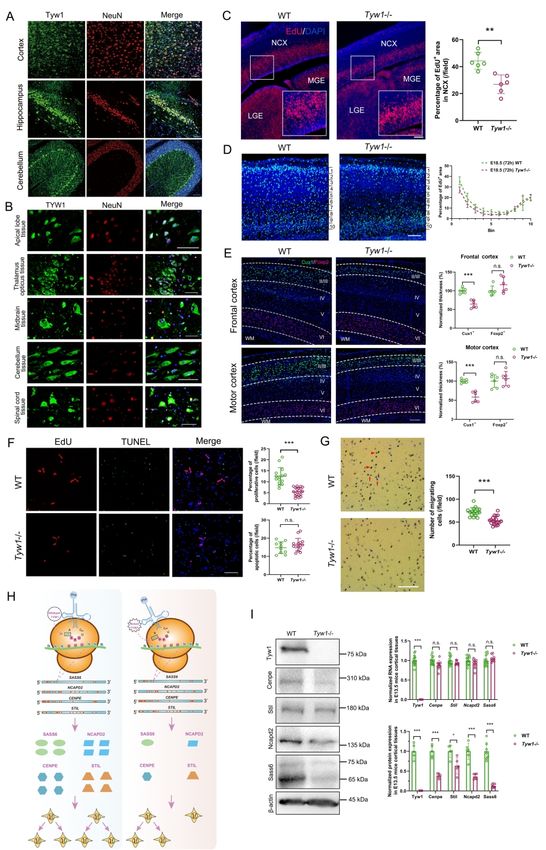
that the defective TYW1, a tRNA hypermodification enzyme, caused primary microcephaly
and problems in motion and cognition by hindering neuronal proliferation and migration.
Furthermore, we developed an algorithm and demonstrated in mouse brains that this
malfunctioning hypermodification specifically perturbed the translation of a subset of
proteins involved in cell cycling. This finding provided a novel and interesting mechanism
for congenital microcephaly. In another cerebral palsy patient with normal intelligence, we
identified a mitochondrial enzyme GPAM, the hypomorphic form of which led to
© The Author(s) (2021). Published by Oxford University Press on behalf of the Guarantors of Brain.
This is an Open Access article distributed under the terms of the Creative Commons Attribution Non-Commercial License
(http://creativecommons.org/licenses/by-nc/4.0/), which permits non-commercial re-use, distribution, and reproduction in any
medium, provided the original work is properly cited. For commercial re-use, please contact journals.permissions@oup.com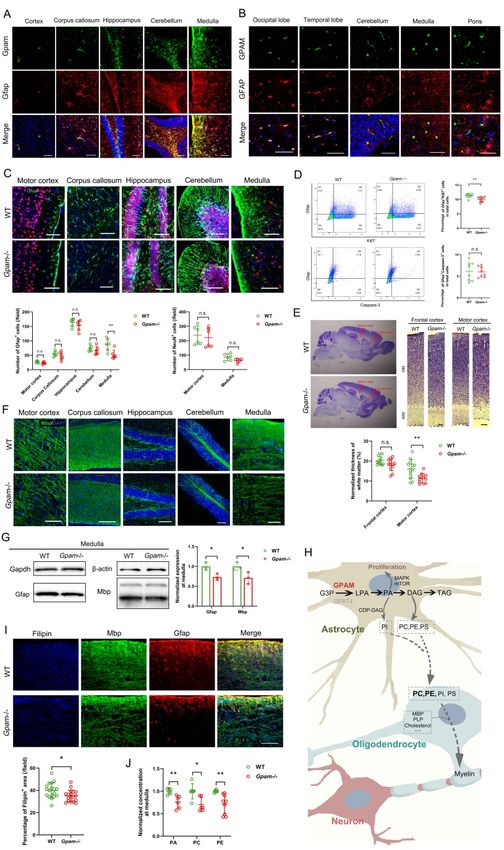
Page 3 of 440                                                   Brain

                hypomyelination of the corticospinal tract in both human and mouse models. In addition, we
                confirmed that the aberrant Gpam in mice perturbed the lipid metabolism in astrocytes,
                resulting in suppressed astrocytic proliferation and a shortage of lipid contents supplied for
                oligodendrocytic myelination. Taken together, our findings elucidate novel aspects of the
                etiology of cerebral palsy and provide insights for future therapeutic strategies.

                                                                                                                 Downloaded from https://academic.oup.com/brain/advance-article/doi/10.1093/brain/awab209/6291242 by guest on 26 October 2021
                Author affiliations:

                1 Laboratory of Medical Systems Biology, Guangzhou Women and Children's Medical
                Center, Guangzhou Medical University, 510623, Guangzhou, China

                2 Department of Rehabilitation, Guangzhou Women and Children's Medical Center,
                Guangzhou Medical University, 510120, Guangzhou, China

                3 Key Laboratory of Neuroregeneration of Jiangsu and Ministry of Education, Nantong
                University, 226001, Nantong, China

                4 Division of Uterine Vascular Biology, Guangzhou Women and Children's Medical Center,
                Guangzhou Medical University, 510623, Guangzhou, China

                5 Department of Genetics and Endocrinology, Guangzhou Women and Children's Medical
                Center, Guangzhou Medical University, 510623, Guangzhou, China

                6 The First Rehabilitation Hospital of Shanghai, Tongji University School of Medicine,
                200029, Shanghai, China

                7 Institute of Cell Biology and Neurobiology, Charité-Universitätsmedizin, 13353, Berlin,
                Germany

                8 Department of Pediatric Neurology, Charité-Universitätsmedizin, 13353, Berlin, Germany

                9 Center for Chronically Sick Children, Charité-Universitätsmedizin, 13353, Berlin,
                Germany

                10 Adelaide Medical School, University of Adelaide, SA5005, Adelaide, Australia

                          ScholarOne, 375 Greenbrier Drive, Charlottesville, VA, 22901 Support (434) 964 4100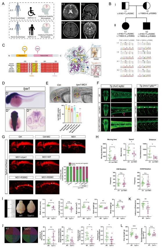
Brain                                           Page 4 of 440

11 Guangdong Provincial Key Laboratory of Research in Structural Birth Defect Disease,
Guangzhou Women and Children's Medical Center, Guangzhou Medical University, 510623,
Guangzhou, China

12 Third Affiliated Hospital of Zhengzhou University, 450052, Zhengzhou, China

                                                                                                        Downloaded from https://academic.oup.com/brain/advance-article/doi/10.1093/brain/awab209/6291242 by guest on 26 October 2021
Correspondence to: Professor Hao Hu

Laboratory of Medical Systems Biology, Guangzhou Women and Children's Medical Center,
Guangzhou Medical University, Guangzhou, China

E-mail: huh@cougarlab.org

Correspondence may also be addressed to: Professor Kaishou Xu

Department of Rehabilitation, Guangzhou Women and Children's Medical Center,
Guangzhou Medical University, Guangzhou, China

E-mail: xksyi@126.com

Professor Dong Liu

Key Laboratory of Neuroregeneration of Jiangsu and Ministry of Education, Nantong
University, Nantong, China

E-mail: liudongtom@gmail.com

Dr Dingding Han, PhD

Laboratory of Medical Systems Biology, Guangzhou Women and Children's Medical Center,
Guangzhou Medical University, Guangzhou, China

         ScholarOne, 375 Greenbrier Drive, Charlottesville, VA, 22901 Support (434) 964 4100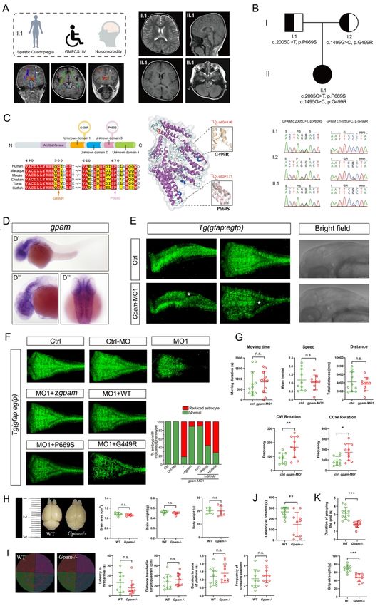
Page 5 of 440                                                   Brain

                Running title: Aetiology of idiopathic cerebral palsy

                Keywords: cerebral palsy; TYW1; GPAM

                Abbreviations: ADL = Activity of Daily Living; ASDs = autism spectrum disorders; CNV =
                copy number variation; CP = Cerebral palsy; DAG = diacylglycerol; DNMs = de novo

                                                                                                                           Downloaded from https://academic.oup.com/brain/advance-article/doi/10.1093/brain/awab209/6291242 by guest on 26 October 2021
                mutations; DRD = DOPA-responsive dystonia; DTI = diffusion tensor imaging; EdU =
                5-Ethynyl-2’-deoxyuridine; GMFCS = Gross Motor Function Classification System;
                GWCMC = Guangzhou Women and Children’s Medical Center; G3P = glycerol-3-phosphate;
                HSP = hereditary spastic paraplegia; ID = intellectual disability; LPA = lysophosphatidic acid;
                MACS = Manual Ability Classification System; Mbp = myelin binding protein; MCPH =
                microcephaly primary hereditary; NDD = neurodevelopmental disorders; PA = phosphatidic
                acid; PC = phosphatidylcholine; PE = phosphatidylethanolamine; PI = phosphatidylinositol;
                PS = phosphatidylserine;      PZM = post-zygotic mutations; TAG =              triacylglycerol; TBI =
                traumatic brain injury; tRNA-yW = tRNA-wybutosine; tRNAPhe = phenylalanine tRNA;
                WISH = whole mount in situ hybridization

                Introduction
                Cerebral palsy (CP) is an umbrella term spanning a group of disorders with compromised
                ambulant performances, and is attributed to non-progressive disturbances in developing fetal
                and infant brains 1. CP can be categorized according to motor signs (spasticity, dyskinesia,
                ataxia, and hypotonia, among which spasticity manifests in ~80% of CP cases), involvement
                of extremities (hemiplegia, monoplegia, diplegia, triplegia, and quadriplegia), and anatomical
                sites of brain lesions (cerebral cortex, pyramidal tract, extrapyramidal system, and
                cerebellum) 2, 3. In CP children who are 2 years or older, severity is reliably classified via the
                five-level Gross Motor Function Classification System (GMFCS)                       4.    The common
                comorbidities of CP include intellectual disability (ID), epilepsy, vision and hearing
                problems, language deficits, autism spectrum disorders (ASDs), sleep disorders, and
                secondary musculoskeletal defects such as scoliosis and hip dislocation           2, 3.   CP is the most
                prevalent physical disability of childhood, with a prevalence of 2.0–3.5 cases per 1,000 live

                          ScholarOne, 375 Greenbrier Drive, Charlottesville, VA, 22901 Support (434) 964 4100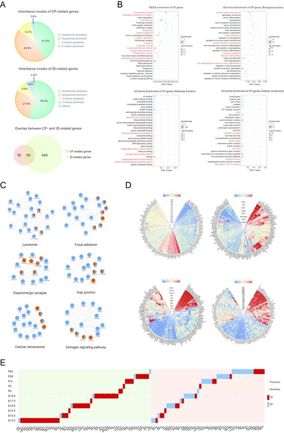
Brain                                                      Page 6 of 440

births in countries with varying degrees of socioeconomic development (more than 20 million
patients around the world), and the incidence of CP at term has remained consistent for over
the past 50 years      1-3, 5-7.   CP poses a critical problem for rehabilitation and welfare systems,
and the lifetime costs for a CP child in the USA are estimated to be $1 million 5. The
conventional risk factors of CP consist of gestational and perinatal insults, including birth

                                                                                                                         Downloaded from https://academic.oup.com/brain/advance-article/doi/10.1093/brain/awab209/6291242 by guest on 26 October 2021
asphyxia, neonatal infections, and teratogens 1, 5. However, two thirds of CP patients are born
at term, and birth asphyxia happens in less than 10% of CP cases 2, 6, 7. Because the estimated
upper limit of unconfirmed causality of CP comprises 80% of cases, it is reasonable that there
exist unknown mechanisms accounting for the majority of CP events 3.

    A growing body of evidence has indicated prevailing genetic contributors to the etiology
of CP, including the reports of familial cases and the high concordance of monozygotic
twins; furthermore, the positive correlation between CP incidence and parental ages suggests
a genetic component, as does the prevalence of congenital malformation observed in CP
patients   8-12.   Large cohorts of case-control studies assessing polymorphisms in a variety of
candidate genes did not identify any significantly associated variants            2, 13, 14.   Therefore, the
genetic underpinnings of CP are likely similar to those of other neurodevelopmental disorders
(NDDs) such as ID and ASD, where causative variants are rarely detected in association
studies based on common variants but with large effect sizes                    15-17.   In line with this
hypothesis, clinically relevant copy number variation (CNV), both de novo and inherited,
were recently identified in four collections of CP patients, resolving 4%–31% of the
cryptogenic cases       18-21.     Meanwhile, gene-panel sequencing and whole-exome sequencing
revealed pathogenic variants, mostly de novo, in an aggregate of six CP studies, yielding
molecular diagnoses for approximately 15% of cases 22-27.

    So far, the known genetic and molecular information on CP etiology has been
insufficient, which impedes improvements in prophylaxis, prognosis, and treatment of CP
patients in the era of precision medicine. Large cohorts need to be recruited to fully decipher
the corresponding gene spectrum due to the high genetic heterogeneity of CP, and geographic
and ethnic biases should be addressed. Whole-genome sequencing, in comparison with
gene-panel sequencing or whole-exome sequencing, can render better coverage of

             ScholarOne, 375 Greenbrier Drive, Charlottesville, VA, 22901 Support (434) 964 4100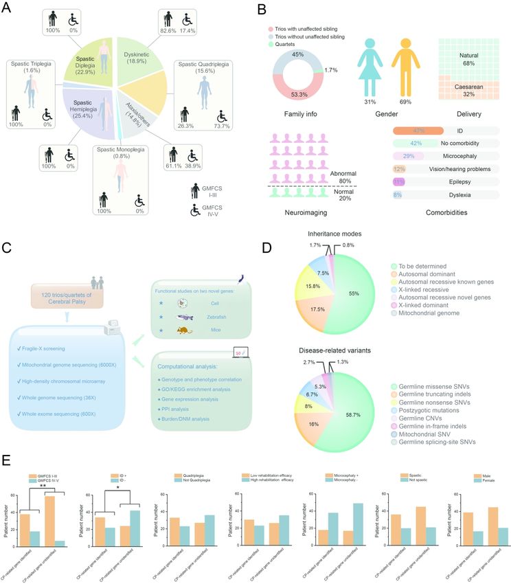
Page 7 of 440                                                    Brain

                predisposing alleles, and somatic mosaicism or post-zygotic mutations (PZM) in CP patients
                must be highlighted as in other NDDs       28, 29.   Hence, targeted investigation into the common
                characteristics of CP-related genes, in addition to detailed mechanistic studies, is warranted
                before the introduction of a gene-based classification system that may have a profound
                impact on clinical practice when treating CP patients.

                                                                                                                     Downloaded from https://academic.oup.com/brain/advance-article/doi/10.1093/brain/awab209/6291242 by guest on 26 October 2021
                Materials and methods

                Cohort recruitment
                The CP families were recruited by the Department of Rehabilitation of GWCMC from
                August 2014 to June 2017 in a cohort study. This study was approved by the ethic committee
                of GWCMC (approval no. 2016061601), and the written informed consents were signed by
                all families involved in this study. The family history and clinical characteristics of CP
                individuals were documented regarding the CP phenotypic profiles. The motor signs (spastic,
                ataxic, dyskinetic, etc.) and anatomical distribution (monoplegia, diplegia, triplegia,
                hemiplegia, and quadriplegia) of movement disorders were evaluated and categorized. Motor
                severity was measured by the Gross Motor Function Classification System (GMFCS) on 5
                levels 30. The common comorbidities, i.e., ID, vision/hearing impairments, epilepsy, dyslexia,
                etc., were recorded for each patient. The fine motor skills were evaluated by the Manual
                Ability Classification System (MACS) 31. Rehabilitation training outcome was determined by
                the Activity of Daily Living (ADL) 32, and the Gross Motor Function Measure (GMFM) 33,
                both of which were measured twice before and after treatments.

                Magnetic Resonance Imaging (MRI) and diffusion tensor imaging
                (DTI)
                All MRI imagings were performed on a 3.0T Siemens scanner (Magnetom Systems, Skyra,
                Munich, Germany). For DTI, an echoplanar sequence with diffusion gradients (b = 1000 s /
                mm2) applied in 64 noncollinear directions was used. Basic parameters included: TR (10000
                ms) / TE (92 ms), Voxel (2.0 × 2.0 × 2.0 mm), FOV (256 × 256), layer thickness (2.0 mm).

                          ScholarOne, 375 Greenbrier Drive, Charlottesville, VA, 22901 Support (434) 964 4100
Brain Page 8 of 440
DTI measurement was three-dimensional reconstruction of color-coded FA graphics through
DTI Studio (DWI standard deviation).
Analysis pipeline of deep sequencing data
The variant prioritization and evaluation of etiologic involvement were performed by two
Downloaded from https://academic.oup.com/brain/advance-article/doi/10.1093/brain/awab209/6291242 by guest on 26 October 2021
pipelines, namely, MERAP and ANNOVAR. In brief, MERAP pipeline first filters all
identified variants through comparison with the disease-associated variants in the Human
Gene Mutation Database (HGMD, 2020.2) and the Online Mendelian Inheritance in Man
(OMIM) to collect those known disease-causing variants. In order to filter out neutral
variants, MERAP uses entries from the dbSNP143
(http://www.ncbi.nlm.nih.gov/projects/SNP/), 1000 Genome
(http://www.1000genomes.org/), NHLBI Exome Sequencing Project (ESP,
http://evs.gs.washington.edu/EVS/), ExAc (http://exac.broadinstitute.org/), and the Chinese
Millionome Database (CMDB) (https://db.cngb.org/cmdb/) as screening databases. In
principle, candidate variants causing recessive traits should not occur in healthy controls as
homozygotes, and the frequency of respective heterozygotes should not exceed 0.1%.
MERAP uses the RefSeq genes (http://www.ncbi.nlm.nih.gov/refseq/) as reference, and
nonsynonymous changes are described in terms of gene ID, base change, protein change,
genomic coordinate, transcript coordinate, protein coordinate, protein length, affiliated with
gene description from the Human Gene Nomenclature Committee (HGNC,
http://www.genenames.org/). Changes destroying conventional splice sites or introducing
novel splice sites are identified by MERAP’s module called SSFinder. To assess the
pathogenicity of missense mutations, MERAP generates a single score integrating the results
of seven different algorithms, including the Grantham score, PhyloP, GERP, SIFT,
PolyPhen2, Mutation-Taster, and the Conserved Domains Database (CDD,
http://www.ncbi.nlm.nih.gov/Structure/cdd/cdd.shtml). With empirical false discovery rate
cutoffs, this score serves as dichotomized pathogenicity predictions even if any two of the
seven algorithms might not coincide, as is often the case. MERAP rules out candidate genes
reported to harbor homozygous loss-of-function (LOF) variants in healthy individuals, which
applies to >1% of the human genes. Typically, if more than three independent truncating
ScholarOne, 375 Greenbrier Drive, Charlottesville, VA, 22901 Support (434) 964 4100Page 9 of 440 Brain
variants are observed in >10 of the exomes listed in the 1000 Genome, Exome Variant Server
(ESP6500), NHLBI GO Exome Sequencing Project (ESP)
(http://evs.gs.washington.edu/EVS/), and ExAC (http://exac.broadinstitute.org/) databases,
the relevant gene is flagged as LOF tolerant. To facilitate the choice between few remaining
candidate genes, MERAP also provides a list of ~4,500 known disease genes extracted from
Downloaded from https://academic.oup.com/brain/advance-article/doi/10.1093/brain/awab209/6291242 by guest on 26 October 2021
OMIM (http://www.ncbi.nlm.nih.gov/omim), ClinVar
(http://www.ncbi.nlm.nih.gov/clinvar/), and HGMD (http://www.hgmd.org/), as well as more
than 8,000 associated disorders and their symptoms. For novel candidate genes without prior
link to disease, MERAP offers information on their interaction with known disease genes,
based on data from Biogrid (http://thebiogrid.org/) and IntAct (http://www.ebi.ac.uk/intact/),
assuming that genes implicated in clinically similar disorders tend to cluster in gene or
protein interaction networks. Variants were considered de novo if neither parent had the
variant, and candidate variants were selected by segregation analysis. The pathogenic and
likely pathogenic genes/variants were defined according to the standards and guidelines of
ACMG 34.
The ANNOVAR pipeline was also used, with additional considerations from the
Residual Variation Intolerance Score (RVIS) and the Combined Annotation-Dependent
Depletion (CADD) score 35. The former ranks genes in terms of intolerance to functional
genetic variation and the latter integrates several well-known tools. We empirically set a
cutoff RVIS score of 50th percentile for known and novel genes and a cutoff CADD score of
20 for novel candidate genes. When defining likely causal variants/genes, we followed the
guidelines designated by ACMG.
All variants of putative clinical relevance were confirmed by the conventional PCR and
Sanger sequencing. Parent-child relationships were confirmed by using the PLINK with
SNPs drawn from WGS which matched the CytoScan SNP repertoire 36. The false positive
rate of WGS was generated by the comparison between Sanger sequencing and WGS, and we
found no false positive variant called by our pipeline. The false negative rate of WGS was
evaluated by the SNP genotyping comparison between WGS and CytoScan high-density
microarray, which resulted in an average false negative rate of less than 0.1%.
ScholarOne, 375 Greenbrier Drive, Charlottesville, VA, 22901 Support (434) 964 4100Brain Page 10 of 440
Zebrafish modeling
All zebrafish experimentation was carried out in accordance with the NIH Guidelines for the
care and use of laboratory animals (http://oacu.od.nih.gov/regs/index.htm) and ethically
approved by the Administration Committee of Experimental Animals, Jiangsu Province,
Downloaded from https://academic.oup.com/brain/advance-article/doi/10.1093/brain/awab209/6291242 by guest on 26 October 2021
China [Approval ID: SYXK (SU) 2007–0021].
Zebrafish lines and whole mount in situ hybridization
The zebrafish embryos and adults were maintained in the Zebrafish Center of Nantong
University under conditions in accordance with our previous protocols 37, 38. The fish lines
Tg(mnx1:GFP)ml2 , Tg(huC:egfp) , Tg(huC:mcherry) and Tg(gfap:egfp) have been described
in the previous work 37, 38. The whole mount in situ hybridization (WISH) was performed as
previously described 39. The templates for generating antisense RNA probes were amplified
from the cDNA library, using specific primers targeting tyw1 and gpam listed as below:
tyw1-Forward: 5’ AAG GCC AGC TGC AAG AAT AA; tyw1-Reverse: 5’ GTG TGG TGT
CTC CAG CAG AA; gpam-Forward: 5’ TCC TGT TCA CAA GTC CCA CA;
gpam-Reverse: 5’ AGT TCT TGC GGA GCA TCC TA.
Morpholino and mRNAs Injections
tyw1 splice-blocking Morpholino (MO; Gene Tools, OR, USA) sequence was 5′ AAC CTT
ATT CCC ACT TAA TGT TAC C. gpam splice-blocking Morpholino sequence was 5′ GGT
GCT ACT TTT CTC CAA GCT TAC C. The sequence of a standard control MO oligo was
5′ CCT CTT ACC TCA GTT ACA ATT TAT A. tyw1 Translation-blocking Morpholino
sequence was 5’ CAG CAT CTC ATG TAC TCT CTC CAT C. gpam Translation-blocking
Morpholino sequence was 5’ ACG TCC ATC CCC TCT CTT CAA ACC A. The MOs
were diluted to 0.3 mM with RNase-free water and injected into the yolk of one to two-cell
stage embryos and then raised in E3 medium at 28.5 ℃. The wild-type and mutated cDNAs
(TYW1 (NM_018264) mut1: p.R389Q; TYW1 mut2: p.R206C; GPAM (NM_001244949)
mut1: p.G499R; GPAM mut2: p.P669S) containing the open reading frame of the zebrafish
and human tyw1 or gpam genes were cloned into pCS2+ vector respectively and then were
ScholarOne, 375 Greenbrier Drive, Charlottesville, VA, 22901 Support (434) 964 4100Page 11 of 440 Brain
transcribed in vitro by using the mMESSAGE mMACHIN Kit (Thermo Fisher Scientific,
MA, USA) after the recombinant plasmids linearized with NotI Restriction Enzyme (NEB,
MA, USA), and then the capped mRNAs were purified by RNeasy Mini Kit (Qiagen,
Frankfurt, Germany). Around 2 nl mRNA were injected at 50 ng/µl into 1/2-cell stage
embryos.
Downloaded from https://academic.oup.com/brain/advance-article/doi/10.1093/brain/awab209/6291242 by guest on 26 October 2021
RNA isolation, reverse transcription, and PCR
Total RNA was extracted from zebrafish embryos by TRIzol reagent according to the
manufacturer’s instructions (Invitrogen, Thermo Fisher Scientific, MA, USA). The reverse
transcription was carried out according to the standard protocols from manufacturer
(Fermentas, Thermo Fisher Scientific, MA, USA). The PCR was carried out as previously
described to validate the efficiency of splicing-blocking effect of specific morpholino 37 by
using the primer sequencing listed as below. tyw1-Forward: 5’ TTA TCG GTG TTG TCG
GGT TT; tyw1-Reverse: 5’ CCT CTG CCA ATC TGT CTT CC; gpam-Forward: 5’ ACA
GTG CGC AAA AAG AGG TC; gpam-Reverse: 5’ TAA GCA CGT TCT CCA CCA CA.
Locomotion analysis in zebrafish larvae, microscopy and statistical analysis
The locomotion analysis of 5 dpf zebrafish larvae was carried out by using the DanioVision
system (Noldus Information Technology, Wageningen, Netherlands). The confocal imaging
was performed by using a TCS-SP8 LSM confocal imaging system (Leica, Wetzlar,
Germany). The zebrafish embryos were embedded as we previously did 37. Photographs of in
situ hybridization results were taken by using a DP70 camera on an Olympus
stereomicroscope MVX10. Statistical comparisons of the data were carried out by student’s
t-test, and P < 0.05 was considered statistically significant.
Mice modeling
All mice experiments in this study were approved by the institutional animal care and use
committee in the Guangzhou Medical University (registration No. 2019-436, 2019-694). In
this study, all mice, either wild-type or mutant, were generated from the C57BL/6J strain and
provided by the Cyagen Biosciences (Guangzhou, China).
ScholarOne, 375 Greenbrier Drive, Charlottesville, VA, 22901 Support (434) 964 4100Brain Page 12 of 440
Generation of knockout mice by CRISPR/Cas9 system
Knockout mouse lines were created by CRISPR/Cas9-mediated genome engineering. To
create a Tyw1 knockout mouse model, exon 2~3 was selected as the target site. gRNA was
designed as the following sequences: gRNA1 (matching reverse strand of gene):
Downloaded from https://academic.oup.com/brain/advance-article/doi/10.1093/brain/awab209/6291242 by guest on 26 October 2021
GCCTAAGGACCACGTTTCGATGG. gRNA2 (matching reverse strand of gene):
CATGTAAGACCTCACGATCAAGG.
To create a Gpam knockout mouse model, exon 2~4 was selected as the target site. gRNA
was designed as the following sequences: gRNA1 (matching reverse strand of gene):
CTCCCACGGGAAAACCACGCAGG. gRNA2 (matching reverse strand of gene):
CCCAAGGGGTCACGCACCACAGG.
Cas9 mRNA and gRNA were co-injected into the cytoplasm of fertilized eggs. The
microinjected zygotes were cultured in medium until the two-cell stage, and then were
transferred into the oviducts of pseudopregnant ICR females at 0.5 d after mating with
vasectomized males. The mutant F0 mice were identified by genomic PCR and the DNA
sequence was confirmed by Sanger sequencing. Subsequent breeding of the mutant F0 mice
generated offspring with desired genotypes for experiments. Gene knockout efficiency was
identified using qPCR (supplementary figure 10).
Behavioral tests
All behavioral tests were carried out on both male and female mice at 8 weeks of age, and the
balanced gender was kept for both wild-type and Tyw1-/- or Gpam-/- mice. Each test was
conducted at fixed day time (between 8:30 am to 18:30 pm) on each training day. Mice were
moved to the testing room 1 h before behavioral testing for acclimation, and those
participating in multiple tests were allowed to rest for at least 3 days between two tests. All
experimental areas were cleaned with 70% ethanol before the tests and between subjects. All
behavioral tests were carried out with the presence of two researchers blinded to the
genotype.
Morris water maze test
ScholarOne, 375 Greenbrier Drive, Charlottesville, VA, 22901 Support (434) 964 4100Page 13 of 440 Brain
Morris water maze test was performed to investigate the learning and memory ability of
mice. Briefly, a platform was placed at the central zone of one quadrant of the pool below the
surface of water. The mice were trained to learn the position of the hidden platform for 4
days. At day 5, the platform was removed. The mice were released into water and allowed to
swim for 60 s to search a virtual quadrant centered on the location of the platform. Duration
Downloaded from https://academic.oup.com/brain/advance-article/doi/10.1093/brain/awab209/6291242 by guest on 26 October 2021
in zone of platform refers to amount of time mice remained in this virtual zone. Frequency of
crossing platform refers to number of times mice crossed the virtual zone, while latency to
first arrival refers to the time needed for mice to reach the virtual zone at first time, while
distance travelled in target quadrant refers to the distance mice swam in the virtual quadrant.
Videos were recorded and analyzed by using the SuperMaze V2.0 software (XinRuan,
Shanghai, China).
Rotarod test
Rotarod test was performed to indicate motor coordination of mice in each group. Firstly,
mice were trained three times at the rate of 30 rpm before test, and each training maintained
for 5 min. In the test, the rotation rate increased gradually to reach 40 rpm within 300 s. The
test was over when a mouse fell off. Each mouse was detected for 3 times and the mean
latency time at rotarod was recorded.
Grip strength test
Grip strength of mice forelimbs was measured with a grip strength test meter (BIOSEB, EB
Instruments). During the test, the mice were placed over the grid allowing only forepaws to
attach to the grid. After the mice paws grasped the grid, their tails were pulled horizontally
until they completely released hold. Each mouse was tested for three times. The readings of
grip strength and duration of grasping the grid were recorded and analyzed by using the
SuperGSM software (XinRuan, Shanghai, China).
Tissue preparation
ScholarOne, 375 Greenbrier Drive, Charlottesville, VA, 22901 Support (434) 964 4100Brain Page 14 of 440
Mice were anaesthetized with 50 mg/kg sodium pentobarbital. After perfused with 0.9%
NaCl and 4% paraformaldehyde, brains were extracted and post-fixed for 24 h, followed with
dehydration by using sucrose. Brains were cut into two hemispheres, and then serial sagittal
sections (30 μm thick) were sliced with freezing microtome (CM3050S, Leica, Germany).
Nissl staining
Downloaded from https://academic.oup.com/brain/advance-article/doi/10.1093/brain/awab209/6291242 by guest on 26 October 2021
Sections were immersed into 75% ethanol (30 s), dH2O (30 s) and cresyl violet (2 min).
Then, sections were dehydrated with gradient ethanol (75%, 95%, 100%) for 30 s
respectively, followed by incubation with xylene and mounted with neutral resins. The
images were taken under an inverted microscope.
5-Ethynyl-2’-deoxyuridine labeling
For proliferation assays, pregnant dams on E13.5 were intraperitoneally injected with
5-Ethynyl-2’-deoxyuridine (EdU, 20 mg/kg body weight) 2 h before sacrifice. Embryonic
brains were harvested, fixed in paraformaldehyde and immersed in 30% sucrose for
dehydration. Brain slices were then stained with Click-iT EdU Imaging Kits (C10339,
Invitrogen, Thermo Fisher Scientific, USA). Cells incorporated with EdU were determined
under a fluorescence microscope. Percentage of EdU+ fluorescence area (indicating the cell
number of EdU+) of E13.5 mouse brain cortex was calculated in 3 random fields per
coverslip. For migration assays, pregnant dams on E15.5 were intraperitoneally injected with
EdU. Brains were harvested after 72 h and stained as above.
Immunostaining
To detect expression of TYW1 and GPAM in human brain tissue, the normal brain tissue
microarrays (BNC17011, Biomax, MD, USA) were prepared. According to the instruction
from Biomax official website, BNC17011 were derived from normal brain tissue of male or
female at the age of 2 to 50, including brain regions such as frontal lobe, apical lobe, occipital
lobe, temporal lobe, midbrain, pons, medulla oblongata, thalamus opticus, cerebellum,
hippocampus, callositas, optic nerve, and spinal cord. The slides were immersed in xylene
ScholarOne, 375 Greenbrier Drive, Charlottesville, VA, 22901 Support (434) 964 4100Page 15 of 440 Brain
and graded ethanol to deparaffinize and rehydrate. After antigen retrieval using microwave
with sodium citrate buffer, 0.3% Triton-X-100 was added to permeabilize the tissue. The
slides were blocked with goat serum and incubated with primary antibodies at 4 ℃ overnight.
After washing with PBS, the sections were incubated with secondary antibodies.
For mouse brain tissues, the perfused brain samples were fixed within 4% paraformaldehyde
Downloaded from https://academic.oup.com/brain/advance-article/doi/10.1093/brain/awab209/6291242 by guest on 26 October 2021
(PFA) in 0.01 M PBS at 4 ℃ for 24 h, then washed by PBS twice , cryoprotected with
sucrose gradients, snap frozen, and sectioned with a cryostat (Leica, Wetzlar, Germany) to
thickness of 30 µm. Then the sections were immuno-stained by using aforementioned
methods, except deparaffinization and rehydration.
Number of Cux1+ or Foxp2+ cells in frontal cortex and motor cortex were counted on
three sagittal cerebrum sections, respectively. Gfap+ and NeuN+ cells in different brain
regions were counted in 3 random fields per section. Data were obtained from at least three
independent experiments and analyzed with ImageJ software.
In order to visualize and measure the cholesterol level, Filipin staining was performed in
tissue sections (10µm thick) by using a cell-based Cholesterol Assay Kit (ab133116, Abcam,
UK). Briefly, the tissue sections were washed (3 × 5 min) with Cholesterol Detection wash
buffer, and then the Filipin III was added to each section and incubated in the dark for 60 min
at room temperature. After washing, the fluorescence images were obtained by a Leica DMi8
(Leica Microsystems, Wetzlar, Germany) fluorescence microscope by using × 20 HC PL
FLUOTAR objective.
Western blot analysis
Total protein was extracted from peripheral blood using cell lysate containing RIPA and
protease inhibitor cocktail. And the brain tissues were lysed with 2% SDS in PBS with PMSF
and proteinase inhibitor cocktail. The BCA assay was used to determine protein
concentration. Proteins were resolved on 7.5%, 10% or 15% tris-glycine gels based on
different molecular weight and transferred to PVDF membranes. After blocking, the
membranes were incubated with primary antibodies, followed by horseradish peroxidase
ScholarOne, 375 Greenbrier Drive, Charlottesville, VA, 22901 Support (434) 964 4100Brain Page 16 of 440
(HRP)-labeled secondary antibodies. Then, blots were visualized by West Pico Plus
Chemiluminescent Substrate (Thermo Fisher Scientific, MA, USA) and scanned using
ChemiDocTM MP system (Bio-Rad, CA, USA). Densitometries of individual blot signals
were quantified using ImageJ software.
qRT-PCR analysis
Downloaded from https://academic.oup.com/brain/advance-article/doi/10.1093/brain/awab209/6291242 by guest on 26 October 2021
Total RNA was extracted from the mice brain tissues by using TRIZOL reagent (Invitrogen,
Thermo Fisher Scientific, MA, USA), and cDNA was synthesized from 1 µg total RNA by
using PrimeScript™ RT Master Mix (RR036Q, Takara, Shiga, Japan). Quantitative PCR was
carried out by using PowerUp™ SYBR™ Green Master Mix (A25778, Applied Biosystems,
Thermo Fisher Scientific, MA, USA) on Biosystems QuantStudio 6 Flex Real-time PCR
system (Applied Biosystems, Thermo Fisher Scientific, MA, USA). β-Actin was used as a
reference gene. For comparison, mRNA expression level of knockout mice were normalized
to those of wild-type mice for each subject. The primers were: Tyw1-Forward: 5’ GTG GGA
CTT GTC GCC TTT G; Tyw1-Reverse : 5’ GGG AAC GAG TGA CCT GCT T;
Stil-Forward: 5’ GAC ACA ATT CAG GAC TGG TAG AC; Stil-Reverse : 5’ GGC ATG
ATC CAC TTT CTG TTC A; Sass6-Forward: 5’ ATT CCT TTA CGC GGA CTT AGC;
Sass6-Reverse: 5’ AAG TAG GCT GAA GAC GAG GAG; Ncapd2-Forward: 5’ AGC CAG
ACA AGC CTC ATT GAC; Ncapd2-Reverse: 5’ TCC ATA GGT GAC GGA TGT CCA;
Cenpe-Forward: 5’ CTT CAG TGG CTG TCT GTG TTC; Cenpe-Reverse: 5’ CCA TCG
CTC TGA TAA ATA GCG TT; β-Actin-Forward: 5’ GGC TGT ATT CCC CTC CAT CG;
β-Actin-Reverse: 5’ CCA GTT GGT AAC AAT GCC ATG T.
Culture of primary neurons
E13.5 mouse embryo brains were taken out from Tyw1+/- pregnant dam. Cortex in each brain
was dissected separately and collected in Hibernate-E supplemented with 2% B27 on ice.
Single cells were obtained by using 0.05% trypsin (containing 0.2 mM EDTA) digestion for
10 min at 37 ℃. After filtration with 70 μm strainer and centrifugation, cells resuspended in
Neurobasal medium with 2% B27 Plus Supplement, 0.25% Glutamax and 25 μM glutamate
ScholarOne, 375 Greenbrier Drive, Charlottesville, VA, 22901 Support (434) 964 4100Page 17 of 440 Brain
were placed in poly-D-lysine coated plates. Adherent neurons were prepared for EdU and
TUNEL staining. To prepare migration assay, isolated cells were initially cultured in
ultra-low attachment plates to form neurospheres, which could be digested by using 0.05%
trypsin without damage of neurites.
Proliferation and apoptosis assays of primary neurons
Downloaded from https://academic.oup.com/brain/advance-article/doi/10.1093/brain/awab209/6291242 by guest on 26 October 2021
At 2 DIV, cells were labeled with 10 μM EdU solution and incubated for 4 h. Cells were
treated by using 4% PFA, followed by 0.5% TritonX-100 permeabilization. After being
washed twice with 3% BSA, cells were incubated with Click-iT reaction cocktail according
to protocol of Click-iT EdU Imaging Kits (C10339, Invitrogen, Thermo Fisher Scientific,
MA, USA) and stained with DAPI. Dead cells were labeled using In Situ Cell Death
Detection Kit, POD (11684817910, Roche, Switzerland).
Migration test of primary neurons
Neurospheres were digested with 0.05% trypsin to obtain single neurons and resuspended in
Neurobasal Medium with 1% B27 Plus Supplement. Cells were seeded on the upper layer of
a cell culture insert with PET track-etched membrane (8 μm pore size, 353097, Corning, NY,
USA) at density of 1 × 10⁵ cells/well. Neurobasal Medium with 2% B27 Plus Supplement
was added into the bottom of the lower chamber. After 16 h, the culture insert was taken out
and the medium was removed carefully. Cells on the upper surface were wiped, while
migrating cells in pore and on the lower surface of membrane were fixed, stained with 0.1%
crystal violet and observed.
Flow cytometry
Whole brain tissues from Gpam-/- mice on P1 (postnatal day 1) were collected, including
brain tissues of their wild-type littermate. After digestion with 4 mg/ml papain and 0.1 mg/ml
DNase in 37 ℃ shaker for 30 min, suspension was diluted using DMEM/F12 medium
containing 5% FBS and filtered with 70 μm strainers. Then, suspension was centrifuged and
the cells were fixed in 4% PFA. 0.3% Triton-X100 was used for permeabilization. After
ScholarOne, 375 Greenbrier Drive, Charlottesville, VA, 22901 Support (434) 964 4100Brain Page 18 of 440
being blocked with 5% goat serum, cells were incubated with mouse anti-Gfap antibody and
rabbit anti-Ki67 antibody or rabbit anti-Caspase-3 antibody at 4 ℃ for 1 h. Cells were
washed with DPBS and incubated with Alexa Fluor 488 goat anti-mouse IgG and Alexa
Fluor 647 goat anti-rabbit IgG at 4 ℃ for 30 min. After being washed for two times, cells
were suspended by using DPBS and analyzed on the flow cytometer (BD FACSCanto, BD
Downloaded from https://academic.oup.com/brain/advance-article/doi/10.1093/brain/awab209/6291242 by guest on 26 October 2021
Biosciences, CA, USA).
Measurement of phosphatidic acid (PA)
Phosphatidic acid was measured by using the PicoProbeTM Phosphatidic Acid Assay Kit
(BioVision, CA, USA). In brief, medulla of wild-type and Gpam-/- mice was homogenized in
PA assay buffer. Lipid extraction was obtained according to the protocol and solubilized in
5% Triton X-100 solution. “Sample background control” and “Sample” were prepared in
parallel. Standard curve was generated by using PA standard solution. Converter mix was
only added in sample and standard wells. All wells were incubated at 45 ℃ for 1 h. Reaction
mix was added in each well and incubate at 37 ℃ for 30 min. Fluorescence was recorded at
Ex/Em = 535/587 nm and PA concentration was expressed as nmol PA per mg tissue weight.
Value of PA concentration was normalized.
Measurement of phosphatidylcholine (PC)
Phosphatidylcholine was measured by using the phosphatidylcholine assay kit (Abcam, UK).
For each individual, about 10 mg brain tissues were washed with cold PBS, resuspended in
the assay buffer, and homogenized on ice. After 10 min incubation, samples were centrifuged
for 5 min at 4 °C at 16,000 g. The supernatant was incubated with the reaction mix including
OxiRed Probe supplemented with hydrolysis enzyme for 30 min. The colorimetric reading
was measured at OD570 nm on a microplate reader.
ScholarOne, 375 Greenbrier Drive, Charlottesville, VA, 22901 Support (434) 964 4100Page 19 of 440 Brain
Measurement of Phosphatidylethanolamine (PE)
Phosphatidylethanolamine was measured by using the phosphatidylethanolamine assay kit
(Abcam, UK) according to the manufacturer’s instruction. Briefly, solubilized lipids were
extracted from brain tissues with 5% Triton X-100, incubated with converter mix at 45°C for
Downloaded from https://academic.oup.com/brain/advance-article/doi/10.1093/brain/awab209/6291242 by guest on 26 October 2021
1 hour, and then with reaction mix at 40 °C for 3 hours. Fluorescence was recorded at Ex/Em
535/587 nm.
Modeling the effect of TYW1 knockout on ribosomal frameshift
The modeling of TYW1-knockout effect on ribosomal frameshift was based upon the fact that
TYW1 is a critical enzyme involved in the synthesis of wybutosine, so the hypomorphic and
null alleles of TYW1 could reduce and even remove the production of wybutosine accordingly
40, 41. It is known that wybutosine at the 37 position of tRNAPhe near the 3’ base of anticodon
can stabilize the codon-anticodon interaction thus drastically reduce the ribosomal frameshift
upon a specific UUU codon 42. We, therefore, hypothesized that the density and distribution
of UUU codons along a specific mRNA should determine the extent of influence exerted by
wybutosine, and in parallel, the relative abundance of wybutosine was another major effector
during this process. Our modeling procedure went as follows:
1. Download the UCSC RefSeq (refGene) gene models of human, mouse, and zebrafish from
https://genome.ucsc.edu/cgi-bin/hgTables. Extract the coding domain sequences (CDS) of
each protein-coding gene. If there exists multiple isoforms, choose the longest ones.
2. Label the location of UUU codons, normalize the location values by the length of CDS. By
doing this, we harvested a series of normalized location values, i.e., L(1), L(2), ..., for each
gene, where 0 < L(n) < 1, n = 1, 2, ....
3. We made the following two assumptions: (1) for a UUU codon at any location, there is a
fixed chance of ribosomal frameshift designated by Pf, given a fixed level of wybutosine; (2)
for a given UUU codon located at L(n), after frameshifting, the remaining activity of a
protein is proportionate to L(n). Thus, the consequence of translating a mRNA holding
ScholarOne, 375 Greenbrier Drive, Charlottesville, VA, 22901 Support (434) 964 4100Brain Page 20 of 440
multiple UUU codons, i.e., the remaining activity of a protein, designated by R, can be
estimated as:
R = ∑ (L(n) * Pf * Pr(n)) + Pr(n), where Pr(1) = 1 and Pr(n) = Pr(n - 1) * (1 - Pf), n = 1, 2, ....
4. For the wild-type (WT) and TYW1-knockout mice (KO), we defined the R values for WT
Downloaded from https://academic.oup.com/brain/advance-article/doi/10.1093/brain/awab209/6291242 by guest on 26 October 2021
and KO to be R(WT) and R(KO), respectively. The R value is determined by the density and
distribution of codon UUU in a given mRNA, also by Pf (the chance of ribosomal frameshift
at a specific location of mRNA, given a fixed level of wybutosine). We set the values of Pf
for R(WT) and R(KO) to be 0.12 and 0.35, respectively 42. Subsequently, we defined the
attenuation coefficient to be R(KO) / R(WT). An attenuation coefficient reflected the extent
of protein activity reduction with TYW1-knockout, and the expected values of attenuation
coefficient ranged from 0 to 1. The lower the value of attenuation coefficient, the more severe
the ribosomal frameshift and protein degradation when TYW1 is knocked out.
5. After generating a list of attenuation coefficients for each human / mouse / zebrafish
protein, we focused on proteins meeting with these criteria: (1) a protein with ortholog in all
three species; (2) a protein with attenuation coefficient less than 0.34 (i.e., 0.12 / 0.35); (3) a
protein with its corresponding gene matching TYW1 in terms of expression profiles in normal
brains; (4) a protein known to be disease-related, with records in HGMD or OMIM.
Data availability
The authors confirm that the data supporting the findings of this study are available within
the article and its supplementary material. This study’s samples and data are available to the
scientific community upon request.
Results
CP cohort recruitment, medical evaluation, and genetic screening
A total of 120 CP families, including 118 trios and 2 quartets, were recruited by the
Department of Rehabilitation of the Guangzhou Women and Children’s Medical Center
ScholarOne, 375 Greenbrier Drive, Charlottesville, VA, 22901 Support (434) 964 4100Page 21 of 440 Brain
(GWCMC) from August 2014 to June 2017 in a cohort study. This study was approved by
the ethics committee of GWCMC, and written informed consents were signed by the
participating families. The following inclusion criteria were adopted: (1) a diagnosis of CP
made by a board of pediatric neurologists, based on early-onset non-progressive motor
disability; (2) an age of two years and older at the time of symptom ascertainment; and (3)
Downloaded from https://academic.oup.com/brain/advance-article/doi/10.1093/brain/awab209/6291242 by guest on 26 October 2021
uneventful pregnancy and delivery (109 CP patients were born at term, six at 36 gestational
weeks, two at 35 weeks, three at 34 weeks, one at 33 weeks, and one at 30 weeks). The
following exclusion criteria were implemented: (1) perinatal insults including potential
asphyxia with an Apgar score < 7, traumatic brain injury (TBI) 43, post-encephalitis brain
lesions 44, and brain tumors; (2) late-onset hereditary spastic paraplegia (HSP) or
DOPA-responsive dystonia (DRD), due to the distinct progressive trajectory and featured
fluctuation of manifestation; (3) maternal infections with a history of fever during pregnancy;
or (4) hyperbilirubinemia of the newborn with high bilirubin levels as a risk for kernicterus 45,
46. The classification and summary of the CP cohort are shown in Fig. 1A and 1B. As
expected, the majority of cohort patients belonged to spastic CP, followed by dyskinetic and
ataxic CP. There were less patients with more severe motor impairments (GMFCS levels IV
and V) than those who were mildly disabled in terms of motion (GMFCS levels I–III). There
was a significant bias for males in this CP cohort, as observed previously in ID and ASD
cohorts 47. The prevalent comorbidities included ID and microcephaly. More details of the CP
patients can be found in Supplementary Table 1 and Supplementary Fig. 1.
As shown in Fig. 1C and Supplementary Table 2, we initiated a comprehensive genetic
assessment via multiple platforms, including targeted PCR for fragile-X, mitochondrial
genome sequencing (6000X), a high-density cytogenetic microarray, whole-exome
sequencing (600X), and whole-genome sequencing (36X). In principle, we defined the
disease-related variants by adopting the ACMG-recommended standards, reinforced by an
in-house variation database of 2,247 ethnic-matched unaffected individuals, and another
database (https://db.cngb.org/cmdb/) of >50,000 individuals of East Asian descent 34, 48.
In sum, we identified CP-related candidate genes and variants in 54 families (~45% of
the 120 CP families), a comparable rate with that of other NDDs 16, 49-52. Interestingly, the
ScholarOne, 375 Greenbrier Drive, Charlottesville, VA, 22901 Support (434) 964 4100Brain Page 22 of 440
unexpected abundance of recessive variants (approximately one quarter) in the candidate
genes, was notably higher than that of previous NDD investigations in outbred populations 50,
53, 54. Whether this was attributed to the specific genetic structure of the ethnic group in the
present study or was due to the characteristic etiology of CP per se, remains to be solved in
future studies 55.
Downloaded from https://academic.oup.com/brain/advance-article/doi/10.1093/brain/awab209/6291242 by guest on 26 October 2021
Of the 75 CP-related variants identified in this cohort, germline point substitutions
accounted for ~68% of instances, followed by germline indels (~18.7%) and CNVs (~5.3%)
(Fig. 1D). Post-zygotic mutations (PZMs) were, for the first time, described in a CP cohort, in
which ~6.7% of the CP-related variants were classified as PZMs. This finding met with the
expectation derived from other NDD studies, and fills a gap in our understanding of CP
etiology 28.
A closer look at the phenotype-genotype correlation in the patients revealed the
following. Patients with more severe physical impairments (GMFCS level IV or V) had a
higher chance of harboring CP-related variants (P = 0.00613; Fisher exact test) than that of
patients less disabled in movement (GMFCS levels I–III); patients with comorbidity of ID
were more likely to have deleterious variants (P = 0.01065; Fisher exact test) compared with
those without ID (Fig. 1E).
In this cohort, we found a total of 129 protein-changing de novo mutations (DNMs) in
the CP patients, the rate of which was significantly higher than that of the in-house 2,247
ethnic-matched unaffected individuals (P = 0.0083; bootstrap 1,000 times). When counting
private mutations of the CP families as the data input of a burden analysis, we concluded that
the 146 private mutations found in the CP patients were significantly more than what we
expected by counting the rate of private mutations in the in-house 2,247 ethnic-matched
controls (P = 0.0142; bootstrap 1,000 times). Paternal ages were revealed to correlate
positively with DNM counts of the patients (r = 0.63, P = 0.025; Pearson correlation test).
We also detected 27 PZMs in 66 CP patients, but no significant correlation with paternal or
maternal ages was established. The aforementioned statistical analyses were based on the
protein-coding exons and conventional splice-sites with > 10X non-redundant coverage
(31,856,576 bp) in all the CP families and the in-house controls.
ScholarOne, 375 Greenbrier Drive, Charlottesville, VA, 22901 Support (434) 964 4100Page 23 of 440 Brain
The details of disease-related variants identified in this study are shown in Supplementary
Table 3 and Supplementary Table 4.
Assessment of the role of de novo mutations
On aggregate, we characterized 26 CP-related de novo events in approximately one fifth of
Downloaded from https://academic.oup.com/brain/advance-article/doi/10.1093/brain/awab209/6291242 by guest on 26 October 2021
the cohort individuals, which included 18 autosomal dominant mutations (two post-zygotic),
two X-linked dominant mutations (one post-zygotic), three de novo CNVs spanning multiple
disease-related genes, and one de novo mutation in the mitochondrial genome
(Supplementary Fig. 2 and 3).
It was noted that there was a PZM observed on a male patient’s (CP_102_1) X
chromosome while the affected gene, TAF1 (OMIM: 313650), is known to be related to
X-linked recessive ID. Also, interestingly, a patient (CP_098_1) had compound heterozygous
variants in ATP8A2 (OMIM: 605870), albeit with one allele of post-zygotic origin.
We found in two spastic diplegia patients (CP_050_1 and CP_095_1) DNMs of SPAST
(OMIM: 604277), a gene frequently identified in CP patients that is related to early-onset
autosomal dominant spastic paraplegia. The hereditary spastic paraplegia (HSP) comprises a
large group of inherited neurological disorders, of which there have been multiple related
genes or loci reported 56, 57. When symptoms begin before the age of two years, the
non-progressive spastic gait (toe walking) of early-onset HSP, such as SPG4 caused by
defective SPAST, may closely resemble that of spastic diplegic CP, thus included in this
cohort.
We identified in three CP patients (CP_039_1, CP_067_1, CP_094_1) DNMs of KIF1A
(OMIM: 601255), which is known to cause autosomal dominant mental retardation and
motor delay. These patients showed variable phenotypes (spastic diplegia and quadriplegia,
GMFCS levels I–V). Nevertheless, all of the three patients manifested ID and severe
microcephaly. Another recurrent gene in this cohort was COL4A1 (OMIM: 120130), in which
we identified DNMs in two CP patients (CP_008_1 and CP_033_1). Both patients presented
ScholarOne, 375 Greenbrier Drive, Charlottesville, VA, 22901 Support (434) 964 4100Brain Page 24 of 440
spastic CP, epilepsy, and severe ID. In addition, one patient (CP_008_1) showed severe
microcephaly and congenital blindness.
Some of the CP patients harbored disease-related variants in genes that are functionally
relevant. For example, the patient CP_107_1 (dyskinetic CP with ID) had a post-zygotic de
novo mutation of BCL11A (OMIM: 606557), while the patient CP_096_1 (mixed-type CP
Downloaded from https://academic.oup.com/brain/advance-article/doi/10.1093/brain/awab209/6291242 by guest on 26 October 2021
(spasticity and dyskinesia) with ID) had a de novo truncating mutation of BCL11B (OMIM:
606558). BCL11A and BCL11B encode zinc-finger proteins that regulate transcription, highly
expressed in brain and bone marrow, and were reported to be related to autosomal dominant
developmental disorders. A similar scenario occurred in two CP patients (CP_055_1 and
CP_061_1), where the former (spastic hemiplegia with severe ID and microcephaly) had a de
novo missense mutation of TUBA1A (OMIM: 602529), and the latter (spastic hemiplegia with
mild motor impairment [GMFCS level I]) had a post-zygotic de novo mutation of TUBB2B
(OMIM: 612850). TUBA1A and TUBB2B code for brain-enriched tubulin components as
subunits of microtubules, and are known to be associated with autosomal dominant brain
malformations.
Identification of variants involved in recessive disease
Altogether, we confirmed recessive-disease-related variants in 30 CP families (one quarter of
the 120 cohort families), including 21 families with autosomal recessive disease, eight
families with X-linked recessive disease, and one CNV located at Xq28 of a male patient
(CP_119_1) inherited from the unaffected mother, where a variety of CNVs have been
reported previously in patients with neuropsychiatric disorders and, in some cases, with
gender bias 58, 59.
We noticed that, among the 21 CP families with autosomal recessive disease, the zygotic
ratio of compound heterozygosity vs. homozygosity was 3:1, which was acceptable in a
largely outbred country. Interestingly, among the six homozygous variants, only one was
found to be autozygous via CMA-based genotyping and family history consulting
(CP_036_1, second-cousin consanguinity). This observation may indicate the existence of
ScholarOne, 375 Greenbrier Drive, Charlottesville, VA, 22901 Support (434) 964 4100Page 25 of 440 Brain
CP-related hotspot variants in a specific population, although other explanations exist and
this hypothesis requires further evidence.
Defects of RARS2 (OMIM: 611524), a gene coding for mitochondrial arginyl-tRNA
synthetase, led to manifestation in two patients (CP_007_1 and CP_110_1). The former
patient had severe spastic quadriplegia with ID, epilepsy, strabismus and amblyopia, probably
Downloaded from https://academic.oup.com/brain/advance-article/doi/10.1093/brain/awab209/6291242 by guest on 26 October 2021
caused by the highly pathogenic compound heterozygous variants of RARS2; whereas the
latter patient, with mild dyskinetic CP, moderate ID, and hearing problem, was likely
associated with a homozygous missense variant that may be less damaging.
The patient CP_105_1 presented ataxic CP with mild ID and microcephaly. A
homozygous truncating variant was found in the gene of SQSTM1 (OMIM: 601530),
encoding a ubiquitin-binding protein. According to a recent report, four families with
autosomal recessive childhood-onset neurodegenerative disorders were found to have
detrimental variants in SQSTM1 60. However, the onset age in our case was much younger (in
the first year since birth) compared with that of the previous report (onset between 7–15 years
of age); therefore, this finding expands the phenotype spectrum related to the gene of
SQSTM1.
TARS (OMIM: 187790) codes for a threonyl-tRNA synthetase and plays dual roles both
as an assembly scaffold of translation initiation components and as a target mRNA selector
61. A very recent report introduced two individuals carrying TARS variants with
trichothiodystrophy 62, while we found a spastic hemiplagia patient (CP_014_1) with biallelic
TARS variants, who presented an overlapping phenotype including developmental delay, but
without a manifestation in the hair or skin (Supplementary Fig. 4).
PTK7 (OMIM: 601890) encodes a tyrosine kinase of the Wnt-signaling pathway, and
altered PTK7 activity in mouse models induces perturbation of neural tube development 63.
Recent studies have found an association of PTK7 variants with neural tube defects and fetal
anomaly in humans 64, 65. We found that the female patient CP_052_1 had compound
heterozygous deleterious variants of this gene, and the patient showed spastic diplegia with
mild motion impairment (GMFCS level I) without other comorbidities reported, although a
ScholarOne, 375 Greenbrier Drive, Charlottesville, VA, 22901 Support (434) 964 4100Brain Page 26 of 440
possibility of spina bifida still remained due to the absence of spinal cord MRI
(Supplementary Fig. 5).
Another two novel candidate genes, namely, TYW1 (OMIM: 611243) and GPAM
(OMIM: 602395), were identified in two CP families with distinct phenotypes (CP_012 and
CP_063, respectively). The former family had multiple patients, manifesting both CP and ID,
Downloaded from https://academic.oup.com/brain/advance-article/doi/10.1093/brain/awab209/6291242 by guest on 26 October 2021
while the latter family had a patient with CP but without ID. Because this dichotomous
manifestation epitomized the two major subgroups of CP (with or without ID), we carried out
detailed functional studies, and present the results in the subsequent parts of this manuscript.
Common features of CP-related genes
Combining CP-related genes from our present cohort and from previous studies, a total of
114 CP-related genes have now been identified up till now (Supplementary Table 5). This
number is far less than the 700+ genes involved in ID and related NDDs 47. About half of
these CP-related genes are defined to be ID-related genes, and this is not unexpected since CP
is on a diagnostic continuum with ID, although CP and ID are regarded as distinct disorders
(Fig. 2A). The inheritance modes of the 114 CP-related genes included autosomal dominant
(43.9%), autosomal recessive (41.2%), X-linked recessive (12.3%), and X-linked dominant
(2.6%). Compared with those of ID-related genes, CP-related genes significantly lack
autosomal recessive genes but have more autosomal dominant genes (P = 0.0006; Fisher
exact test) (Fig. 2A). We thus speculate that there is a great chance for novel autosomal
recessive CP-related genes to be identified in future studies.
We found that the 114 CP-related genes were enriched in a variety of pathways and
protein-protein interaction modules, involving multiple aspects of brain development and
functioning (Fig. 2B). We noticed that there were at least six functional modules that
reflected the essential and coordinated roles of CP-related genes in living organisms (i.e.,
lysosomes, gap junctions, dopaminergic synapses, focal adhesions, cellular senescence, and
estrogen signaling modules) (Fig. 2C). Estrogen signaling pathways regulate a plethora of
physiological processes in mammals, including cellular homeostasis and behavior. Given the
significant gender bias in populations of CP, ID, and ASD, it may be worthwhile to explore
ScholarOne, 375 Greenbrier Drive, Charlottesville, VA, 22901 Support (434) 964 4100You can also read